HAL Id: cea-01938173
https://hal-cea.archives-ouvertes.fr/cea-01938173
Submitted on 28 Nov 2018
HAL is a multi-disciplinary open access
archive for the deposit and dissemination of
sci-entific research documents, whether they are
pub-lished or not. The documents may come from
teaching and research institutions in France or
abroad, or from public or private research centers.
L’archive ouverte pluridisciplinaire HAL, est
destinée au dépôt et à la diffusion de documents
scientifiques de niveau recherche, publiés ou non,
émanant des établissements d’enseignement et de
recherche français ou étrangers, des laboratoires
publics ou privés.
Explain Disease
Raymond Monnat Jr., Yannick Saintigny
To cite this version:
Raymond Monnat Jr., Yannick Saintigny. Werner Syndrome Protein - Unwinding Function to Explain
Disease. Science of aging knowledge environment [electronic resource] : SAGE KE, 2004, 13, pp.re3.
�cea-01938173�
Werner syndrome (WS) is one of three heritable hu-man genetic instability/cancer predisposition syn-dromes that result from mutations in a member of the gene family encoding human RecQ helicases. Cellular defects are a prominent part of the WS phe-notype. Here we review recent work to identify in vi-vo functions of the WS protein and discuss how loss of function leads to cellular defects. These new re-sults provide clues to the origin of cell lineage-spe-cific defects in WS patients and suggest a broader role for Werner protein function in determining dis-ease risk in the general population.
Introduction
Werner syndrome (http://sageke.sciencemag.org/cgi/content/ full/sageke;2001/1/ns2) (WS) is growing up. After an uncertain childhood that included almost a half century of neglect (1), re-search on WS has entered a vigorous adolescence. Much of the continuing interest in this autosomal recessive disease (caused by loss-of-function mutations in the WRN http://sageke. sciencemag.org/cgi/genedata/sagekeGdbGene;189 gene) has fo-cused on the appearance of premature aging in association with an elevated risk of age-associated diseases such as cancer, atherosclerotic cardiovascular disease, diabetes mellitus, and osteoporosis in affected individuals (2-4). The presumption is that a deeper understanding of WS will provide useful new in-formation about the pathogenesis of these clinically important, age-associated disease processes, as well as useful new insights into more general aspects of the biology of human aging (5, 6).
Our focus in this Review is on functions of the WRN protein (http://sageke.sciencemag.org/cgi/content/full/2002/13/re2), a DNA helicase, at the cellular level. Our aim is to indicate how loss of function promotes cell- and lineage-specific defects in vivo.
Genetic instability is an important consequence of the loss of WRN function and one of the first abnormal phenotypes to be identified in cells from WS patients (7). Genetic instability after the loss of WRN function is a plausible “intermediate” phenotype for experimental analyses, because it identifies an immediate con-sequence of the loss of WRN function at the cellular level. This phenotype also provides a conceptually useful way to think about the origins of the lineage-specific defects observed in affected in-dividuals. Thus, we consider how genetic instability might arise in the absence of WRN function and the consequences of genetic in-stability in specific cell lineages during and after development. Al-so discussed are emerging data that indicate a larger role for WRN function in human health and disease than is suggested by the
rari-ty of the WS clinical phenorari-type.
We emphasize in this discussion the role of WRN in homol-ogous recombination (HR). Homolhomol-ogous recombination, as sug-gested by its name, involves the exchange of genetic informa-tion between homologous (identical) DNA sequences in the genome. In germ cells, these exchanges promote the reshuffling of genetic information between generations, whereas in somatic cells, HR promotes DNA repair and the successful completion of DNA replication. The recent identification of a role for the WRN protein in HR in human somatic cells was unexpected. A reconsideration of molecular, biochemical, and cytological data on WS in the context of HR begins to explain mechanistic links among recombination, cell viability, and mutagenesis in WS cells. The most surprising aspect of this story is the conclusion that WS disease pathogenesis might be driven by a recombina-tion defect. This conclusion is the opposite of what has been widely assumed—that WS is a hyperrecombination syn-drome—and thus of particular heuristic value. The assumption that WS is a hyperrecombination syndrome came initially from the identification of chromosomal rearrangements and exten-sive deletions in WS cells and cell lines (see below), and the as-sumption that these genomic rearrangements were the result of excessive recombination. As we discuss below, the deletion mu-tator phenotype appears to be a consequence of defective, not excessive, HR. A few of the most important aspects of the WRN gene, WRN protein, and WS phenotype are summarized below to begin this discussion. Readers are also referred to recent re-views in SAGE KE [see Fry Review (http://sageke.sciencemag. org/cgi/content/full/sageke;2002/13/re2) and Cheng Perspective (http://sageke.sciencemag.org/cgi/content/full/sageke;2003/31/p e22)] and in print that provide additional details and viewpoints on many of the topics discussed here (4, 8-13).
The WRN Gene and Protein
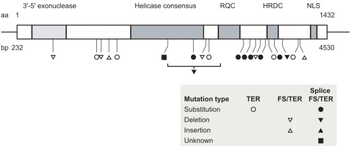
Positional cloning of the WRN (http://sageke.sciencemag.org/ cgi/genedata/sagekeGdbGene;189) gene in 1996 provided a strong stimulus for research on in vivo function and disease patho-genesis. The WRN locus, situated at chromosomal position 8p12, encodes a 162-kD member of the human RecQ helicase family (14). The five members of this family all possess 3′-to-5′ helicase activity as well as adenosine triphosphatase activity (13, 15). WRN is unique among the human RecQ helicases in possessing an additional 3′-to-5′ exonuclease activity (16-18) (Fig. 1).
The biochemical similarity of human RecQ helicases to ho-mologs identified in prokaryotes (http://sageke.sciencemag.org/ cgi/content/full/2003/40/nw137) and single-celled eukaryotes suggested the potential for functional parallelism and helped to identify several model organisms in which to investigate RecQ helicase function [reviewed in (12, 13)]. Potential roles for the human RecQ helicases in genome stability assurance were em-phasized by the identification of three human RecQ helicase de-ficiency syndromes: WS, Bloom syndrome (caused by
muta-Werner Syndrome Protein—Unwinding
Function to Explain Disease
Raymond J. Monnat Jr. and Yannick Saintigny
(Published 31 March 2004)
Raymond J. Monnat Jr. is in the Departments of Pathology and Genome Sciences at the University of Washington, Seattle, WA 98195, USA. Yannick Saintigny is at the Laboratoire d’Etude de la Recombinaison, Département de Radiobiologie et Radiopathologie, Commissariat à l’Energie Atomique, 92265 Fontenay aux Roses Cedex, France. E-mail: [email protected] (R.J.M.)
tions in the BLM http://sageke.sciencemag.org/cgi/genedata/ sagekeGdbGene;8 gene), and a subset of Rothmund-Thomson syndrome. In each syndrome, genetic instability and a predispo-sition to neoplasia result from the loss of function of a different human RecQ helicase protein (14, 19-21) (see “A Jump-Start for Replication” http://sageke.sciencemag.org/cgi/content/ abstract/2002/13/nw45).
Analyses of WRN ac-tivities on defined nucle-ic acid substrates indnucle-icate that WRN can unwind and/or degrade several types of DNA metabolic intermediates. These in-clude three- and four-way DNA junctions and gapped, branched, or un-paired DNA regions [re-viewed in (10, 22)]. DNA molecules containing three- or four-stranded junctions arise as part of DNA replication, as a consequence of DNA re-pair, and during HR. The increasingly rich body of in vitro biochemical data on WRN has been com-plemented by protein teraction studies that in-dicate physical and/or functional cross-talk be-tween WRN and general nucleic acid metabolic proteins such as replica-tion protein A (http://
sageke.sciencemag.org/cgi/content/full/sageke;2002/13/re2) and more specialized proteins involved in DNA synthesis, re-combination, or repair (10, 22). The WRN mutations identified in WS patients all truncate and promote loss of the WRN pro-tein from patient cells (23-26). The absence of WS-associated missense mutations that selectively inactivate either the WRN exonuclease or helicase activity is noteworthy: This finding suggests that it is the loss of the helicase and exonuclease func-tions of WRN that leads to WS. We discuss this point further below in light of new experimental results.
The Importance of Cellular Phenotype
Three consistent cellular defects have been identified after the loss of WRN function. This triad includes (i) cell proliferation defects that have been best defined in the fibroblast cell lineage (27, 28); (ii) selective sensitivity to a small number of DNA-damaging agents (29-33); and (iii) genetic instability (observed at both the cytogenetic and molecular genetic level) in different cell lineages in vivo and in vitro (34-36). These aspects of the WS cell phenotype are experimentally tractable, can be easily quantified, and appear to directly reflect WRN function. Thus, cellular phenotype has been an important focus in attempts to understand WRN function.
Analysis of WRN function at the cellular level has also been conceptually important as a way to integrate and interrelate the
molecular, biochemical, cytogenetic, and cytological data on WRN function. Further, understanding WRN function at the cellular level is likely to be directly relevant to disease pathogenesis. Thus, an important goal and milestone for current WS research is to develop a sophisticated molecular-level model of WRN function at the level of single cells that is both quantitative and predictive.
WRN Function in HR
The past 5 years have seen a resurgence of interest in HR in hu-man somatic cells (37-40). This renewed interest has been driv-en by several factors, including (i) data indicating the quantita-tive importance of HR in mammalian DNA break repair [see, for example, (41)]; (ii) the critical role played by HR in mam-malian development (42, 43) and cell viability (44); and (iii) da-ta from bacteria and single-celled eukaryotes that indicate a critical role for HR proteins and HR function in DNA repair and the rescue of stalled replication forks (45-47). A final stim-ulus for renewed interest in mammalian HR comes from the fields of human genetics and genomics: An increasing number of human diseases are being identified that might represent de-fects in recombination [reviewed in (48, 49)], and a better un-derstanding of HR might provide more efficient ways to modify human genes for therapeutic gain. Moreover, many of the pro-teins mediating human HR have polymorphic variants with the potential to affect expression and/or function, and several vari-ants have already been associated with disease risk, most no-tably for neoplasia (48, 50).
Although there have been hints for some time that WRN might play a role in HR [see, for example, (51)], the nature of this involvement only became clear when the behavior of “di-rect repeat” recombination reporter (DR reporter) substrates was examined in WS and control cells (Fig. 2) (52). The use of
aa 1 1432
3'-5' exonuclease
Mutation type TER FS/TER FS/TER Substitution Deletion Insertion Unknown Splice Helicase consensus RQC HRDC NLS bp 232 4530
Fig. 1. Domain structure of WRN and spectrum of WS-associated WRN mutations. The central box
indi-cates the WRN open reading frame with amino acid residue numbering indicated on top and cDNA base pair coordinates below. The positions of five protein motifs are indicated by shaded boxes and labels: (i) exonuclease domain, (ii) RecQ helicase domain, (iii) RecQ consensus (RQC) domain, and (iv) the heli-case and RNaseD-C-terminal (HRDC) domain and the C-terminal nuclear localization signal (NLS). The positions and molecular types of mutations identified in WS patients are indicated by symbols below. All mutations thus far identified truncate the WRN open reading frame and cause loss of the nuclear local-ization signal. These data have been compiled and are available in the WRN Mutation and Polymorphism Database (www.pathology.washington.edu/research/werner/ws_wrn.html), developed and maintained at the University of Washington. TER, termination; FS, frameshift; splice FS/TER, mutations that interfere with splicing and lead to frameshifts with downstream stop codon(s) in the new reading frame.
this type of chromosomally integrated substrate allows the fre-quency and rate of spontaneous and damage-induced nation to be quantified and the molecular nature of recombi-nants to be analyzed to provide mechanistic insight into mam-malian recombination pathways (40, 53, 54).
Two classes of genetically active recombinant molecules can be recovered after DR reporter plasmids undergo recombina-tion: (i) conversion-type recombinant molecules, in which the DR reporter is intact and one of the two reporter alleles has been converted to an active form; and (ii) crossover or “popout” molecules, in which a single active allele
re-mains with the loss of intervening DNA. Ei-ther or both recombinant classes can be recov-ered depending on the structure of the initial or substrate DR reporter plasmid. These two types of recombinant molecules can be gener-ated by several different pathways (Fig. 2). Human and other mammalian cells show a clear preference for the generation of conver-sion-type events, the most conservative of the potential outcomes that generate active re-porter alleles (53, 55-57).
Analyses of spontaneous HR in WS cells using this type of DR reporter have revealed a 25-fold reduction in the rate of generation of viable recombinant daughter cells that retain growth potential as compared with normal cells. This reduction is observed despite an ap-parently normal rate of generation of recombi-nant molecules in WS cells. Related to this finding is a reduction in the proportion of vi-able recombinant cells that harbor conversion-type recombinant molecules (52). This WS-as-sociated HR defect can be rescued by expres-sion of catalytically active WRN protein or by the expression of the RusA bacterial resolvase protein, suggesting that WRN, like RusA, functions in the resolution of HR products. Resolution or postsynapsis is the stage of HR when successful recombinant molecules are disentangled and segregated to daughter cells. The reduced cell survival that accompanies the WS HR defect can also be suppressed by
dom-inant negative RAD51 (http://sageke.sciencemag.org/cgi/ genedata/sagekeGdbGene;121), the key protein involved in nucle-oprotein filament formation, strand invasion, and the search for homology in the early stages of HR (58) (Fig. 3). Expression of this dominant negative form inhibits HR. The simplest explanation for these results is that HR initiation is normal in WS cells, but a portion of the products of RAD51-dependent HR cannot be suc-cessfully resolved in the absence of WRN function.
Recent data have allowed us to take this story one step further and to address the roles of the WRN exonuclease and helicase functions in HR and in cell survival after DNA damage. By ex-pressing WRN proteins in which single amino acid substitutions were used to inactivate the WRN exonuclease and/or helicase functions, we showed that both catalytic activities were essential for WRN to function in HR resolution. To our surprise, single missense mutant forms of WRN that lacked exonuclease or heli-case activity supported normal cell survival levels after DNA damage in the absence of HR. The WS cell phenotypes of an HR
defect and reduced cell survival after DNA damage did not differ between WS cells that lacked detectable WRN or that expressed catalytically inactive WRN at physiological levels (58, 59).
These results indicate that catalytic functions [as opposed to postulated scaffolding functions (http://sageke.sciencemag.org/ cgi/content/full/2003/31/pe22)] of WRN are critical for HR, and that HR and cell survival can in some cases be separated. Our results thus differ from recently published work by Chen et al. proposing an important structural role for WRN in determining the outcome of break repair events (60). We think the most
like-ly explanation for this discrepancy is Chen et al.’s use of fibrob-lasts immortalized by expression of the catalytic subunit of telomerase, hTERT (http://sageke.sciencemag.org/cgi/ genedata/sagekeGdbGene;205). Although hTERT expression suppresses the growth defect of primary WS fibroblasts, as does the SV40 T antigen expressed in the cells used in our experi-ments (61-64), hTERT also appears to suppress other important aspects of the WS cellular phenotype, such as selective drug sensitivity and the WS HR defect. The expression of hTERT al-so appears to alter the outcome of al-some classes of repair event in WS cells, such as plasmid rejoining [see (65)], that is likely to depend on nonhomologous DNA end joining (http://sageke. sciencemag.org/cgi/content/full/2003/8/re3) (NHEJ), a repair pathway in which DNA ends are joined without regard for the presence of homologous DNA sequences.
An important conclusion from our experimental results is that both of the WRN catalytic activities need to be lost in order to generate the WS cellular phenotype. This conclusion
pro-5' neor
mutant
neo+ hghr neo+
Gene conversion Recombination 3' neor
mutant
neo–/G418s
neo+/G418r
hghr
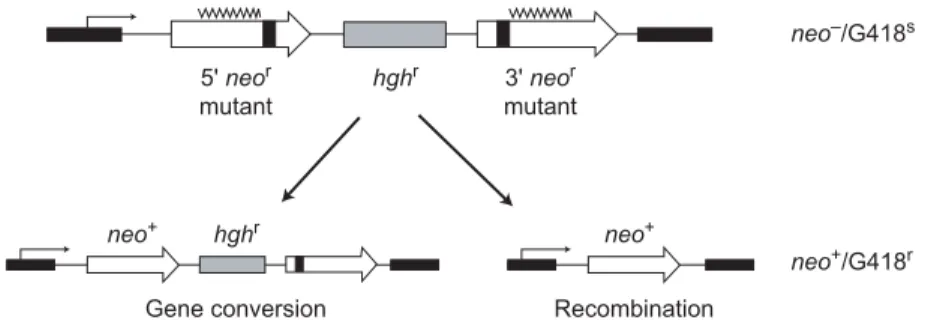
Fig. 2. DR reporter plasmid structure and recombinant classes. Structure of a DR
re-porter plasmid, pNeoA (57). Open arrows indicate direct repeat neomycin phospho-transferase (neo) genes inactivated by linker insertions (solid boxes), with overlying wavy lines indicating the region of homology between linker insertion sites. Two major classes of recombinant molecules represent the outcome of recombination pathways that by virtue of gene conversion (left) or crossing over (right) generate an active re-porter allele (neo+) that allows growth in the presence of the drug G418, a neomycin
analog. The two outcomes are distinguished by the structure of the resulting recombi-nant molecule and by whether cells also retain or lose resistance to hygromycin (hygr,
hygromycin resistance cassette; G418s, G418-sensitive; G418r, G418-resistant). Gene
conversion (left), the nonreciprocal transfer of genetic information between two homol-ogous DNA molecules, is thought to be initiated in most cases by DNA breakage. Breaks can be repaired by the classical two-ended double-strand break repair pathway or via single-ended invasion and synthesis-dependent strand annealing. Noncrossover conversion-type events predominate among mitotic recombinant products in mam-malian cells. Crossover or popout-type recombinants (right) can arise as a result of in-trachromosomal recombination, unequal sister chromatid exchange, or single-strand annealing. These recombination pathways and the genetic requirements for each are discussed in detail in (40, 54, 116).
vides a satisfying explanation for the spectrum of WRN tions in WS patients that, as noted above, lack missense muta-tions that selectively inactivate the WRN exonuclease or heli-case activities. The identification of a requirement for both WRN catalytic activities in HR also raises the interesting prospect that inherited or somatically acquired WRN missense mutations could selectively cripple the WRN HR function with-out appreciably affecting cell viability. Such mutations, if they are segregating in the human population, would likely confer a phenotype different from WS [see (59) for further discussion]. Resolution and Its Consequences
A role for WRN in the resolution of HR products was unexpect-ed. Successful resolution is important if HR products are to be topologically disentangled and
accurately segregated to generate viable recombinant daughter cells (66, 67). In addition to topologi-cally disentangling recombinant DNA duplexes, resolution deter-mines the proportion of crossover and noncrossover recombinants. Crossover recombinants have the potential to promote gene or chromosomal rearrangement and the loss of genetic information. Thus, not surprisingly, the gener-ation of different types of resolu-tion products is under tight con-trol, with mammalian HR strong-ly favoring the generation of non-crossover products (40).
How the resolution of recom-binant molecules is managed at the molecular level is best under-stood in prokaryotes. Successful resolution appears to involve at least three discrete steps: (i) the recognition and binding of DNA molecules containing recombina-tion juncrecombina-tions such as Holliday junctions or D loops; (ii) branch migration of the DNA junctions; and (iii) junction cleavage at or near crossover points, followed
by ligation of the now-separated DNA molecules to form intact recombinant DNA duplexes (46, 66-68). In Escherichia coli, a dedicated molecular machine known as the RuvABC complex binds, branch-migrates, and cleaves Holliday junction-contain-ing products at crossover points to give rise to different classes of recombinant DNA molecules (68).
The comparable proteins that mediate resolution in eukary-otes have been intensively sought and are now just beginning to come into focus (67). Two human RecQ helicases, WRN and BLM, can bind and branch-migrate model recombination sub-strates such as Holliday junctions, one of the desired activities for a resolution complex (69, 70). The exonuclease activity of WRN might also be useful for resolving certain types of recom-binant products such as D loops (Fig. 4A). However, neither WRN nor BLM can cleave model recombination junctions in vitro, and neither appears to be present in mammalian cell
frac-tions that possess the ability to branch-migrate and cleave mod-el HR junction substrates (71, 72).
Some types of eukaryotic recombination junctions might be cleaved and resolved by the heteromeric protein Mus81-Eme1/Mms4. This endonuclease, first identified in parallel analyses in budding and fission yeast, consists of two proteins that interact to form a structure-specific endonuclease [re-viewed in (67, 73)]. Human homologs exist for both proteins, and both proteins appear to be involved in recombination and the response to DNA damage. For example, Mus81 contributes to one of the recombination resolution activities identified by biochemical fractionation of mammalian cell extracts (72, 74). Human Mus81 localizes to sites of DNA damage in the nuclei of HeLa cells and colocalizes in the nucleolus with WRN and
BLM (75). Moreover, depletion of Mus81 mRNA by RNA in-terference leads to a reduction in the generation of recombi-nants (in the same recombination reporter cell lines that were originally used to identify the HR defect in WS cells) and a loss of cell viability (52, 76).
A recent extensive analysis of murine Eme1 indicates that it is part of a structure-specific endonuclease with a preference for 3ρ flap substrates. This type of DNA structure, in which a nicked DNA duplex contains a single-stranded DNA tail that is displaced at the nick, can arise in the context of DNA replica-tion or DNA repair. Murine embryonic stem (ES) cells that lack Eme1 are sensitive to DNA cross-linking agents such as cis-platin (cis-Pt) and mitomycin-C, and exhibit an increased inci-dence of damage-dependent sister chromatid exchange and ele-vated levels of both spontaneous and damage-induced chromo-somal aberrations as compared with wild-type ES cells (77).
Strand breaks Stalled/broken forks DNA damage Failed resolution/ restart Resolution/ restart Cell death Genetic stability Survival Homologous recombination WRN+ WRN– + wt WRN + SMRad51 + RusA
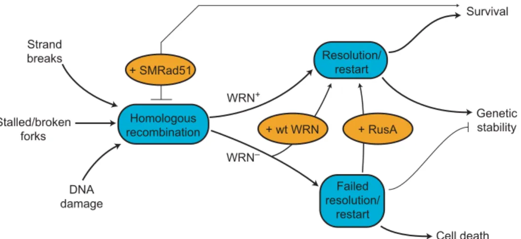
Fig. 3. Model of WRN function in HR. DNA damage, replication, or repair can initiate HR (left).
WRN promotes HR resolution or replication restart to ensure cell viability and genetic stability (WRN+arrow). In the absence of WRN (WRN−), HR resolution and/or replication restart fails,
leading to mitotic arrest, cell death, and genetic instability. Experimental tests of this model are shown in ovals: Reexpressing WRN protein (+wt WRN) improves both cell survival and the recov-ery of viable mitotic recombinants, as does expression of the bacterial resolvase protein RusA (+RusA). The dependence of WRN phenotypes on RAD51 pathway function and products can be revealed by expressing a dominant negative form of mammalian RAD51 protein (+SMRAD51) that suppresses mitotic recombination in WRN and controls cells while improving WRN cell sur-vival after cis-Pt-induced damage. Anticipated consequences of sursur-vival in the absence of HR function are mutagenesis and genetic instability (58).
These and other data indicate that the Mus81-Eme1/Mms4 en-donuclease is a plausible candidate for one of the eukaryotic resolution endonuclease activities, despite uncertainty as to the likely in vivo substrates and the quantitative importance of this resolution pathway (67).
The recent identification of RAD51C, one of the mammalian RAD51 paralogs, as a candidate resolution protein is intriguing (78). The RAD51 paralogs were originally identified on the basis of sequence conservation and the ability to complement the x-ray sensitivity displayed by certain mutant hamster cell lines. These proteins clearly play a role in the maintenance of genome stabili-ty and might have functional roles in one or more repair path-ways, although the mechanistic details remain obscure (79). RAD51C appears to be part of at least two different heteromeric paralog complexes. One of these complexes, RAD51C-XRCC3, is important for both HR resolution and the replication of dam-aged DNA (78-81). Precisely how RAD51C acts to promote res-olution, despite apparently lacking branch migration or endonu-clease activity (78), is puzzling and points to the participation of RAD51C-XRCC3 in one or more resolution complexes. The number, composition, and substrate preference of these resolution complexes may be rapidly discovered, now that several participat-ing proteins, as well as mammalian cell fractions that exhibit res-olution activity, have been identified.
Controlling Resolution to Suppress Gene Rearrangements
Recent evidence indicates that RecQ heli-cases might also play a role in determin-ing the proportion of crossover products in mammalian HR and in mitotic and meiot-ic HR in yeast (82, 83). Biochemmeiot-ical in-sight into one mechanism by which crossover suppression might occur in hu-man cells was revealed in recent analyses of the resolution of recombination sub-strates by recombinant human BLM and DNA topoisomerase IIIα (84). This pair of proteins appears to work in concert to re-solve substrates containing double Holli-day junctions without the exchange of flanking markers. Junction “dissolution,” as this type of resolution has been termed, thus differs from the more familiar RuvC-or Mus81-like cleavage reaction discussed above (67, 84). These recent results paral-lel and extend earlier analyses of E. coli RecQ, in which similar catenation-decate-nation activities were first described (85). One satisfying aspect of this result is that it explains one of the cytogenetic hall-marks of Bloom syndrome: the abnormal-ly high levels of sister chromatid ex-changes in cells and cell lines from BLM patients. Sister chromatid exchange crossover products arise at high frequency once crossover product formation is no longer effectively suppressed after the loss of BLM function (40, 84, 86).
The results summarized above indicate that the WRN and BLM human RecQ
he-licases might have complementary roles in suppressing gene re-arrangement or loss in somatic cells. WRN appears to promote the successful resolution of HR products to favor the generation of viable conversion-type recombinants, whereas BLM acts to suppress the generation of crossover products. This model also explains why BLM and WRN, which are both envisioned to be acting in HR, have divergent loss-of-function HR phenotypes. The Role of HR in DNA Replication
Many of the proteins that mediate HR in mammalian cells might have an additional role in insuring the completion of DNA replication (http://sageke.sciencemag.org/cgi/content/ full/sageke;2003/8/re3). The stalling or disruption of DNA replication forks appears to be common in virtually all organ-isms and can be accentuated by many forms of DNA damage (45, 47, 87). The most desirable outcome of this apparently un-avoidable event is to promote the successful restart of replica-tion, while suppressing DNA breakage and genome rearrange-ment. DNA damage that involves both DNA strands (for exam-ple, DNA interstrand cross-links) are potent blocks to replica-tion fork progression that may be efficiently and preferentially repaired by HR (88). Many other forms of DNA damage trigger HR, and thus might use HR for purposes of repair or to promote damage tolerance (by, for example, lesion bypass) (40, 47).
One example of the latter role for HR proteins and HR
func-Reinvade/end join Cleave Restart Bypass
A B C
WRN helicase Bypass polymerase WRN exonuclease DNA damage WRN exo + heli RAD52
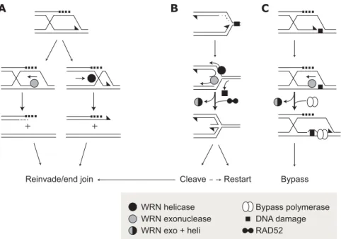
Fig. 4. Potential recombination resolution and replication restart pathways using WRN
ex-onuclease or helicase activity. (A) Degradation or unwinding of recombination intermediates (a D loop is shown) by the WRN exonuclease or helicase activities could topologically disen-tangle molecules to promote resolution. (B) Degradation or unwinding of replication forks stalled by DNA damage could remove bound proteins, stabilize the fork for restart after re-pair, or promote regression and cleavage to generate free DNA ends. Lagging strand un-winding is shown as an example. WRN acting with RAD52 could promote a stable regressed fork that could be cleaved by Holliday junction resolvases to promote end invasion or used directly to reinitiate replication (90). (C) Disassembly or unwinding of a replication complex stalled at template DNA by WRN exonuclease (shown) or helicase activities could promote assembly of a bypass complex (open ovals) containing one or more specialized DNA poly-merases. All three pathways depicted here are likely to be error-prone (45, 47, 87).
tion is in the processing or stabilization of stalled replication forks to either promote or inhibit the initiation of HR. Many of the postulated DNA intermediates in stalled replication forks resemble recombination intermediates, and thus might serve as substrates for recombination protein recognition and processing (40, 45, 47, 67, 87). An explicit role for the E. coli RecQ pro-tein in stalled replication fork processing has been proposed (89), and similar roles can be envisioned for
WRN acting either alone or in conjunction with other proteins such as mammalian RAD52 (http://sageke.sciencemag.org/ cgi/genedata/sagekeGdbGene;122) to facili-tate fork repair or stabilization (Fig. 4, B and C) (90). The WRN exonuclease activity alone, or WRN helicase in conjunction with other nuclease activities, could also act to promote the bypass of DNA damage (Fig. 4, B and C).
Roles for RecQs in the Cross-Talk Between HR and NHEJ
The identification of an HR defect in WS cells provides a ready explanation for the loss of conversion-type recombinants, re-duced cell viability, and selective sensitivity to DNA damaging agents that might require HR for repair (58). How, given this picture, do we make sense of reports that indicate biochemical and functional interactions be-tween WRN and proteins that function in the NHEJ pathway, such as Ku and DNA-PKcs? [See “Twisted Logic: Discoveries tangle Werner syndrome helicase story” (http:// s a g e k e . s c i e n c e m a g . o r g / c g i / c o n t e n t / abstract/2002/12/nw40) and “Break Danc-ing: Werner syndrome protein might keep rowdy enzymes from doing a number on tat-tered DNA ends” (http://sageke.sciencemag. org/cgi/content/abstract/2002/3/nw8.)] The
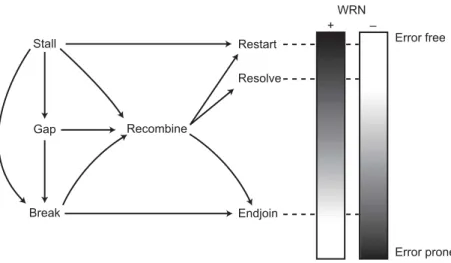
interrelation of WRN function, HR, and NHEJ is perhaps easi-est to understand in the context of a model of substrate traffick-ing or flux durtraffick-ing DNA replication (S phase) (Fig. 5).
In WRN+cells, DNA damage or S-phase intermediates that initiate HR or that require HR function are successfully converted to appropriate products. The successful resolution of these prod-ucts insures high cell viability and minimizes genetic instability or gene rearrangements. In WS cells, in contrast, a portion of HR resolution events fail, giving rise to cells that undergo mitotic ar-rest and/or harbor DNA breaks or gaps. Such DNA breaks and gaps are substrates for recombination (40, 91). However, in the ab-sence of WRN function, many of these “second tries” are again likely to fail, setting up a futile cycle that leads to mitotic death or apoptosis. DNA breaks that arise directly or indirectly from reso-lution failure can, in contrast, be captured and joined by NHEJ to restore chromosome integrity and insure high cell viability, albeit at the expense of mutation (92).
In this model, NHEJ function is downstream of WRN func-tion in HR. This provides an explanafunc-tion for the WRN cell phe-notype, which most closely resembles an HR (as opposed to NHEJ) defect (52, 58, 93, 94). Close coordination of NHEJ and HR function in normal cells makes good teleologic sense by
providing redundant ways to recognize, process, and resolve po-tentially dangerous DNA ends to insure chromosomal integrity and cell viability. The suggestion that WRN might be playing somewhat different roles in HR and in NHEJ, and that the bal-ance might be altered in WRN-deficient or NHEJ-deficient cells, is also consistent with data indicating substantial cross-talk between HR and NHEJ (95-101).
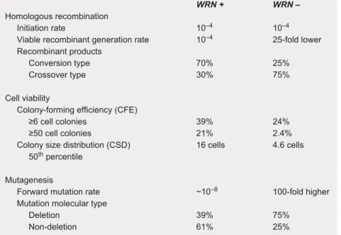
Some quantitative rigor can be brought to this model by exam-ining HR, cell viability, and mutagenesis data as a function of WRN status (Fig. 6). These data were all derived from the same set of well-characterized human WS and control SV40-trans-formed fibroblast cell lines (52, 58, 102). Consequences of the loss of WRN function in fibroblast-lineage cells include (i) a sub-stantial reduction in the probability of continued cell division, as measured by colony forming efficiency (CFE) or more sensitive colony size distribution (CSD) assays; (ii) a decrease in the rate of generation of recombinant daughter cells, together with the appar-ent loss of conversion-type recombinants; and (iii) a 10- to 100-fold increase in mutagenesis at the X-linked HPRT locus (a conve-nient marker for examining mutagenesis, because loss-of-function mutations at this locus confer resistance to an otherwise toxic compound) (52, 58, 102). Molecular characterization of the result-ing HPRT mutations arisresult-ing in WS cells indicates that they are predominantly large deletions involving the loss of from 1 to >100 kb of chromosomal DNA (102, 103). Most likely, these deletions originate from disrupted DNA replication intermediates or HR re-pair products that have been captured by NHEJ with the associat-ed loss of intervening DNA (103).
One of the appealing aspects of the picture of WRN function
Stall WRN + – Gap Break Recombine Restart Resolve Endjoin Error free Error prone
Fig. 5. S-phase substrate trafficking and outcomes. As depicted in Fig. 3, several types
of DNA damage, including replication fork stalling, DNA gaps that result from fork stall, and DNA breaks, can initiate HR. HR proteins can funnel these substrates into classi-cal HR and correctly resolve the products to insure high cell viability and chromosomal integrity (center; Fig. 4, A and C). HR proteins together with additional proteins such as DNA polymerases and resolvases may be able to act in concert to promote the restart of replication forks stalled by DNA damage either directly in the absence of HR (top) or via HR (center; see also Fig. 4, B and C). Replication stall and gapped and broken DNA molecules, if not successfully resolved by replication restart or HR, can be captured by NHEJ to insure chromosomal integrity, albeit at the expense of gene rearrangement, loss, or mutagenesis (40, 47). The shaded boxes to the right indicate the disposition of substrate in the presence or absence of WRN function.
summarized in Fig. 3, Fig. 4, and Fig. 5 is the ability to reconcile, and in part explain, many of the molecular, biochemical, cytolog-ical, and cytogenetic features of WS and of WRN function. This functional “snapshot” also begins to reveal the complexity of WRN functional roles in vivo and of functional cross-talk among the nucleic acid metabolic processes that can be influenced by WRN. The consequences of the loss of WRN function in fibrob-last-lineage cells are likely to be reflected in other cell types and lineages in vivo, although this assumption needs to be document-ed. It will be important to replicate the quantitative and molecular data summarized in Fig. 6, using primary cells to assess how the consequences of WRN loss of function are further modified by DNA damage checkpoints and apoptosis, which can be compro-mised or lost in immortalized cell lines.
WRN Function in Cell Lineages
WRN and the other human RecQ helicases appear to be ubiqui-tously expressed, whereas HR function appears to be largely limited to dividing cells. These observations lead to an in-evitable question: If the functional picture
represented in Fig. 3, Fig. 4, and Fig. 5 is ac-curate, why are dividing cell lineages not se-lectively affected by the loss of WRN func-tion? We think the most likely explanation is the following (104). All cell lineages (or at least the precursors to all cell lineages) un-dergo mitotic division during development. An absence of WRN function during devel-opment thus has the potential to affect all cell lineages by reducing the number of cells available for lineage construction and by “seeding” cell lineages with mutant progeny. How cell loss, genetic instability, and muta-tion accumulamuta-tion affect a given cell lineage is likely to depend on (i) how mitotically “deep” the mature cell lineage is; (ii) how much cell editing (or loss of lineage precur-sor cells) occurs during and after develop-ment; (iii) whether there is redundancy of function that is lineage-specific; and (iv) how large the functional reserve is for a given lin-eage, tissue, or organ.
For example, the central nervous system (CNS) might be protected from the effects of loss of WRN function by virtue of strin-gent cell editing during development and by a comparative absence of mitotic activity during adult life. Cell editing of neuronal precursors can occur as part of normal CNS development, in which a large number of precursors are generated during the initial stages of neurogenesis, and cells in excess of the number needed to complete develop-ment undergo programmed cell death. Cell editing also appears to reflect the selective loss of precursors that lack required func-tional properties or are damaged. However, subtle defects might emerge over time if WRN is important for neural stem cell func-tion during adult life (105). Conversely, cell lineages that divide continuously throughout
adult life, such as those that make up the skin, gut, and bone marrow, might be tolerant of the loss of WRN function by virtue of mutation expansion-limiting lineage architecture (106), a combination of stringent cell editing and large re-serves of stem or lineage repopulating cells. The cell lineages or tissues most susceptible to a loss of WRN function might be those that retain cell division potential, lack stringent cell editing during and after development, and are tolerant of and thus able to accumulate at least some types of genetic instabil-ity. The fibroblast lineage and other mesenchymal or mesoder-mally derived cell lineages might be selectively affected by the loss of WRN function, as suggested by the clinical and cell phenotype of WS, for precisely these reasons.
This line of reasoning leads to two important conclusions. First, we need to know more about the “normal biology” of human cell lineages before we will be able to understand and predict the in vivo consequences of loss of function of WRN or of related proteins. Second, we clearly need new and exper-imentally tractable cell culture and animal models in which to
WRN + WRN –
Homologous recombination
Initiation rate 10–4 10–4
Viable recombinant generation rate 10–4 25-fold lower
Recombinant products
Conversion type 70% 25% Crossover type 30% 75%
Cell viability
Colony-forming efficiency (CFE)
≥6 cell colonies 39% 24% ≥50 cell colonies 21% 2.4% Colony size distribution (CSD) 16 cells 4.6 cells 50th percentile
Mutagenesis
Forward mutation rate ~10–8 100-fold higher
Mutation molecular type
Deletion 39% 75% Non-deletion 61% 25%
Fig. 6. Quantitative aspects of the WS fibroblast phenotype. This integrated data set
was developed from the same SV40-transformed fibroblast cell lines. With a loss of WRN function, the recombination initiation rate (recombinant molecules/cell doubling) is unaffected. However, the rate of successful HR resolution as measured by the rate of generation of viable recombinant daughter cells/cell doubling falls 25-fold, together with an apparent loss of conversion-type recombinants (52). The HR defect is mirrored by a decline in cell viability in the absence of DNA damage as measured by CFE and CSD assays that measure, respectively, the generation of colonies of 6 or more or 50 or more cells (CFE), or the 50th percentile for number of cells in colonies after a defined growth interval (CSD) (58). In conjunction with the loss of viable recombinant daughter cells and reduced cell division potential, the rate of forward spontaneous mutation at the X-linked HPRT gene climbs 10- to 100-fold per cell per generation. The predomi-nant type of mutation identified in HPRT-deficient WS cell lines was the large deletion [(102, 103); see text for additional discussion]. The mitotic recombination rate is at least two orders of magnitude higher that the rate of forward HPRT mutations in WRN+and
WRN−cells, and the recombination defect in WRN−cells is accompanied by a large
in-crease in HPRT mutagenesis. These results indicate that the recombination initiation rate is comparable in WS and control cells and that in WRN+cells, recombination
events rarely give rise to mutations. The recombination and viability deficits obersved in WS cells are further accentuated by DNA damage (58, 59).
study normal lineage biology and to identify lineage-specific aspects of WRN function.
WRN As a Modulator of Disease Risk in Populations The WRN locus resembles many other human genes in that a large number of genetic variants are present in the population [see com-pilation and links in (24) and at the Environmental Genome Pro-ject (EGP) Web site (http://egp.gs.washington.edu)]. A subset of these are clearly disease-associated mutant alleles segregating in the human population, whereas the majority are single-nucleotide polymorphisms (SNPs) or sequence variants of uncertain func-tional importance. The number of these variants is surprisingly large: Recently completed resequencing of the WRN exons, pro-moter region, and downstream untranslated region at the Universi-ty of Washington as part of the EGP revealed 375 WRN sequence variants in 90 different DNA samples contained in the EGP’s Polymorphism Discovery Resource (http://locus.umdnj. edu/nigms/products/pdr.html). These variants included a large number of SNPs in addition to nonsynonymous coding region substitutions and a previously reported nonsense mutant allele.
The potential of WRN sequence variants to modify human disease risk outside the context of WS is most clearly under-stood for WRN heterozygotes. Heterozygous carriers of single mutant WRN alleles appear to be present worldwide at frequen-cies ranging up to 1:100 [reviewed in (4)]. Frequency estimates of heterozygotes in the United States are in the range of ~1:250, leading to estimates of >106 carriers of mutant alleles in the United States alone. WRN heterozygotes exhibit genetic insta-bility in vivo (107), and lymphoblastoid cell lines derived from otherwise healthy WRN heterozygotes display an intermediate sensitivity to killing by DNA-damaging agents that selectively kill WRN-deficient cells (31, 32).
Heterozygote effects have the potential to influence cancer risk or the outcome of cancer therapy. Several strategies might be helpful in establishing these links (108). For example, anal-ysis of DNA derived from cells within clonal tumors might re-veal inherited or somatically acquired WRN mutations that played a role in either the origin or progression of cancer. It will be interesting to see if sporadic tumors of the types ob-served in WS patients (109, 110) are enriched for mutations or epigenetic events such as hypermethylation (http://sageke. sciencemag.org/cgi/content/full/2001/1/oa3), with associated silencing of WRN expression. The identification of an aberrant or exaggerated response to chemotherapy in tumors that are heterozygous or deficient in WRN function would provide a second route to identification of a role for the WRN pathway in tumor biology. Of note, several clinically important chemotherapeutic agents such as camptothecin, mitomycin-C, and cis-Pt selectively kill cells that are WRN-deficient or hap-loinsufficient (31-33, 111). The functional consequences of the much larger number of polymorphic WRN variants and po-tential disease associations outside neoplasia are also just be-ginning to be explored (24, 112-115).
Concluding Remarks
WS is an example of the growing number of heritable human diseases in which a defect in genome stability assurance or ge-nomic “housekeeping” leads to an elevated risk of one or more secondary disease phenotypes such as cancer. WS is further dis-tinguished among the heritable housekeeping defects as one of the increasing number of human cancer predispositions that
ap-pear to result from a defect in HR (49). In returning to the metaphor with which we began, all parents know that change and surprise are key features of adolescence. Our “adolescent” WS research promises an immediate future that will be chal-lenging, surprising, and rewarding!
References
1. G. M. Martin, A brief history of research on the Werner syndrome. Gann
Monogr. Cancer Res. 49, 1-10 (2001).
2. C. J. Epstein, G. M. Martin, A. L. Schultz, A. G. Motulsky, Werner’s syn-drome: A review of its symptomatology, natural history, pathologic features, genetics and relationship to the natural aging process. Medicine 45, 177-221 (1966).
3. M. Goto, Hierarchical deterioration of body systems in Werner’s syndrome: implications for normal ageing. Mech. Ageing Dev. 98, 239-254 (1997). 4. G. D. Schellenberg, T. Miki, C.-E. Yu, J. Nakura, in The Metabolic &
Molecu-lar Basis of Inherited Disease, C. R. Scriver, A. L. Beaudet, W. S. Sly, D.
Valle, Eds. (McGraw-Hill, New York, ed. 8, 2001), pp. 785-797.
5. G. M. Martin, Genetic syndromes in man with potential relevance to the pathobiology of aging. Birth Defects Original Article Ser. 14, 5-39 (1978). 6. V. A. Bohr, Werner syndrome and its protein: clinical, cellular and molecular
advances. Mech. Ageing Dev. 124, 1073-1082 (2003).
7. H. Hoehn, E. M. Bryant, K. Au, T. H. Norwood, H. Boman, G. M. Martin, Var-iegated translocation mosaicism in human skin fibroblast cultures.
Cyto-genet. Cell Genet. 15, 282-298 (1975).
8. R. M. Brosh Jr., V. A. Bohr, Roles of the Werner syndrome protein in path-ways required for maintenance of genome stability. Exp. Gerontol. 37, 491-506 (2002).
9. M. Fry, The Werner syndrome helicase-nuclease-one protein, many mys-teries. Sci. SAGE KE 2002, re2 (2002) (http://sageke.sciencemag.org/ cgi/content/full/sageke;2002/13/re2).
10. P. L. Opresko, W.-H. Cheng, C. von Kobbe, J. A. Harrigan, V. A. Bohr, Wern-er syndrome and the function of the WWern-ernWern-er protein; what they can teach us about the molecular aging process. Carcinogenesis 24, 791-802 (2003). 11. W.-H. Cheng, V. A. Bohr, Diverse dealings of the Werner helicase/nuclease.
Sci. SAGE KE 2003, pe22 (2003)
(http://sageke.sciencemag.org/cgi/con-tent/full/sageke;2003/31/pe22).
12. H. Nakayama, RecQ family helicases: roles as tumor suppressor proteins.
Oncogene 21, 9008-9021 (2002).
13. I. D. Hickson, RecQ helicases: caretakers of the genome. Nat. Rev. Cancer
3, 169-178 (2003).
14. C.-E. Yu, J. Oshima, Y.-H. Fu, E. M. Wijsman, F. Hisama, S. Ouais, J. Naku-ra, T. Miki, G. M. Martin, J. Mulligan, et al., Positional cloning of the Wern-er’s syndrome gene. Science 272, 258-262 (1996).
15. A. J. van Brabant, R. Stan, N. A. Ellis, DNA helicases, genomic instability, and human genetic disease. Annu. Rev. Genomics Hum. Genet. 1, 409-459 (2000).
16. S. Huang, B. Li, M. D. Gray, J. Oshima, I. S. Mian, J. Campisi, The prema-ture aging syndrome protein, WRN, is a 3′to 5′exonuclease. Nat. Genet.
20, 114-116 (1998).
17. J.-C. Shen, M. D. Gray, J. Oshima, A. S. Kamath-Loeb, M. Fry, L. A. Loeb, Werner syndrome protein I: DNA helicase and DNA exonuclease reside on the same polypeptide. J. Biol. Chem. 273, 34139-34144 (1998).
18. A. S. Kamath-Loeb, J.-C. Shen, L. A. Loeb, M. Fry, Werner syndrome pro-tein II: Characterization of the integral 3′Æ5′DNA exonuclease. J. Biol.
Chem. 273, 34145-34150 (1998).
19. N. A. Ellis, J. Groden, T.-Z. Ye, J. Straughen, D. J. Lennon, S. Ciocci, M. Proytcheva, J. German, The Bloom’s syndrome gene product is homolo-gous to RecQ helicases. Cell 83, 655-666 (1995).
20. S. Kitao, A. Shimamoto, M. Goto, R. W. Miller, W. A. Smithson, N. M. Lindor, Y. Furuichi, Mutations in RECQ4L cause a subset of cases of Rothmund-Thomson syndrome. Nat. Genet. 22, 82-84 (1999).
21. L. L. Wang, A. Gannavarapu, C. A. Kozinetz, M. L. Levy, R. A. Lewis, M. M. Chintagumpala, R. Ruiz-Malanado, J. Contreras-Ruiz, C. Cunniff, R. P. Er-ickson, et al., Association between osteosarcoma and deleterious muta-tions in the RECQL4 gene in Rothmund-Thomson syndrome. J. Natl.
Can-cer Inst. 95, 669-674 (2003).
22. C. Z. Bachrati, I. D. Hickson, RecQ helicases: suppressors of tumorigene-sis and premature aging. Biochem. J. 374, 577-606 (2003).
23. M. J. Moser, J. Oshima, R. J. Monnat Jr., WRN mutations in Werner syn-drome. Hum. Mutat. 13, 271-279 (1999).
24. P. W. Wagner, R. J. Monnat Jr., Werner Syndrome Mutation and Polymor-phism Database, Release 2. http://www.pathology.washington.edu/re-search/werner/ws_wrn.html (1 July 2003).
25. M. Goto, Y. Yamabe, M. Shiratori, M. Okada, T. Kawabe, T. Matsumoto, M. Sug-imoto, Y. Furuichi, Immunological diagnosis of Werner syndrome by down-reg-ulated and truncated gene products. Hum. Genet. 105, 301-307 (1999). 26. M. J. Moser, A. S. Kamath-Loeb, J. E. Jacob, S. E. Bennett, J. Oshima, R. J.
Monnat Jr., WRN helicase expression in Werner syndrome cell lines.
Nu-cleic Acids Res. 28, 648-654 (2000).
27. G. M. Martin, C. A. Sprague, C. J. Epstein, Replicative life-span of cultivat-ed human cells. Effects of donor’s age, tissue, and genotype. Lab. Invest.
23, 86-92 (1970).
28. T. O. Tollefsbol, H. J. Cohen, Werner’s syndrome: an underdiagnosed disor-der resembling premature aging. Age 7, 75-88 (1984).
29. E. Gebhart, R. Bauer, U. Raub, M. Schinzel, K. W. Ruprecht, J. B. Jonas, Spontaneous and induced chromosomal instability in Werner syndrome.
Hum. Genet. 80, 135-139 (1988).
30. E. Gebhart, M. Schinzel, K. W. Ruprecht, Cytogenetic studies using various clastogens in two patients with Werner syndrome and control individuals.
Hum. Genet. 70, 324-327 (1985).
31. C. E. Ogburn, J. Oshima, M. Poot, R. Chen, K. E. Hunt, K. A. Gollahon, P. S. Rabinovitch, G. M. Martin, An apoptosis-inducing genotoxin differentiates heterozygotic carriers for Werner helicase mutations from wild-type and homozygous mutants. Hum. Genet. 101, 121-125 (1997).
32. M. Poot, K. A. Gollahon, P. S. Rabinovitch, Werner syndrome lymphoblas-toid cells are sensitive to camptothecin-induced apoptosis in S-phase.
Hum. Genet. 104 , 10-14 (1999).
33. M. Poot, J. S. Yom, S. H. Whang, J. T. Kato, K. A. Gollahon, P. S. Rabi-novitch, Werner syndrome cells are sensitive to DNA cross-linking drugs.
FASEB J. 15, 1224-1226 (2001).
34. D. Salk, K. Au, H. Hoehn, G. M. Martin, Cytogenetic aspects of Werner syn-drome. Adv. Exp. Med. Biol. 190, 541-546 (1985).
35. M. I. Melaragno, D. Pagni, M. d. C. Smith, Cytogenetic aspects of Werner’s syndrome lymphocyte cultures. Mech. Ageing Dev. 78, 117-122 (1995). 36. R. Melcher, R. von Golitschek, C. Steinlein, D. Schindler, H. Neitzel, K.
Kainer, M. Schmid, H. Hoehn, Spectral karyotyping of Werner syndrome fi-broblast cultures. Cytogenet. Cell Genet. 91, 180-185 (2000).
37. J. Thacker, The role of homologous recombination processes in the repair of severe forms of DNA damage in mammalian cells. Biochimie 81, 77-85 (1999).
38. L. H. Thompson, D. Schild, Homologous recombinational repair of DNA en-sures mammalian chromosome stability. Mutat. Res. 477, 131-153 (2001). 39. C. Richardson, M. Jasin, Recombination between two chromosomes:
impli-cations for genomic integrity in mammalian cells. Cold Spring Harbor
Symp. Quant. Biol. 65, 553-560 (2000).
40. T. Helleday, Pathways for mitotic homologous recombination in mammalian cells. Mutat. Res. 532, 103-115 (2003).
41. R. D. Johnson, M. Jasin, Sister chromatid gene conversion is a prominent double-strand break repair pathway in mammalian cells. EMBO J. 19, 3398-3407 (2000).
42. T. Tsuzuki, Y. Fujii, F. Sakumi, Y. Tominaga, K. Nakao, M. Sekiguchi, A. Mat-sushiro, Y. Yoshimura, T. Morita, Targeted disruption of the Rad51 gene leads to lethality in embryonic mice. Proc. Natl. Acad. Sci. U.S.A. 93, 6236-6240 (1996).
43. D.-S. Lim, P. Hasty, A mutation in mouse rad51 results in an early embryon-ic lethal that is suppressed by a mutation in p53. Mol. Cell. Biol. 16, 7133-7143 (1996).
44. E. Sonoda, M. S. Sasaki, J. M. Buerstedde, O. Bezzubova, A. Shinohara, H. Ogawa, M. Takata, Y. Yamaguchi-Iwai, S. Takeda, Rad51-deficient verte-brate cells accumulate chromosomal breaks prior to cell death. EMBO J.
17, 598-608 (1998).
45. M. M. Cox, M. F. Goodman, K. N. Kreuzer, D. Sherratt, S. J. Sandler, K. J. Marians, The importance of repairing stalled replication forks. Nature 404, 37-41 (2000).
46. A. Kuzminov, Recombinational repair of DNA damage in Escherichia coli and bacteriophage 8. Microbiol. Mol. Biol. Rev. 63, 751-813 (2000). 47. P. McGlynn, R. G. Lloyd, Recombinational repair and restart of damaged
replication forks. Nat. Rev. Mol. Cell Biol. 3, 859-870 (2002).
48. A. J. Pierce, J. M. Stark, F. D. Araujo, M. E. Moynahan, M. Berwick, M. Jasin, Double-strand breaks and tumorigenesis. Trends Cell Biol. 11, s52-s59 (2001).
49. L. H. Thompson, D. Schild, Recombinational DNA repair and human dis-ease. Mutat. Res. 509, 49-78 (2002).
50. B. Kuschel, A. Auranen, S. McBride, K. L. Novik, A. Antoniou, J. M. Lip-scombe, N. E. Day, D. F. Easton, B. A. J. Ponder, P. D. P. Pharoah, et al., Variants in DNA double-strand break repair genes and breast cancer sus-ceptibility. Hum. Mol. Genet. 11, 1399-1407 (2002).
51. R. Z. Cheng, S. Murano, B. Kurz, R. J. Shmookler Reis, Homologous re-combination is elevated in some Werner-like syndromes but not during nor-mal in vitro or in vivo senescence of mamnor-malian cells. Mutat. Res. 237, 259-269 (1990).
52. P. R. Prince, M. J. Emond, R. J. Monnat Jr., Loss of Werner syndrome pro-tein function promotes aberrant mitotic recombination. Genes Dev. 15, 933-938 (2001).
53. L. B. K. Herzing, M. S. Meyn, Novel LacZ-based recombination vectors for mammalian cells. Gene 137, 163-169 (1993).
54. F. Pâques, J. E. Haber, Multiple pathways of recombination induced by
double-strand breaks in Saccharomyces cerevisiae. Microbiol. Mol. Biol.
Rev. 63, 349-404 (1999).
55. R. J. Bollag, A. S. Waldman, R. M. Liskay, Homologous recombination in mammalian cells. Annu. Rev. Genet. 23, 199-225 (1989).
56. R. J. Bollag, R. M. Liskay, Direct-repeat analysis of chromatid interactions during intrachromosomal recombination in mouse cells. Mol. Cell. Biol. 11, 4839-4845 (1991).
57. M. S. Meyn, High spontaneous intrachromosomal recombination rates in ataxia-telangiectasia. Science 260, 1327-1330 (1993).
58. Y. Saintigny, K. Makienko, C. Swanson, M. J. Emond, R. J. Monnat Jr., Ho-mologous recombination resolution defect in Werner syndrome. Mol. Cell.
Biol. 22, 6971-6978 (2002).
59. C. Swanson, Y. Saintigny, M. J. Emond, R. J. Monnat Jr., The Werner syn-drome protein has separable recombination and viability functions. DNA
Repair, in press.
60. L. Chen, S. Huang, L. Lee, A. Davalos, R. H. Schiestl, J. Campisi, J. Oshi-ma, WRN, the protein deficient in Werner syndrome, plays a critical struc-tural role in optimizing DNA repair. Aging Cell 2, 191-199 (2003). 61. F. S. Wyllie, C. J. Jones, J. W. Skinner, M. F. Haughton, C. Wallis, D.
Wyn-ford-Thomas, R. G. A. Faragher, D. Kipling, Telomerase prevents the accel-erated cell ageing of Werner syndrome fibroblasts. Nat. Genet. 24, 16-17 (2000).
62. T. Matsumura, M. Nagata, R. Konishi, M. Goto, Studies of SV40-infected Werner syndrome fibroblasts. Adv. Exp. Med. Biol. 190, 313-330 (1985). 63. L. I. Huschtscha, K. V. A. Thompson, R. Holliday, The susceptibility of
Werner’s syndrome and other human skin fibroblasts to SV40-induced transformation and immortalization. Proc. R. Soc. London Ser. B 229, 1-12 (1986).
64. H. Saito, R. E. Moses, Immortalization of Werner syndrome and progeria fi-broblasts. Exp. Cell Res. 192, 373-379 (1991).
65. T. M. Rünger, C. Bauer, B. Dekant, K. Möller, P. Sobotta, C. Czerny, M. Poot, G. M. Martin, Hypermutable ligation of plasmid DNA ends in cells from pa-tients with Werner syndrome. J. Invest. Dermatol. 102, 45-48 (1994). 66. D. M. J. Lilley, M. F. White, The junction-resolving enzymes. Nat. Rev. Mol.
Cell Biol. 2, 433-443 (2001).
67. W.-D. Heyer, K. T. Ehmsen, J. A. Solinger, Holliday junctions in the eukary-otic nucleus: resolution in sight? Trends Biochem. Sci. 28, 548-557 (2003). 68. S. C. West, Processing of recombination intermediates by the RuvABC
proteins. Annu. Rev. Genet. 31, 213-244 (1997).
69. A. Constantinou, M. Tarsounas, J. K. Karow, R. M. Brosh Jr., V. A. Bohr, I. D. Hickson, S. C. West, Werner’s syndrome protein (WRN) migrates Holliday junctions and co-localizes with RPA upon replication arrest. EMBO Rep. 1, 80-84 (2000).
70. J. K. Karow, A. Constantinou, J.-L. Li, S. C. West, I. D. Hickson, The Bloom’s syndrome gene product promotes branch migration of Holliday junctions.
Proc. Natl. Acad. Sci. U.S.A. 97, 6504-6508 (2000).
71. A. Constantinou, A. A. Davies, S. C. West, Branch migration and Holliday junction resolution catalyzed by activities from mammalian cells. Cell 104, 259-268 (2001).
72. A. Constantinou, X.-B. Chen, C. H. McGowan, S. C. West, Holliday junction resolution in human cells: two junction endonucleases with distinct sub-strate specificities. EMBO J. 21, 5577-5585 (2003).
73. N. M. Hollingsworth, S. J. Brill, The Mus81 solution to resolution: generating meiotic crossovers without Holliday junctions. Genes Dev. 18, 117-125 (2004).
74. X.-B. Chen, R. Melchionna, C.-M. Denis, P.-H. L. Gaillard, A. Blasina, I. V. de Weyer, M. N. Boddy, P. Russell, J. Vialard, C. H. McGowan, Human Mus81-associated endonuclease cleaves Holliday junctions in vitro. Mol.
Cell 8, 1117-1127 (2001).
75. H. Gao, X.-B. Chen, C. H. McGowan, Mus81 endonuclease localizes to nu-cleoli and to regions of DNA damage in human S-phase cells. Mol. Biol.
Cell 14, 4826-4834 (2003).
76. V. Blais, H. Gao, C. A. Elwell, M. N. Boddy, P.-H. L. Gaillard, P. Russell, C. H. McGowan. RNAi inhibition of Mus81 reduces mitotic recombination in hu-man cells. Mol. Biol. Cell 15, 552-562 (2004).
77. J. Abraham, B. Lemmers, M. P. Hande, M. E. Moynahan, C. Chahwan, A. Ciccia, J. Essers, K. Hanada, R. Chahwan, A. K. Khaw, et al., Eme1 is in-volved in DNA damage processing and maintenance of genomic stability in mammalian cells. EMBO J. 22, 6137-6147 (2003).
78. Y. Liu, J.-Y. Masson, S. Shah, P. O’Regan, S. C. West, RAD51C is required for Holliday junction processing in mammalian cells. Science 303, 243-246 (2004).
79. J. Thacker, M. Z. Zdzienicka, The mammalian XRCC genes: their roles in DNA repair and genetic stability. DNA Repair 2, 655-672 (2003).
80. M. Brenneman, B. M. Wagener, C. A. Miller, C. Allen, J. A. Nickoloff, XRCC3 controls the fidelity of homologous recombination: roles for XRCC3 in late stages of recombination. Mol. Cell 10, 387-395 (2002).
81. J. Henry-Mowatt, D. Jackson, J. Y. Masson, P. A. Johnson, P. M. Clements, F. E. Benson, L. H. Thompson, S. Takeda, S. C. West, K. W. Caldecott, XR-CC3 and Rad51 modulate replication fork progression on damages
verte-brate chromosomes. Mol. Cell 11, 1109-1117 (2003).
82. G. Ira, A. Malkova, G. Liberi, M. Foiani, J. E. Haber, Srs2 and Sgs1-Top3 suppress crossovers during double-strand break repair in yeast. Cell 115, 401-411 (2003).
83. B. Rockmill, J. C. Fung, S. S. Branda, G. S. Roeder, The Sgs1 helicase reg-ulates chromosome synapsis and meiotic crossing over. Curr. Biol. 13, 1954-1962 (2003).
84. L. Wu, I. D. Hickson, The Bloom’s syndrome helicase suppresses crossing over during homologous recombination. Nature 426, 870-874 (2003). 85. F. G. Harmon, R. J. DiGate, S. C. Kowalczykowski, RecQ helicase and
topoisomerase III comprise a novel DNA strand passage function: a con-served mechanism for control of DNA recombination. Mol. Cell 3, 611-620 (1999).
86. J. German, Bloom syndrome: a Mendelian prototype of somatic mutational disease. Medicine 72, 393-406 (1993).
87. B. Michel, Replication fork arrest and DNA recombination. Trends Biochem.
Sci. 25, 173-178 (2000).
88. M. L. G. Dronkert, R. Kanaar, Repair of DNA interstrand cross-links. Mutat.
Res. 486, 217-247 (2001).
89. J. Courcelle, P. C. Hanawalt, RecQ and RecJ process blocked replication forks prior to the resumption of replication in UV-irradiated Escherichia coli.
Mol. Gen. Genet. 262, 543-551 (1999).
90. K. Baynton, M. Otterlei, M. Bjørås, C. von Kobbe, V. A. Bohr, E. Seeberg. WRN interacts physically and functionally with the recombination mediator protein RAD52. J. Biol. Chem. 278, 36476-36486 (2003).
91. F. Fabre, A. Chan, W.-D. Heyer, S. Gangloff, Alternate pathways involving Sgs1/Top3, Mus81/Mms4, and Srs2 prevent formation of toxic recombina-tion intermediates from single-stranded gaps created during DNA replica-tion. Proc. Natl. Acad. Sci. U.S.A. 99, 16887-16892 (2002).
92. M. R. Lieber, Y. Ma, U. Pannicke, K. Schwarz, Mechanism and regulation of human non-homologous DNA end-joining. Nat. Rev. Mol. Cell Biol. 4, 712 720 (2003).
93. P. R. Prince, C. E. Ogburn, M. J. Moser, M. J. Emond, G. M. Martin, R. J. Monnat Jr., Cell fusion corrects the 4-nitroquinoline 1-oxide sensitivity of Werner syndrome fibroblast cell lines. Hum. Genet. 105, 132-138 (1999). 94. S. M. Yannone, S. Roy, D. W. Chan, M. B. Murphy, S. Huang, J. Campisi, D.
J. Chen, Werner syndrome protein is regulated and phosphorylated by DNA-dependent protein kinase. J. Biol. Chem. 276, 38242-38248 (2001). 95. M. Takata, M. S. Sasaki, E. Sonoda, C. Morrison, M. Hashimoto, H. Utsumi,
Y. Yamaguchi-Iwai, A. Shinohara, S. Takeda, Homologous recombination and non-homologous end-joining pathways of DNA double-strand break repair have overlapping roles in the maintenance of chromosomal integrity in vertebrate cells. EMBO J. 17, 5497-5508 (1998).
96. C. Richardson, M. Jasin, Coupled homologous and nonhomologous repair of a double-strand break preserves genome integrity in mammalian cells.
Mol. Cell. Biol. 20, 9068-9075 (2000).
97. C. Allen, J. Halbrook, J. A. Nickoloff, Interactive competition between ho-mologous recombination and non-hoho-mologous end joining. Mol. Cancer
Res. 1, 913-920 (2003).
98. J. Prudden, J. S. Evans, S. P. Hussey, B. Deans, P. O’Neill, J. Thacker, T. Humphrey, Pathway utilization in response to a site-specific DNA double-strand break in fission yeast. EMBO J. 22, 1419-1430 (2003).
99. M. Frank-Vaillant, S. Marcand, Transient stability of DNA ends allows non-homologous end joining to recede non-homologous recombination. Mol. Cell
10, 1189-1199 (2003).
100. A. J. Pierce, P. Hu, M. Han, N. A. Ellis, M. Jasin, Ku DNA end-binding protein modulates homologous repair of double-strand breaks in mam-malian cells. Genes Dev. 15, 3237-3242 (2001).
101. C. Lundin, K. Erixon, C. Arnaudeau, N. Schultz, D. Jenssen, M. Meuth, T. Helleday, Different roles for nonhomologous end joining and homolo-gous recombination following replication arrest in mammalian cells. Mol.
Cell. Biol. 22, 5869-5878 (2002).
102. K. Fukuchi, G. M. Martin, R. J. Monnat Jr., Mutator phenotype of Werner syndrome is characterized by extensive deletions. Proc. Natl. Acad. Sci.
U.S.A. 86, 5893-5897 (1989).
103. R. J. Monnat Jr., A. F. M. Hackmann, T. A. Chiaverotti, Nucleotide se-quence analysis of human hypoxanthine phosphoribosyltransferase gene deletions. Genomics 13, 777-787 (1992).
104. R. J. Monnat Jr., in From Premature Gray Hair to Helicase—Werner
Syndrome: Implications for Aging and Cancer, M. Goto, R. W. Miller,
Eds. (Japan Scientific Societies Press, Tokyo, Japan, 2001), pp. 83-94. 105. F. H. Gage, J. Neurosci. 22, 612-643 (2004).
106. M. A. Nowak, F. Michor, Y. Iwasa, The linear process of somatic evolu-tion. Proc. Natl. Acad. Sci. U.S.A. 100, 14966-14969 (2003).
107. M. J. Moser, W. L. Bigbee, S. G. Grant, M. J. Emond, R. G. Langlois, R. H. Jensen, J. Oshima, R. J. Monnat Jr., Genetic instability and hemato-logic disease risk in Werner syndrome patients and heterozygotes.
Cancer Res. 60, 2492-2496 (2000).
108. R. S. Houlston, I. P. M. Tomlinson, Detecting low penetrance genes in cancer: the way ahead. J. Med. Genet. 37, 161-167 (2000).
109. M. Goto, R. W. Miller, Y. Ishikawa, H. Sugano, Excess of rare cancers in Werner syndrome (adult progeria). Cancer Epidemiol. Biomarkers Prev.
5, 239-246 (1996).
110. R. J. Monnat Jr., in World Health Organization Classification of
Tu-mours. Pathology and Genetics of Tumors of Soft Tissue and Bone, C.
Fletcher, K. Unni, F. Mertens, Eds. (IARC Press, Lyon, France, 2002), pp. 273-274.
111. M. Okada, M. Goto, Y. Furuichi, M. Sugimoto, Differential effects of cyto-toxic drugs on mortal and immortalized B-lymphoblastoid cell lines from normal and Werner’s syndrome patients. Biol. Pharm. Bull. 21, 235-239 (1998).
112. L. Ye, T. Miki, J. Nakura, J. Oshima, K. Kamino, H. Rakugi, H. Ikegami, J. Higaki, S. D. Edland, G. M. Martin, et al., Association of a polymorphic variant of the Werner helicase gene with myocardial infarction in a Japanese population. Am. J. Hum. Genet. 68, 494-498 (1997). 113. E. Castro, C. E. Ogburn, K. E. Hunt, R. Tilvis, J. Louhija, R. Penttinen, R.
Erkkola, A. Panduro, R. Riestra, C. Piussan, et al., Polymorphisms at the Werner locus: I. Newly identified polymorphisms, ethnic variability of 1367Cys/Arg, and its stability in a population of Finnish centenarians.
Am. J. Med. Genet. 82, 399-403 (1999).
114. E. Castro, S. D. Edland, L. Lee, C. E. Ogburn, S. S. Deeb, G. Brown, A. Panduro, R. Riestra, R. Tilvis, J. Louhija, et al., Polymorphisms at the Werner locus: II. 1074Leu/Phe, 1367Cys/Arg, longevity, and atheroscle-rosis. Am. J. Med. Genet. 95, 374-380 (2000).
115. G. Passarino, P. Shen, J. B. Van Kirk, A. A. Lin, G. De Benedictis, L. L. Cavalli Sforza, P. J. Oefner, P. A. Underhill, The Werner syndrome gene and global sequence variation. Genomics 71, 118-122 (2001). 116. L. S. Symington, Role of RAD52 epistasis group gene in homologous
recombination and double-strand break repair. Microbiol. Mol. Biol. Rev.