HAL Id: hal-01875995
https://hal.sorbonne-universite.fr/hal-01875995
Submitted on 18 Sep 2018
HAL is a multi-disciplinary open access
archive for the deposit and dissemination of
sci-entific research documents, whether they are
pub-lished or not. The documents may come from
teaching and research institutions in France or
abroad, or from public or private research centers.
L’archive ouverte pluridisciplinaire HAL, est
destinée au dépôt et à la diffusion de documents
scientifiques de niveau recherche, publiés ou non,
émanant des établissements d’enseignement et de
recherche français ou étrangers, des laboratoires
publics ou privés.
Distributed under a Creative Commons Attribution| 4.0 International License
Social environment mediates cancer progression in
Drosophila
Erika H. Dawson, Tiphaine Bailly, Julie dos Santos, Celine Moreno, Maëlle
Devilliers, Brigitte Maroni, Cédric Sueur, Andreu Casali, Beata Ujvari,
Frédéric Thomas, et al.
To cite this version:
Erika H. Dawson, Tiphaine Bailly, Julie dos Santos, Celine Moreno, Maëlle Devilliers, et al.. Social
environment mediates cancer progression in Drosophila. Nature Communications, Nature Publishing
Group, 2018, 9, pp.3574. �10.1038/s41467-018-05737-w�. �hal-01875995�
Social environment mediates cancer progression in
Drosophila
Erika H. Dawson
1,2, Tiphaine P. M. Bailly
1, Julie Dos Santos
1, Céline Moreno
1, Maëlle Devilliers
3,
Brigitte Maroni
3, Cédric Sueur
4,5, Andreu Casali
6, Beata Ujvari
7,
Frederic Thomas
8, Jacques Montagne
3& Frederic Mery
1The influence of oncogenic phenomena on the ecology and evolution of animal species is becoming an important research topic. Similar to host–pathogen interactions, cancer nega-tively affects host fitness, which should lead to the selection of host control mechanisms, including behavioral traits that best minimize the proliferation of malignant cells. Social behavior is suggested to influence tumor progression. While the ecological benefits of sociality in gregarious species are widely acknowledged, only limited data are available on the role of the social environment on cancer progression. Here, we exposed adultDrosophila, with colorectal-like tumors, to different social environments. We show how subtle variations in social structure have dramatic effects on the progression of tumor growth. Finally, we reveal thatflies can discriminate between individuals at different stages of tumor development and selectively choose their social environment accordingly. Our study demonstrates the reci-procal links between cancer and social interactions and how sociality may impact health and fitness in animals and its potential implications for disease ecology.
DOI: 10.1038/s41467-018-05737-w OPEN
1Evolution, Génomes, Comportement & Ecologie, CNRS, IRD, Université Paris-Sud, Université Paris-Saclay, 91198 Gif-sur-Yvette, France.2Unité mixte
internationale de Modélisation Mathématique et Informatique des Systèmes Complexes. (UMI IRD/ Sorbonne Université, UMMISCO), 32 Avenue Henri Varagnat, 93143 Bondy Cedex, France.3Institut for Integrative Biology of the Cell (I2BC), CNRS, Université Paris-Sud, CEA, UMR 9198, 91190 Gif-sur-Yvette, France.4Département Ecologie, Physiologie et Ethologie, Centre National de la Recherche Scientifique, 67037 Strasbourg, France.5Institut Pluridisciplinaire Hubert Curien, Université de Strasbourg, 67037 Strasbourg, France.6Institut de Recerca Biomèdica de Lleida Fundació Dr. Pifarré (IRBLleida), 25198 Lleida, Spain.7Centre for Integrative Ecology, School of Life and Environmental Sciences, Deakin University, Waurn Ponds 3216, Australia.8CREEC, MIVEGEC, UMR
IRD/CNRS/UM 5290, 34394 Montpellier, France. These authors contributed equally: Erika H. Dawson, Tiphaine P. M. Bailly, Julie Dos Santos, Frederic Thomas, Jacques Montagne, Frederic Mery. Correspondence and requests for materials should be addressed to F.T. (email:frederic.thomas2@ird.fr) or to J.M. (email:Jacques.montagne@i2bc.paris-saclay.fr) or to F.M. (email:frederic.mery@egce.cnrs-gif.fr)
123456789
I
n gregarious species, sociality not only offers important posi-tive benefits associated with reducing predation risk1 and increasing foraging efficiency2, but also provides additional adaptive benefits by reducing overall metabolic demand3, pro-viding thermal advantages4, decreasing stress responses5 and increasing disease avoidance6. It is therefore, generally accepted that an individual’s social environment affects a large range of behavioral, psychosocial, and physiological pathways. Limited empirical evidence suggests that extreme social environments such as complete isolation or overcrowding of conspecifics in a group can potentially induce and accelerate pathological dis-orders. For example, in mammals, social isolation has been associated with faster progression of type 2 diabetes7, cardiovas-cular or cerebrovascardiovas-cular disorders8, and, notably, early and faster mammary cancer development. Moreover, social overcrowding has been found to induce psychiatric and metabolic disorders9. Few human studies have attempted to explore the role of social interactions on cancer progression (though see ref.10,11for non-human animal studies), and the topic remains controversial. Adverse psycho-social factors, including traumatic life events, high levels of depressive symptoms, or low levels of social sup-port, have been related to higher rates of, for example, breast and colon cancers12,13. However, these community based studies or meta-analyses often suffer from the complexity of inter-correlated factors. For example, low sample sizes, high risk behaviors asso-ciated with stress (e.g., smoking), and the heterogeneity and retrospective origins of these studies make it difficult to find a conclusive causal relationship between cancer progression and social conditions.Increasing evidence demonstrates that oncogenic phenomena are extremely prevalent in host populations, and not just in post-reproductive individuals as previously believed14. While cancer is generally viewed as a senescence-related malady, it also exists at sub-clinical levels in humans and other animals15. Even at early stages, tumors will impose a heavy burden on the body16 (e.g., through tolerance mechanisms), which will undoubtedly have indirectfitness consequences (such as vulnerability to predation), and as a result is likely to be a strong selective force from early on in the lifetime of an organism. Despite cancer (both transmissible and non-transmissible) being an emerging important factor influencing life history traits, even early in life17–19, little is known regarding the reciprocal links between the social environment and the development and progression of this illness.
Drosophila has the potential to be a powerful model system to address the relationship between social group composition and tumor progression. Social interactions are an important life his-tory trait, particularly in femaleflies who use social information to make fitness enhancing decisions20–22. More importantly, behavioral and physiological processes are influenced by social interactions. In Drosophila, social isolation leads to a reduced lifespan23, an increase in aggression24–26, a reduced need for sleep27,28, and a decrease in the number of fibers in the mush-room bodies, a center for integration of information in the fly brain29. Finally, tumor-like over-proliferation of tissues occurs naturally in Drosophila30,31and induced tumors influence fitness traits in individuals19.
Here, we use an established colorectal-like tumor model32to explore the reciprocal relationship between social environment and cancer progression. Using genetic tools available for D. melanogaster, tumors can be induced during a precise adult developmental stage and subsequently followed over the lifespan of the fly. The tumors are generated by inducing clones in intestinal progenitor cells that are homozygous mutants for the two Drosophila adenomatous polyposis coli (APC) genes and that express an oncogenic form of the proto-oncogene Ras. Loss-of-function of the APC tumor suppressor and expression of
oncogenic Ras are critical steps towards malignancy in the human colorectal tract33. In the present study, we first exposed tumor-bearing Drosophila females to various social environments for 21 days and measured tumor progression and social interactions. We then tested the hypothesis that flies with cancer should choose social environments that limit cancer progression. Flies kept in isolation exhibit faster tumor progression thanflies kept in homogeneous groups. More importantly, we also found that cancerousflies, kept in homogeneous groups, develop tumors at a lower rate compared to heterogeneous groups, where a single cancerous fly was kept with other non-cancerous conspecifics, suggesting a strong impact of social group composition on cancer growth. Finally, we show that flies can discriminate between individuals at different stages of tumor development and selec-tively choose their social environment accordingly. Thesefindings highlight the importance of the relationship between social interactions and the development of tumor growth, which may consequently affect the evolutionary ecology of non transmissible diseases.
Results
Biological model. Flies bearing heat shock (HS)-induced MARCM (Mosaic analysis with a repressible cell marker) clones34, induced in 3-day old adult virgin females intestinal progenitor cells, were used. The clones were mutant for both Drosophila APC genes, Apc and Apc2, and expressed the oncogenic form of Ras, RasV12and the GFP marker (Apc-Ras clones)32. These compound Apc-Ras clones, but not clones expressing either RasV12 or
mutated for the APC genes alone, expand as aggressive intestinal tumor-like overgrowths that reproduce many hallmarks of human colorectal cancer32. One and two weeks after induction of the MARCM recombination, GFP-positive cells were dispersed along the midgut (Supplementary Fig. 1A-B), while 3 weeks after recombination, GFP-positive cells were condensed mostly in one single group in the anterior midgut (Supplementary Fig. 1C) or in the Malpighian tubules. The frontier between groups of tumoral cells and the surrounding control cells was however, difficult to precisely delineate (Supplementary Fig. 1D). Conversely, and as previously shown32, neutral clones were always dispersed along the midgut at any time after HS induced recombination. Thus, the number of GFP-positive gut cells was monitored over time every 7-day by flow cytometry from flies bearing either Apc-Ras or neutral clones (Supplementary Fig. 2), hereafter referred to as cancerous and controlflies, respectively. In accordance with the images of dissected guts (Supplementary Fig. 1A-C), a clear increase in the number of GFP-positive tumor cells was observed 3 weeks after clone induction (Supplementary Fig. 2J (ANOVA) F1,33= 8.6; P = 0.006). The presence of tumor cells (Apc-Ras clones) had little impact onfly performance and survival over the 3 weeks of the experimental study32 (Supplementary note 1; Supplementary Fig. 4).
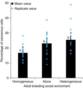
Cancer progression and social environment. To investigate the impact of the social environment on tumor progression, we exposed adult cancerous females for 21 days, post-induction, to various social environments in 40 ml food tubes. Individual virgin cancerous females were either kept in tubes alone (social isola-tion), in groups composed of seven other female cancerousflies (homogeneous groups) or in groups with seven non-cancerous control females (heterogeneous groups). Tumor progression was significantly affected by the social environment (Wald χ2
2= 6.7, P= 0.031): after 21 days we observed that tumor progression was markedly higher in cancerous flies kept in isolation than in cancerous flies kept in homogeneous groups (Fig.1). More sur-prisingly, we also observed that cancerous individuals kept within
a group of control flies showed an increased number of tumor cells compared to cancerousflies grouped together (Fig.1).
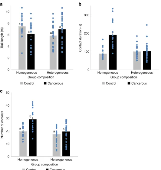
Social interactions. We then analyzed how social interactions were affected by tumor progression and group composition. Using a video tracking setup, we followed the locomotion and interactions of groups offlies (3 weeks post-induction) placed in an arena for 1 h. For social interaction measures we used homogeneous groups of eight control or eight cancerousflies, and a heterogeneous group consisting of seven control and one can-cerousfly which were kept together for 21 days post-induction. Social interaction analyses confirmed that control and cancerous flies had similar locomotor activity, independent of their social environment ((ANOVA) Fig. 2a; log (trail length): group com-position: F1,84= 2.64, P = 0.1; fly state: F1,84= 0.13, P = 0.7; fly state × group composition: F1,84= 3.8, P = 0.061). However, the length of the interaction that afly had with another was strongly affected by group composition and fly state ((ANOVA)contact duration: group composition: F1,84= 14.8, P < 10−3; fly state: F1,84= 26.8, P < 10−3;fly state × group composition: F1,84= 22.9, P < 10−3). In homogeneous groups, cancerous flies had longer interactions compared to homogeneous control groups (Fig.2b). However, when placed in a group of controlflies (heterogeneous group), cancerous individuals showed a strong decrease in contact duration (Fig.2b). Similarly, the average number of contacts per fly also differed depending on the social context and fly state ((ANOVA)number of contacts: group composition: F1,84= 17.5, P < 10−3; fly state: F1,84= 11.4, P = 0.001; fly state × group composition: F1,84= 4.4, P = 0.038). Groups of cancerous flies had a higher number of contacts than groups of control flies. Once again, cancerous individuals showed a decrease in the number of contacts when placed with controlflies (heterogeneous group) compared to when in a group with other cancerousflies (Fig.2c). Taken together this suggests that, individuals are more aggregated in a homogeneous group of cancerousflies than in a heterogeneous group or a homogeneous group of control flies. We thus concluded that, for a cancerousfly, the composition of the social group strongly affects the level of social interaction. However, our measure of social contact was constrained by the small size of the arena and therefore did not allow us to
disentangle the direction of the social contact i.e., whether specific fly states (cancerous or control) show avoidance or attraction towards other individuals within a group.
Cancer progression and social environment choice. Based on the results described above we tested whether cancerous and/or control flies would show variation in their choice of social environment and whether this was dependent on the level of their tumor progression. Using a similar protocol to Saltz35, we assessed social preference by putting two small mesh cages, each containing 8“stimulus flies” (cancerous or control) in a plastic, transparent box. The small mesh cages were placed on top of a small petri-dish containing standard food. We introduced a“focal fly” (cancerous or control) into the enclosed box and recorded their position over 7 h, i.e., whether the fly was found on one of the two mesh cages. Focal and stimulus flies were tested at dif-ferent ages post HS-induction.
Cancerous flies appeared, on average, more attracted than control flies to other cancerous individuals and we observed a general decrease of preference by cancerous and controlflies for the cancerous group with age of the focal fly (Fig. 3; focal fly: Wald χ2
1= 4.1, P = 0.04; age: Wald χ21= 17.6, P < 10−3; age × focal fly: Wald χ21= 2.7, P = 0.1). Furthermore, we find that at 21 days-post-HS, both control and cancerflies prefer to associate with control over cancerflies (Fig.3).
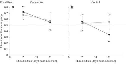
To understand whether the preferences seen in the dual choice test, were due to avoidance or attraction, young (7 days post HS) focalflies were given a choice between a stimulus group in a mesh cage (8 flies) and an empty mesh cage using a similar experimental design. Cancerous flies showed, on average, attraction for the social group, independent of the age or state of the stimulusflies (Fig. 4a; intercept: Waldχ2
1= 8.1, P < 10−3; stimulus: Waldχ21= 0.06, P = 0.79; stimulus age: Wald χ21= 1.4, P= 0.23; stimulus × stimulus age: Wald χ2
1= 0.6, P = 0.44). While controlflies showed, on average, no clear attraction for the social group, they clearly avoided 3-week-old cancerflies (Fig.4b; intercept: Waldχ21= 4.4, P = 0.036; stimulus: Wald χ21= 2.6, P = 0.1; stimulus age: Wald χ2
1= 3.37, P = 0.066; stimulus × stimulus age: Waldχ21= 6.61, P = 0.01).
Discussion
Here, we show that social environment can significantly shape the development of an intestinal-like cancer type in Drosophila. Consistent with previous studies on mammals10,36, cancerous flies kept in isolation exhibit faster tumor progression than flies kept in groups of other cancerous individuals. However, more importantly we found that variation in group composition also leads to increased proliferation of tumor cells, thus highlighting how subtle variations in social structure may have dramatic effects on the progression of non-transmissible diseases. Other tumor models have been reported in adult Drosophila37,38, which could be used in future projects to evaluate whether the social impact on tumor progression is a general or a tumor-specific effect.
Despite the opportunity to interact with others, individual cancerous flies, kept in groups with control flies, developed tumors at similar rates to when cancerous flies were bred in isolation. Social interaction analyses revealed that despite similar locomotor activities, cancerous flies interacted considerably less with controlflies compared to when they were housed with other cancerous conspecifics. This reduction in social contact may potentially be perceived as a form of social isolation by cancerous flies, which could result in increased tumor progression, analo-gous to when flies are kept in true isolation. In humans, it has been proposed that the subjective perceived feeling of social
50 40 Mean value Replicate value 30 P
ercentage of cancerous cells
20 a b b 10 0
Homogeneous Alone Heterogeneous Adult breeding social evironment
Fig. 1 Gut tumor progression as a function of social environment. FACS analysis of GFP-positive cells in guts dissected from 21 days post-HS cancerous females as a function of social environment. Blue dots indicate mean value for each replicate. Error bars: standard error of the mean.N = 15 measures for each treatment. Letters are Tukey’s post-hoc classification
isolation may impact psychological and physiological traits as much as real social isolation39. Furthermore, the social environ-ment choice experienviron-ment suggests that control flies may actively recognize and avoid cancerous individuals, especially when tumor progression is significant. Potentially, this may be a result of tumor-induced changes in cuticular hydrocarbons, pheromone profiles or even the gut microbiome. The most parsimonious explanation for this observation is infection avoidance40,41, a behavior that has also recently been observed in Drosophila42. Even if not contagious, cancerous flies may show particular behaviors, or produce chemical cues, which are generally asso-ciated with being sick.
Wefind no such avoidance behavior in cancerous flies towards other cancerous individuals, suggesting that the benefits of slower cancer progression outweigh the costs of joining potentially infectious groups. Moreover, it also suggests that thefitness costs of joining sick individuals are lower than those of joining a group of healthy individuals. Firstly, with respect to predation risk, it is potentially worse to be the only sick individual within a healthy group than in a group with other vulnerable individuals (dilution effect). Secondly, since natural selection favors reproduction and not survival per se, and the probability to reproduce is reduced for ill individuals within a healthy group (because of avoidance of
sick partners and reduced competitive ability), it is better for sick individuals to join other ill individuals because sexual partners will be less selective and/or the disadvantage of sexual competi-tion is reduced43,44.
These findings offer new perspectives on the reciprocal rela-tionship between disease and social behavior. While we observe that social structure has profound effects on disease progression, our study also suggests that disease might play a fundamental role in influencing group composition. We observed that cancerous flies, exhibit strong social attraction towards each other, especially at the beginning of tumor development, which decreases over time. At least two reasons could explain this change in preference. Firstly, at day 7 (when tumor progression is still minimal; Sup-plementary Figure 1a), it seems likely that flies are relatively unaffected by any pathological effects so that the decisions that maximize fitness related traits (i.e., slowing cancer progression and maximizing reproduction19can still be made, while at later stages of cancer progression (when tumor size is much more significant; Supplementary Figure 1c), the impact on normal functioning is likely to be high, and therefore the ability to make such decisions is lost. Secondly, it makes sense to be more selective during the initial stages of tumor progression, when the fitness benefits of reduced cancer progression are best maximized
10 a 300 200 100 0 8 a a a a 6 T rail length (m) Number of contacts Contact dur ation (s) 4 2 0 50 40 30 20 10 0 Homogeneous Heterogeneous Group composition Control Cancerous Homogeneous Heterogeneous Group composition Control Cancerous Homogeneous Heterogeneous Group composition Control Cancerous a a a b b c
Fig. 2 Social interactions for 21 day post-HS females in homogeneous (8 cancerousflies or 8 control flies) or heterogeneous groups (1 cancerous and 7 controlflies). a Total locomotion trail. Statistical analyses were carried out on log trail length, but for simpler graphical presentation, we present the untransformed data.b The mean contact duration and c mean number of contacts an individual has, averaged across replicates. Blue dots indicate mean value for each replicate. Error bars: standard error of the mean.N = 27 heterogeneous groups and N = 18 homogeneous group for each fly state. Letters are Tukey’s post-hoc classification
(e.g., better reproductive output). In later stages, the“damage is done” and cancer will be too advanced to maximally reap these benefits. These findings raise questions on the very early impact of internal oncogenic process on individual behavior and natural selection pressures on this process45. While, at this stage, tumors are not found to affect the survival of flies, cancer may affect fitness in other ways (e.g., reproductive competitiveness, vulner-ability to predators etc). A previous study showed that female Drosophila, bearing colorectal tumors, have earlier oviposition periods suggesting thatflies are adapted to minimize the costs of cancer on fitness19. Further studies would be necessary to determine the exact proximate factors responsible for the effect found here, as well as the extent to which generalizations can be made across other cancer types (or indeed other illnesses) and animal species.
Our findings highlight the importance of social structure on disease progression, beyond the context of transmission. This is
the first time that a direct link between social environment, specifically group composition, and cancer progression has been shown, while removing all other confounding psycho-sociological parameters that are frequently encountered in human studies. More generally, this study brings new light to how sociality impacts health andfitness in animals and its potential implication in human disease therapy. Moreover, we provide essential data for the emerging topic of evolutionary ecology of cancer, and demonstrate the importance of cancerous tumor progression in the intestine as afitness-limiting factor that potentially influences life history adaptations and strategies.
Methods
Drosophila stocks and genetics. yw, HS-flp;esg-gal4, UAS-GFP;FRT82B,Tub-Gal80 (line 1), yw,HS-flp;UAS-RasV12, FRT82B, Apc2N175K, ApcQ8(line 2) and yw,
HS-flp, FRT82B (line 3) flies32were balanced over co-segregating SM5-TM6B
balancers. In all experiments, cancerousflies were HS-flp;esg-gal4,UAS-GFP/UAS-RasV12;FRT82B,Tub-Gal80/FRT82B,Apc2N175K,ApcQ8(offspring 1 of line 1 crossed
to line 2), whereas controls were HS-flp;esg-gal4,UAS-GFP;FRT82B,Tub-Gal80/ FRT82B (offspring 2 of line 1 crossed to line 3). MARCM clones32were randomly
generated in heterozygousflies by flipase-induced exchange of pairing chromo-some arms, resulting in mosaic individuals where homozygous Apc2N175K, ApcQ8
mutant cells lacked the Gal80 repressor. This allowed Gal4 activity and the sub-sequent expression of GFP and RasV12for clones located in intestinal progenitor
cells. MARCM control clones are wild type for both Apc genes and do not express RasV12. MARCM clones were generated by heat shocking 3-day old femaleflies at
37 °C for 1 h32. Several attempts were made to use non-induced (no HS) offspring
flies as controls, however, a few flies developed tumors without HS making this an inadequate control for our study.
Flow cytometry. These intestinal tumors are polyclonal and tumoral cells are often intercalated with healthy cells (Supplementary Figure 3), making it hard to delineate the limits of a tumor. For this reason we chose to quantify tumor size by flow cytometry (FACS) instead of measuring the area, which would only provide a rough estimate. Flies were starved overnight, provided only with water prior to the quantification of GFP-positive cells used to estimate tumor progression. The entire midgut and the Malpighian tubules (hereafter referred to generally as gut dissec-tions), were sampled and dissected in PBS (phosphate buffer saline) as described46.
Both tissues exhibited tumor-like structures. Each replicate consisted of the guts and Malpighian tubules offive flies. Each fly was taken from a separate tube of the same social environment treatment (for example, one replicate of the homogeneous treatment consisted offive guts and Malpighian tubules of cancerous flies, with eachfly randomly taken from five different homogeneous tubes) and digested by collagenase (125μg in 60 μl PBS) (Sigma-Aldrich) for 2 h at 27 °C with strong agitation. Sixty microliter Trypsin 10× (Sigma-Aldrich) was then added by gentle pipetting and nuclei were stained with Hoechst 33342 (0.5μg/ml) for approxi-mately 1 h. Dissociated cells werefiltered through a cell strainer snap cap (70 μm size). To set the enzyme digestion protocol, we used Mex-gal4 > UAS-GFP (intestinal cells), esg-gal4 > UAS-GFP (stem cell and progenitors) and tumor-bearing guts, and checked the efficiency of the cell dissociation by direct 0.8 0.7 0.6 ns ns ** ** ** * 0.5 0.4 Pref erence f or cancerous g roup 0.3 0.2 0.1 0 0 7 14 21
Days post induction Cancerous Control
Fig. 3 Dual choice experiment: proportion of lands on the mesh cage containing stimulus cancerousflies as a function of age. N = 12–21 per treatment. Stars indicate deviation from random choice (binomial test per state and age): ns:P > 0.05; *P < 0.05, **P < 0.01; Error bars: standard error of the mean
0.8 a
Focal flies: Cancerous Control
** ** * * Attr action f or the social g roup 0.7 0.6 0.5 ns ns ns ns 0.4 0.3 0.2 0.1 0 0 7 14
Stimulus flies (days post-induction) Stimulus flies (days post-induction) Stimulus flies : Cancerous Control
21 0 7 14 21
b
Fig. 4 Attraction vs. aversion experiment. Proportion of lands on stimulus cage (vs. empty cage) by a focal cancerousflies and b focal control flies as a function of age of the stimulusflies. N = 16 per treatment. Stars indicate deviation from random choice (binomial test per state and age) ns: P > 0.05; *P < 0.05, **P < 0.01
observation with a GFP dissecting microscope every 30 min. The collagenase treatment was very efficient to digest the extracellular matrix, though the post-trypsin treatment was required to end up with single cells only. For each sample, 50,000 R2 cells were processed on a Partec PAS III and analyzed using the FlowMax software (Supplementary Figure 2, 3).
Social environment. Flies were sexed at emergence and control or cancerous females were kept in groups until the third day post emergence. Control and cancerous virgin females were heat shocked at 37 °C for 1 h. Flies were then put into their social groups and introduced into new 40 ml food tubes. Social groups created for experiments measuring tumor size consisted of either a cancerousfly in isolation, a group of eight all cancerous individuals (homogeneous groups) or a group of one cancerous individual in a group of seven non-cancerous individuals (heterogeneous group). Flies were partially wing-clipped on the right or left wing to distinguish their genotype. Previous behavioral studies have shown that wing clipping has no effect on social interactions47. Flies were then kept at 25 °C on
standard food (changed every 3 days) until 21 days post-HS (induction). Flies were housed in small tubes (40 ml) to promote social interactions and limit the possi-bility of complete social isolation for any givenfly. Tumor size was estimated with flow cytometry. Data (the number of tumor cells relative to the total number of cells counted) were analyzed using a generalized linear model (binomial distribu-tion, Pearson correction for over-dispersion) and Tukey’s post-hoc tests. Social interactions. Again, femalesflies were put into groups according to the protocol described above, except this time social groups consisted of a group of eight all controlflies (homogeneous), a group of eight all cancerous flies (homo-geneous) and a single cancerousfly with seven control flies (heterogeneous). A group offlies to be tracked was composed of eight flies taken from different food tubes to ensure that they had never previously interacted. They were introduced into a semi-opaque white polyoxymethylene (Delrin) arena (diameter 100 mm; height 5 mm), covered with a transparent Plexiglas for 1 h. We simultaneously tracked four groups of eightflies over the 1 h. The tracking apparatus consisted of four synchronizedfirewire cameras (Guppy pro, Allied vision technologies), each filming one interaction arena that was backlit by a 150 × 150 mm IR backlight (R&D vision). We used Vision software to analyze spatial data (open-source C-trax 0.3.748) that allowed us to collect ten positions per second for eachfly49,50in the
group over 1 h video experiments. Tracking corrections were made post C-trax analysis withfixerrors Matlab toolbox (Ctrax-allmatlab version 0.2.11) using Matlab software 7.11.0 to ensure that the identity of eachfly was maintained when individuals were close to one another. For eachfly, we calculated the total length of the path, the distance to otherflies, the number of contacts with other flies (a contact was considered when the distance between the centers of two individuals was smaller than, or equal to, one mean body length of the individuals for 1 s or more) and the duration of each contact. Interactions were considered between all individuals within a group. We averaged each measure for allflies of the same cancer state within a group to obtain a single value for one replicate. For each measure we performed a general linear model and included the measures of group composition (homogeneous vs. heterogeneous),fly state (cancerous or control) and the interaction group composition ×fly state as fixed explanatory variables. Tukey post-hoc contrasts among treatments were tested.
Social environment choice. Flies were sexed at emergence and control or can-cerous females were kept in groups until the third day post emergence. Control and cancerous virgin females were heat shocked at 37 °C during 1 h and kept in groups until the day of the experiment. The experimental setup consisted of a 17 × 12 × 5 cm plastic box in which 2 small 2 × 2 × 2 cm mesh cages were introduced and each placed on a 3 cm diameter petri dish containing standard food. The two cages were positioned at opposite ends of the box. Groups of eightflies (hereafter referred to as stimulusflies) were placed in the mesh cages. In the dual choice experiment, one mesh cage contained controlflies while the other contained cancerous flies. In the attraction vs. avoidance experiment only one of the two cages contained stimulus flies. A focal fly (control or cancerous), taken from a separate tube from the stimulusflies, was introduced in the box 15 h before starting the experiment. The position of thefly was then visually recorded every 30 min between 10 am and 5 pm. A choice was only recorded when thefly was positioned on a mesh cage or the associated petri dish. For the dual choice experiment, focal and stimulusflies were of the same age (7, 14, or 21 days post-induction), whereas for the attraction vs. aversion experiment the focalfly was always 7 days old post-induction and the stimulusflies were either 7 or 21 days post-induction. The number of times a focal fly was observed on a cancerous stimulus cage (for the dual choice experiment) or the stimulus cage (for the attraction vs. aversion experiment) compared to the total number of cage landings were then analyzed with a general linear model and a binary logistic regression. For the dual choice experiment, wefirst compared the behavior of cancerous vs. controlflies: state of the focal fly was included as a fixed factor andfly age was included as a covariate. For the attraction vs. aversion experiment, we separately analyzed the behavior of each focalfly (i.e., cancerous or control) as a function of stimulusfly state and age. Finally, a binomial test, for each independent measure, was performed to test for a significant deviation from ran-dom choice.
Data availability
The data and computer code uses to support thefindings of this study are available from the corresponding author upon request.
Received: 25 October 2017 Accepted: 19 July 2018
References
1. Beauchamp, G. Function and structure of vigilance in a gregarious species exposed to threats from predators and conspecifics. Anim. Behav. 116, 195–201 (2016).
2. Dalerum, F. et al. Foraging competition, vigilance and group size in two species of gregarious antelope. S. Afr. J. Wildl. Res. 38, 138–145 (2008). 3. Nadler, L. E., Killen, S. S., McClure, E. C., Munday, P. L. & McCormick, M. I.
Shoaling reduces metabolic rate in a gregarious coral reeffish species. J. Exp. Biol. 219, 2802–2805 (2016).
4. McFarland, R. et al. Social integration confers thermal benefits in a gregarious primate. J. Anim. Ecol. 84, 871–878 (2015).
5. Apfelbeck, B. & Raess, M. Behavioural and hormonal effects of social isolation and neophobia in a gregarious bird species, the European starling (Sturnus vulgaris). Horm. Behav. 54, 435–441 (2008).
6. Behringer, D. C., Butler, M. J. & Shields, J. D. Ecology: avoidance of disease by social lobsters. Nature 441, 421 (2006).
7. Nonogaki, K., Nozue, K. & Oka, Y. Social isolation affects the development of obesity and type 2 diabetes in mice. Endocrinology 148, 4658–4666 (2007). 8. Venna, V., Xu, Y., Doran, S., Patrizz, A. & McCullough, L. Social interaction
plays a critical role in neurogenesis and recovery after stroke. Transl. Psychiatry 4, e351 (2014).
9. Lin, E.-J. D. et al. Social overcrowding as a chronic stress model that increases adiposity in mice. Psychoneuroendocrinology 51, 318–330 (2015).
10. Hermes, G. L. et al. Social isolation dysregulates endocrine and behavioral stress while increasing malignant burden of spontaneous mammary tumors. Proc. Natl Acad. Sci. USA 106, 22393–22398 (2009).
11. McClintock, M. K., Conzen, S. D., Gehlert, S., Masi, C. & Olopade, F. Mammary cancer and social interactions: identifying multiple environments that regulate gene expression throughout the life span. J. Gerontol. B 60, 32–41 (2005).
12. Chida, Y., Hamer, M., Wardle, J. & Steptoe, A. Do stress-related psychosocial factors contribute to cancer incidence and survival? Nat. Clin. Pract. Oncol. 5, 466–475 (2008).
13. Lin, Y. et al. Striking life events associated with primary breast cancer susceptibility in women: a meta-analysis study. J. Exp. Clin. Cancer Res. 32, 32–53 (2013).
14. Madsen, T. et al. Cancer prevalence and etiology in wild and captive animals. in Ecology and Evolution of Cancer (eds Ujvari, B., Roche, B. & Thomas, F.) 11–46 (Academic Press, London, UK, 2017).
15. Ujvari, B., Roche, B. & Thomas, F. Ecology and Evolution of Cancer. (Academic Press, London, UK, 2017).
16. Peck, S., Corkrey, R., Hamede, R., Jones, M. & Canfield, P. Hematologic and serum biochemical changes associated with devil facial tumor disease in Tasmanian devils. Vet. Clin. Pathol. 45, 417–429 (2016).
17. Vittecoq, M. et al. Animal behaviour and cancer. Anim. Behav. 101, 19–26 (2015).
18. Ujvari, B., Gatenby, R. A. & Thomas, F. The evolutionary ecology of transmissible cancers. Infect. Genet. Evol. 39, 293–303 (2016). 19. Arnal, A. et al. Cancer brings forward oviposition in thefly Drosophila
melanogaster. Ecol. Evol. 7, 272–276 (2017).
20. Sarin, S. & Dukas, R. Social learning about egg-laying substrates in fruitflies. Proc. R. Soc. B 276, 4323–4328 (2009).
21. Battesti, M., Moreno, C., Joly, D. & Mery, F. Spread of social information and dynamics of social transmission within Drosophila groups. Curr. Biol. 22, 309–313 (2012).
22. Mery, F. et al. Public versus personal information for mate copying in an invertebrate. Curr. Biol. 19, 730–734 (2009).
23. Ruan, H. & Wu, C. F. Social interaction-mediated lifespan extension of Drosophila Cu/Zn superoxide dismutase mutants. Proc. Natl Acad. Sci. USA 105, 7506–7510 (2008).
24. Hoffmann, A. A. A laboratory study of male territoriality in the sibling species Drosophila melanogaster and D. simulans. Anim. Behav. 35, 807–818 (1987).
25. Hoffmann, A. A. The influence of age and experience with conspecifics on territorial behavior in Drosophila melanogaster. J. Insect Behav. 3, 1–12 (1990).
26. Zhou, C., Rao, Y. & Rao, Y. A subset of octopaminergic neurons are important for Drosophila aggression. Nat. Neurosci. 11, 1059–1067 (2008).
27. Ganguly-Fitzgerald, I., Donlea, J. & Shaw, P. J. Waking experience affects sleep need in Drosophila. Science 313, 1775–1781 (2006).
28. Donlea, J. M., Ramanan, N. & Shaw, P. J. Use-dependent plasticity in clock neurons regulates sleep need in Drosophila. Science 324, 105–108 (2009). 29. Technau, G. M. Fiber number in the mushroom bodies of adult Drosophila
melanogaster depends on age, sex and experience. J. Neurogenet. 1, 113–126 (1984).
30. Salomon, R. N. & Jackson, F. R. Tumors of the testis and midgut in agingflies. Fly 2, 265–268 (2008).
31. Gonzalez, C. Drosophila melanogaster: a model and a tool to investigate malignancy and identify new therapeutics. Nat. Rev. Cancer 13, 172–183 (2013).
32. Martorell, Ò. et al. Conserved mechanisms of tumorigenesis in the Drosophila adult midgut. PLoS ONE 9, e88413 (2014).
33. Smith, G. et al. Mutations in APC, Kirsten-ras, and p53—alternative genetic pathways to colorectal cancer. Proc. Natl Acad. Sci. USA 99, 9433–9438 (2002).
34. Lee, T. & Luo, L. Mosaic analysis with a repressible cell marker (MARCM) for Drosophila neural development. Trends Neurosci. 24, 251–254 (2001). 35. Saltz, J. B. Natural genetic variation in social environment choice: context‐
dependent gene–environment correlation in Drosophila melanogaster. Evolution 65, 2325–2334 (2011).
36. Moreno-Smith, M., Lutgendorf, S. K. & Sood, A. K. Impact of stress on cancer metastasis. Future Oncol. 6, 1863–1881 (2010).
37. Caussinus, E. & Gonzalez, C. Induction of tumor growth by altered stem-cell asymmetric division in Drosophila melanogaster. Nat. Genet. 37, 1125–1129 (2005).
38. Patel, P. H., Dutta, D. & Edgar, B. A. Niche appropriation by Drosophila intestinal stem cell tumours. Nat. Cell Biol. 17, 1182–1192 (2015). 39. Cacioppo, J. T. & Cacioppo, S. Social relationships and health: the toxic effects
of perceived social isolation. Soc. Personal. Psychol. Compass 8, 58–72 (2014). 40. Kiesecker, J. M., Skelly, D. K., Beard, K. H. & Preisser, E. Behavioral reduction
of infection risk. Proc. Natl Acad. Sci. USA 96, 9165–9168 (1999). 41. Loehle, C. Social barriers to pathogen transmission in wild animal
populations. Ecology 76, 326–335 (1995).
42. Vale, P. F. & Jardine, M. D. Infection avoidance behavior: viral exposure reduces the motivation to forage in female Drosophila melanogaster. Fly 11, 3–9 (2017).
43. Campbell, L., Head, M., Wilfert, L. & Griffiths, A. An ecological role for assortative mating under infection? Conserv. Genet. 18, 983–994 (2017). 44. Thomas, F. et al. Assortative pairing in Gammarus insensibilis (Amphipoda)
infected by a trematode parasite. Oecologia 282, 259–264 (2015). 45. Tissot, T. et al. Host manipulation by cancer cells: expectations, facts, and
therapeutic implications. Bioessays 38, 276–285 (2016).
46. Tauc, H. M., Tasdogan, A. & Pandur, P. Isolating intestinal stem cells from adult Drosophila midguts by FACS to study stem cell behavior during aging. J. Vis. Exp. 94, 52223 (2014).
47. Battesti, M. et al. Ecology of information: social transmission dynamics within groups of non-social insects. Proc. R. Soc. B 282, 20142480 (2015). 48. Branson, K., Robie, A. A., Bender, J., Perona, P. & Dickinson, M. H.
High-throughput ethomics in large groups of Drosophila. Nat. Methods 6, 451–457 (2009).
49. Gargano, J. W., Martin, I., Bhandari, P. & Grotewiel, M. S. Rapid iterative negative geotaxis: a new method for assessing age-related locomotor decline in Drosophila. Exp. Gerontol. 40, 386–395 (2005).
50. Tinkerhess, M. J., Ginzberg, S., Piazza, N. & Wessells, R. J. Endurance training protocol and longitudinal performance assays for Drosophila melanogaster. J. Vis. Exp. e3786 (2012).
Acknowledgements
This work was supported by the ANR (Blanc project EVOCAN to F.T. and project DROSONET to F.M. and C.S.), the CNRS (INEE and INSB), Fondation ARC (1555286 to J.M. and F.M.), The French league against Cancer (M27218 to J.M.), IDEEV program (to F.M.), by an International Associated Laboratory Project France/Australia, by the French-Australian Science Innovation Collaboration Program Early Career Fellowship (B.U.), by André Hoffmann (Fondation MAVA), Fyssen Foundation (to F.M. and E.H. D.) and the French Government (fellowship 2015–155 to M.D.). We thank F. Bastin, J.C. Sandoz and A. Couto for their help with confocal imaging.
Author contributions
E.H.D., T.B., C.M., F.T., J.M., and F.M. designed the experiments. E.H.D., T.B., J.D.S., C.M., M.S., B.M., and J.M. performed the experiments. C.S., J.M., and F.M. analyzed the data. E.H.D., J.M., F.T., A.C., C.S., B.U., and F.M. wrote the manuscript.
Additional information
Supplementary Informationaccompanies this paper at https://doi.org/10.1038/s41467-018-05737-w.
Competing interests:The authors declare no competing interests.
Reprints and permissioninformation is available online athttp://npg.nature.com/ reprintsandpermissions/
Publisher's note:Springer Nature remains neutral with regard to jurisdictional claims in published maps and institutional affiliations.
Open Access This article is licensed under a Creative Commons Attribution 4.0 International License, which permits use, sharing, adaptation, distribution and reproduction in any medium or format, as long as you give appropriate credit to the original author(s) and the source, provide a link to the Creative Commons license, and indicate if changes were made. The images or other third party material in this article are included in the article’s Creative Commons license, unless indicated otherwise in a credit line to the material. If material is not included in the article’s Creative Commons license and your intended use is not permitted by statutory regulation or exceeds the permitted use, you will need to obtain permission directly from the copyright holder. To view a copy of this license, visithttp://creativecommons.org/ licenses/by/4.0/.