HAL Id: hal-02635353
https://hal.inrae.fr/hal-02635353
Submitted on 27 May 2020
HAL is a multi-disciplinary open access
archive for the deposit and dissemination of
sci-entific research documents, whether they are
pub-lished or not. The documents may come from
teaching and research institutions in France or
abroad, or from public or private research centers.
L’archive ouverte pluridisciplinaire HAL, est
destinée au dépôt et à la diffusion de documents
scientifiques de niveau recherche, publiés ou non,
émanant des établissements d’enseignement et de
recherche français ou étrangers, des laboratoires
publics ou privés.
Morphometric analysis and taxonomic revision of
Anisopteromalus Ruschka (Hymenoptera: Chalcidoidea:
Pteromalidae) - an integrative approach
Hannes Baur, Yvonne Kranz-Baltensperger, Astrid Cruaud, Jean Yves
Rasplus, Alexander V. Timokhov, Vladimir E. Gokhman
To cite this version:
Hannes Baur, Yvonne Kranz-Baltensperger, Astrid Cruaud, Jean Yves Rasplus, Alexander V.
Tim-okhov, et al.. Morphometric analysis and taxonomic revision of Anisopteromalus Ruschka
(Hy-menoptera: Chalcidoidea: Pteromalidae) - an integrative approach. Systematic Entomology,
Wiley-Blackwell, 2014, 39 (4), pp.691-709. �10.1111/syen.12081�. �hal-02635353�
Morphometric analysis and taxonomic revision
of Anisopteromalus Ruschka (Hymenoptera:
Chalcidoidea: Pteromalidae) – an integrative approach
H A N N E S B A U R
1, Y V O N N E K R A N Z - B A L T E N S P E R G E R
1,
A S T R I D C R U A U D
2, J E A N - Y V E S R A S P L U S
2,
A L E X A N D E R V . T I M O K H O V
3and V L A D I M I R E . G O K H M A N
41Abteilung Wirbellose Tiere, Naturhistorisches Museum der Burgergemeinde Bern, Bern, Switzerland,2INRA, UMR1062 CBGP, Montferrier-sur-Lez, France,3Department of Entomology, Moscow State University, Moscow, Russia and4Botanical Garden, Moscow State University, Moscow, Russia
Abstract. We use an integrative taxonomic approach to revise the genus
Anisopteromalus. In particular, we apply multivariate ratio analysis (MRA), a rather new statistical method based on principal component analysis (PCA) and linear dis-criminant analysis (LDA), to numerous body measurements and combine the data with those from our molecular analysis of Cytb and ITS2 genetic markers (on a subset of species) and all available published data on morphology, karyology, behaviour, host associations and geographic distribution. We demonstrate that the analysis of quan-titative characters using MRA plays a major role for the integration of name-bearing types and thus for the association of taxa with names. Six species are recognized, of which two are new: A. cornis Baur sp.n. and A. quinarius Gokhman & Baur sp.n. For Anisopteromalus calandrae (Howard), a well-known, cosmopolitan parasitoid of stored-product pests, we have selected a neotype to foster continuity and stability in the application of this important name. The species was sometimes confused with the related A. quinarius sp.n., another cosmopolitan species that is frequently encountered in similar environments. We also show that several species originally described or later put under Anisopteromalus actually belong to different genera: Cyrtoptyx camerunus (Risbec) comb.n.; Meraporus glaber (Szelényi) comb.n.; Dinarmus schwenkei (Roomi, Khan & Khan) comb.n. Neocatolaccus indicus Ayyar & Mani is confirmed as a junior synonym of Oxysychus sphenopterae (Ferrière) syn.n. and Anisopteromalus calandrae brasiliensis (Domenichini) stat.rev. must be considered as a valid but doubtful taxon. This published work has been registered in ZooBank, http://zoobank.org/urn:lsid: zoobank.org:pub:BDFE96D3-D0F4-4012-90F5-9A087F7F5864.
Introduction
In systematics, data from multiple sources are becoming more and more easily available to taxonomists working at the species level. This has led to interest in a method for combining these data, which recently has been termed integrative taxonomy (for a review and the distinction from iterative taxonomy,
Correspondence: Hannes Baur, Abteilung Wirbellose Tiere, Naturhis-torisches Museum der Burgergemeinde Bern, Bern, Switzerland. E-mail: hannes.baur@nmbe.ch
see Yeates et al., 2011). The term is adopted when species delimitation is based on results from a variety of disciplines, for instance morphology, DNA analysis, cytogenetics, behaviour or biochemistry. Detailed procedures have been devised for integrating conflicting datasets into sound hypotheses (e.g. Schlick-Steiner et al., 2010).
In most studies adopting an integrative approach, attention is usually paid just to species limits per se, based on the evaluation of the sample at hand (e.g. Chesters et al., 2012). Only rarely is the main task of a taxonomic revision also considered (e.g. Steiner et al., 2010), that is, the association
© 2014 The Authors. Systematic Entomology published by John Wiley & Sons Ltd on behalf of The Royal Entomological Society. 691 This is an open access article under the terms of the Creative Commons Attribution License, which permits use, distribution and reproduction
of taxa with names. This leads to cases where information is often very limited and data integration becomes a real challenge: for example, when name-bearing types are damaged and character sets are thus incomplete; when types are too fragile or old for molecular analyses; and when types are lost (or inaccessible) and nominal taxa are based on nothing more than a set of qualitative characters. In combination such challenges cause major, and widespread, problems in evaluating available evidence to support necessary taxonomic revisions.
Although focusing on morphometry, we are here pursuing such an integrative approach for establishing species boundaries and identities within Anisopteromalus Ruschka (Hymenoptera: Chalcidoidea: Pteromalidae: Pteromalinae), a small genus of parasitic wasps currently comprising seven species (Noyes, 2013). The genus was erected for Anisopteromalus mollis Ruschka which is the type species by monotypy, a species syn-onymized with A. calandrae (Howard) by Graham (1969). The genus is easily recognized by a combination of characters, most notably the female antenna with three anelli, the structure of the propodeum and the extended hind margin of the first gastral tergite (Graham, 1969; Bouˇcek & Rasplus, 1991). Species of
Anisopteromalus occur mainly in the Old World, where they
were recorded from tropical Africa (Risbec, 1956; Rasplus, 1988), Asia (Roomi et al., 1973; Sureshan, 2010) and Western Europe (Szelényi, 1981). They usually parasitize beetle larvae (e.g. Chrysomelidae: Bruchinae, Anobiidae, Curculionoidea) feeding on stored grain and legume seeds (Fabaceae: Faboideae and Caesalpinioideae), but have sometimes been reared also from lepidopteran hosts (e.g. Gelechiidae, Pyralidae) (Noyes, 2013).
While for most Anisopteromalus species hardly anything has been published beside the original description or an occasional host record, A. calandrae is a well-known, cosmopolitan para-sitoid of various stored-product pests. It has been the subject of numerous studies spanning a wide variety of topics, such as bio-logical control (Hou et al., 2004; Ngamo et al., 2007; Ni et al., 2008; Chaisaeng et al., 2010), impact of pesticides and herbi-cides (Perez-Mendoza et al., 1999; Lacoume et al., 2009; Yoon
et al., 2009), life-history traits (Bressac et al., 2009; Lebreton et al., 2009a, 2010; Chaisaeng et al., 2010), behaviour
includ-ing learninclud-ing (Ryoo et al., 1996; Lebreton et al., 2009b; Belda & Riudavets, 2010; Ishii & Shimada, 2010) and physiology (Zhu
et al., 1999; Howard & Baker, 2003). The ISI Web of Science
database (Thomson Reuters) cites more than 100 papers in this respect and many of the older works can be retrieved from the Universal Chalcidoidea Database (Noyes, 2013).
Despite the multitude of studies, little critical attention has been paid to the systematics of Anisopteromalus. As mentioned before, most species have not been re-considered since their description. Furthermore, the taxonomic status of A. calandrae is doubtful. Gokhman et al. (1998) first suspected that two sib-ling species might be hidden under that name. In fact, inves-tigation of the karyotypes of two laboratory strains revealed a marked difference in the number and shape of chromosomes. One strain originating from Moscow, Russia (called the MSU strain) showed five chromosomes in its haploid set (n = 5). All of those chromosomes were metacentric except for the last
submetacentric one. Moreover, they could be subdivided into two size classes. In the other strain, cultured at Imperial Col-lege at Silwood Park, Ascot, Berkshire, UK (origin Slough, Berkshire; called the ICSP strain) with n = 7, all chromosomes were metacentric and demonstrated a continuous gradation in length. Such differences in the karyotype are usually a clear indication of different species (Gokhman, 2009). Gokhman
et al. (1998, 1999) also found significant differences in
mor-phology, courtship behaviour and several important life-history traits. Because the crossing between MSU and ICSP strains also failed, there could be little doubt that more than one taxon was involved. Later on, similar differences between some other pop-ulations presumably belonging to A. calandrae were also found (Gokhman & Timokhov, 2002) and their different host prefer-ences were revealed also (Timokhov & Gokhman, 2003). How-ever, the presence of the two closely related and widely dis-tributed species with contrasting life-history strategies in the genus Anisopteromalus got relatively little support from the expert community for a number of years (but see Quicke, 2002), although it is becoming increasingly accepted now (see, e.g., Sasakawa et al., 2012, 2013). Of course, taxonomic ambiguity within such an important taxon needs to be solved.
As mentioned above, we focused on morphometric data and their integration with molecular data as well as all relevant pub-lished information on morphology, karyology, behaviour, distri-bution and ecology. We also considered all valid names and their junior synonyms (in total 16 nominal taxa) and checked their name-bearing types whenever possible. Beside the type mate-rial, more than one thousand specimens from various collections and from several cultured strains of supposed ‘A. calandrae’ were studied. For the morphometric study we applied multi-variate ratio analysis (MRA), a recently developed method that allows the interpretation of results from principal component analysis (PCA) and linear discriminant analysis (LDA) in terms of body ratios (Baur & Leuenberger, 2011) and that is thus espe-cially suited for analysing body measurements in a taxonomic context (reviewed in László et al., 2013). Furthermore, the MRA algorithms offer separate analyses of shape and size as well as an estimation of the extent of shape change with size (i.e. allo-metry in the sense of Gould, 1966). Allometric variation of body ratios was first observed for Pteromalidae by Janzon (1986) who discussed its impact on species delimitation in the
Pteroma-lus albipennis group. MRA also allowed the incPteroma-lusion of the
available name-bearing types, which were not usable for DNA sequencing because the age of specimens (usually more than 20, up to 100 years old) is likely to hamper successful extraction and amplification. We also used the mitochondrial cytochrome b (Cytb) and the nuclear internal transcribed spacer 2 (ITS2) for differentiating entities within the ‘A. calandrae’ complex.
Material and methods
Specimens and character selection
For the morphological investigation we used a total of more than 1300 specimens. Morphometric analysis was based on a
subset of 289 dry-mounted females. We focused on females because they are usually easier to separate and more readily available (e.g. Graham, 1969; Bouˇcek & Rasplus, 1991). Males were nevertheless studied when necessary, for instance to solve the identity of a nominal taxon that was based on the male sex. The examined material originated from: The Natural History Museum, London, UK (BMNH); Centre de Biologie pour la Gestion des Populations, Montferrier-sur-Lez, France (CBGP); Swiss Federal Institute of Technology, Entomology Collection, Zurich, Switzerland (ETHZ); Muséum d’histoire naturelle, Geneva, Switzerland (MHNG); Muséum national d’Histoire naturelle, Paris, France (MNHN); Natural History Museum, Vienna, Austria (NHMV); Naturhistorisches Museum der Burgergemeinde Bern, Berne, Switzerland (NMBE); Queensland Museum, Brisbane, Australia (QMB); Bohart Museum of Entomology, University of California, Davis, California, USA (UCD); United States National Museum, Washington DC, USA (USNM); Zoological Institute of the Russian Academy of Sciences, St. Petersburg, Russia (ZIN);
Zoological Museum of Moscow State University, Moscow, Russia (ZMMU).
Gibson (1997) is followed for terminology of morphologi-cal structures. The list of morphometric characters used in the analyses is given in Table 1. Table S1 gives an overview of the basic descriptive statistics for each measurement (in μm) and species as well as the sample sizes. The selected charac-ters correspond to those used in the taxonomy of Pteromalidae for calculating standard ratios (e.g. Graham, 1969). Most mea-surements were made with an Olympus SZ11 stereomicroscope at different magnifications using a calibrated eye-piece micro-meter (12 mm subdivided into 120 units) and were taken by Y. Kranz-Baltensperger. For some of the name-bearing types, each character was photographed with a Keyence VHX 2000 digital photo-microscope and a VH-Z20R/W zoom lens at a magnifi-cation of 200× (i.e. 1000 μm corresponded to 888 pixels) and was measured by H. Baur using ImageJ v1.47v (Schneider et al., 2012); body parts on the images were enlarged 3–4 times before measuring. For all measurements, we ensured that the points of
Table 1. Abbreviation, name, definition and magnification of the 20 measurements used for the morphometric analyses of Anisopteromalus females.
Abbreviation Character name Definition Magnification
eye.b Eye breadth Greatest breadth of eye, lateral view 150× eye.d Eye distance Shortest distance between eyes, dorsal view 150× eye.h Eye height Greatest length of eye height, lateral view 150× gst.b Gaster breadth Greatest breadth of gaster, distance between the outermost lateral edges
of the gaster, dorsal view
100×
gst.l Gaster length Length of gaster along median line from posterior edge of nucha to tip of ovipositor sheath, dorsal view
70×
hea.b Head breadth Greatest breadth of head, dorsal view 100× hea.h Head height Distance between lower edge of clypeus and lower edge of anterior
ocellus, frontal view
100×
msc.b Mesoscutum breadth Greatest breadth of mesoscutum just in front of level of tegula, dorsal view
100×
msc.l Mesoscutum length Length of mesoscutum along median line from posterior edge of pronotum to posterior edge of mesoscutum, dorsal view
150×
msp.l Malar space Distance between the point where malar sulcus enters mouth margin and malar sulcus enters lower edge of eye, lateral view (Graham, 1969)
150×
mss.l Mesosoma length Length of mesosoma along median line from anterior edge of pronotum collar to posterior edge of nucha, dorsal view
70×
mv.l Marginal vein Length of marginal vein, distance between the point at which the submarginal vein touches the leading edge of the wing and the point at which stigmal vein and postmarginal vein unite (Graham, 1969)
150×
ool.l OOL Shortest distance between posterior ocellus and eye margin, dorsal view (Graham, 1969)
150×
pdl.flg Pedicel + flagellum Combined length of pedicel plus flagellum, outer aspect (Graham, 1969)
100×
pol.l POL Shortest distance between posterior ocelli, dorsal view (Graham, 1969) 150× ppd.l Propodeum length Length of propodeum measured along median line from anterior edge
to posterior edge of nucha, dorsal view
150×
scp.l Scape length Length of scape exclusive of radicle, outer aspect (Graham, 1969) 150× sct.l Scutellum length Length of scutellum along median line from posterior edge of
mesoscutum to posterior edge of scutellum, dorsal view
150×
stv.l Stigmal vein Length of stigmal vein, distance between the point at which stigmal vein and postmarginal vein unite apically, and the distal end of the stigma (Graham, 1969)
150×
reference were equidistant from the lens of the microscope and that the diaphragm of the lens was fully open. To avoid addi-tional variation due to fluctuating asymmetry (e.g. Palmer & Strobeck, 1986; Bechshøft et al., 2008), measurements of paired characters were usually taken on the left-hand side. The Keyence microscope with a VH-Z100UR/W zoom lens was used for mak-ing stack-images of body parts, except for the forewmak-ings, which were removed and embedded in Hoyer’s medium on slides prior to stack-imaging using a Leica DFC420 camera on a Zeiss Axioskop 40 light microscope and the ImageAccess software (Imagic AG, Glattbrugg, Switzerland).
Morphometric analysis
We applied multivariate ratio analysis (MRA) of Baur & Leuenberger (2011) to our data. MRA comprises a set of tools for analysing size and shape of body measurements in a multi-variate mathematical framework that is entirely consistent with the customary usage of body lengths and ratios in taxonomic works (e.g. descriptions, diagnoses). In systematic studies, MRA offers several advantages over conventional explorative multivariate methods, such as principal component analysis (PCA) and linear discriminant analysis (LDA). We refer to László et al. (2013) who gave an overview of the various MRA tools and applied them to a particular taxonomic problem in some other Hymenoptera. Following Baur & Leuenberger (2011) we first calculated an isometric size axis (isosize), defined as the geometric mean of all variables. We then per-formed a shape PCA (i.e. a PCA in the space of all ratios) for evaluating how well the morphometric pattern corresponds to the groups obtained by qualitative morphology and karyology. In order to decide how many shape components to retain we inspected the scree graph (Rencher, 2002: 398–399). We also plotted isosize against shape PCs, because the correlation of size with shape is a measure of the amount of allometry in the data (e.g. Klingenberg, 1998). Estimation of the extent of allometric variation is important, because body ratios sometimes correlate with size. Depending on the magnitude of this correlation, the use of ratios may then hold the risk that discrimination of groups is – indirectly – more or less based on size rather than shape (e.g. Janzon, 1986; Seifert, 2008). For this reason, we also employed two graphical tools, the PCA ratio spectrum and allometry ratio spectrum, respectively. Finally, we used the LDA ratio extractor to extract the best ratios for use in the identification key and diagnoses, and calculated the standard distance as well as the measure𝛿.
The R language and environment for statistical computing was used for data analysis (R Core Team, 2013; v3.0.2). In partic-ular, we employed slightly modified versions of the R-scripts provided by Baur & Leuenberger (2011, under ‘Supplemen-tary material’). Pearson product-moment correlation coefficients were calculated with the function ‘cor()’ using the default set-tings. Scatterplots were generated with the package ‘ggplot2’ (Wickham, 2009). A few specimens, especially some of the name-bearing types, lacked one body part or another. In order
to be able to include all specimens in the multivariate analy-ses, missing values were imputed with the R package ‘mice’, using the default settings of the function ‘mice()’. For the cal-culation of ratios used in the description and Table 2, specimens with missing values were excluded, because imputed values may sometimes produce outliers when calculating ratios.
Molecular analysis
Taxonomic sampling
We sampled six specimens of Anisopteromalus calandrae and three specimens of A. quinarius belonging to the main strains used in laboratories (Table S2). Specimens were collected alive and stored in 95% ethanol. Adult specimens were identified to species by V.E. Gokhman, A.V. Timokhov and J.-Y. Ras-plus. DNA vouchers are deposited at the CBGP collection, Montferrier-sur-Lez, France. Four outgroup taxa belonging to the pteromalid genera Nasonia (three species) and
Pachycre-poideus (one species) were included to root phylogenetic trees.
Cytochrome b (Cytb) and ITS2 sequences from Nasonia species were downloaded from GenBank (Table S2).
DNA extraction, amplification and sequencing
Total DNA was extracted using standard phenol-chloroform methods (Sambrook et al., 1989). Due to repeated failures of amplification of the Cytochrome c oxidase I Folmer fragment (COI, standard barcode) for most specimens, we used a long fragment of the Cytochrome b gene (Cytb) instead. To inves-tigate potential mitochondrial introgression we also sequenced one nuclear gene, the internal transcribed spacer ITS2 rRNA.
Genes were amplified as follows:
1 Cytb: we used primers CP1 (forward) (5′-GAT GAT GAA ATT GGA TC-3′: Harry et al., 1998), CB1 (forward) (5′-TAT GTA CTA CCA TGA GGA CAA ATA TC-3′: Jermiin & Crozier, 1994) and Tser (reverse) (5′-TAT TTC TTT ATT ATG TTT TCA AAA C-3′: Simon et al., 1994). Using the Promega Taq package, 30 cycles of amplification were performed as follows in 25-μL reaction volumes: denaturation step at 92∘C for 1 min, annealing at 48∘C for 1 min and 30 s, and extension at 72∘C for 1 min.
2 ITS2: we used forward primer (5′-TGT GAA CTG CAG GAC ACA TG-3′) and reverse primer (5′-AAT GCT TAA ATT TAG GGG GTA-3′: Campbell et al., 1993). 29 cycles of amplification were performed as follows in 25-μL reaction volumes: denaturation step at 94∘C for 1 min, annealing at 50∘C for 1 min, and extension at 72∘C for 2 min.
PCR products were purified with QIAquick PCR purification kit (Qiagen, Venlo, The Netherlands) and directly sequenced on an ABI 377 automated sequencer using TaqFS and dye-labeled terminators (Perkin–Elmer). CP1 and Tser were used as sequencing primers for Cytb. Both strands for each overlapping fragment were assembled using the sequence-editing software Geneious v5.5.7 (Drummond et al., 2012). All sequences were deposited in GenBank (Table S2).
Sequence data analyses
All gene regions were aligned with MAFFT 6.864 (Katoh
et al., 2005) using the L-INS-i option. Cytb alignment was
translated to amino acids using MEGA 4 (Tamura et al., 2007) to detect frame-shift mutations and premature stop codons, which may indicate the presence of pseudogenes. Pair-wise nucleotide sequence divergences were calculated using a Kimura 2-parameter (K2P) model of substitution (Kimura, 1980) in MEGA 4, using the ‘pairwise-deletion’ of gaps option. The most appropriate model of evolution for each gene region was identified using the Akaike information criterion imple-mented in MrAIC.pl 1.4.3 (Nylander, 2004). We performed Maximum likelihood (ML) analyses of the two gene regions using MPI-parallelized RAxML 7.2.8. (Stamatakis, 2006a). GTRCAT approximation of models was used for ML bootstrap-ping (Stamatakis, 2006b) (1000 replicates). Analyses were con-ducted on a 150-core Linux Cluster at CBGP.
Data resources
Morphometric raw data files, R-scripts used for calcu-lating the MRA, alignments and photographs of mea-sured characters, as well as of type specimens and their labels, are deposited in the Dryad Data Repository at doi: http://doi.org/10.5061/dryad.km728.
Results
Morphometric analysis
For the MRA we focused on six groups that could be sepa-rated by qualitative characters (A. apiovorus, A. caryedophagus,
A. ceylonensis, A. cornis) or by karyotype (A. calandrae, A. quinarius). At this stage prior to the morphometric and
molecular analysis, we deliberately avoided the concept of species and rather interpreted the groups in the sense of oper-ational taxonomic units (OTU). We first performed a series of shape PCAs to see how well the OTUs were supported by vari-ation in shape (Figs 1–3). A PCA type of analysis is convenient here because it does not require a priori assignment of OTUs to particular groups but assumes instead that all OTUs belong to one single group. A PCA thus avoids bias with respect to particular groupings (e.g. Pimentel, 1979; Peters & Baur, 2011). According to the scree graph (not shown), only the first and second shape PC were relevant in all analyses reported below.
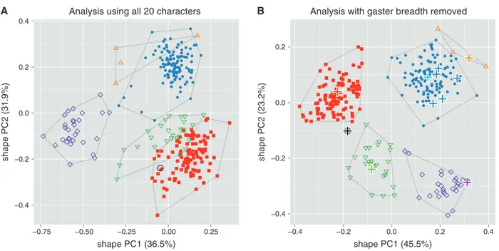
Figure 1A shows a scatterplot of the first two shape PCs including all twenty variables and six OTUs. Only the OTU
A. apiovorus was clearly distinct while the others were at least
slightly overlapping. This result was surprising, as all OTUs could be rather well separated by qualitative morphology. We therefore suspected a significant amount of error variance in some of the variables and checked the variables with matrix
−0.4 −0.2 0.0 0.2 0.4 −0.75 −0.50 −0.25 0.00 0.25 shape PC1 (36.5%) shape PC2 (31.9%) −0.4 −0.2 0.0 0.2 −0.4 −0.2 0.0 0.2 0.4 shape PC1 (45.5%) shape PC2 (23.2%) B A
Anisopteromalus shape PCA
Analysis using all 20 characters Analysis with gaster breadth removed
Fig. 1. Scatterplot of first against second shape PC of females of all six OTUs of Anisopteromalus. (A, B) Shape PCA, including all 20 variables
(A) and with variable gaster breadth omitted (B). Closed symbols: blue dots, A. calandrae; red squares, A. quinarius. Open symbols: violet diamonds,
A. apiovorus; green upside down triangles, A. caryedophagus; black circle, A. ceylonensis; orange triangles, A. cornis. In (B), name-bearing types are
marked with bold plus signs, the light blue plus sign indicates the position of the neotype of A. calandrae. The variance explained by each shape PC is given in parentheses.
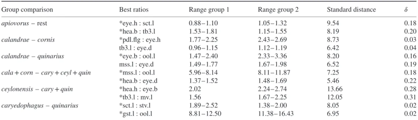
−0.2 −0.1 0.0 0.1 0.2 −0.4 −0.2 0.0 0.2 0.4 shape PC1 (62.3%) shape PC2 (8.2%) −0.4 −0.2 0.0 0.2 0.4 −0.4 −0.2 0.0 isosize shape PC1 (62.3%)
A. calandrae versus A. quinarius shape PCA
PCA Ratio Spectrum for shape PC1 0.31 -0.73 eye.b stv.l msc.l eye.h sct.l pol.l mss.l hea.b tb3.l gst.l hea.h msc.b mv.l pdl.flg ppd.l scp.l eye.d msp.l ool.l B A
Allometry Ratio Spectrum 0.32 0.023 eye.b msc.l eye.h stv.l sct.l mss.l hea.b pol.l tb3.l msc.b gst.l hea.h mv.l pdl.flg ppd.l scp.l msp.l eye.d ool.l D C
Fig. 2. Size and shape analysis of females of OTUs Anisopteromalus calandrae and A. quinarius using all variables except gaster breadth. (A, B)
Shape PCA, scatterplot of first against second shape PC (A), scatterplot of isosize against first shape PC (B). Symbols: blue dots, A. calandrae; red squares, A. quinarius; in parentheses the variance explained by each shape PC. (C, D) Ratio spectra, PCA ratio spectrum (C), allometry ratio spectrum (D); horizontal bars in the ratio spectra represent 68% bootstrap confidence intervals based on 1000 replicates.
scatterplots and Pearson product-moment correlation coeffi-cients. Correlation among body measurements of related taxa should usually be positive and high (e.g. Hills, 1978; Pimentel, 1979), hence its lack may indicate measurement error or mor-phological artefacts. Indeed, gaster breadth correlated much less strongly than all other variables (Table S3 and Figure S1), with coefficients ranging from −0.18 to 0.30, including many around 0. Closer inspection revealed many specimens with a moderately to strongly deformed gaster (dorso-ventrally
or laterally collapsed), mainly in specimens of A. apiovorus,
A. caryedophagus, A. ceylonensis and A. cornis. The reasons for
these artefacts were unclear, as the preservation history of the specimens was unknown. The observed damage nevertheless led us to remove gaster breadth from all further analyses. As a result, in the new graph (Fig. 1B) of a shape PCA with the remaining 19 variables all groups were neatly separated. The plot also shows the position of the available name-bearing types (marked with bold plus signs). Five of them represent some
Table 2. First and second-best ratios found by the LDA ratio extractor for separating various groupings of Anisopteromalus females.
Group comparison Best ratios Range group 1 Range group 2 Standard distance 𝛿
apiovorus – rest *eye.h : sct.l 0.88–1.10 1.05–1.32 9.54 0.18
*hea.b : tb3.l 1.53–1.81 1.15–1.55 8.19 0.20
calandrae – cornis *pdl.flg : eye.h 1.77–2.25 2.43–2.69 8.73 0.03 tb3.l : eye.d 0.96–1.15 1.12–1.19 6.42 0.04
calandrae – quinarius *eye.b : ool.l 1.47–2.40 2.33–3.36 8.20 0.16 mss.l : eye.d 1.49–1.77 1.67–1.98 6.52 0.19
cala + corn – cary + ceyl + quin *mss.l : ool.l 5.96–8.14 8.11–11.87 7.25 0.18 *hea.b : eye.d 1.37–1.52 1.48–1.69 5.46 0.22
ceylonensis – cary + quin *hea.h : eye.b 2.02 2.24–2.74 13.66 0.28 *tb3.l : mv.l 1.56 1.67–2.25 12.05 0.31
caryedophagus – quinarius *sct.l : stv.l 1.89–2.52 1.38–2.00 8.05 0.02 *gst.l : ool.l 8.81–12.50 11.38–16.43 6.95 0.02
Ratios marked with * have very little or no overlap and were thus eligible for use in the identification key and the diagnoses.
previous synonyms of A. calandrae and grouped rather closely with the neotype of A. calandrae that we are designating below (see ‘Description of species’ section).
The OTUs A. apiovorus, A. caryedophagus, A. ceylonensis and
A. cornis were quite distinct based on qualitative morphology
alone and were also well supported by morphometric analy-sis. Therefore, we focused on A. calandrae and A. quinarius that differ in their karyotypes with the haploid chromosome num-ber of either n = 7 or n = 5, respectively, but were otherwise generally similar in qualitative morphology. The question thus was how well they were distinguished, not only in shape, but also in size. The results of a shape PCA including only these two OTUs confirmed the pattern in the first plot (Fig. 1B), as they were well separated along the first shape PC (Fig. 2A). In a scatterplot of isosize against the first shape PC (Fig. 2B)
A. quinarius was on average slightly larger than A. calandrae,
although the size ranges were broadly overlapping. The plot thus revealed a moderate amount of allometric variation. A similar trend was visible by comparing the PCA ratio trum and the allometry ratio spectrum. In a PCA ratio spec-trum, only ratios calculated with variables lying at the oppo-site ends of the spectrum are relevant for a particular shape PC (see also Baur & Leuenberger, 2011; László et al., 2013 for the interpretation of ratio spectra). In the same manner, the most allometric ratios are found in an allometry ratio spectrum. Now, the PCA as well as the allometry ratio spectrum were dominated by the same ratio, eye.b : ool.l (Fig. 2C, D), that is, the most important ratio concerning the first shape PC was also the most allometric one.
The LDA ratio extractor is a tool for finding the best ratios for separating some groups (see Baur & Leuenberger, 2011: 816–818 for how this algorithm works). In contrast to a PCA, group membership had to be specified beforehand. The results are compiled in Table 2 showing various comparisons. The ranges of first and second best ratios were often not or only just overlapping between the respective groupings. We were able to integrate such ratios in the identification key and diagnoses (see below), as they represent important diagnostic features.
The best ratio for discrimination of A. calandrae from
A. quinarius, eye.b : ool.l, happened to be the same as the one
that dominated the PCA and allometry ratio spectrum (Fig. 2C, D). Note that this is by coincidence, as the best separating ratios must not necessarily correspond to the most important ratios of a PCA ratio spectrum (see also Peters & Baur, 2011 for the conceptual difference between a PCA and a LDA based type of analysis). For instance, the second best ratio, mss.l : eye.d, that still separated most of the specimens in this comparison (compare Table 2), was neither in the PCA nor in the allometry ratio spectrum among the dominant ratios (Fig. 2C, D). In fact, the respective variables were lying rather closely to each other in the spectra and thus the ratio had a negligible influence. Now, of the two best discriminating ratios, eye.b : ool.l and mss.l : eye.d, only the former showed allometric behaviour (i.e. correlated with isosize), while the latter did not. This furthermore demonstrates that allometric variation generally had a marginal impact on the discrimination of the OTUs – in other words, separation of A. calandrae and A. quinarius was attributable to true shape differences, not merely to an indirect size effect.
For all comparisons,𝛿 – a measure of how well shape discrim-inates in comparison to size (see Baur & Leuenberger, 2011: 818, formula 14) – was close to 0 (0.02–0.31), again indicat-ing that separation of OTUs was mainly due to shape rather than size. Furthermore, standard distances (Baur & Leuen-berger, 2011: 817, formula 12) were relatively high, ranging from 5.46–13.66, which reflects the good separation of OTUs observed in the shape PCA (Figs 1B, 2A).
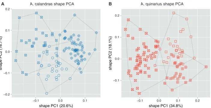
For the exploration of within-group variation we performed a MRA of the three reared strains of A. calandrae (Fig. 3A) and
A. quinarius (Fig. 3B). The scatterplots were in both cases rather
homogenous, indicated by the low variation explained by the first two shape PCs (37.3 and 52.9%, respectively). In each OTU, two strains were almost distinct with respect to the first shape PC, but they were partly covered by the third. The analysis did thus not allow any further subdivision of the two OTUs.
Molecular analysis
Cytb (980 bp) and ITS2 (655 aligned bp) were successfully
−0.2 −0.1 0.0 0.1 0.2 −0.1 0.0 0.1 shape PC1 (20.6%) shape PC2 (16.7%) −0.1 0.0 0.1 0.2 −0.1 0.0 0.1 0.2 shape PC1 (34.8%) shape PC2 (18.1%)
A. quinarius shape PCA
B
A A. calandrae shape PCA
Fig. 3. Shape PCA for exploring female variation within Anisopteromalus calandrae and A. quinarius using all variables except gaster breadth. The
analyses included three cultured laboratory strains for each OTU. (A) Scatterplot of first against second shape PC of A. calandrae: cross, strain Bamberg USA; plus signs, strain Savannah USA; open circles, strain Slough (ICSP) UK. (B) Scatterplot of first against second shape PC of A. quinarius: cross, strain Moscow (MSU) Russia; plus signs, strain Michurinsk Russia; open squares, strain Fresno USA. The variance explained by each shape PC is given in parentheses.
Alignment of Cytb was straightforward due to a lack of length variation, and no stop codons or frame shifts were detected. The intraspecific K2P distance range for Cytb was 0.001–0.024 (mean 0.014) for A. calandrae and 0.006–0.036 (mean 0.025) for A. quinarius. The intraspecific K2P distance range for
ITS2 was 0.000–0.002 (mean 0.001) for A. calandrae and
0.002–0.006 (mean 0.004) for A. quinarius. Even though these OTUs were morphologically difficult to discriminate, minimum interspecific divergences between A. calandrae and A. quinarius (Cytb: 0.169, ITS2: 0.271) were very high, largely exceeding maximum intraspecific divergences. Models chosen by MrAIC were as follows: GTR + I for Cytb and SYM + Γ for ITS2. As the SYM model is not implemented in RAxML we used GTR instead.
The results of the molecular analysis confirmed the discrimi-nation of A. calandrae from A. quinarius by MRA. Indeed, phy-logenetic analyses of Cytb and ITS2 (Fig. 4) recovered the same well-supported clusters of sequences, which corresponded to both morphologically delineated OTUs (Fig. 2A). Furthermore, comparison between Cytb and ITS2 genetic clusters revealed no mitochondrial introgression between these closely related species.
Status of OTUs
Our morphometric and molecular analyses unambiguously revealed that all OTUs formed distinct and well-supported taxa. We can thus conclude that the six OTUs examined in this
study represent valid species, A. apiovorus, A. calandrae,
A. caryedophagus, A. ceylonensis, A. cornis sp.n. and
A. quinarius sp.n. The morphometric analysis furthermore
con-firmed the synonymy of five nominal taxa with A. calandrae (see Fig. 1B). Below, we provide a key to females of all species and descriptions for the two new species as well as for A. calandrae. Information on the other species and a discussion of a few doubtful nominal taxa hitherto associated with Anisopteromalus can be found in Appendix S1. Nomenclatural changes discussed therein are as follows: Cyrtoptyx camerunus (Risbec) comb.n.;
Meraporus glaber (Szelényi) comb.n.; Dinarmus schwenkei
(Roomi, Khan & Khan) comb.n.; Anisopteromalus calandrae
brasiliensis (Domenichini) stat.rev., valid subspecies; Neoca-tolaccus indicus Ayyar & Mani, junior synonym of Oxysychus sphenopterae (Ferrière) syn.n.
Key to females
1. Head breadth equal to or more than 1.53× metatibia length
and eye height equal to or less than 1.1× scutellum length. Forewing speculum medially with a patch of about 10–30 setae (Fig. 5A), setea on wing disc whitish. Scutellum projecting beyond anterior margin of dorsellum (Fig. 5C). First funicular segment subcylindrical, proximally distinctly broader than third anellus and provided with 2–3 rows of longitudinal sensilla (Fig. 5E) . . . A. apiovorus Rasplus – Either head breadth less than 1.53× metatibia length or eye height more than 1.1× scutellum length. Forewing speculum
A. calandrae (strain Tours red-eye) Ivory Coast A. calandrae (strain Tours wild-type) Ivory Coast
A. calandrae (strain Krasnodar-2) Russia A. calandrae (strain Krasnodar-1) Russia A. calandrae (strain Savannah) USA
A. calandrae (strain Bamberg) USA
0.1
Anisopteromalus maximum likelihood tree
ITS2 Cytb
A. quinarius (strain Michurinsk) Russia A. quinarius (strain Fresno) USA A. quinarius (strain MSU) Russia Nasonia longicornis (strain IV7) USA
N. giraulti (strain RV2) USA N. vitripennis (strain HiCD12 & R5 11) Canada
Pachycrepoideus vindemmiae (strain Rennes) France 100 100 100 92 98 92 98 100 100 100 94 79 75
Fig. 4. ML trees from the analyses of ITS2 and Cytb sequences of several strains of the OTUs Anisopteromalus calandrae and A. quinarius. Bootstrap
supports higher than 70% are indicated at nodes. Scale bar indicates substitution per site for both trees.
bare (Fig. 5B), setea on wing disc dark. Scutellum projecting at level of anterior margin of dorsellum (Fig. 5D). First funicular segment subconical, proximally at most slightly broader than third anellus and provided with 1–2 rows of longitudinal sensilla (Fig. 5F) . . . 2 2. Mesosoma length equal to or less than 8.14× OOL and head breadth equal to or less than 1.52× eye distance. Gaster with hind margin of first tergite curving backwards and medially strongly produced (Fig. 5G) . . . 3 – Mesosoma length more than 8.14× OOL or head breadth more than 1.52× eye distance. Gaster with hind margin of first tergite at most slightly curving backwards but medially not produced (Fig. 5H) . . . 4 3 . Pedicel plus flagellum 1.77–2.25× eye height; flagellum dis-tinctly clavate (Fig. 5I). Head and mesosoma olive-green, cov-ered with relatively short, inconspicuous whitish setae (Fig. 5K) . . . A. calandrae (Howard) – Pedicel plus flagellum 2.43–2.69× eye height; flagellum almost filiform (Fig. 5J). Head and mesosoma blue-green, covered with relatively long, conspicuous whitish setae (Fig. 5L) . . . A. cornis Baur sp.n. 4. Scutellum strongly curved in lateral view (Fig. 6A). Head height 2.02× as long as eye breadth. Metatibia only 1.56× as long as marginal vein . . . A. ceylonensis Sureshan – Scutellum weakly curved in lateral view (Fig. 6B). Head height distinctly greater, 2.24–2.74× as long as eye breadth. Metatibia longer, 1.67–2.25× as long as marginal vein . . . 5 5. Gena compressed, with a short carina near mouth margin (Fig. 6C). Propodeum with anterior plica consisting of small deep pits, costula indistinct (Fig. 6E). Scutellum length equal to or less than 2.00× stigmal vein and gaster length equal to or more than 11.38× OOL . . . A. quinarius Gokhman & Baur
sp.n.
– Gena terete, not carinate near mouth margin (Fig. 6D). Propodeum with anterior plica short and strongly bent inwards, joining a strong costula (Fig. 6F). Either scutellum length more than 2.00× stigmal vein or gaster length less than 11.38× OOL . . . A. caryedophagus Rasplus
Description of species (for A. apiovorus, A. caryedophagus and A. ceylonensis, descriptions are provided in Appendix S1)
Anisopteromalus calandrae (Howard) (Figs 5D, F, G, I, K,
7A, B, D)
Pteromalus calandrae Howard in Comstock, 1881: 273:
Neotype ♀ in NMBE, here designated, labelled ‘USA Georgia Savannah Sitophilus oryzae leg. ix. 1978 [print]; chromosomes = 7 strain lab culture e.p. iii.2002 Sitophilus granarius A. Timokhov, Moscow [print]; 1551 Baur [print]; Neotype♀ Anisopteromalus calandrae Howard lab. H. Baur 2014 [hand, label with red left and right border]’ (entire; glued on a card rectangle); type locality: USA: Georgia, Savannah (examined by Baur). For discussion see under
Remarks.
Pteromalus oryzae Cameron, 1891: 184–185: Syntypes♂♂
and♀♀ lost (Bouˇcek et al., 1979: 435), but see also Cotes (1896: 11); type locality: INDIA. Synonymized with A.
calandrae by Bouˇcek et al. (1979: 435).
Because the original description is inconclusive (Cameron mainly described some colour characters that apply to many
Anisopteromalus species), we are inclined to accept the
synonymy of Bouˇcek et al. (1979). Moreover, the species described here as new can be ruled out: first, A. cornis
A C E
B D F
G I K
H J L
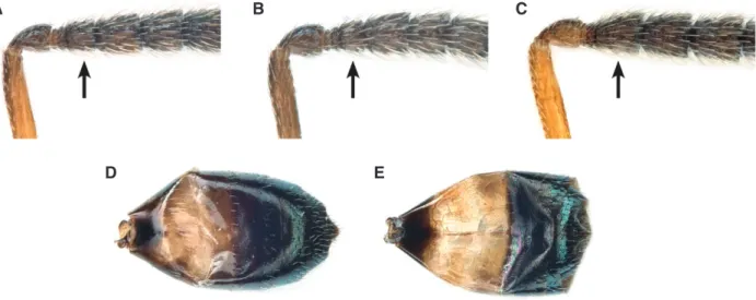
Fig. 5. Diagnostic characters of Anisopteromalus, females. (A, B) Forewing with speculum, A. apiovorus (A; actually the setae are whitish in this
species but they appear darkened because of the contrasting background), A. quinarius sp.n. (B); (C, D) mesosoma with scutellum projection in lateral view, A. apiovorus (C), A. calandrae (D); (E, F) proximal funicular segments, A. apiovorus (E), A. calandrae (F); (G, H) gaster in dorsal view with posterior margin of first tergite emphasized with a magenta line, A. calandrae (G), A. quinarius sp.n. (H); (I, J) antenna, A. calandrae (I), A. cornis sp.n. (J); (K, L) mesosoma in dorsal view, A. calandrae (K), A. cornis sp.n. (L). Arrows mark important character states.
sp.n.occurs in the Afrotropical region while P. oryzae was described from India; second, A. quinarius sp.n. naturally attacks anobiid beetles, while P. oryzae was recorded as a parasite of the rice weevil Sitophilus oryzae.
Meraporus vandinei Tucker, 1910: 343–344: Lectotype♀
in USNM, catalogue no. 13389, here designated, labelled ‘Plano TX [print] 7. 27. 09 [hand]; emerged on [print] XI. 23. 09 [hand]; Hunter No [print] 1821 [hand]; ESTucker col-lector [print]; Paratype No. [print] 13389 [hand] U.S.N.M. [print]’ (entire; glued on a card point, with a card rectangle put underneath by Baur); type locality: USA: Texas, Plano (examined by Baur). Synonymized with A. calandrae by
Mani (1938: 103), synonymy confirmed by MRA (see above).
Tucker (1910: 343) stated that his new species was based on 45 specimens collected by himself from Plano and by Van Dine from some other places. He then singled out one female and one male as ‘Type’ and considered the rest to be ‘Paratypes’. However, following the International Code of Zoological Nomenclature (ICZN) all specimens must be considered as syntypes. We received only 6♂ and 33♀ that stood under this name in the USNM. As lectotype we have taken a female with most body parts intact, and not the female with the additional label ‘Meraporus vandinei (n. sp.)
A C E
B D F
Fig. 6. Diagnostic characters of Anisopteromalus, females. (A, B) Mesosoma with scutellum shape in lateral view, A. ceylonensis (A; holotype female,
original position mirrored and photo slightly modified with Photoshop), A. caryedophagus (B); (C, D) gena near mouth corner in postero-lateral view,
A. quinarius sp.n. (C), A. caryedophagus (D); (E, F) median area of propodeum, A. quinarius sp.n. (E), A. caryedophagus (F). Arrows mark important
character states.
A
B
C
D
E
Fig. 7. Diagnostic characters of Anisopteromalus, males. (A–C) Proximal funicular segments, A. calandrae, average-sized specimen (A), the same,
small specimen (B), A. quinarius sp.n. (C); (D, E) gaster in dorsal view, A. calandrae (D), A. quinarius sp.n. (E). Arrows mark important character states.
Tucker [hand]’ and that emerged on September 11, 1909. This specimen was heavily damaged and lacked the head and wings, but otherwise it was conspecific with the lectotype, as well as all the other paralectotypes we have seen.
Anisopteromalus mollis Ruschka, 1912: 243–245: Lectotype ♀ in NHMV, here designated, labelled ‘2298 Mehlfruchtbörse [hand]; ex Laemophloeus ferrugineus [hand]; Anisopteromalus mollis m. type. female [hand]; Anisoptero = malus [sic] mollis [hand] det. Ruschka [print]; Type [print, red]’ (entire; lectotype the uppermost specimen on the pin; lectotype and one paralectotype♀ remounted by
Baur on card rectangles); type locality: AUSTRIA: Vienna (examined by Baur). Synonymized with A. calandrae by Graham (1969: 435), synonymy confirmed by MRA (see above).
Aplastomorpha pratti Crawford, 1913: 252–253:
Lecto-type ♀ in USNM, catalogue no. 15314, here designated, labelled ‘Dallas TX [print], Nov., 1906 [hand]; U.S.D.A. No. [print] 6076 [hand]; Bred from No [print] 3715 [hand]; WDHunter collector [print]; Paratype No. [print] 15314 [hand] U.S.N.M. [print]’ (right antenna beyond third anellus and parts of mid and hind legs lacking; glued on a card point,
with a card rectangle put underneath by Baur); type locality: USA: Texas, Dallas (examined by Baur). Synonymized with
A. calandrae by Mani (1938: 103), synonymy confirmed by
MRA (see above).
We consider the entire series of 2♂ and 9♀ housed in the USNM to be syntypes, because no specimen was fixed as holotype in the original description (compare Crawford, 1913: 252–253). One female belonged to Dinarmus, as also stated by an identification label of Gahan. The rest of the specimens were clearly conspecific. We have taken as lectotype a female with most body parts intact, and not the female with the additional label ‘Aplastomorpha pratti Type Crfd [hand]’. This specimen was in a slightly worse condition, lacking for instance the metatibiae that constitute one of the characters used in our morphometric analyses.
Neocatolaccus australiensis Girault, 1913: 306: Lectotype ♀ in QMB, catalogue no. 1956, here designated, labelled
‘TYPE [print, orange label]; Syntype 1♂ 2♀♀ Hy. 1956 [hand, red label]; Neocatolaccus australiensis Gir.,♀ types [last word red underlined, Girault hand]’ (head lacking, wings glued to card point and crumpled; glued at tip of a card point; on the same card point two male paralectotypes, one with head missing. Mounted separately on a slide two female and two male heads labelled ‘TYPE [print] Hy/1956 [hand] A. A. Girault [print]; Queensland Museum. [print] Neocato-lac♀ -cus. [sic] australiensis ♂ wing [hand]’; one of the heads might belong to the lectotype, the other one to a paralecto-type female mounted on a separate pin; association of those heads with specimens was impossible); type locality: AUS-TRALIA: Queensland, Nelson (? = Gordonvale) (examined by Baur). Synonymized with A. calandrae by Bouˇcek (1988: 414), synonymy confirmed by MRA (see above). As pointed out by Dahms (1983: 102), Girault (1913) published the cata-logue number as ‘1596’ which is wrong. The correct number is ‘1956’ as indicated above.
Bruchobius medius Masi, 1917: 176–177: Holotype ♀ in
BMNH, catalogue no. 5.699, labelled ’64 [hand, on card point]; Mahe, ’08-9 Seychelles Exp. [print]; Holo- [hand] Type [print; round label with red rim]; Percy Sladen Trust. Exped. B.M. 1913-170. [print]; Brucho = bius medius ♀ Masi [hand]; B.M. TYPE HYM. [print] 5.699;♀ Anisoptero-malus calandrae (How.) [hand] det. Z. Bouˇcek, 197 [print] 5 syn. nov. [hand]’ (right flagellum beyond first segment and left hindwing lacking; card point mounted with micro-pin on rectangular tag; head remounted on card point by Baur); type locality: SEYCHELLES: Mahé, Cascade Estate (examined by Baur). Synonymized with A. calandrae by Bouˇcek (1976: 22), synonymy confirmed by MRA (see above).
Neocatolaccus mamezophagus Ishii & Nagasawa, 1942:
67–68: Syntypes♂♂ and ♀♀ in Tokyo College of Agriculture and Forestry; type locality: JAPAN. Synonymized with
A. calandrae by Tachikawa (1966: 99).
The type material could not be traced, even with the help of Japanese colleagues. The original description clearly rules out that N. mamezophagus is the same as any of the new species described here. First, females of A. cornis sp.n. have the pedicel distinctly shorter than first funicular segment,
while it is stated by Ishii & Nagasawa that the pedicel is slightly longer than the first funicular segment; second, males of A. quinarius sp.n. have the first funicular segment at least as long and usually longer than the second, while the first segment of N. mamezophagus is said to be slightly shorter than the second. The character states of N. mamezophagus match specimens of A. calandrae as defined here, hence we accept the synonymy of Tachikawa (1966).
Diagnosis, female. Head and mesosoma olive-green with
slight bronze tinges in places, setae whitish, inconspicu-ous (Fig. 5K). Gena terete, not carinate near mouth margin. Flagellum distinctly clavate (Fig. 5I), first funicular seg-ment subconical, basally slightly broader than third anellus, provided with 1–2 rows of longitudinal sensilla (Fig. 5F). Scutellum projecting at level of anterior margin of dorsel-lum (Fig. 5D), in lateral view weakly curved. Forewing with setae on wing disc dark. Speculum bare, closed in distal and proximal part. Anterior plica of propodeum short and evenly curved, joining sometimes an indistinct costula. Posterior margin of first gastral tergite curving backwards and strongly produced (Fig. 5G). Head breadth 1.24–1.51× metatibia length and 1.38–1.52× eye distance; head height 2.46–3.39× eye breadth; eye height 1.09–1.30× scutellum length; pedicel plus flagellum 1.77–2.25× eye height; mesosoma length 5.96–8.14× OOL; scutellum length 1.48–1.96× stigmal vein; metatibia length 1.66–2.21× marginal vein; gaster length 7.78–12.02× OOL.
Description, female. Antenna with scape testaceous, pedicel
testaceous, fuscous on upper side, anelli fuscous and rest of flagellum testaceous, fuscous on upper side. Legs with tibiae yellowish.
Head 1.06–1.19× as broad as mesoscutum. POL 1.22–1.87× OOL. Eyes 1.42–1.80× as high as broad, separated by 1.29–1.59× their height. Malar space 0.46–0.63× eye height. Head in frontal view 1.10–1.30× as broad as high. Antenna with scape 0.77–1.00× as long as eye height. Combined length of pedicel plus flagellum 0.86–1.03× head breadth. First and second anellus strongly transverse, third transverse, about as long as second anellus.
Mesosoma 1.16–1.36× as long as broad. Mesoscutum 1.84–2.42× as broad as long. Hind margin of scutellum broadly rounded. Upper mesepimeron strongly narrowing below, reaching at most basal third of mesopleuron. Basal setal line complete. Costal cell with dorsal surface with at most a short row of setae distally, lower surface with a patch of setae in distal half and a single row of setae running to proximal part, costal setal line complete. Forewing disc moderately pilose. Marginal vein 1.35–1.96× as long as stigmal vein. Stigma subcircular to oval, medium-sized. Propodeum 0.42–0.60× as long as scutellum. Median carina fine, straight. Median area in anterior part evenly reticulate with inner corner of anterior plica with a rather deep and reticulate depression along the anterior plicae. Nucha subglobose, not distinctly separated from rest of propodeum, often at least with some traces of alutaceous sculpture.
Metasoma: Gaster 1.41–2.54× as long as broad, 1.13–1.66× as long as mesosoma, and 0.69–1.22× as long as mesoscutum. Posterior margin of second gastral tergite incised medially to almost straight. Posterior margin of third gastral tergite straight.
Material examined. Beside the above-mentioned
name-bearing types we examined the following specimens:
Paralecto-types, Anisopteromalus mollis, 2♂, 1♀, same data as lectotype; Aplastomorpha pratti, USA: 2♂, 7♀, Texas, Dallas, xi.1906
(W.D. Hunter), paratype no. 15314 (USNM); USA: 1♀, Texas, Dallas, xi.1906 (W.D. Hunter), type no. 15314 (USNM);
Mer-aporus vandinei, USA: 1♂, 1♀, Texas, El Campo, 22.vi.1909
(D.L. Van Dine), paratype (USNM); USA: 3♂, 26♀, Texas, Plano, 26.vii.1909 (E.S. Tucker), paratype no. 13389 (USNM); USA: 1♂, 1♀, Texas, Plano, 26.vii.1909 (E.S. Tucker), type no. 13389 (USNM); USA: 2♀, Louisiana, Welsh, 2.viii.1909 (D.L. Van Dine), paratype (USNM); USA: 1♂, 2♀, Louisiana, Lake Arthur, 29.vii.–2.viii.1909 (Rosenfeld and D.L. Van
Dine), paratype (USNM); Neocatolaccus australiensis, 2♂, 1♀,
same data as lectotype. Further, non-type material is listed in Appendix S1.
Biology. The species has a relatively broad host range. It
prefers to parasitize Sitophilus (Dryophthoridae) and
Calloso-bruchus spp. (Chrysomelidae: Bruchinae); however, it can easily
be reared (e.g. under laboratory conditions) on certain Anobi-idae, like Lasioderma serricorne (see Gokhman & Timokhov, 2002; Timokhov & Gokhman, 2003). Many other host records (see Noyes, 2013) need to be verified.
Distribution. Cosmopolitan (Noyes, 2013).
Remarks.The females of A. calandrae can be most easily recognized by the characters given in the key and diagnosis. The species is most similar to A. cornis sp.n. From A. quinarius
sp.n.it is further distinguished by the pilosity of the forewing, for which we refer to the description of each species.
In order to assess the taxonomic status of A. calandrae, it is necessary to concentrate on the other cosmopolitan species,
A. quinarius sp.n. Not only have the two species been confused
over a long time, but they both are also likely to occur in human dwellings. However, these species usually occupy somewhat dif-ferent habitats due to their difdif-ferent host preferences (Gokhman & Timokhov, 2002; Timokhov & Gokhman, 2003): A. calandrae inhabits mills and grain bins where it is usually associated with
Sitophilus spp.; A. quinarius sp.n. inhabits houses and
ware-houses (e.g. containing stored fruit or tobacco) being associated with Stegobium or Lasioderma beetles there. We believe that these differences were an important factor that hampered the discovery of A. quinarius sp.n. (see Discussion).
A further complication is that the male holotype of
A. calandrae is now lost (Peck, 1963: 733), which was recently
confirmed by the curator of the Chalcidoidea collections at the USNM, M. Gates (personal communication). We have therefore studied characters of the males of the two species that were
relevant with respect to Howard’s original description. They can be distinguished as follows:
• A. calandrae: First funicular (third flagellar) segment always shorter than the second (Fig. 7A), in small specimens only about as long as combined length of first and second anellus (Fig. 7B). Pale band in proximal half of gaster dirty yellow (Fig. 7D).
• A. quinarius sp.n.: First funicular (third flagellar) segment about as long as the second, always much longer than combined length of first and second anellus (Fig. 7C). Pale band in proximal half of gaster bright yellow (Fig. 7E).
Now, Howard in Comstock (1881: 273), who apparently examined a very small specimen, noted in his brief but concise description ‘joint 5 [corresponding to the third flagellar segment] small, equal in length to the two ring joints’ and ‘abdomen [cor-responding to the gaster] yellow-brown at base’. A comparison of Howard’s statements with our description of the male char-acters should make it clear that both concepts of A. calandrae match almost perfectly.
With regard to the overall morphological similarity with the other cosmopolitan species, A. quinarius sp.n., we designated a neotype for A. calandrae in order to guarantee stability in the application of the name. This is even more important, because
A. calandrae is the type species of Aplastomorpha Crawford,
the only synonym of Anisopteromalus (see Noyes, 2013). We selected a specimen from Savannah (Georgia), which might appear to be quite far away from Hempstead, Waller County (Texas), the place where the holotype of A. calandrae was collected (Howard in Comstock, 1881: 273). Given that the species is very easily disseminated from one site to another by transportation of stored products, the distance between the two localities is certainly not a problem here. Taking material of the laboratory culture from Savannah also had the advantage that a specimen in perfect condition coming from the centre of the analysed size and shape space was available (cf. Fig. 1B). The Savannah strain could furthermore be included in our genetic analyses (Fig. 4). Finally, in both cases the specimens were originally reared from Sitophilus oryzae-infested crop – that is, both demonstrably had the same host preference.
Anisopteromalus cornis Baur sp.n. (Fig. 5J, L)
http://zoobank.org/urn:lsid:zoobank.org:act:94339C6E-0AF1-4883-836D-BE6AB2EF145B
Holotype ♀ in MHNG, here designated, labelled ‘MALI Sikasso ex stock de mil 1 X 1986 [print, blue label]; B. Sauphanor leg. 6241 [print, blue label]; 2119 Baur [print]; Holotype♀ Anisopteromalus cornis sp. n. Baur lab. H. Baur 2014 [hand, label with red left and right border]’ (entire; glued on card rectangle with metasoma detached from rest of body); type locality: MALI: Sikasso (examined by Baur).
Diagnosis, female. Head and mesosoma blue-green with
Gena terete, not carinate near mouth margin. Flagellum fili-form (Fig. 5J), first funicular segment subconical, basally about as broad as third anellus, provided with 1–2 rows of longi-tudinal sensilla. Scutellum projecting at level of anterior mar-gin of dorsellum, in lateral view weakly curved. Forewing with setae on wing disc dark. Speculum bare, open below but some-times closed in proximal part. Anterior plica of propodeum short and evenly curved, joining an indistinct costula. Posterior margin of first gastral tergite curving backwards and strongly produced. Head breadth 1.15–1.26× metatibia length and 1.37–1.40× eye distance; head height 3.15–3.59× eye breadth; eye height 1.05–1.09× scutellum length; pedicel plus flagellum 2.43–2.69× eye height; mesosoma length 6.19–7.96× OOL; scutellum length 1.64–1.74× stigmal vein; metatibia length 1.90–2.29× marginal vein; gaster length 7.98–11.94× OOL.
Description, female. Antenna with scape testaceous, pedicel
fuscous on upper side, anelli fuscous and rest of flagellum testaceous, fuscous on upper side. Legs with tibiae yellowish.
Head 0.98–1.14× as broad as mesoscutum. POL 1.33–1.64× OOL. Eyes 1.65–1.87× as high as broad, separated by 1.53–1.64× their height. Malar space 0.59–0.74× eye height. Head in frontal view 1.09–1.19× as broad as high. Antenna with scape 1.05–1.10× as long as eye height. Combined length of pedicel plus flagellum 1.06–1.26× head breadth. First and second anellus strongly transverse, third conspicuous, slightly longer than second anellus.
Mesosoma 1.20–1.30× as long as broad. Mesoscutum 1.92–2.16× as broad as long. Hind margin of scutellum broadly rounded. Upper mesepimeron strongly narrowing below, reaching at most basal third of mesopleuron. Basal setal line complete. Costal cell with dorsal surface with a short row of setae distally, lower surface with a patch of setae in distal half and a single row of setae running to proximal part, costal setal line complete. Forewing disc moderately pilose. Marginal vein 1.41–1.78× as long as stigmal vein. Stigma subcircular to slightly elongate, small. Propodeum 0.51–0.57× as long as scutellum. Median carina fine, straight. Median area in anterior part evenly reticulate with inner corner of anterior plica with a rather deep and reticulate depression along the anterior plicae. Nucha subglobose, not distinctly separated from rest of propodeum, alutaceous.
Metasoma: Gaster 1.71–2.89× as long as broad, 1.29–1.50× as long as mesosoma, and 0.63–1.05× as long as mesoscutum. Posterior margin of second gastral tergite weakly incised medi-ally. Posterior margin of third gastral tergite straight.
Etymology. The specific name ‘cornis’ is derived from Latin
and means ‘horned’. It refers to the relatively long antennae.
Material examined. Beside the above-mentioned holotype
we examined the following specimens: Paratypes, GHANA: 1♂, 1♀, East Gonja District, Salaga, 1944 (G.S. Cottérell), ex moth borer on millet seeds (BMNH); MALI: 7♂, 3♀, Sikasso Province, Sikasso, x.1986 (B. Sauphanor), ex host on stock
millet (CBGP; MHNG; NMBE); NIGERIA: 1♂, 1♀, Kaduna State, Zaria, Samaru, ex Sitotroga sp., 20.iii.1958 (BMNH).
Biology. Reared from Sitotroga sp. (Lepidoptera:
Gelechi-idae).
Distribution. Afrotropical region.
Remarks. The species is very close in habitus to A. calandrae
from which it can be most easily separated by its relatively long flagellum.
Anisopteromalus quinarius Gokhman & Baur sp.n. (Figs
5B, H, 6C, E, 7C, E)
http://zoobank.org/urn:lsid:zoobank.org:act:62905F79-1E91-4D21-88D5-F252B18229F3
Holotype♀ in NMBE, here designated, labelled ‘RUSSIA Moscow (in Apartment) Stegobium paniceum leg. ix.1995 [print]; chromosomes = 5 strain lab culture e.p. iii.2002 Sitophilus granarius A. Timokhov, Moscow [print]; 1956 Baur [print]; Holotype ♀ Anisopteromalus quinarius sp. n. Gokhman & Baur lab. H. Baur 2014 [hand, label with red left and right border]’ (entire; glued on card point with metasoma detached from rest of body); type locality: RUSSIA: Moscow (examined by Baur).
Diagnosis, female. Head and mesosoma olive-green with
slight bronze tinge in places, setae whitish, inconspicuous. Gena compressed, with a short carina near mouth margin (Fig. 6C). Flagellum almost filiform, first funicular segment subconical, basally about as broad as third anellus, provided with 1–2 rows of longitudinal sensilla. Scutellum project-ing at level of anterior margin of dorsellum, in lateral view weakly curved. Forewing with setae on wing disc dark. Specu-lum bare, open below but sometimes closed in proximal part (Fig. 5B). Anterior plica of propodeum consisting of small deep pits, costula indistinct (Fig. 6E). Posterior margin of first gastral tergite curving backwards, not produced medi-ally (Fig. 5H). Head breadth 1.24–1.51× metatibia length and 1.48–1.69× eye distance; head height 2.34–2.74× eye breadth; eye height 1.09–1.32× scutellum length; pedicel plus flagellum 1.63–1.97× eye height; mesosoma length 8.11–11.87× OOL; scutellum length 1.38–2.00× stigmal vein; metatibia length 1.67–2.25× marginal vein; gaster length 11.38–16.43× OOL.
Description, female. Antenna with scape testaceous, pedicel
light brown, first and second anellus testaceous, rest of flagel-lum fuscous. Legs with tibiae yellowish testaceous or slightly infuscate in basal half.
Head 1.01–1.25× as broad as mesoscutum. POL 1.71–2.46× OOL. Eyes 1.45–1.64× as high as broad, separated by 1.12–1.35× their height. Malar space 0.40–0.53× eye height. Head in frontal view 1.13–1.26× as broad as high. Antenna
with scape 0.68–0.85× as long as eye height. Combined length of pedicel plus flagellum 0.84–0.99× head breadth. First and second anellus strongly transverse, third conspicuous, about as long as or longer than first two anelli (shorter in Fresno strain).
Mesosoma 1.18–1.43× as long as broad. Mesoscutum 1.66–2.17× as broad as long. Hind margin of scutellum broadly rounded. Upper mesepimeron strongly narrowing below, reach-ing at most basal third of mesopleuron. Basal setal line complete. Costal cell with dorsal surface with a patch of setae in distal half and a single row of setae in proximal half, lower surface with a large patch of setae in distal half and two to three rows of setae in proximal part, costal setal line complete. Forewing disc thickly pilose. Marginal vein 1.09–1.90× as long as stigmal vein. Stigma variable in shape, from subcircular (Fresno strain) to sub-rectangular (MSU strain), large. Propodeum 0.40–0.52× as long as scutellum. Median carina fine, straight. Median area evenly reticulate with inner corner of anterior plica with slight and evenly reticulate depression. Nucha subglobose, not distinctly separated from rest of propodeum, smooth or weakly alutaceous. Metasoma: Gaster 1.39–2.86× as long as broad, 1.23–1.67× as long as mesosoma, and 0.61–1.21× as long as mesoscutum. Posterior margin of second and third gastral tergite incised medially.
Etymology. The specific name ‘quinarius’ is derived from
Latin and means ‘of five’. It refers to the haploid chromosome number, which is only five in this species. The name ‘quinarius’ is treated as a noun in apposition and does not change with gender.
Material examined. Beside the above-mentioned holotype
we examined the following specimens: Paratypes, RUSSIA: 3♂, 22♀, Moscow Province, Moscow, ex Stegobium paniceum (Lin-naeus, 1758), ix.1995, cultured iii.2002 on Sitophilus granarius (Linnaeus, 1758), in apartment (NMBE); 22♂, 10♀, Moscow Province, Moscow, ex Stegobium paniceum (Linnaeus, 1758), ix.1995, cultured iii.2002 on Lasioderma serricorne (Fabricius, 1792), in apartment (NMBE); 6♀, Moscow Province, Moscow, ex Stegobium paniceum (Linnaeus, 1758), ix.1995, cultured iii.2002 on Sitophilus granarius (Linnaeus, 1758), in apartment (USNM; UCD); 6♂, Moscow Province, Moscow, ex
Stegob-ium paniceum (Linnaeus, 1758), ix.1995, cultured iii.2002 on Lasioderma serricorne (Fabricius, 1792), in apartment (USNM;
UCD); 5♀, Moscow Province, Moscow, ex Stegobium paniceum (Linnaeus, 1758), ix.1995, cultured iii.2002 on Sitophilus
gra-narius (Linnaeus, 1758), in apartment (BMNH); 1♂, 10♀,
Moscow Province, Moscow, ex Stegobium paniceum (Linnaeus, 1758), ix.1995, cultured iii.2002 on Sitophilus granarius (Lin-naeus, 1758), in apartment (ZIN; ZMMU); 12♂, 12♀, Moscow Province, Moscow, ex Stegobium paniceum (Linnaeus, 1758), ix.1995, cultured iii.2002 on Lasioderma serricorne (Fabricius, 1792), in apartment (ETHZ; MHNG; NHMV); 4♂, 19♀, Tambov Province, Michurinsk, ex Stegobium paniceum (Linnaeus, 1758), viii.2000, cultured iii.2002 on Sitophilus
granarius (Linnaeus, 1758), laboratory culture (NMBE); 2♂,
10♀, Tambov Province, Michurinsk, ex Stegobium paniceum
(Linnaeus, 1758), viii.2000, cultured iii.2002 on Sitophilus
granarius (Linnaeus, 1758), laboratory culture (ZIN; ZMMU);
USA: 3♂, 24♀, California, Fresno, ex Lasioderma
serri-corne (Fabricius, 1792), 1999, cultured iii.2002 on Sitophilus granarius (Linnaeus, 1758), laboratory culture (NMBE); 2♂,
8♀, California, Fresno, ex Lasioderma serricorne (Fabri-cius, 1792), 1999, cultured iii.2002 on Sitophilus granarius (Linnaeus, 1758), laboratory culture (ZIN; ZMMU). Further, non-type material is listed in Appendix S1.
Biology. Host preferences of this species contrast somewhat
with those of A. calandrae. Specifically, it prefers to attack cer-tain Anobiidae, for example Stegobium paniceum or Lasioderma
serricorne; however, it can easily be reared (e.g. under
labora-tory conditions) on Sitophilus spp. (Dryophthoridae; see, e.g., Timokhov & Gokhman, 2003).
Distribution. Cosmopolitan.
Remarks. The females of A. quinarius sp.n. can be
recog-nized by the characters given in the key and diagnosis. For the separation of males from A. calandrae, see ‘Remarks’ in that species description.
Discussion
Morphometry and data integration
Multivariate ratio analysis (MRA) revealed six distinct clusters that fully corresponded to the species delineation based on qualitative characters (Fig. 1B). For A. calandrae and A. quinarius sp.n. our DNA analyses as well as the pub-lished data on karyotype, behaviour and life-history strategies (summarized in Gokhman & Timokhov, 2002) also con-firmed the morphometric differentiation of the two species (see Fig. 2 for interspecific and Fig. 3 for intraspecific vari-ation). Concerning the molecular markers we would like to point to the fact that the comparison between ITS2 and
Cytb genetic clusters showed no mitochondrial
introgres-sion (Fig. 4). Consequently, Cytb should be valuable for identification of the cultured strains of ‘A. calandrae’ and accurate assignment of specimens to either A. calandrae or
A. quinarius sp.n.
The associations of names with species as delimited here was straightforward for A. apiovorus, A. caryedophagus, A. ceylonensis, A. cornis sp.n. and A. quinarius sp.n., because
type specimens were available in good condition and thus could be measured and included in the morphometric analyses. The same was true for other nominal taxa where name-bearing types were available (Fig. 1B). All of these names could be confirmed as synonyms of A. calandrae. Cases where the type material was lacking posed rather more difficulties. Here, we took the available evidence from the original description and integrated it with our data. Once again morphometric character ratios proved to be most helpful. For instance measurements taken