HAL Id: hal-01392845
https://hal.archives-ouvertes.fr/hal-01392845
Submitted on 4 Nov 2016HAL is a multi-disciplinary open access
archive for the deposit and dissemination of sci-entific research documents, whether they are pub-lished or not. The documents may come from teaching and research institutions in France or abroad, or from public or private research centers.
L’archive ouverte pluridisciplinaire HAL, est destinée au dépôt et à la diffusion de documents scientifiques de niveau recherche, publiés ou non, émanant des établissements d’enseignement et de recherche français ou étrangers, des laboratoires publics ou privés.
Multilayered structure of tension wood cell walls in
Salicaceae sensu lato and its taxonomic significance
Barbara Ghislain, Eric-André Nicolini, Raïssa Romain, Julien Ruelle, Arata
Yoshinaga, Mac H. Alford, Bruno Clair
To cite this version:
Barbara Ghislain, Eric-André Nicolini, Raïssa Romain, Julien Ruelle, Arata Yoshinaga, et al.. Mul-tilayered structure of tension wood cell walls in Salicaceae sensu lato and its taxonomic significance. Botanical Journal of the Linnean Society, Linnean Society of London, 2016, 182 (4), pp.744-756. �10.1111/boj.12471�. �hal-01392845�
Multilayered structure of tension wood cell walls in Salicaceae sensu
lato and its taxonomic significance
Barbara Ghislain1*, Eric-André Nicolini2, Raïssa Romain1, Julien Ruelle3, Arata Yoshinaga4, Mac H. Alford5, Bruno Clair1
1 CNRS, UMR EcoFoG, AgroParisTech, Cirad, INRA, Université des Antilles, Université de Guyane, 97310 Kourou, France
2 CIRAD, AMAP, botAnique et bioinforMatique de l’Architecture des Plantes, Campus Agronomique BP 701, 97387 Kourou, French Guiana, France
3 INRA, Laboratoire d’Etude des Ressources Forêt-Bois (LERFoB), 54280 Champenoux, Nancy, France
4 Laboratory of Tree Cell Biology, Graduate School of Agriculture, Kyoto University, Sakyo-ku, Kyoto 606-8502, Japan
5 Department of Biological Sciences, University of Southern Mississippi, 118 College Drive #5018, Hattiesburg, Mississippi 39406, U.S.A.
Multilayered tension wood cell walls in Salicaceae *Corresponding author: barbara.ghislain@ecofog.gf
2
Abstract
The circumscription of Salicaceae has recently been enlarged to include a majority of the species formerly placed in the polyphyletic tropical family Flacourtiaceae. Several studies have reported a peculiar and infrequently formed multilayered structure of tension wood in four of the tropical genera. Tension wood is a tissue produced by trees to restore their vertical orientation, and most studies have focused on trees developing tension wood by means of cellulose-rich, gelatinous fibres, as is known in Populus L. and Salix L. (Salicaceae s.s.). This study aims to determine if the multilayered structure of tension wood is an anatomical characteristic common in other Salicaceae, and if so, how its distribution correlates to phylogenetic relationships. Therefore, we studied the tension wood of 14 genera of Salicaceae as well as two genera of Achariaceae, one genus of Goupiaceae, and one genus of Lacistemataceae, families closely related to Salicaceae or formerly placed in Flacourtiaceae. Opposite wood and tension wood were compared with light microscopy and 3D Laser Scanning Confocal Microscopy. The results indicate that a multilayered structure of tension wood is common in the family except in Salix, Populus, and one of their closest relatives, Idesia
polycarpa Maxim. We suggest that tension wood may be a useful anatomical character in
understanding phylogenetic relationships in Salicaceae. Further investigation is still needed on the tension wood of several other putatively close relatives of Salix and Populus, in particular
Bennettiodendron Merr., Macrohasseltia L.O.Williams and Itoa Hemsl.
Keywords
3
Introduction
Until recently, morphological and anatomical characters were the primary sources of data for inferring phylogenetic relationships. More recently, DNA data have provided the majority of characters for our analyses of relationships, but reference to morphological characters remains useful for many reasons, from pedagogy to comparative evolutionary studies to studies of organisms where DNA data are inaccessible (e.g., fossils, rare organisms). Wood anatomy has certainly proved useful (Tippo, 1946; Lens et al., 2007; Christenhusz et al., 2010), especially characters linked to cell organization, cell types, or pitting. Here we show that fibre cell wall of reaction tissues, in this case tension wood, may also be useful phylogenetically.
Trees are able to optimize their orientation thanks to the production of asymmetrical maturation stress around the tree. In angiosperms, a special wood with very high tensile stress, called tension wood, is produced on the upper side of the tilted stem. This high tensile stress allows for the active bending of the tree axis (Du & Yamamoto, 2007; Alméras & Fournier, 2009). Tension wood exhibits anatomical differences from normal wood, for example, a lower frequency of vessels (Jourez, Riboux, & Leclercq, 2001; Ruelle et al., 2006). However, the strongest differences are observable at the fibre wall level. The secondary wall of a normal wood cell is composed of three sub-layers (S1, S2, S3) made of cellulose microfibrils oriented at different angles and embedded in a matrix of lignin and hemicelluloses. Whereas S1 and S3 are thin, with cellulose microfibrils oriented to nearly 80° compared to the fibre axis, the S2 layer is much thicker and the angle of microfibrils range from 10 to 20°. In tension wood the cell wall is generally modified by the presence of an inner unlignified layer, the so-called gelatinous layer or G-layer, replacing the S3 and part of or the entire S2 layer. It has been recently shown that this layer can be later lignified (Roussel & Clair, 2015). This partially explains why many species were known to produce tension wood lacking G-layers (Onaka, 1949; Fisher & Stevenson, 1981; Clair et al., 2006). Whether lignified or not, tension wood cell wall is homogeneous and characterized by a much lower (up to nil) cellulose microfibril angle compared to normal wood (Chaffey, 2000; Ruelle et al., 2006).
A peculiar fibre wall structure was first discovered in xylem fibres of Homalium foetidum Benth., Homalium luzoniense Fern.-Vill. and Olmediella betschleriana Loes. (Bailey & Kerr, 1935; Daniel & Nilsson, 1996), in which the secondary wall appears multilayered. This peculiar cell wall structure was later demonstrated to occur only in tension wood (Clair et al., 2006; Ruelle et al., 2007a; Fig. 1). Daniel & Nilsson (1996) described this structure in Homalium
foetidum as a succession of thick layers separated by thin layers with elevated levels of lignin.
In Laetia procera Eichler, Ruelle et al. (2007a) described the thick layers as lightly lignified. Similar cell wall structures were also reported in reaction phloem fibres (Nanko, Saiki, & Harada, 1982; Nakagawa, Yoshinaga, & Takabe, 2012, 2014). In phloem fibres of Populus ×
canadensis Moench, Nanko et al. (1982) observed that the maximum number of layers was on
the upper side of the tilted axis and decreases to normal phloem on the other side. They concluded that the number of layers is related to the intensity of tension wood. Nakagawa et al. (2012) observed multi-layered fibres in opposite phloem in Mallotus japonicus (L.f.) Müll.Arg. (Euphorbiaceae), but they demonstrated an increase in the number of layers from opposite phloem to reaction phloem. In addition, Mallotus japonicus and Hevea brasiliensis (Willd. ex A.Juss.) Müll.Arg. (Euphorbiaceae) were reported to form a multilayered secondary wall structure in their tension wood fibres (Encinas & Daniel, 1997; Nakagawa et al., 2012) while
Dipterocarpus C.F.Gaertn. (Dipterocarpaceae), Dillenia L. (Dilleniaceae), Laurelia Juss.
(Atherospermataceae), and Elateriospermum Blume (Euphorbiaceae) formed it in wood fibres, without specifying it was tension wood (Daniel and Nilsson 1996).
The observation of this multi-layered structure raises many questions about its role and the benefits for the plant compared to usual G-layers. Clair et al. (2006) showed that tensile stress
4
measured on Casearia javitensis Kunth, a species with multilayered G-layer, is among the highest compared to the other 21 species measured, but they did not show a gap compared to other tension wood types. Ruelle et al. (2007b) obtained similar results for Laetia procera, compared to 10 species.
Interestingly, this atypical structure is reported in five species of Salicaceae, all belonging to the former Flacourtiaceae: Homalium luzoniense and Olmediella betschleriana (Bailey & Kerr, 1935), Homalium foetidum (Daniel & Nilsson, 1996), Casearia javitensis (Clair et al., 2006) and Laetia procera (Ruelle et al., 2007a). These results contrast with observations recorded from Salicaceae s.s. (Salix and Populus). Indeed, tension wood has been extensively studied in
Populus, considered as a model plant for studies of angiosperms (Pilate et al., 2004). These
numerous studies report observations of tension wood cell walls with various techniques such as transmission electron microscopy (Araki et al., 1982; Yoshinaga et al., 2012), atomic force microscopy, scanning electron microscopy (Clair & Thibaut, 2001), confocal Raman microscopy (Gierlinger & Schwanninger, 2006), UV or bright field microscopy (Yoshinaga et
al., 2012) and phase contrast microscopy (Abedini et al., 2015; Chang et al., 2015). All of these
observations describe tension wood cell walls in Populus as a single walled G-layer (Fig. 1).
Salix is also known to have single walled G-layers (Gritsch et al., 2015). In this paper, we will
name these single layered G-layers as “usual G-layer” in contrast with “multilayered G-layer” or “multilayered fibres” for G-layers composed of two or more layers (Fig. 1). Both may be lignified or not.
Salicaceae s.s., composed of the genera of Populus and Salix, has been recently enriched with numerous genera from the former Flacourtiaceae (Chase et al., 2002). The latter family was hard to characterize because it served as a depository, or “garbage bag,” for taxa with uncertain affinities (Chase et al., 2002). Several studies have provided molecular and/or morphological data that support the realignment of most of the genera and species to Salicaceae or Achariaceae (Chase et al., 2002; Alford, 2005; Xi et al., 2012), but there is still argument about whether the non-cyanogenic Flacourtiaceae should be treated in a Salicaceae s.l. or subdivided even further into Samydaceae, Scyphostegiaceae, and a Salicaceae sensu medio (Alford, 2005; Samarakoon, 2015). Alford (2005) argues for the latter because he could find no morphological characters that supported Salicaceae s.l. while several synapomorphies supported recognition of Samydaceae, Scyphostegiaceae, and Salicaceae sensu medio. Given these questions of circumscription as well as the variation in tension wood cell walls, tension wood characters may prove to be a unifying feature or synapomorphy of Salicaceae s.l. which was later lost in
Salix, Populus, and their closest relatives.
This study aims to answer how this particular multilayered tension wood is distributed among the species newly classified in the Salicaceae. The topic of this study is two-fold: (1) to clarify the expression of this peculiar tension wood, and (2) to generate new anatomical data for the Salicaceae. Here, we investigate (1) the characteristics of multilayered tension wood and (2) whether the multilayered cell wall is a characteristic of all former Flacourtiaceae newly integrated in Salicaceae. Achariaceae, Goupiaceae, and Lacistemataceae, three families formerly included in or closely linked to the Flacourtiaceae, were also examined for a broader understanding of the distribution of this anatomical character.
Materials and methods
Naturally tilted branches or main axes of Salicaceae were collected in natural forest in four places in French Guiana and Guadeloupe or were provided by the Lyon Botanical Garden
5
(France), the Nancy Botanical Garden (France), the Strasbourg University Botanical Garden (France), and the experimental unit of Villa-Thuret (INRA, France) (Table 1). Thirty-one species belonging to 14 genera from Salicaceae were studied. The selected genera were drawn from five of the nine tribes of the family, encompassing the major morphological groups. Some of the species are represented by several individuals. Additionally, two species of Achariaceae, one species of Goupiaceae, and three species of Lacistemataceae were added to this study because of their former inclusion in or close relationship to Flacourtiaceae. Finally, because of the surprising results obtained from Idesia collected in a botanical garden, a three-year-old tree of Idesia polycarpa Maxim. collected at the Kitashirakawa Experimental Station of the Field Science Education and Research Center of Kyoto University in Japan, was artificially tilted to ensure the production of tension wood. The list of the species and their provenances are given in Table 1. Table 2 lists the genera belonging to Salicaceae and highlights the genera observed in the study.
Tension wood was confirmed by the presence of eccentric growth with more wood produced on the upper side of the axis. Sample preparation and observations were performed in Kourou, French Guiana.
3D Laser Scanning Confocal Microscopy
Tension wood samples were observed with a 3D Laser Scanning Confocal Microscope (Keyence VK-9710K). This technique allows for the observation of the topography of the surface of a sample with a resolution of 10 nm. Observations were made on dry blocks after smoothing the surface with a diamond knife on a rotary microtome. This sample preparation produces a nearly perfect surface. However, changes in organization or composition from layer to layer create some topographic traces at the surface of the sample, allowing an easy identification of the cell wall layers or sub-layers.
Optical Microscopy
Wood samples were kept wet until sectioning. Sections 20-50 µm thick were produced with a sliding microtome and stained with Safranin and Alcian blue 8GX in order to observe lignin distribution. Mounted on glass slides, they were observed under bright field with an optical microscope (Olympus BX2, Japan). Unstained sections 2-3 µm thick were produced from some species for UV microscopy in order to validate results obtained via Safranin/Alcian blue staining. These sections were observed under the same microscope but equipped with a Mercury lamp (USH102D USHIO, USA) that generates light filtered with Fluorescence Filter Cubes U-MNU2 (Olympus, Japan, excitation filter: 360–370 nm, dichromatic mirror: 400 nm, emission filter: 420 nm). Lignified cell wall autofluoresce, whereas unlignified layers like the G-layer remain dark (Roussel & Clair, 2015).
Results
Fig. 2 presents tension wood fibres of some of the studied species observed with Laser Scanning Confocal Microscopy (see [Supplementary Material] for other species), and Fig. 3 presents the anatomical sections of seven species stained with Safranin/Alcian blue and observed in bright field with optical microscopy.
A multilayered structure in tension wood cell walls is observable in all former Flacourtiaceae, except in Idesia polycarpa and Idesia polycarpa var. vestitata, and is absent in all studied species from Salicaceae s.s. (Populus and Salix), Achariaceae, Goupiaceae, and Lacistemataceae (Fig. 2). A thick S2 layer is sometimes present and should not to be confused
6
with the G-layer, which often stands because it detaches from other layers during sectioning (Clair, Thibaut, & Sugiyama, 2005).
The maximum number of layers in a multilayered wall varies from species to species. Most of the species show only two layers, but can be up to six layers in Casearia sylvestris Sw. or
Neoptychocarpus apodanthus (Kuhlm.) Buchheim (Table 2).
For a given species, the number of layers also varies. In particular, monolayered G-layer, i.e. usual G-layers, can be found in the vicinity of multilayered G-layers for some species. In a single tension wood specimen, the number of layers increases progressively from cell to cell from one to multiple layers. Consequently, usual G-layers are hardly found near to multilayered fibres. In a limited number of samples, the multilayered fibres are scarce and hard to find amid the usual G-layers (for instance Dovyalis caffra (Hook.f. & Harv.) Warb. from Villa-Thuret Experimental garden, INRA). Whenever multilayered tension wood fibres occur they are often found in intense zones of tension wood, i.e. centered in the arc of tension wood.
For all species, the thin interlayers of multilayered tension wood cell wall appear lignified as described by Ruelle et al. (2007a). In some of the species, thick layers of multilayered tension wood cell wall appeared lignified with the Safranin/Alcian blue staining (confirmed on unstained samples under UV light) (Fig. 3, Table 2). For instance, all multilayers of Banara
guianensis Aubl. appear lignified while only some of them were lignified in Casearia sylvestris.
Multilayers appear unlignified in a few species like Azara dentata Ruiz & Pav. and Carrierea
calycina Franch. (Table 2). Interestingly, thick layers of the tension wood cell wall of Dovyalis caffra are unlignified on the sample from Villa-Thuret Experimental garden (INRA) but partly
lignified on the sample from the Strasbourg University Botanical Garden. Similarly, the thick layer of tension wood cell wall of Laetia procera looks partially lignified in our sampling, whereas Ruelle et al. (2007a) found these layers unlignified with Safranin/Alcian blue staining, although slightly lignified with Wiesner reaction.
Discussion
Tension wood. Despite the scarcity of previous observations in the literature (Ruelle et al.,
2007a), we confirm that the multilayered wall occurs in the tension wood fibres of a large number of genera and species (11 genera and 21 species). When multilayered cell walls are formed, they are always observed on the upper side of the tilted axis, i.e. only in tension wood. We did not observe multilayered cell walls in axes without tension wood even when the given species is able to form multilayered wall cells. Thus, the position of sampling in the plant body for observing the presence or absence of such multilayered cells is critical. In this study, we collected only axes highly susceptible to have formed tension wood during past radial growth (tilted branches or bent main axis). So the present investigations give a clear idea of the distribution of the multilayered cells in tension wood of these species of Salicaceae.
Variability in the number of layers and in the distribution of multilayered wall in tension wood.
The number of layers of the tension wood cell wall varies between species, but also within a species or within a single individual. Within a sample, tension wood with the usual G-layer can occur near multilayered fibres. Similar observations was made by Daniel & Nilsson (1996), although the authors did not identify this peculiar structure to be a characteristic of tension wood. At the species level it is interesting to note that the tilted branch of Laetia procera showed here a variation from 1–5 layers, whereas 4–8 layers were observed by Ruelle et al. (2007b) on naturally tilted trees. It is suspected that the number of layers of one species would be linked to
7
the intensity of reaction wood formation, as it has been shown for reaction phloem fibres (Nanko et al., 1982). It also seems that some species may have a higher maximal number of layers. Nevertheless, those conclusions cannot be reached at this point and would require a more complete study with artificially tilted stems grown in a controlled environment. It would also be interesting to investigate if the formation of the usual G-layer or multilayered G-layer depends on the mechanical stimulus. For example, in Dovyalis caffra from Villa-Thuret Experimental garden (INRA), most fibres formed a typical G-layer and very few multilayered fibres occurred. One can wonder whether the degree of mechanical stimulus has an effect on the resulting fibre wall structure in some species, and this would have a bearing on the distinction between the former Flacourtiaceae and Salicaceae s.s.
Tension wood and lignification. In Homalium foetidum (Daniel & Nilsson, 1996), the thick
layers show weak lignification whereas in Laetia procera (Ruelle et al., 2007a), some multilayered fibres are even more lignified than other multilayered fibres. The role of lignification in tension wood is not fully understood yet and is still being investigated (Roussel & Clair, 2015). It is, for instance, not clear whether lignification would occur in some species and not in others or whether it is a result of a peculiar environment or mechanical stimulus. Does Dovyalis caffra lignify or not in reaction to the environment or, for instance, due to the plant ontogeny (Roussel & Clair, 2015) or some other unknown factor ? It can nonetheless be noted that we did not observe an obvious pattern of lignification at the level of a species or a genus. A better understanding of the triggers and the role of lignification in tension wood will be necessary to further interpret our observations.
Tension wood fibre wall structure and phylogeny.
Although our sample does not include all genera and species of Salicaceae, our results give a clearer representation of the family on the basis of this anatomical character. Chase et al. (2002) proposed nine tribes constituting the family (Table 3), primarily following Lemke (1988). We studied 14 genera in five tribes. The seven species studied within tribe Saliceae (Populus and
Salix) did not show multilayered walls. Conversely, among the 12 genera and 24 species studied
from the four other tribes, all species except Idesia polycarpa exhibited multilayered tension wood fibre walls. Thereby, among the genera observed in this study (representing 27% of the genera in this last group of tribes), nearly all of them showed multilayered tension wood cell walls, supporting the idea that this anatomical character would also be present in most of the other non-studied genera.
Nevertheless, the particular exception is Idesia polycarpa, a species for which we could not find multilayered tension wood even within a zone of severe tension wood produced in an artificially tilted tree. Idesia polycarpa is therefore probably unable to form multilayered tension wood cell walls like both Salix and Populus. Indeed, long ago Hallier (1908, 1912) suggested that Idesia polycarpa is closely connected to Salicaceae, an idea relayed by Miller (1975) and supported by the phylogenetic analysis of morphological and DNA data (Leskinen & Alström-Rapaport, 1999; Alford, 2005). Miller (1975) previously suggested a close relationship between Salicaceae and some Flacourtiaceae and attested to the close relationships in wood anatomy between Idesia, Itoa, Salix, and Populus.
Chase et al. (2002) clearly show the close relationships betweenIdesia, Benettiodendron Merr., Itoa Hemsl, Poliothyrsis and Salix and Populus, and Alford (2005) added Carrierea Franch., Macrohasseltia L.O.Williams, and Olmediella Baill. to this clade (Fig. 4). In this clade, only Poliothyrsis sinensis, Carrierea calycina, and Olmediella betschleriana (Bailey & Kerr, 1935)
8
the tension wood of Bennettiodendron spp., Macrohasseltia macroterantha, and Itoa orientalis since no information about their tension wood is currently available. Due to the particular position of Idesia polycarpa and the limited phylogenetic resolution in this clade, the hypothesis is proposed that Idesia polycarpa and both Salix and Populus may have a recent common ancestor which lost the ability to form multilayered tension wood.
Therefore, on the basis of the multilayered tension wood cell wall, Salicaceae appear structured in two parts: the Salicaceae s.s. without multilayered tension wood fibre walls and the former Flacourtiaceae, with Idesia polycarpa (and maybe Bennettiodendron, Macrohasseltia
macroterantha, and Itoa orientalis) positioned in between. The results clearly distinguish a core
ex-Flacourtiaceae on the basis of tension wood. Tension wood is therefore an additional character, together with morphological and genetic characters, useful for understanding the evolution of Salicaceae.
Whilst this anatomical feature was almost systematically present in the former Flacourtiaceae newly classed in Salicaceae s.l., it appears that several species of Euphorbiaceae, a family in the same order of angiosperms (Xi et al., 2012), developed it as well (Daniel & Nilsson, 1996; Encinas & Daniel, 1997; Nakagawa et al., 2012; unpublished results). Additional sampling there, too, may provide broader insights to the evolutionary significance of this character.
Funding
This work was supported by the French National Research Agency in the framework of the project “StressInTrees” (ANR-12-BS09-0004). BG benefits from an “Investissements d’Avenir” grant managed by French National Research Agency (CEBA, ANR-10-LABX-25-01).
Acknowledgment
We thank Richard Bellanger from the experimental unit of Villa- Thuret (INRA), Frédérique Dumont from the Monaco Exotic Garden, Frédéric Pautz, Maxime Rome and David Scherberich from the Lyon Botanical Garden, Alain Rousteau (EcoFoG, Université des Antilles) for the collect in Guadeloupe, Jean-François Gonot and Laurent Péru from the Nancy Botanical Garden and Frédéric Tournay from the Strasbourg University Botanical Garden.
Literature cited
Abedini R, Clair B, Pourtahmasi K, Laurans F, Arnould O. 2015. Cell wall thickening in
developing tension wood of artificially bent poplar trees. IAWA Journal 36: 44–57.
Alford MH. 2005. Systematic studies in Flacourtiaceae. Dissertation, Cornell University, USA. Alford MH. 2006. Gerrardinaceae: A new family of African flowering plants unresolved
among Brassicales, Huerteales, Malvales, and Sapindales. Taxon 55: 959–964.
Alméras T, Fournier M. 2009. Biomechanical design and long-term stability of trees:
Morphological and wood traits involved in the balance between weight increase and the gravitropic reaction. Journal of Theoretical Biology 256: 370–381.
Araki N, Fujita M, Saiki H, Harada H. 1982. Transition of the fiber wall structure from
normal wood to tension wood in Robinia pseudoacacia L. and Populus euramericana Guinier.
9
Bailey IW, Kerr T. 1935. The visible structure of the secondary wall and its significance in
physical and chemical investigations of tracheary cells and fibers. Journal of the Arnold
Arboretum 16: 273–300.
Chaffey N. 2000. Microfibril orientation in wood cells: new angles on an old topic. Trends in Plant Science 5: 360–362.
Chang SS, Quignard F, Alméras T, Clair B. 2015. Mesoporosity changes from cambium to
mature tension wood: a new step toward the understanding of maturation stress generation in trees. New Phytologist 205: 1277–1287.
Chase MW, Zmarzty S, Lledó MD, Wurdack KJ, Swensen SM, Fay MF. 2002. When in
doubt, put it in Flacourtiaceae: A molecular phylogenetic analysis based on plastid rbcL DNA sequences. Kew Bulletin 57: 141–181.
Christenhusz MJM, Fay MF, Clarkson JJ, Gasson P, Morales Can J, Jiménez Barrios JB, Chase MW. 2010. Petenaeaceae, a new angiosperm family in Huerteales with a distant
relationship to Gerrardina (Gerrardinaceae). Botanical Journal of the Linnean Society 164: 16– 25.
Clair B, Ruelle J, Beauchêne J, Prévost MF, Fournier M. 2006. Tension wood and opposite
wood in 21 tropical rain forest species. 1. Occurence and efficiency of G-layer. IAWA Journal
27: 329–338.
Clair B, Thibaut B. 2001. Shrinkage of the gelatinous layer of poplar and beech tension wood. IAWA Journal 22: 121–131.
Clair B, Thibaut B, Sugiyama J. 2005. On the detachment of the gelatinous layer in tension
wood fiber. Journal of Wood Science 51: 218–221.
Daniel G, Nilsson T. 1996. Polylaminate concentric cell wall layering in fibres of Homalium foetidum and its effect on degradation by microfungi. In: Donaldson LA, eds. Third Pacific on regional conference on recent advances in wood anatomy. Rotorua: New Zealand Forest
Research Institute, 369–372.
Du S, Yamamoto F. 2007. An overview of the biology of reaction wood formation. Journal of Integrative Plant Biology 49: 131–143.
Encinas O, Daniel G. 1997. Degradation of the Gelatinous Layer in Aspen and Rubberwood
by the Blue Stain Fungus Lasiodiplodia Theobromae. IAWA Journal 18: 107–115.
Fisher JB, Stevenson JW. 1981. Occurrence of reaction wood in branches of dicotyledons and
its role in tree architecture. Botanical Gazette 142.
Gierlinger N, Schwanninger M. 2006. Chemical imaging of poplar wood cell walls by
confocal Raman microscopy. Plant Physiology 140: 1246–1254.
Gritsch C, Wan Y, Mitchell RAC, Shewry PR, Hanley SJ, Karp A. 2015. G-fibre cell wall
development in willow stems during tension wood induction. Journal of Experimental Botany
10
Hallier H. 1908. Uber Juliana, eine terebinthaceen-gattung mit cupula, und die wahren
stammeltern de kaitzchenbliltler. Beihefte zum Botanischen Centralblatt: 81–265.
Hallier H. 1912. L’origine et le système phylétique des angiospermes exposés à l’aide de leur
arbre généalogique. Archives Néerlandaises des Sciences Exactes et Naturelles Série 3: 146– 234.
Jourez B, Riboux A, Leclercq A. 2001. Anatomical characteristics of tension wood and
opposite wood in young inclined stems of poplar (Populus euramericana cv ‘Ghoy’). IAWA
Journal 22: 133–157.
Lemke DE. 1988. A synopsis of Flacourtiaceae. Aliso 12: 28–43.
Lens F, Schönenberger J, Baas P, Jansen S, Smets E. 2007. The role of wood anatomy in
phylogeny reconstruction of Ericales. Cladistics 23: 229–294.
Leskinen E, Alström-Rapaport C. 1999. Molecular phylogeny of Salicaceae and closely
related Flacourtiaceae: evidence from 5.8 S, ITS 1 and ITS 2 of the rDNA. Plant Systematics
and Evolution 215: 209–227.
Miller RB. 1975. Systematic anatomy of the xylem and comments on the relationships of
Flacourtiaceae. Journal of the Arnold Arboretum 56: 20–102.
Nakagawa K, Yoshinaga A, Takabe K. 2012. Anatomy and lignin distribution in reaction
phloem fibres of several Japanese hardwoods. Annals of Botany 110: 897–904.
Nakagawa K, Yoshinaga A, Takabe K. 2014. Xylan deposition and lignification in the
multi-layered cell walls of phloem fibres in Mallotus japonicus (Euphorbiaceae). Tree Physiology 34: 1018–1029.
Nanko H, Saiki H, Harada H. 1982. Structural modification of secondary phloem fibers in
the reaction phloem of Populus euramericana. Mokuzai Gakkaishi 28: 202–207.
Onaka F. 1949. Studies on compression and tension wood. Wood Research: 1–88.
Pilate G, Déjardin A, Laurans F, Leplé JC. 2004. Tension wood as a model for functional
genomics of wood formation. New Phytologist 164: 63–72.
Roussel J-R, Clair B. 2015. Evidence of the late lignification of the G-layer in Simarouba
tension wood, to assist understanding how non-G-layer species produce tensile stress. Tree
Physiology, in press. doi: 10.1093/treephys/tpv082.
Ruelle J, Clair B, Beauchêne J, Prévost MF, Fournier M. 2006. Tension wood and opposite
wood in 21 tropical rain forest species. 2. Comparison of some anatomical and ultrastructural criteria. IAWA Journal 27: 341–376.
Ruelle J, Yoshida M, Clair B, Thibaut B. 2007b. Peculiar tension wood structure in Laetia procera (Poepp.) Eichl. (Flacourtiaceae). Trees 21: 345–355.
11
Ruelle J, Beauchene J, Thibaut A, Thibaut B. 2007a. Comparison of physical and
mechanical properties of tension and opposite wood from ten tropical rainforest trees from different species. Annals of Forest Science 64: 503–510.
Samarakoon T. 2015. Phylogenetic relationships of Samydaceae and taxonomic revision of the species of Casearia in South-Central Asia. Dissertation, University of Southern Mississippi,
USA.
Tippo O. 1946. The Role of Wood Anatomy in Phylogeny. American Midland Naturalist 36:
362–372.
Xi Z, Ruhfel BR, Schaefer H, et al. 2012. Phylogenomics and a posteriori data partitioning
resolve the Cretaceous angiosperm radiation Malpighiales. Proceedings of the National
Academy of Sciences 109: 17519–17524.
Yoshinaga A, Kusumoto H, Laurans F, Pilate G, Takabe K. 2012. Lignification in poplar
tension wood lignified cell wall layers. Tree Physiology 32: 1129–1136.
Fig. 1. Comparison of normal wood (A, C, E, G) and tension wood (B, D, F, H) fibre wall in Populus (A, B, C, D) and Laetia procera. (E, F, G, H) observed in bright field after staining with Safranin/Alcian blue (A, B, E, F) and with 3D Laser Scanning Confocal Microscope (C, D, G, H). Populus tension wood is characterised by a typical unlignified G-layer and Laetia tension wood by a multi-layered G-layer. Arrows indicate the G-layer and/or its thickness (g) and an artefact of residual traces of the diamond knife (k).
Fig. 2. Transverse sections of tension wood fibres observed with 3D Laser Scanning Confocal Microscope . Scale bar : 10µm. A-B: Achariaceae, C: Goupiaceae, D: Lacistemataceae, E-R: Salicaceae s.l. A: Carpotroche sp., B:
Kiggelaria africana, C: Goupia sp., D: Lacistema aggregatum, E: Idesia polycarpa, F: Populus alba (thick S2
layer), G: Salix lucida, H: Azara dentata, I: Banara guianensis, J: Carrierea calycina, K: Casearia cf. decandra, L: Dovyalis caffra, M: Homalium guianense, N: Laetia procera, O: Neoptychocarpus apodanthus, P: Poliothyrsis
sinensis, Q: Ryania speciosa, R: Xylosma benthamii. A-G: One layer observed in the G-layer, H-R: More than two
layers observed in the G-layer.
Fig. 3. Transverse sections of opposite wood (A, C, E, G, I, K) and tension wood (B, D, F, H, J, L) stained with Safranin/Alcian blue. Scale bar: 20 µm. A-B: Achariaceae, C-L: Salicaceae s.l.. (A, B) Carpotroche sp., (C, D)
Banara guianensis, (E, F) Carrierea calycina, (G, H) Idesia polycarpa var.vestitata, (I, J) Laetia procera, (K, L) Salix lucida. (H,L): One layer observed in the G-layer, (B,D,F,J): More than two layers observed in the G-layer.
Note the presence of lignin in the G-layer.
Fig. 4. Schematic representation of phylogenetic relationships in Salicaceae s.l., based on Chase et al. (2002) and Alford (2005). Arrow shows immediate relatives of Salix and Populus according to Alford (2005). Studied species and species from the literature are written in black. Species with multilayered tension wood are underlined.
Table 1. Species of trees used in this study. Samples were collected in Europe (France), the Caribbean (Guadeloupe), South America (French Guiana) and Asie (Japan). Location of sampling performed in French Guiana are presented as: Place name, nearest city, FG. N = number of trees collected, V= number of vouchers collected, FG = French Guiana, JP = Japan, FSERC = Field Science Education and Research Center.
Genera Species Family N V Sampling location
Carpotroche sp. Achariaceae 3 3 Paracou, Sinamary, FG
Kiggelaria africana Achariaceae 2
Lyon botanical garden
Villa-Thuret Experimental garden (INRA), Antibes
12
Lacistema aggregatum Lacistemataceae 1 1 Paracou, Sinamary, FG
Lacistema pubescens Lacistemataceae 1 1 Paracou, Sinamary, FG
Lacistema sp. Lacistemataceae 1 1 Montagne des singes, Kourou, FG
Azara dentata Former Flacourtiaceae 1 Lyon Botanical Garden
Banara guianensis Former Flacourtiaceae 1 Montagne des singes, Kourou, FG
Carrierea calycina Former Flacourtiaceae 1 Strasbourg University Botanical Garden
Casearia cf decandra Former Flacourtiaceae 3 3 Paracou, Sinamary, FG
Casearia commersoniana Former Flacourtiaceae 1 1 Paracou, Sinamary, FG
Casearia grandiflora Former Flacourtiaceae 1 1 Piste Paul Isnard, Saint-Laurent, FG
Casearia guianensis Former Flacourtiaceae 2 2 Crique Passoura, Kourou, FG
Casearia javitensis Former Flacourtiaceae 2 2 Crique Passoura, Kourou, FG Montagne des singes, Kourou, FG
Casearia pitumba Former Flacourtiaceae 3 3 Montagne des singes, Kourou, FG
Casearia sylvestris Former Flacourtiaceae 3 3 Paracou, Sinamary, FG
Dovyalis caffra Former Flacourtiaceae 2
Strasbourg University Botanical Garden Villa-Thuret Experimental garden (INRA), Antibes
Homalium guianense Former Flacourtiaceae 1 1 Crique Passoura, Kourou, FG
Homalium racemosum Former Flacourtiaceae 1 Guadeloupe
Idesia polycarpa
polycarpa var.vestitata Former Flacourtiaceae 2
Kyoto University FSERC(JP) Strasbourg University Botanical Garden
Laetia procera Former Flacourtiaceae 1 1 Montagne des singes, Kourou, FG
Neoptychocarpus apodanthus Former Flacourtiaceae 1 1 Paracou, Sinamary, FG
Poliothyrsis sinensis Former Flacourtiaceae 2
Strasbourg University Botanical Garden Villa-Thuret Experimental garden (INRA), Antibes
Ryania speciosa
speciosa var.bicolor Former Flacourtiaceae 2
Paracou, Sinamary, FG
Montagne des singes, Kourou, FG
Xylosma benthamii Former Flacourtiaceae 1 Paracou, Sinamary, FG
Xylosma congesta Former Flacourtiaceae 1 Villa-Thuret Experimental garden (INRA), Antibes
Xylosma flexuosa Former Flacourtiaceae 1 Strasbourg University Botanical Garden
Xylosma japonica Former Flacourtiaceae 1 Strasbourg University Botanical Garden
Populus alba Salicaceae s.s. 1 Nancy Botanical Garden
Populus nigra (italica) Salicaceae s.s. 1 Nancy Botanical Garden
Populus trichocarpa Salicaceae s.s. 1 Nancy Botanical Garden
Populus deltoides × nigra Salicaceae s.s. 1 Nancy Botanical Garden
Salix lucida Salicaceae s.s. 1 Nancy Botanical Garden
Salix myrsinifolia Salicaceae s.s. 1 Nancy Botanical Garden
Salix purpurea Salicaceae s.s. 1 Nancy Botanical Garden
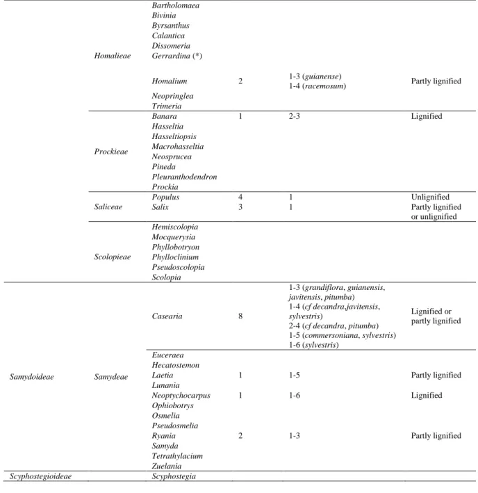
Table 2. Genera studied among Salicaceae, based on Chase et al. (2002). The column 'Described species' indicates the number of species observed in a given genus. Tension wood cell wall, the number of layers min-max of the G-layer and the presence of lignification of the G-G-layer are described. (*) This genus has now been moved to a distantly related family (Alford, 2006).
Sub family Tribes Genera Described
species
Number of layers Tension wood
lignification
Incertae sedis Oncoba
Salicoideae Abatieae Abatia Aphaerema Bembicieae Bembicia Flacourtieae Azara 1 1-3 Unlignified Bennettiodendron Carrierea 1 1-2 Unlignified
Dovyalis 1 1-3 Partly lignified
or unlignified Flacourtia Idesia 1 1 Unlignified Itoa Lasiochlamys Ludia Olmediella Poliothyrsis 1 1–3 Unlignified Priamosia Tisonia Xylosma 4 1-3 (flexuosa) 2-3 (japonica) 1-4 (benthamii, congesta) Partly lignified
13 Homalieae Bartholomaea Bivinia Byrsanthus Calantica Dissomeria Gerrardina (*) Homalium 2 1-3 (guianense)
1-4 (racemosum) Partly lignified
Neopringlea Trimeria Prockieae Banara 1 2-3 Lignified Hasseltia Hasseltiopsis Macrohasseltia Neosprucea Pineda Pleuranthodendron Prockia Saliceae Populus 4 1 Unlignified
Salix 3 1 Partly lignified
or unlignified Scolopieae Hemiscolopia Mocquerysia Phyllobotryon Phylloclinium Pseudoscolopia Scolopia Samydoideae Samydeae Casearia 8 1-3 (grandiflora, guianensis, javitensis, pitumba) 1-4 (cf decandra,javitensis, sylvestris) 2-4 (cf decandra, pitumba) 1-5 (commersoniana, sylvestris) 1-6 (sylvestris) Lignified or partly lignified Euceraea Hecatostemon
Laetia 1 1-5 Partly lignified
Lunania
Neoptychocarpus 1 1-6 Lignified
Ophiobotrys Osmelia Pseudosmelia
Ryania 2 1-3 Partly lignified
Samyda Tetrathylacium Zuelania
Scyphostegioideae Scyphostegia
Table 3. Number and percentage of the studied genera among Salicaceae and of the presence of a multilayered wall in tension wood fibres of the studied genera.
Tribe (or genus) Genera Studied genera Studied genera with multilayered wall
Number Number % Number %
(Oncoba) 1 0 0 ─ ─ Flacourtieae 14 6 42 5 (- Idesia) 83 Homalieae 9 1 11 1 100 Saliceae 2 2 100 0 0 Scolopieae 6 0 0 ─ ─ Abatieae 2 0 0 ─ ─ Bembicieae 1 0 0 ─ ─ Prockieae 8 1 12 1 100 Samydeae 13 4 30 4 100 (Scyphostegia) 1 0 0 ─ ─ 57 14 24 11 78
Supplementary material 1. Transverse sections of tension wood fibres observed with 3D Laser Scanning Confocal Microscope . Scale bar : 10µm. A-B: Lacistemataceae, C-S: Salicaceae s.l. A: Lacistema pubescens, B: L. sp (thick S2 layer), C: Idesia polycarpa var. vestitata, D: Populus nigra (italica), E: P. trichocarpa, F: P. deltoides × nigra
14
(thick S2 layer), G: Salix myrsinifolia, H: S. purpurea (thick S2 layer), I: Casearia commersoniana, J: C.
grandiflora, K: C. guianensis, L: C. javitensis, M: C. pitumba, N: C. sylvestris, O: Homalium racemosum, P: Ryania speciosa var. bicolor, Q: Xylosma congesta, , R: X. flexuosa, S: X. japonica. A-H: One layer observed in