916 Notes Eur. J. Clin. Microbiol. Infect. Dis.
Influence of Hepatitis G Virus
Infection on Liver Disease
I.D. Diamantis 1., E.
Kouroumalis 2,
M. Koulentaki 2, E. Fasler-Kan 1,
RA. Schmid 3, H.H. Hirsch 4, H. Bt~hler 3,
K. Gyr 1, M. Battegay 1
The influence of hepatitis G virus (HGV) infection on disease activity in hepatitis C related and unrelat- ed liver disease was investigated in 254 individu- als using an EIA polymerase chain reaction assay for HGV. One hundred patients had chronic hepa- titis C, 26 primary biliary cirrhosis, and 30 alcohol- ic liver cirrhosis. In addition, 51 hepatitis B surface antigen (HBsAg)-positive and 47 anti-hepatitis C vi- rus (HCV)-positive blood donors were screened. Hepatitis G virus was detected in 18% of patients with chronic hepatitis C, 13% of patients with al- coholic liver cirrhosis, 11% of patients with primary biliary cirrhosis, 10% of anti-HCV-positive blood do- nors, and 2% of HBsAg-positive blood donors. Vi- rus load and alanine aminotransferase (ALT) levels did not differ significantly in patients with HCV alone versus patients coinfected with HCV and HGV. However, mild liver fibrosis correlated with HGV coinfection. Hepatitis G virus did not influence ALT levels or liver damage in liver disease unrelat- ed to viral infection.
Hepatitis G virus (HGV) (1) belongs to the Fla- viviridae family and has a genomic organization similar to that of the hepatitis C virus (HCV) (2). Hepatitis G virus has been detected in patients with liver diseases such as chronic and acute hep- atitis (1, 3-5) and in patients with risk factors of
1 Outpatient Department of Internal Medicine and Depart- ment of Research, Petersgraben 4, University Hospital Ba- sel, 4031 Basel, Switzerland.
2 Department of Gastroenterology, University Hospital Hera- klion, Crete, Greece.
3 Department of Internal Medicine, Waid Hospital, Zfirich, Switzerland.
4 Institute for Medical Microbiology of the University of Ba- sel, Basel, Switzerland.
parenteral exposure, e.g. multiple transfusions (6) and intravenous drug use (7). Hepatitis G virus was also detected in patients with no evidence of liver disease and without known risk factors, such as volunteer blood donors. Recent reports suggest that coinfection of HCV-infected patients with H G V does not influence the outcome of HCV- related hepatitis or the response to interferon ther- apy (8, 9). The aims of this study were (i) to study the prevalence of H G V infection in patients with chronic liver disease and in blood donors found positive for hepatitis B surface antigen (HBsAg) or anti-HCV; (ii) to examine whether H G V had an influence on H C V replication; and (iii) to an- alyze whether H G V / H C V coinfection influences the degree of liver disease.
Patients and Methods. Data and sera from 254 pa- tients were analyzed. Of these patients, 100 had histologically proven chronic hepatitis C (Waidspi- tal Ztirich, Switzerland), 26 had primary biliary cir- rhosis, and 30 had alcoholic liver cirrhosis (Univer- sity Hospital of Heraklion, Greece). Samples from 47 anti-HCV-positive and 51 HBsAg-positive blood donors were also obtained from the Univer- sity Hospital of Heraklion, Greece.
Total nucleic acids were extracted from serum ac- cording to published methods (10) and subjected to reverse transcriptase reaction. For the detection of H G V sequences, two sets of primers were used. One set derives from the NS5a region (5'- CTC TTT GTG GTA GCC GAG A G A T-3', po- sition 6904 and 5'-CGA ATG AGT CAG A G G ACG G G G TAT-3' position 7059) and gives rise to a 156bp polymerase chain reaction (PCR) fragment, and the other derives from the 5'-non coding region (5'-NCR) (5'-CGG CCA A A A GGT G G T G G A TG-3' position 100, and 5'- C G A C G A GCC T G A CGT C G G G-3' position 285) and gives rise to a 186bp PCR fragment. An Expand PCR system (Boehringer, Mannheim, Germany) was used, with 45 cycles of 1 min at 94~ 1 rain and 25 sec at 55~ and 1 rain at 72~ To confirm the specificity of amplified products, and E I A PCR employing an EIA PCR labelling and detection system was used (Boehringer Mannheim). The specific probes for the hybridi- zation were biotinylated at the 5'-end. The probe for the NS5a region was 5'-biotin-GTT ACT GAG AGC AGC TCA GAT-3' position NS5a 152- 172 and for the 5'-NCR region 5'-biotin-GGT AGC CACTAT A G G TGG G-3'.As positive con- trol HGV, ribonucleic acid (RNA) transcripts were used, a generous gift from Genelabs Technol- ogies, USA. Values higher than the cutoff level (3
Vo1.16,1997 Notes 917
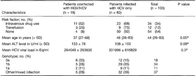
Table 1: Characteristics of 100 patients with chronic hepatitis C.
Characteristics
Patients coinfected Patients infected with HGWHCV with HCV only
(n = 18) (n = 82)
Total (n = 100)
P value
Risk factor; no. (%)
Intravenous drug use 11 (32) 23 (68)
Transfusion 3 (25) 9 (75)
None 4 (8) 50 (92)
Mean age in years (• SD) 37 (27-68) 46 (26-83) Mean ALT level in U/ml (_+ SD) 133 + 78 108 _+ 103 Mean HCV viral load in Eq/ml 264049 _+ 293635 301096 _+ 616829 Genotype; no. (%) 3a 6 (33) 12 (15) t b 5 (28) 29 (35) l a 2 (11) 9 (11) Other/mixed infection 5 (28) 32 (39) 34 (34) 12 (12) 54 (54) 44 (26-83) 18 34 11 37 0.05 a 0.09 a 0.3 a a Wilcoxon test.
ALT, alanine aminotransferase.
times the mean value of 3 negative controls) in the E I A PCR assay were defined as positive. The H C V viral load was measured using the Am- pticor system (Roche, Switzerland). Antibodies against H C V and HBsAg were detected by immu- noassays. Histological examination was per- formed using standard criteria, with fibrosis as- sessed semiquantitatively (11). The Innolipa strip assay (Innogenetics, Belgium) was used for detec- tion of H C V and analysis of genotype. For the sta- tistical analysis, the Yates corrected chi-square method and the Wilcoxon test were applied.
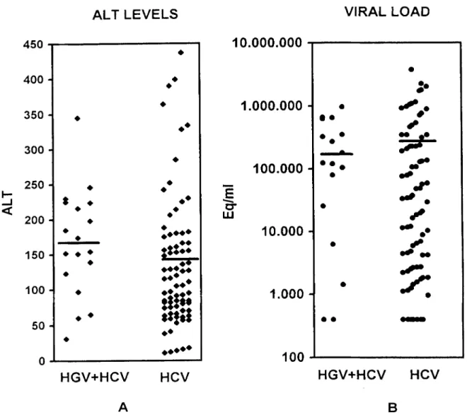
Results and Discussion~ Of 100 patients with his- tologically proven chronic hepatitis C, 34 had used intravenous drugs, 12 had a history of trans- fusions, and the remaining 54 had no known risk factors (Table 1). Altogether, 18 of 100 (18%) HCV-infected patients were coinfected with HGV. The highest prevalence was found in intra- venous drug users (32%). Patients infected with H C V alone were older (p = 0.05). Thirty-three per- cent of coinfected patients but only 15% of pa- tients with H C V infection alone had H C V geno- type 3a, whereas 28% of coinfected and 35% of patients infected with H C V alone had genotype lb. Genotype la was found in an equal proportion of both groups (11%). Other genotypes, including infections with a mixture of genotypes, were found in 28% of coinfected patients and in 39% of patients with H C V infection alone. Ninety-four of the 100 patients had elevated ALT levels. Al- though H G V / H C V coinfected patients had slightly higher ALT levels, the difference was not significant (p = 0.09,Wilcoxon test, Table i and Fig-
ure 1A). Virus load was slightly higher in patients with H C V infection alone, but the difference did not reach significance (p = 0.3, Wilcoxon test, Ta- ble 1 and Figure 1B). In both groups ALT and vi- rus load values were distributed over a broad range. Of the 18 patients coinfected with H G V and HCV, 16 (90 %) had histologically proven mild liv- er fibrosis; only two patients showed a severe fi- brosis. Both patients with severe liver fibrosis and H G V / H C V coinfection had a mixture of H C V genotypes. All six patients with genotype 3a, all five with genotype lb, and both with genotype la had mild fibrosis. Forty-seven of 82 (58 %) patients with H C V infection alone had mild fibrosis, and 35 (42%) had severe fibrosis. The difference re- garding fibrosis in H G V / H C V versus HCV-infect- ed patients was statistically significant (p = 0.02, Yates corrected chi-square test).
Twenty-six patients with primary biliary cirrhosis (median age 63 years, range 36-78 years) were an- alyzed. Only four individuals had elevated ALT levels. Histologically, eight patients had grade IV liver disease, four had grade III, seven grade II, and the remaining four grade I. Three of the 26 pa- tients were infected with H G V (11.5 %). However, ALT was elevated in only one HGV-infected pa- tient without severe disease. Thirty patients with alcoholic liver disease were examined, two of whom had elevated ALT levels but were infected with HCV. Hepatitis G virus was detected in four patients (13 %), all of whom had normal ALT lev- els. The histological degree of liver injury was not different in H G V positive or negative patients. Fifty-one HBsAg-positive and 47 anti-HCV-pos- itive blood donors were examined. Four of the
918 Notes Eur. J. Clin. Microbiol. Infect. Dis.
A L T L E V E L S
V I R A L L O A D
I-.- _,1 4 5 0 4 0 0 3 5 0 3 0 0 2 5 0 2 0 0 1 5 0 100 5 0 0 $ 9 o 9 o 9 0 9 0 0 9 1 4 910.000.000
1.000.000
E
100.000
10.000
I9 9 1 4 9 I
9 9 1 4 9
1.000
:tli*
*ill!
9 * 9 1 4 9100
9.,.,.
O 9 o o 9 ~ .ip 9oOe 9
9 0 0 2H G V + H C V
H C V
H G V + H C V
H C V
A
B
Figure 1: Alanine aminotransferase levels and HCV viral load values in individual patients with chronic hepatitis C coinfect- ed with HGV or infected with HCV alone. Figure 1A shows the alanine aminotransferase levels and 1B the viral load values. The mean value is indicated by a horizontal line. Alanine aminotransferase is given in U/I and the viral load in equivalent vi- ral copies per milliliter.
HBsAg-positive blood donors and two of the anti-HCV-positive donors had elevated ALT lev- els. Only one of 51 (2%) HBsAg-positive blood donors was H G V positive, with a normal ALT lev- el. In the group of anti-HCV-positive donors, five were H G V positive (10%), all of whom had nor- mal ALT levels.
I n this study we found that (i) H G V is prevalent in patients with chronic hepatitis C and different risk factors as well as in patients with other liver diseases; (ii) viremia did not differ significantly in HGV/HCV-coinfected versus HCV-infected pa- tients; and (iii) H G V / H C V coinfection was signif- icantly associated with mild fibrosis.
The prevalence of H G V is higher in HCV-infect- ed patients with risk factors, such as intravenous drug use and transfusions (7,12). Interestingly, pa- tients infected with H G V were younger than pa- tients infected with H C V alone. Possibly, hepati- tis G, in contrast to hepatitis C, is a self-limited vi- ral infection that is cleared from the serum after a number of years, resulting in infection by H C V alone after years or decades. ~[lais hypothesis is supported by our previous finding that drug users who started intravenous heroin use before 1980 had a lower prevalence of H G V than those who started after 1980 (7). Alternatively, this observa- tion may be explained by the notion that H G V was introduced only recently in these popula- tions. Hepatitis G virus and H C V share the paren-
Vol. 16, 1997 Notes 919
teral mode of transmission, but the high preva- lence found in patients with alcoholic liver cirrho- sis, patients with primary biliary cirrhosis, and other low-risk groups investigated in our study suggests that other modes of transmission prob- ably occur.
Virus load and ALT levels were slightly but not significantly different in HCV-infected versus HGV/HCV-coinfected patients. Possibly, as both vi- ruses belong to the Flaviviridae family and have a high degree of sequence homology, interferences may occur at many stages, e.g., at the replication level or during the production of viral proteins. In this study we found that H G V / H C V coinfection may lead to a rather mild course of liver disease. Similarly, Tanaka et al. (8) observed a tendency for less severe liver fibrosis in HGV/HCV-coinfected patients. Our observation, that all patients with H C V genotypes 3a and l b coinfected with H G V had only mild fibrosis, indicates that this difference is probably not due to genotypes, since lb is usu- ally correlated with severe disease (13).
One limitation of this study may be that the two groups of patients with chronic hepatitis, the one coinfected with H G V / H C V and the other with H C V alone, were different regarding age, mode of acquisition, and possibly other factors. This may have influenced our results regarding severity of fibrosis. In particular, the older age in the group of patients with H C V infection alone may have negatively influenced the degree of liver fibrosis. Neither ALT levels nor the severity of liver pathol- ogy was influenced in alcoholic liver disease or pri- mary biliary cirrhosis, suggesting that H G V infec- tion plays rather a secondary role in the develop- ment of these diseases, with no etiologic link. The higher prevalence of H G V infection among these patients as compared to the normal population may indicate that a liver with pathology enables the persistence of H G V infection.
In conclusion, the data presented in this study sup- port the hypothesis that H G V infection does not worsen the outcome of H C V infection and may even have a favorable effect.
Acknowledgment
The authors thank Genelabs Technologies, Redwood City, CA, USA, for providing the HGV positive control, Alana Althage for critical reading of the manuscript, and Mich6le Girard for excellent secretarial assistance.
This study was supported by a grant from the Swiss National Science Foundation (32-43'413.95) and the Kanton of Basel- Stadt.
References
1. Linnen J, Wages J, Zhang-Keck ZY, Kyrk EF, Krawczyn- ski KZ, Alter H, Koonin E, Gallagher M, Alter M, Hadziy- annis S, Karayiannis P, Fung K, Nakatsuji Y, Shih JW, Young L, Piatak M, Hoover C, Fernandez J, Chen S, Zou JC, Morris T, Hyams KC, Ismay S, Lifson JD, Hess G, Foung SK, Thomas H, Bradley D, Margolis H, Kirn JP: Molecular cloning and disease association of hepatitis G virus: a transfusion-transmissible agent. Science 1996, 271:505-509.
2. Simons LN, Leary TP, Dawson GJ, Pilot-Matias T J, Muerhoff AS, Schlauder GG, Desai SM, Mushahwar IK: Isolation of novel virus-like sequences associated with human hepatitis. Nature Medicine 1995, 1: 564-569. 3. Yoshiba M, Okamoto H, Mishiro S: Detection of the GBV-
C hepatitis virus genome in serum from patients with ful- minant hepatitis of unknown aetiology. Lancet 1995, 346: 1131-1132.
4. Nakatsuji Y, Shih JW-K, Tanaka E, Kiyosawa K, Wages J, Kim JP, Alter HJ: Prevalence and disease association of hepatitis G virus infection in Japan. Journal of Viral Hepatitis 1996, 3:307-316.
5. Alter M J, Galiagher M, Morris TT, Moyer, LA, Meeks EL, Krawczynski K, Kim JP, Margolis HS: Acute non A-E hep- atitis in the United States and the role of hepatitis G vi- rus infection. New England Journal of Medicine 1997, 336: 741-746.
6. Alter H J, Nakatsuji Y, Melpolder J, Wages J, Wesley R, Shih JW, Kim JP: The incidence of transfusion associated hep- atitis G virus infection and its relation to liver disease. New England Journal of Medicine 1997, 336: 747-754. 7. Diamantis ID, Bassetti S, Erb P, Ladewig D, Gyr K, Bat-
tegay M: High prevalence and coinfection rate of hepa- titis G and C infections in i.v. drug users. Journal of Hep- atology 1997, 26: 794-797.
8. Tanaka E, Alter H J, Nakatsuji Y, Shih JW, Kim JP, Mat- sumoto A, Kobayashi M, Kiyosawa K: Effect of hepati- tis G virus infection on chronic hepatitis C. Annals of In- ternal Medicine 1996, 125: 740-743.
9. Berg T, Dirla U, Naumann U, Heuft HG, Kuether S, Lo- beck H, Schreier E, Hopf U: Responsiveness to interfer- on alpha treatment in patients with chronic hepatitis C coinfected with hepatitis G virus. Journal of Hepatology 1996, 25: 763-768.
10. Chomczynski P, Sacchi N: Single-step method of RNA iso- lation by acid guanidinium thiocyanate-phenol-chloroform extraction. Annals of Biochemistry 1987, 162: 156-159. 11. Knodel RG, Ishak KG, Black WC, Chen TS, Craig R,
Kaplowitz N, Kiernan TW: Formulation and application of a numerical scoring system for assessing histological ac- tivity in asymptomatic chronic active hepatitis. Hepatol- ogy 1981, 1: 431-435.
12. Schreier E, H6hne M, KLinkel U, Berg, T, Hopf U: Hepa- titis GBV-C sequences in patients infected with HCV con- taminated immunoglobulin and among i.v. drug users in Germany. Journal of Hepatology 1996, 25: 385-389. 13. Pawlotsky JM, Tsakiris L, Roudot-Thoraval F, Pellet C,
Stuyver L, Duval J, Dhumeaux D: Relationship between hepatitis C virus genotypes and sources of infection in patients with chronic hepatitis C. Journal of Infectious Diseases 1995, 171: 1607-1610.