Susceptibility of Normal and Transformed Cell Lines to Cytostatic and Cytocidal Effects
Exerted by Macrophages
1, 2R. Keller3, 4
SUMMARY-Activated, nonimmune macrophages exerted pro-found effects on the proliferation and viability of eukaryotic target cells in vitro. Pronounced macrophage-mediated cyto-stasis was exerted on every rapidly proliferating cell line ex-amined, irrespective of transformation, species derivation, cell type, or growth characteristics. However, the magnitude of cytostasis effected differed markedly among the 40 cell lines tested. There was no evident correlation between susceptibility to cytostasis and degree of transformation. Transformed cell lines with high and with low malignant attributes were affected equally. A comparable pattern was discerned for cytocidal effects of macrophages, in which the susceptibility of trans-formed targets was independent of the degree of
malignancy.-J Natl Cancer Inst 56: 369-374, 1976.
Recent work (1-5) has shown that apart from the im-portant function of mononuclear phagocytes in patho-logic processes, the presentation of antigen to immuno-competent cells, and resistance against intracellular microorganisms, mononuclear phagocytes also exert pro-found effects on eukaryotic cells. These in vitro studies have yielded voluminous data indicating that activated, nonimmune macrophages (AM) can affect target cells in a variety of ways, and that they can discriminate between normal and transformed cells and resting and replicating cells. These destructive capabilities apparently involve the metabolic state of a macrophage population, as well as the proportion of effectors to targets. The findings thus far indicated that the cytostatic effects on targets exerted by AM are independent of target species (syngeneic, allo-geneic, or xenogeneic), target cell type (epithelial or lym-phoid), target growth characteristics (monolayer or sus-Pension), or transformation (normal vs. neoplastic tissue) (4-7). There is general agreement (1-4, 8-10) that inter-action of AM with tumor targets results in an altered morphology and decrease in the number of cells, attesting to the Al\tI's capacity to kill tumor targets in vitro. How-ever, further investigation on many cell lines derived from normal and transformed tissue disclosed that macro-phage cytocidal target cell effects are not invariably correlated with the malignant attributes of cell lines derived from transformed tissue.5,6 The present work
further explores whether the macrophage-mediated cyto-cidal effect is selective for tumors.
MATERIALS AND METHODS
Target cell lines.-Rat: Cells derived from normal rat kidney (NRK) and rat kidney cells infected with B77
Rauscher murine leukemia virus (B77=NRK) (11), a gift
from Dr. T. Graf, were grown in Eagle's minimum essen-tial medium (MEM) (12) modified as follows: 280 mg glutamine/liter, 100 mg calcium/liter, 1 g NaHCOs/liter, 2 g glucose/liter, and 1 mg biotin/liter, and supple-mented with 100 U penicillin/ml, 50ft streptomycin/ml (modified MEM), 10% fetal calf serum (FCS), and 10% tryptose phosphate (Difco Laboratories, Detroit, Mich.). DA rat tumors were those described in (4) or were newly induced with polyoma virus, dimethylbenz[a]anthracene
(DMBA), or 3-methylcholanthrene (MCA). These tumor cells were grown in modified MEM supplemented with 10% newborn calf serum (NCS). DA rat adult and embryo fibroblasts were repeatedly newly established (6) and grown in modified MEl\tI supplemented with 10% FCS.
Mouse: The A9 cell (having little ability to grow pro-gressively in vivo), its highly malignant counterpart A9HT (high incidence of take) (13), and the hybrid cell lines between these L-cell derivatives and malignant mouse tumors, such as Ehrlich (spontaneous carcinoma), SEWA (polyoma-induced sarcoma), MSWBS (MCA-in-duced sarcoma), and YACIR (an immunoresistant deriva-tive of the YAC tumor, a Moloney virus-induced lym-phoma) (13, 14), provided by Dr. George Klein, were grown in nonmodified Eagle's MEM. BALB/c3T3 cells and BALB / c simian virus 40 (SV40)-transformed 3T3 cells, originally obtained from Dr. Stuart Aaronson, were maintained in modified l\-fEM with 10% FCS. Suspension cultures of the transplantable murine mast cell tumor P-815 X2, obtained from Drs. R. Schindler and M. Bertschmann, were grown as described in (15); the me-dium was supplemented with 10% NCS instead of horse serum.
Human: Three adherent cell lines derived from hu-man mammary carcinomas were used: BT-20, supplied by Dr.
J.
Fogh, was grown in modified MEM supple-mented with 10% FCS; MPZ-2 and MPZ-4 were freshly established from a biopsy of human breast cancer, pro-vided by Dr.J.
R. Riittner and grown in modified MEM supplemented with 15% FCS. Two human melanoma lines were used. SK-melanoma-l (MEL-I) cells, obtained from Dr. K. T. Brunner, were grown in suspension in modified MEM supplemented with 10% FCS. Melanoma cells (RPM I 7932), provided by Dr. W. D. Terry, were grown in adherent culture in modified MEM with 10% FCS. SK-OS-5, derived from an osteogenic sarcoma and supplied by Dr.J.
Fogh, was grown in adherent culture1Received June 2, 1975; accepted September 15, 1975.
2Supported by grants 3.516.71 and 3.234.74 from the Swiss Na-tional Science Foundation.
s Immunobiology Research Group, University of Zurich, Schon-leinstrasse 22, CH-8032 Zurich, Switzerland.
4I thank the following colleagues for providing cell lines: Dr. S. Aaronson, National Cancer Institute (NCI), National Institutes of Health (NIH), Bethesda, Md.; Dr. A. C. Allison, Clinical Research Centre, Harrow, England; Dr. M. Bertschmann, Theodor Kocher-Institut, Bern, Switzerland; Dr. K. T. Brunner, Institut Suisse des Recherches Experimentales sur Ie Cancer, Lausanne, Switzerland; Dr. J. Fogh, Sloan-Kettering Institute for Cancer Research, Rye, N.Y.; Dr. T. Graf, Max Planck-Institut flir Virusforschung, Tiibin-gen, W. Germany; Dr. G. Klein, Institute of Tumor Biology, Karo-Hnska Institutet, Stockholm, Sweden; Dr. J. R. Riittner, Patho-logisches Institut, University of Zurich; Dr. Ch. Sauter, Medizinische Universitatshlinik, Kantonsspital, Zurich; Dr. R. Schindler, Patho-logisches Institut der Universitat, Bern; and Dr. W. D. Terry, NCI, NIH. I also thank Dr. Maurice Landy, Schweizerisches Forschungs-institut, Davos, Switzerland for his helpful criticism of this manu-script, and Miss R. Keist and MissE. Miiller for their expert tech-nical assistance.
5Bregnard A, Gehring WJ, Keller R, et al: In preparation.
6Keller R: Unpublished data.
in modified MEM with 10% FCS. The Burkitt's lym-phoma cell line RAJI, obtained from Dr. G. Klein, was grown in modified MEM supplemented with 10% FCS. BEN, a tumor cell line producing carcinoembryonic anti-gen, was supplied by Dr. Ch. Sauter and grown in ad-herent culture in modified MEM with 10% FCS. Cell lines were repeatedly checked for the possibility of myco-plasma contamination, but none was found.
Origin, culture conditions, and properties of other cell lines used were described in (6).
Macrophage monolayers were prepared as described in (4, 6). Peptone-induced peritoneal cells from inbred DA rats were seeded into plastic petri dishes. After 30 min-utes at 37° C, the nonadhering cells were removed by intensive washing. After this procedure, at least 96% of the cells in the monolayer of approximately 2X 1~ cells showed the characteristics of macrophages. To these macrophage monolayers, target cells (2X 10⁵/dish) were immediately added and the cultures were maintained at the appropriate temperature in a humid atmosphere of 5% CO2 and 95% air.
Measurement of cytostasis.-Residual target cell pro-liferation was· assessed after varying intervals of macro-phage-target cell interaction by: I) exposure for 60 min-utes at 37° C to I ,p.Ci 3H-methylthymidine (3H-TDR)/ dish (5,000 mCi/mmole; The Radiochemical Centre, Amersham, Buckinghamshire, England), and processing as described in (4, 6); or 2) by exposure for 8 hours at 37° C to 0.1 ,p.Ci 1251-5-iodo-2'-deoxyuridine (125IUDR, sp act, 8-10 p,Ci/p.g; The Radiochemical Centre). After careful washing, the cells were washed twice with 1.5% perchloric acid and radioactivity was measured in an automatic gamma counter (Tracerlab, Inc., Waltham, Mass.).
Assessment of target cell viability.-Two methods were used to assess target cell viability. In experiments per-formed to assess the capacity of target cells to reestablish growth, 2X10⁵ targets were cultured in the presence of 2xl~ AM (6). After 72 hours, the cells were harvested by trypsinization, washed, counted, and diluted to a concentration of 300/3.5 ml; aliquots of this volume were also dispensed to 30-ml Falcon culture flasks. After 10 days' incubation in 5% CO2 at 37° C, we assessed target cell proliferation by adding 1 p.Ci 3H-TDR for 60 min-utes at 37° C and by processing the cells as described. Lines derived from the A9 cell were easily distinguished morphologically from AM. Thus the number of target cells remaining in culture after a 72-hour interaction with AM could be counted after trypsinization.
In other experiments, the release of 125IUDR from prelabeled targets was a measure of macrophage-medi-ated target cell damage. Subconfluent cultures of target cells grown in 250-ml Falcon tissue culture flasks con-taining 10 ml medium were pulsed for 8 hours with Ip.Ci 125IUDR/flask (O.Ip.Ci/ml) in the presence of 2'-fluoro-5-deoxyuridine (FUDR) at a concentration of 10-5M.The
FUDR, a known inhibitor of thymidylate synthetase, was included to increase 125IUDR incorporation into DNA
(16), in place of 3H-TDR.
In many target cell lines, incubation with a combina-tion of 125IUDR and FUDR in the concentracombina-tions and time indicated resulted in adequate specific labeling with-out marked signs of toxici ty; these particular cell lines were used here. Following incubation with 125IUDR for 8 hours, the cells were washed twice with phosphate-buffered saUne to remove unincorporated isotope. The prelabeled cells (2X 10⁵/dish) were then added to macro-phage monolayers and incubated for varying intervals,
after which the cells were removed by a pipette and then centrifuged. Supernatants and cells were enumerated separately in an automatic Tracerlab gamma counter. Results are expressed as a percent of cytocidal capacity, calculated as follows:
Experimental dpm-control dpm X 100, total dpm
where dpm
=
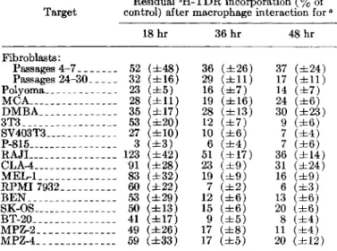
disintegrations per minute. RESULTSCytostatic Target Cell Effects Mediatedby Macrophages Following interaction for different intervals with 10 macrophages per target cell, the 3H-TDR incorporation, a measure of residual target cell proliferative capacity, was profoundly diminished in all of the target cell lines examined (table I). However, there were considerable differences in the susceptibility of different target cell lines to macrophage-mediated cytostasis. Proliferation of several cell lines (i.e., polyoma virus-induced syngeneic rat tumor cells, xenogeneic mouse SV403T3, and espe-cially P-815 mastocytoma cells) was blocked even in the early phase of interaction and remained at a very low rate as interaction proceeded. Other cell lines were initi-ally more resistant to macrophage-mediated cytostasis but were inhibited during the later phases of interaction; this was seen with 3T3, RPMI 7932, BEN, BT-20, MPZ-2, MPZ-4, SK-OS, and MEL-I cells. Proliferation of RAJI and several other human lymphoblastoid cell lines was often enhanced in the early phase of interaction with macrophages, but was subsequently diminished as the interaction proceeded. DMBA- and MCA-induced syn-geneic rat tumor cells, although effectively inhibited in the early phase, retained proliferative capacity during continuous interaction with macrophages. Proliferation of fibroblasts, derived from rat embryo (or adult) tissues and exposed to macrophages during in vitro passages 4-7 and 24-30, was distinctly diminished in the early phase but often remained unchanged or was even enhanced as interaction proceeded; such reversed reaction to macro-phages was observed especially with recently explanted fibroblasts. Thus in all target cell lines examined,
pro-TABLE I.-Marked inhibition of proliferation of normal and neoplastic target cells(2Xl05) in the presence of2X106DA rat macrophages
Residual3H-TDR incorporation(%of Target control) after macrophage interaction for"
18 hr 36 hr 48 hr Fibroblasts : Passages 4-7 _______ 52 (±48) 36 (±26) 37 (±24) Passages 24-30 _____ 32 (±16) 29 (±11) 17 (±11) Polyoma_____________ 23 (±5) 16 (±7) 14 (±7) MCA________________ 28 (±11) 19 (±16) 24 (±6) DMBA______________ 35 (±17) 28 (±13) 30 (±23) 3T3 _________________ 53 (±20) 12 (±7) 9 (±6) SV403T3 _____________ 27 (±1O) 10 (±6) 7 (±4) P-815________________ 3 (±3) 6 (±4) 7 (±6) RAJL _______________ 123 (±42) 51 (±17) 36 (±14) CLA-4_______________ 91 (±28) 23 (±9) 31 (±24) MEL-l ______________ 83 (±32) 19 (±9) 16 (±9) RPMI 7932 __________ 60 (±22) 7 (±2) 6 (±3) BEN ________________ 53 (±29) 12 (±6) 13 (±6) SK-OS _______________ 50 (±13) 15 (±6) 20 (±6) BT-20 _______________ 41 (±17) 9 (±5) 8 (±4) MPZ-2 ______________ 49 (±26) 17 (±8) 11 (±4) MPZ-4 ______________ 59 (±33) 17 (±5) 20 (±12)
NORMAL AND TRANSFORMED CELL LINES 371
liferation was clearly inhibited when the proportion of macrophages was in the majority, but the pattern of inhibition varied markedly from one cell line to another. Results similar to those with 3H-TDR were obtained when targets, which interacted for varying intervals with macrophages, were pulse labeled with 125IUDR.
15 21. 36 1.8 72hours
TEXT-FIGURE I.-Decrease in number of neoplastic and normal target
cells (initially 2X10⁵) during interaction with macro ph ages
(2X IOn). Target cell lines: D = DMB-induced DA rat tumor
cells; .0.=polyoma-induced DA rat tumor cells; O=mouse SV403T3
cells; 0 =mouse 3T3 fibroblasts; \7=CHO hamster fibroblasts.
Closed symbols arc targets alone; open symbols are targets in the presence of AM. 53 (±9) 35 (±6) 22 (±9) 29 (±6) Cloning efficacya NRK B77 ts, 38°C ts, 33° C Target cell lines
aPercent of control as represented by the capacity for 3R-TDR Incorporation. Values are means(±SD)of 10 determinations.
TABLE3.-Differing effects by AM on capacity of normal and
transformed targets to reestablish growth
cells were equally affected, irrespective of whether or not they were permissive.
Experiments, in which loss of capacity to reestablish growth after prolonged culture with AM was taken as a measure of macrophage-mediated cytocidal target cell effects, revealed a significant cloning reduction in all cell lines examined (table 3). However, cytocidal effects were most pronounced in transformed cells, and no dif-ference occurred between cells grown under permissive or nonpermissi ve condi tions.
It was recently shown that the fusion of highly nant mouse cells with normal cells or cells of low malig-nancy yields a hybrid with suppressed malignant char-acteristics (14). Among such hybrids, the A9 series is particularly interesting since hybrids with the slightly malignant L-cell subline showed suppressed malignancy, but partners of the highly malignant L-cell subline re-tained undiminished malignancy (13). Thus it appeared that a comparison between pairs of A9, A9HT, and the various A9-tumor hybrids might give information rele-vant to the present issue.
Accordingly, the macrophage effects on five paired lines consisting of a slightly malignant (A9) and highly malig-nant (A9HT) counterpart were assessed. The data in text-figure 2show that in every cell line examined, inter-action with AM resulted in a marked diminution of 3H-TDR incorporation. However, when compared with most previously examined cell lines, several L-cell hybrids were surprisingly resistant to macrophage-mediated cyto-stasis; this became especially evident after 4 and 15hours. After this interval, proliferation of a few cell lines was even stimulated by AM. Again, the susceptibility of the cell lines to macrophage-mediated cytostasis seemed inde-pendent of the degree of malignancy.
When macrophage-mediated cytocidal effects. were as-sessed by an enumeration of the targets remaining after various intervals, all cell lines were comparably affected (text-fig. 3). In the absence of effectors, the cell numbers increased progressively; however, in their presence, cell numbers remained low and decreased as interaction pro-ceeded (table 4). Again, no clear distinction was found in the susceptibility of cells with low or high malignancy within each comparable pair.
•
-:
•
•
.... o "" Q)1
zCytostatic and Cytocidal Macrophage Effects on Targets of Low and High Tumorigenicity
Enumeration of targets remaining after AM interac-tion of some lines revealed that, irrespective of whether targets were derived from normal or from transformed tissues, their number decreased as interaction proceeded (text-fig. 1). Since these experiments resolved neither the issue whether macrophage cytostatic and cytocidal effects were related nor whether cytocidal effects were indeed tumor specific, further experiments were made on other cell lines.
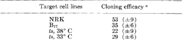
Cell lines derived from NKR, its virus-transformed counterpart, and from a temperature-sensitive mutant
(ts 339) permissive at 33° C but not at 38° C (11), pro-vided a useful model to probe the extent to which macro-phage-mediated cytostatic and cytocidal effects are re-lated to malignancy. Proliferation rate among these cells differed considerably, but was high enough in each instance to enable reliable quantitation of 3H-TDR in-corporation into target cell DNA (table 2). These data show that in everyone of these cell lines the residual capacity to 3H-TDR decreased as the period of inter-action with AM increased. Moreover, the findings showed that NRK cells, although significantly inhibited, were less susceptible to macrophage-mediated cytostasis than were the transformed lines. However, the transformed
TABLE2.-Correlation between degree of macrophage-mediated cytostasis and growth characteristics of different rat cell lines
Residua13H-TDR incorporation(%of control) by targets post culture with AM in ratio of
Targeta Proliferative capacity 10 AM/target after interaction with AM forb
(dpm at 60 hI')
12hI' 24hI' 36hI' 48hI' 60hI'
4hI'
NRK 16,000-26,000 82 (±2l) 50 (±15) 38 (±12) 30 (±8) 32 (±3) 32 (±5)
B77 34,000-88,000 63 (±1O) 31 (±6) 14 (±3) 11 (±1 ) 11 (±3) 10 (±2)
ts, 38° C 48,000-120,000 52 (±9) 16 (±9) 8 (±2) 8 (±4) 8 (±4) 8 (±2)
ts, 33° C 82,000-212,000 40 (±19) 13 (±4) 8 (±5) 10 (±7) 5 (±3) 4 (±l)
aB1J =NRK cells infected withBnavian sarcoma VIrus; Is =NRK cells infected with temperature-sensitive mutant Is 339. permissive at 33° C and reverted to a normal phenotype at 38° C.
TABLE4.-Effect of AM on capacity of pairs of targets of low and high tumorigenicity to reestablish growth
Tumorigenicity Number of targetsa
Low: A9___________________________________ 14 A9 IMSWBS _ _ _ _______________________ 4 A9ISEWA. ___________________________ 2 A9/EA_ ______________________________ 14 A9
IY
ACIR . ________ 7 High: A9HT________________________________ 13 A9HTIMSWBS_ _ _ ____________________ 12 A9HTISEWA_ ________________________ 9 A9HTIEA_ ___________________________ 11 A9HTIY
ACIR ________________________ 1aAbsolute No. of targets (Xl()4; initially 2 XlOo)remaining in culture after 96-hr interaction with AM.
tumor cells, retained a high proportion of the label for at least 48 hours, whereas others (especially MEL-I, BEN, BT-20, and rat fibroblasts) often showed considerable spontaneous release. This leakage is probably an expres-sion of 125IUDR- and/or FUDR-mediated toxicity. Al-though these differences in resistance to the labeling procedure may considerably affect the outcome of cyto-cidal tests, the measurements of the label release from target cells proved reproducible and provided informa-tion. The results in table 5 show that macrophage-mediated release of 125IUDR from prelabeled target cells was consistently observed after 18 hours; after an 8-hour interaction, no such release was detectable. In many cell lines, and most evident in those with low spontaneous release (RPMI 7932, SK-OS, and RAJ I), specific release increased as interaction proceeded. However, other cell lines including P-8I5, MEL-I, 3T3, SV403T3, BT-20, BEN, MPZ-2, MPZ-4, and CLA-4, were also susceptible to cytocidal macrophage effects. No difference was dis-cerned between "normal" 3T3 fibroblasts and their virus-transformed counterparts. Among the cells examined, recently explanted syngeneic rat fibroblasts and D:MBA-induced syngeneic rat tumors were resistant; cytocidal effects were not consistently detectable.
A comparison of cytostatic and cytocidal macrophage effects on these targets (tables 1, 5) indicates that the
TABLE5.-Susceptibility of various target cell lines to macrophage-mediated cytocidal effects
Cell line
Percent of cytocidal activity effected
(12bIUDR release) ata 18 hr 36 hr 48 hr Fibroblasts Passages 4-7 _______ 15 (±14) 9 (±7) 8 (±7) Passages 24-30 _____ 17 (±8) 19 (±12) 21 (±!)) Polyoma _____________ 5 (±S) ~) (±6) 21 (±1O) MCA________________ 14 (±16) 12 (±9) U (±8) DMBA ______________ 9 (±5) 9 (±6) 8 (±15) 3T3 _________________ 13 (±5) 9 (±9) 10 (±8) SV403T3 ____________ 16 (±4) 23 (±5) 16 (±5) P-815 _______________ 20 (±5) 28 (±1O) 28 (±4) RAJI _______________ . 3 (±3) 30 (±7) 37 (±7) CLA-4 _______________ 6 (±4) 17 (±~)) 18 (±7) M EL-l ______________ 23 (±8) 38 (±6) 45 (±5) RPMI7932 __________ 11 (±4) 44 (±1O) 50 (±7) BEN ________________ 3 (±3) 10 (±1O) 13 (±17) SK-OS ______________ 23 (±4) 48 (±1O) 48 (±7) BT-20 _______________ 20 (±6) 49 (±17) 59 (±6) MPZ-2 ______________ 30 (±11) 28 (±11) 26 (±6) MPZ-4 ______________ 9 (±6) 12 (±7) 12 (±6)
"Values are means (±SD) of 10-15 experiments, each performed in trivlicatc
•
;:::;- '\7252 0 I-< .... § 0 (J ...•
0 ~ § 0 ~•
.... <U I-<'t,
D. 0 P-I-< 08
(J.s
~
p:: A•
Eo-<•
~ I ~ 15 24 48 72 96 hoursTEXT-FIGURE 2.-Macrophage-mediated (2X10⁶ ) inhibition of target
cell (2X10⁵) proliferation is not dependent on degree of
tumori-genicity. Target cell lines: .=A9; O=A9HT; .=A9/SEWA;
\7=A9HT/SEWA; .=A9/MSWBS; O=A9HT/MSWBS; .=A9/
YACIR; L.:,.=A9HT/YACIR; +=A9/EA; <:>=A9HT/EA. Closed
symbols are slightly malignant derivatives; open symbols are highly malignant derivatives.
24 6holrs
TEXT-FIGURE 3.-ln pairs of L-cell derivatives with low (A9) and
high (A9HT) tumorigenicity, the number of targets (initially
2X10⁵) was similarly affected during interaction with 2X10⁶ DA
rat macrophages. Target cell lines: 0= A9; \7 = A9 / SEWA;
O=A9/MSWBS; L.:,.=A9/YACIR; <:>=A9/EA; ¢=A9HT; y=A9HT/
SEWA; ijJ=A9HT/MSWBS; ,6.=A9HT/YACIR; t=A9HT/EA.
Open symbols are targets alone; closed symbols are targets in the presence of AM.
Cytocidal Effects Mediated by Macrophages
Among the cell lines suitable for cytocidal experiments (i.e., those which incorporated and subsequently retained an appropriate amount of 125IUDR), considerable differ-ences were seen in label retention. Some cell lines, e.g., RAJI, RPMI 7932, SK-OS, or polyoma virus-induced rat
280 260 ,-... 240 ..;:t 0 220
~
'-" (/) 200 M I"""'l Q) 180 (J ~ 160 Q) bO )..4 (Ij 140 ~ 4-1 0~
)..4 Q) 100<>
..c,
§
Z 80*
O~
6 40~
,
20~
10NORMAL AND TRANSFORMED CELL LINES
373
changes in these parameters during interaction were not consistently parallel. A close parallelism in the degree and time course of cytostatic and cytocidal manifestations was especially notable in the Iymphoblastoid lines RAJI and CLA-4 and in rat fibroblasts. However, no such parallelism in cytostatic and cytocidal effects was seen in polyoma virus-induced DA rat tumor and P-815 mouse mastocytoma cells.
DISCUSSION
Prior in vitro studies (4, 6) in this laboratory
con-si~tently demonstrated that interaction of macrophages
WIth targets first affects target cell proliferation and is often accompanied by a decrease in the number of tar-gets; the capability of remaining targets to reestablish growth was diminished. These studies led to the provi-sional conclusion that macrophage-mediated cytostasis transcends species, cell type, and growth characteristics and is exerted on all rapidly replicating cells, whether derived from normal or transformed tissues (4-7). The data reported here encompass more target cell lines and show that prolonged interaction with a majority of mac-rophages (effector to target cell ratio is 10: I) results in a distinct inhibition of target cell proliferation in every cell line examined, but the degree of cytostasis evoked by macrophages differs markedly from one cell line to an-other. The results were similar irrespective of whether 3H-TDR or 125IUDR was used as a pulse label to assess the residual proliferative capacity. Since macrophages do not replicate under tissue culture conditions and remain in the G1 or Gil phase of the cell cyde, incorporation of these labels is sharply restricted to target cells.
The data once again demonstrate that there are major differences in the susceptibility of cell lines. When a large array of cell lines is ranked by their susceptibility to macrophage-mediated cytostasis, normal replicating lymphoid cells (7), various virus-transformed lines (rat polyoma and B77 , mouse SV40), P-815 mouse mastocytoma cells, and some cell lines derived from human malignant tumors are especially sensitive; derivatives of the mouse
~ fibro~last,t?ough consistently blocked after prolonged
InteractIOn WIth macrophages, showed large differences in their initial sensitivity not correlated with their degree of malignancy. Other lines such as 3T3, CHO,
DMBA-and ~1CA-inducedrat tumor cells, the lymphoblastoid
cell hnes, and recently explanted fibroblasts derived from normal adult or embryonic DA rat tissue, were resistant to macrophage-mediated cytostasis. Accordingly, it is evident that in rapidly replicating cell lines, factors other than capacity for in vivo malignancy or in vitro trans-formation determine susceptibility to macrophage-mediated cytostasis.
Earlier studies (4) showed that indicators of immune cytotoxicity such as those widely employed in lymphocyte-target studies (i.e., release of 51Cr or uptake of trypan ?lue) were unsuitable for detection of macrophage-Induced ch~ngesin target cell viability. Accordingly, in some expenments, the number of target cells remaining after various intervals of interaction with macrophages was counted for quantitation of cytocidal macrophage effects on targets; the number of remaining targets shows some diminution by 24 hours and is further decreased as
the i~te!action proceeds. However, many targets often
remaIn In culture even after a 96-hour interaction with macrophages (table 4).
125IUDR has been used more frequently as a label of target cells for the detection of lymphocyte-mediated
cytotoxicity (17-21). The main advantages of '125IUDR a.re low spo~taneousrelease and little, if any, reutiliza-tlon. Ac~or~Ingly,release of the isotope from prelabeled targets lI~dIcates cell death and lysis (22-25); since 125IUDR IS. a gamma-emitting isotope, little preparation
?f sa~plesIS needed before counting. However, 125IUDR
IS toxIC (26, 27) a~d often blocks further cell replication.
The pre~entstudIes show, however, that despite
consid-erabl~ dIfferences amonp- the various cell lines, toxici ty
contnbuted by the labelIng procedure can be held within acceptable limits, provided the isotope concentration is kept low and incubation is limited to 8 hours. Under
such ~onditio~s,theincl~sionof FUDR to increase
pref-erentIally th~IncorporatIOn of IUDR into DNA in place of 3H-TDR IS a necessary precondition. vVith the use of these mo~ifications, target cell lines were sufficiently labeled WIthout pross signs of toxicity; i.e., 125IUDR-labeled cells rephcated at a rate similar to that of un-labeled controls. Thus the present work shows that this method ofmeas~ringcytocidal (and cytostatic) capacities of effectors applIes to targets long established in culture
or~ecentlyde.rived from n.ormal tissues, growing adherent
or In suspenSIOn, or shOWIng growth attributes typical of normal or transformed cells.
The accu~ate me.asu~ementof viability in a variety of
targetsan~Its applIcatlon. to macrophage-mediated effects
are essent~al for furthenng the understanding of the
processes Involved in the interaction between macro-phages and oth~r euka~yotecells. Our work clearly
dem-o~strates t?at InteractIOn of nonimmune macrophages
WIth. a vanety of prelabeled targets is accompanied by a conSIstent release of the label. Such release is not usually detectable before 18 hours, but often increases as inter-action . proceeds. Beyond the principal demonstration of
t?e eXIstenc~of the potent cytocidal or cytolytic
capaci-tles of n?l1l~~une macrophages, the present findings, though stl.ll l~mlted,. a~einformative in many ways. Thus
the ~ata I~dIcat~ dIstInct. ~i~erences among the targets
exam~nedIn theIr susceptIbIlIty to macrophage-mediated
cytoCIdal effects. For example, carcinogen-induced tumor cells and recent explants of normal fibroblasts, both of syngeneic origin, seem particularly resistant.
O~ the basis of morphologic grading of
macrophage-me~~hated target. cell cytotoxicity, neoplastic cells are
unIquely susceptIble as contrasted to their normal coun-terparts (3,4~10). How~ver,both previous (6,28,29) and present findIngs shOWIng that transformed cells are affected ~q~allywhether they are grown at permissive or
~onpermI~sIve t~mper~tures,or whether they differ
con-sidera~ly III theIr .malIgnant potential, now indicate that
the pnor .concluSIOI1S were overgeneralized. These data
and espec~allythose ab~:)Ut.recent explants of rat
fibro-blasts IndIcate a quantItatIve rather than a qualitative difference in susceptibility of normal and transformed targets to macrophage-mediated cytocidal effects. Further studies with cells recently derived from normal tissues
~especially primary explants) are necessary to clarify this
Issue.
I~ many tar~et cell lines, macrophage-mediated
cyto-statIC and cytoCIdal effects showed considerable parallel-ism. For example, recent explants of DA rat fibroblasts and carcinogen-induced DA rat cell lines, as well as human Iymphoblastoid lines, were relatively resistant to both effects. However, in polyoma-induced rat and P-815
~ouse cells, no such parallelism was observed; these cell
hnes, were hi~hly suscepti~le to cytostatic macrophage
e~ectsbut reSIstant to cytoCIdal macrophage effects. This
are not necessarily closely related. Earlier works (5, 7,30) have convincingly shown that the effects on cell prolifera-tion, both enhancing and blocking, are mediated by solu-ble factors released from macrophages in in vitro culture. However, close contact with targets seems necessary for cytocidal and/or cytolytic effects of macrophages (31).
Most of the present comparative studies have been per-formed in parallel with the same macrophage populations for all targets. This is important since comparisons of results of series of experiments have repeatedly disclosed large quantitative differences (6, 7). Such differences as previously observed in macrophage cytostatic activity were probably sometimes due to differing degrees of macrophage functional activities. Similarly, the present variability in cytostatic (table 1) and cytocidal (table 5)
macrophage-mediated effects is mainly due to variations in activity manifested by macrophages harvested from different groups of inbred DA rats. Other variables, e.g., target cell characteristics, did not significantly affect the outcome of the interaction. The quantitative differences encountered in the degree of macrophage-mediated cyto-stasis and cytolysis did not prejudice the principal con-clusions discussed earlier.
Interpretation of the present findings is further com-plicated by the likelihood that most of the lines generally utilized as representative of "normal" cells, such as 3T3 or eRO cells, are not the unaltered host cells we seek. In this respect, recent findings demonstrating that inocu-lation of the "normal" BALB / c3T3 cell line attached to glass beads leads to malignant hemangioendotheliomas, are significant (32). Thus we have reasons for believing that the currently accepted view that postconfluence in-hibition of cell division, low saturation density, and an-chorage dependence are in vitro properties characteristic only of normal nonmalignant cells, should now be reas-sessed. These developments imply that eukaryotic cells once established as cell lines (i.e., no longer under host regulatory control) have the potential for malignancy. Accordingly, the findings obtained with established cell lines in vitro are not the direct and extrapolatable infor-mation as previously viewed.
Despite these ambiguities, one salient point does emerge. Among a larger number of transformed cell lines with considerably differing malignant potential, there is no ,correlation between the degree of transformation and the susceptibility to macrophage cytocidal effects.
REFERENCES
(1) KELLER R, JONES VE: Role of activated macrophages and
antibody in inhibition and enhancement of tumour growth in rats. Lancet 2:847-849, 1971
(2) ALEXANDER P, EVANS R: Endotoxin and double-stranded RNA render macrophages cytotoxic. Nature [New BioI] 232:76-78, 1971
(3) HIBBS JB, LAMBERT LH, REMINGTON JS: Macrophage mediated nonspecific cytotoxicity-possible role in tumour resistance. Nature [New BioI] 235:48-50, 1972
(4) KELLER R: Cytostatic elimination of syngeneic rat tumor cells in vitro by nonspecifically activated macrophages. J Exp Med 138:625-644, 1973
(5) - - : Cytostatic and cytocidal effects of activated macro-phages. In Immunobiology of the Macrophage, (Nelson DS, ed.).New York, Academic Press, 1976, chapt 19
(6) - - - : Modulation of cell proliferation by macrophages:
A possible function apart from cytotoxic tumour rejection. Br J Cancer 30:401-415, 1974
(7) - - : Major changes in lymphocyte proliferation evoked by activated macrophages. Cell Immunol 17:542-551, 1975
(8) - - : Beziehungen zwischen Tumorwachstum und
Im-munitat. Schweiz Moo Wochenschr 102:1148-1151, 1972 (9) HIBBS JB: Macrophage nonimmunologic recognition: Target
cell factors related to contact inhibition. Science 180:868-870, 1973
(10) HOLTERMANN OA, KLEIN E, CASALE GP: Selective cytotoxicity
of peritoneal leukocytes for neoplastic cells. Cell Immunol 9:339-352, 1973
(11) GRAF T, FRIIS RR: Differential expression of transformation in rat and chicken cells infected with an avian sarcoma virus
ts mutant. Virology 56:359-374, 1973
(12) EAGLE H: Amino acid metabolism in mammalian cell culture.
Science 130:432-437, 1959
(13) KLEIN G, BREGULA U, WIENER R, et al: The analysis of
ma-lignancy by cell fusion. I. Hybrids between tumour cells
and L cell derivatives. J Cell Sci 8:659-672, 1971
(14) WIENER F, KLEIN G, HARRIS H: The analysis of malignancy by cell fusion. IV. Hybrids between tumour cells and a malig-nant L cell derivative.
J
Cell Sci 12:253-261, 1973(15) SCHINDLER R, DAY M, FISCHER GA: Culture of neoplastic mast cells and their synthesis of 5-hydroxytryptamine and hista-mine in vitro. Cancer Res 19:47-51, 1959
(16) HARTMANN KU, HEIDELBERGER C: Studies on fluorinated pyri-midines. XIII. Inhibition of thymidylate synthetase. J BioI Chern 236:3006-3013, 1961
(17) COHEN AM, BURDICK JF, KETCHAM AS: Cell-mediated cyto-toxicity: An assay using 125I-iododeoxyuridine-Iabeled target
cells.
J
Immunol 107:895-898, 1971(18) SEEGER RC, OWEN
11:
Measurement of tumor immunity invitro with 1251-iododeoxyuridine-Iabeled target cells. Trans-plantation 15:404-408, 1973
(19) SEEGER RC, RAYNER SA, OWEN
11:
An analysis of variablesaffecting the measurement of tumor immunity in vitro with 125I-iododeoxyuridine-labeled target cells. Studies of im-munity to primary Moloney sarcomas. Int J Cancer 13:697-713, 1974
(20) LE MEVEL BP, WELLS SA: A microassay for the quantitation
of cytotoxic antitumor antibody: Use of
1251-iododeoxy-uridine as a tumor cell label. J Natl Cancer Inst 50:803-806, 1973
(21) OLDHAM RK, HERBERMAN RB: Evaluation of cell-mediated
cytotoxic reactivity against tumor associated antigens with
12;,I-iododeoxyuridine-Iabeled target cells.
J
Immunol Ill:1862-1871, 1973
(22) OLDHAM RK, SIWARSKI D, McCoy JL, et al: Evaluation of a
cell-mediated cytotoxicity assay utilizing
1251-iododeoxy-uridine-labeled tissue-culture target cells. Natl Cancer Inst Monogr 37:49-58, 1973
(2J) HUGHf:S WL, COMMERFORD SL, GITLIN D, et al:
Deoxyribo-nucleic acid metabolism in vivo. I. Cell proliferation and
death as measured by incorporation and elimination of iododeoxyuridine. Fed Proc 23:640-648, 1964
(24) ElDINOFF ML, CHEONG L, RICH MA: Incorporation of
un-natural pyrimidine bases into deoxyribonucleic acid of
mammalian cells. Science 129:1550-1551, 1959
(25) HEINIGER HJ, FREIDRICH G, FREINENDEGEN LE, et aI: ReutiIiza-tion of 5-1251-iodo-2'-deoxyuridine and 3H-thymidine in re-generating liver of mice. Proc Soc Exp BioI Med 137: 1381-1384, 1971
(26) MATHIAS AP, FISCHER GA, PRUSOFF WH: Inhibition of the growth of mouse leukemia cells in culture by 5-iododeoxy-uridine. Biochim Biophys Acta 36:560-561, 1959
(27) JAFFE
11,
PRUSOFF WH: The effect of 5-iododeoxyuridine uponthe growth of some transplantable rodent tumors. Cancer Res 20:1383-1388, 1960
(28) CLEVELAND RP, MELTZER MS, ZBAR B: Tumor cytotoxicity in
vitro by macrophages from mice infected with
Myco-bacterium bovis strain BCG. J Nat! Cancer Inst 52:
1887-1895, 1974
(29) KAPLAN AM, MORAHAN PS, REGELSON W: Induction of macro-phage-mediated tumor-cell cytotoxicity by pyran copolymer. J Nat! Cancer Inst 52:1919-1923, 1974
(JO) CALDERON J, WILLIAMS RT, UNANUE ER: An inhibitor of cell
proliferation released by cultures of macrophages. Proc Natl Acad Sci USA 71:4273-4277, 1974
(Jl) HIBBS JB: Heterocytolysis by maerophages activated by bacillus
Calmette-Guerin: Lysosome exocytosis into tumor cells.
Science 184:468-471, 1974
(J2) BOONE CW: Malignant hemangioendotheliomas produced by
subcutaneous inoculation of BALB/3T3 cells attached to glass beads. Science 188:68-70, 1975