Antimicrobial resistance patterns and genotypes of
Salmonella
enterica serovar Hadar strains associated with human infections
in Switzerland, 2005
–2010
N. CERNELA, M. NÜESCH-INDERBINEN, H. HÄCHLER A N DR. STEPHAN*
Institute for Food Safety and Hygiene, Vetsuisse Faculty, University of Zurich, Zurich, Switzerland
Received 3 September 2012; Final revision 29 January 2013; Accepted 12 February 2013; first published online 16 April 2013
SUMMARY
Salmonella Hadar ranks in the top ten serovars reported from humans in Switzerland. In this study, all 64 S. Hadar strains isolated from different patients from 2005 to 2010 in Switzerland were characterized by (i) assessing phenotypic antimicrobial resistance profiles using the disk diffusion method and (ii) by genotyping using pulsed-field gel electrophoresis (PFGE) in order to evaluate the relationship of the strains. The annual incidences varied between 0·32/100 000 in 2005 (highest incidence) and 0·065/100 000 in 2007 (lowest incidence). In total 71·8% of the isolates were resistant to nalidixic acid. Although 40·6% of the strains were resistant to the β-lactam antibiotic ampicillin, they remained susceptible to the third-generation cephalosporin cefotaxime. Genotyping revealed a primary cluster consisting of 42 strains, sharing a similarity of >92%, with a subcluster of 18 strains with indistinguishable patterns. Resistance profiles allowed further differentiation within this subcluster providing a link of two strains to an outbreak in Spain.
Key words: genotypes, human infections, resistance profiles, Salmonella Hadar.
I N T R O D U C T I O N
Non-typhoidal Salmonella are commonly acquired from contaminated food and are an important cause of gastroenteritis and bacteraemia, posing a world-wide threat to public health [1,2]. It is estimated that Salmonella causes 93·8 million human infections and 155 000 deaths annually worldwide [3]. Although more than 2500 serovars have been identified, S. Enteritidis and S. Typhimurium are responsible for the majority of reported cases of salmonellosis. For Europe, the distribution of other Salmonella serovars is reported to vary widely geographically and over time [4].
S. Hadar ranks in the top ten serovars isolated from humans in Switzerland over recent years and has been reported to be associated with poultry [5]. This serovar has been responsible for several foodborne outbreaks, e.g. in Italy in 1994 [6] and 2000 [7], and in Spain in 2005 [8].
As with other non-typhoidal Salmonella, infections due to S. Hadar often manifest themselves as self-limiting gastroenteritis, but severe invasive infections can also occur which require antimicrobial treatment. Drugs for Salmonella infection include fluoroquino-lones, trimethoprim-sulfamethoxazole, ampicillin, or third-generation cephalosporins [9]. The emergence and spread of resistant bacteria and subsequent failure of treatment of infections is a significant and increas-ing global public health problem [10]. Resistance to antimicrobials used to treat invasive Salmonella is also increasing, including extended spectrum cepha-losporins and quinolones [11]. S. Hadar is reported * Author for correspondence: Professor R. Stephan, Institute for
Food Safety and Hygiene, Vetsuisse Faculty University of Zurich, Winterthurerstr. 272, CH-8057 Zurich, Switzerland.
(Email: [email protected]) doi:10.1017/S095026881300054X
less frequently compared to other serovars; nonethe-less, these isolates have been described as a reservoir of antimicrobial resistance [12].
In this study, all 64 S. Hadar strains isolated from different patients from 2005 to 2010 in Switzerland were further characterized by (i) assessing phenotypic antimicrobial resistance profiles using the disk diffusion method and (ii) by genotyping using pulsed-field gel electrophoresis (PFGE) after macrorestriction with XbaI in order to evaluate the relationship of the strains. M E T H O D S
Strains
Human salmonellosis is a notifiable disease in Switzerland and isolates have to be submitted to the National Centre for Enteropathogenic Bacteria (NENT). NENT, in cooperation with the Swiss Federal Office of Public Health, collects clinical data and conducts final serological identification by slide agglutination with commercial antisera according to the Kauffmann–White scheme [13]. From 2005 to 2010, 10 048 cases of Salmonella infection were reported to the Swiss Federal Office of Public Health [14]. During this period, 64 human isolates of S. Hadar were collected and selected for this study. Multiple isolates from the same patient were excluded, isolates from individual family members were regarded as distinct isolates. All samples were collected and submitted by diagnostic laboratories, hospitals or family doctors throughout Switzerland.
Antimicrobial susceptibility testing
The strains were tested for antimicrobial resistance by the disk diffusion method according to the Clinical and Laboratory Standards Institute [15]. The panel of antibiotic disks (Becton, Dickinson and Company, USA) consisted of ampicillin (AM10), amoxicillin/clavulanic acid (AMC30), cephalothin (CF30), cefotaxime (CTX30), ciprofloxacin (CIP5), gentamycin (GM10), tetracycline (Te30), streptomy-cin (S10), chloramphenicol (C30), kanamystreptomy-cin (K30), nalidixic acid (NA30), sulfamethoxazole (SMZ), and trimethoprim (TMP5). The strains were classified as resistant, intermediate or susceptible to each antimi-crobial agent according to CLSI criteria [15]. For calculations of percentages, intermediate susceptibility was considered as resistant. Strains displaying resist-ance to three or more antimicrobial compounds of different classes were defined as multidrug resistant.
Genotyping
PFGE was performed by following the CDC PulseNet protocol (http://www.cdc.gov/pulsenet/protocols.htm) with minor modifications. In brief, strains were grown on blood agar at 37 °C overnight. Colonies from blood agar were re-suspended in cell suspension buffer (OD600 = 1). The bacterial cell suspension was mixed with 400μl of 1·4% pulsed-field certified agarose (Bio-Rad, Germany) and cells were lysed by proteinase K treatment (0·1 mg/ml at 55 °C) overnight. After lysis the plugs were washed twice for 30 min in ultrapure water and four times for 1 h in 0·5 × Tris-EDTA (TE) buffer. After washing with TE buffer, DNA agarose plugs were incubated overnight in the presence of XbaI (Roche, Germany) according to the manufac-turer’s instructions. Restricted DNA in plug slices was separated in a 1% SeaKem Gold (BioConcept, Switzerland) agarose gel at 6 V/cm in 0·5 × Tris-borate-EDTA buffer cooled to 12 °C in a CHEF-DR III system (Bio-Rad). The pulse times were ramped from 2 s to 64 s for 20 h at an angle of 120°. Gels were stained with ethidium bromide and visualized under UV light transillumination with Gel Doc (Bio-Rad) and analysed with BioNumerics software (Applied Maths, Belgium). Pairwise similarities between the XbaI pulsed-field patterns were calculated using the DICE similarity coefficient. Clustering was based on the unweighted pair-group method with averages (UPGMA), setting tolerance and optimization each at 1·5%. S. Braenderup strain H9812 (ATCC BAA 664) was used as a reference strain.
R E S U LT S
Clinical and epidemiological data
Sixty-one (95%) S. Hadar strains were isolated from stool samples. Of the remaining strains, one originated from a blood sample, one from urine and one isolate was of unknown origin.
During the whole study period S. Hadar was ranked in the top ten serovars reported from humans in Switzerland. Of the total annual Salmonella iso-lates, this serovar accounted for 1·3% in 2005, 0·6% in 2006, 0·3% in 2007, 0·4% in 2008, 0·8% in 2009 and 0·5% in 2010. The annual incidence of S. Hadar was highest in 2005 with a value of 0·32/100 000. In 2006 the annual incidence was 0·13/100 000, followed by the lowest incidence of 0·065/100 000 in 2007, 0·1/ 100 000 in 2008, 0·14/100 000 in 2009 and finally, 0·076/100 000 in 2010.
Age was known for 62 of the 64 patients (Fig. 1). In the45 years age group, eight strains were identified accounting for 12·5% of the study collection. In the 6–14 years age group, four (6·3%) strains were found. The majority of 47 (73·4%) cases was found in the 15–65 years age group; 7·8% of the strains orig-inated from people aged >65 years (five strains).
Of all patients, only three had a documented history of foreign travel. One patient aged >65 years had visi-ted Kenya. Two patients were infants in the <5 years age group; one had been to Macedonia and the other to Egypt. Two patients were members of the same family and both were in the 15–65 years age group. Antimicrobial susceptibility
A total of 28 (43·8%) strains were resistant and 30 (46·9%) strains revealed multidrug resistance (Fig. 1). Only six (9·4%) strains showed full suscepti-bility to all antimicrobials.
Resistance rates were high for the quinolone nali-dixic acid (71·8%). Only two strains were resistant to ciprofloxacin. The frequency for resistance to tetra-cycline was 70·3%. For the class of aminogylcosides, resistance to streptomycin was detected in 75% of the strains. All strains were fully susceptible to genta-mycin and three (4·7%) strains were resistant to kanamycin.
A high percentage (40·6%) of the strains were resist-ant to the β-lactam antibiotic ampicillin and 29·7% revealed resistance to the first-generation cephalos-porin cephalothin. Four isolates were resistant (inter-mediate) to the combination of amoxicillin with the β-lactamase inhibitor clavulanic acid and all strains were fully susceptible to the third-generation cephalos-porin cefotaxime. Combined resistance to ampicillin and nalidixic acid was exhibited by 24 strains (37·5%). We found 21 sulfamethoxazole- and three trimethoprim-resistant strains, which amounted to 32·8% and 4·7%, respectively. Three strains were resistant to both antimicrobials. All isolates except two (3·1%) were susceptible to chloramphenicol.
From 2005 to 2010 no major shift in resistance patterns was observed. However, resistance rates were maintained at high levels. In different years 80–100% of the isolated strains were resistant to at least one antimicrobial.
Genetic relationship of SwissS. Hadar isolates PFGE analysis with the XbaI restriction enzyme revealed 34 banding patterns in the 64 isolates
(Fig. 2). In general, PFGE analysis of the different strains showed a close clonal relationship. One pri-mary cluster was observed, consisting of 42 strains sharing a similarity of >92%. Within this cluster 18 strains shared a similarity of 100%. Eight of these indistinguishable strains were isolated in 2005, which amounted to 33·3% of the 24 isolates collected in 2005. In the following years, this pulsotype appeared less frequently (in 2006 two strains were detected, in 2007 there was one strain, in 2008 we found three, in 2009 there were four, and in 2010 one strain was found). Except for strains N05-1373 and N05-1374, which originated from two individuals from the same family, no connection between the patients could be established. Samples had been obtained and submitted to NENT from all over Switzerland. Age groups and gender were distributed evenly and all strains originated from stool samples.
Two of these 18 strains (N05-1374, N06-1737) showed the resistance pattern AM-CF-S-NA-TE; four strains (N05-216, N05-1556, N05-1957, N09-1491) showed the resistance pattern AM-CF-S-NA-TE-SMZ and the rest of these 18 strains were heterogeneous in regard to their resistances.
D I S C U S S I O N
On average, the annual incidence of salmonellosis in Switzerland for 2005–2010 was 21·78/100000 [14]. From this period, isolates from 64 cases of human infections with S. Hadar were selected and screened for antibiotic resistance and for genetic relationship. Strains were from medical institutions from all over Switzerland.
In an analysis of the global distribution of the 15 most frequently identified serovars of Salmonella isolated from humans, S. Hadar was found in a pro-portion of 1·5% in Europe [16]. In Switzerland, S. Hadar ranked in the top ten serovars reported from humans during to whole study period.
Although salmonellosis is a notifiable disease, it is normally self-limiting in otherwise healthy individuals and hence many infections go undetected by the authorities, accounting for an unknown number of unreported cases. It has been suggested that reported laboratory-confirmed Salmonella incidences should be adjusted for under-ascertainment using multipliers ranging from 3·2 to 14·3 for European countries and as high as 38 for the USA [3]. Consequently, it must be assumed that the true population burden of disease
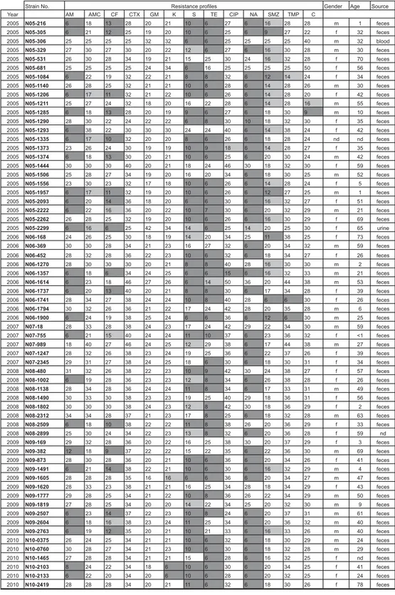
Fig. 1. Resistance profiles and anamnestic data of the 64 S. Hadar strains. Strains resistant, intermediate or susceptible to a specific antibiotic are highlighted in grey, light grey, or white, respectively. AM, ampicillin; AMC, amoxicillin-clavulanic acid; CF, cephalothin; CTX, cefotaxime; GM, gentamicin; K, kanamycin; S, streptomycin; TE, tetracycline; CIP, ciprofloxacin; NA, nalidixic acid; SMZ, sulfamethoxazole; TMP, trimethoprim; C, chloramphenicol; m, male; f, female; nd, no data.
caused by S. Hadar is greater than that suggested by ourfindings.
We found that the majority of strains were resistant to nalidixic acid, tetracycline andβ-lactam antibiotics, excluding third-generation cephalosporins. The emerg-ence and spread of multidrug resistance in patho-genic as well as commensal bacteria in general and in Salmonella spp. in particular is an increasing global concern [10,17,18]. Dissemination of resistance genes through bacterial populations needs to be monitored globally as well as locally [19]. Moreover, there is evi-dence that less frequent serovars, such as S. Hadar, are reservoirs of antibiotic resistance genes [12]. The considerable percentage (46·9%) of multidrug-resistant strains in our strain collection is in accord with the results of a European multi-centre study, which describes 37% multidrug-resistant Salmonella isolates [20].
Reisistance to nalidixic acid in salmonellae is of great concern, as it has been reported to be an indi-cator for reduced susceptibility to the drug of choice, ciprofloxacin, resulting in possible treatment failure [21–23]. Consequently, the 71·8% nalidixic acid-resistant S. Hadar of this study must be considered as having reduced susceptibility to ciprofloxacin, and thus potentially treatment-resistant. Interestingly, the strain that was isolated from blood, and presumably associated with invasive infection, was susceptible to both nalidixic acid and ciprofloxacin.
Reports on the emergence of extended-spectrum β-lactamase (ESBL)-resistant salmonellae give rise to concern [24]. Genes encoding for ESBLs, predomi-nantly blaTEM, blaSHV and blaCTX variants have
been detected in various Salmonella serovars isolated from humans as well as from poultry and poultry pro-ducts [25]. However, all the S. Hadar isolates in this study were susceptible to cefotaxime. So far, resistance to ESBL antibiotics is not yet an issue in S. Hadar iso-lates in Switzerland. Nevertheless, continued surveil-lance for the presence of ESBL in Salmonella spp. is needed in order to minimize the risks to future treat-ment [26].
PFGE analysis showed a close clonal relationship for this serovar, similar to what is also known for S. Enteritidis. In the primary cluster, consisting of 42 strains sharing a similarity of >92%, a subcluster of 18 strains with indistinguishable patterns was observed. By combining the PFGE and the resistance patterns of these strains, two strains (N05-1374, N06-1737) isolated in 2005 and early 2006 are indis-tinguishable from a S. Hadar strain responsible for a chicken meat-borne outbreak in Spain [8], suggesting a possible exposure of these patients in Spain.
AC K N O W L E D G E M E N T S
We thank Grethe Sägesser for her help in strain collec-tion and technical support and the Swiss Federal
Fig. 2. Pulsed-field gel electrophoresis (PFGE) patterns of the 64 S. Hadar strains. The primary PFGE cluster (92% similarity) is indicated by light grey.
Office of Public Health for financial support. We also thank Silvia Herrera Leon for providing the pattern of the S. Hadar outbreak strain from 2005 in Spain. D E C L A R AT I O N O F I N T E R E S T
None.
R E F E R E N C E S
1. Rabsch W, Tschape H, Baumler AJ. Non-typhoidal sal-monellosis: emerging problems. Microbes and Infection 2001; 3: 237–247.
2. Olsen SJ, et al. The changing epidemiology of sal-monella: trends in serovars isolated from humans in the United States, 1987–1997. Journal of Infectious Disease 2001; 183: 753–761.
3. Majowicz SE,et al. The global burden of nontyphoidal salmonella gastroenteritis. Clinical Infectious Disease 2010; 50: 882–889.
4. Huehn S, et al. Virulotyping and antimicrobial resist-ance typing of Salmonella enterica serovars relevant to human health in Europe. Foodborne Pathogen and Diseases 2010; 7: 523–535.
5. Anon. European Food Safety Authority (EFSA) (http:// www.efsa.europa.eu/de/scdocs/doc/1533.pdf). Accessed 29 January 2013.
6. Fantasia M, et al. Conventional and molecular approaches to isolates of Salmonella Hadar from spora-dic and epidemic cases. Journal of Applied Microbiology 1997; 82: 494–498.
7. Di Giannatale E,et al. Investigation of an outbreak of Salmonella enterica subsp. enterica serovar Hadar food illness in the Abruzzi region of Italy. Veterinary Italian 2008; 44: 405–427.
8. Lenglet A. Over 2000 cases so far in Salmonella Hadar outbreak in Spain associated with consumption of pre-cooked chicken, July–August, 2005. Eurosurveillance 2005; 11: 10(8).
9. Hohmann EL. Nontyphoidal salmonellosis. Clinial In-fectious Disease 2001; 32: 263–269.
10. Anon. World Health Organisation: tackling antibiotic resistance from a food safety perspective in Europe. Copenhagen, 2011.
11. Parry CM. Antimicrobial drug resistance in Salmonella enterica. Current Opinion in Infectious Diseases 2003; 16: 467–472.
12. Bagger-Skjøt L,et al. Less frequent Salmonella serovars as a reservoir of antimicrobial resistance. Journal of Antimicrobial Chemotherapy 2007; 59: 814–815. 13. Grimont PAD, Weill FX. Antigenic Formulae of the
Salmonella Serovars, 9th edn. WHO Collaborating
Center for Reference and Research on Salmonella, Institut Pasteur: Paris, 2007.
14. Anon. Swiss Federal Office for Public Health (http:// www.bag.admin.ch/k_m_meldesystem/00733/00813/). Accessed 29 January 2013.
15. CLSI. Clinical and Laboratory Standards Institute: Performance Standards for Antimicrobial Susceptibility Testing; Eighteenth Informational Supplement. CLSI document M100-S18 2008, Wayne, PA.
16. Hendriksen RS,et al. Global monitoring of salmonella serovar distribution from the World Health Organiz-ation global foodborne infections network country data bank: Results of quality assured laboratories from 2001 to 2007. Foodborne Pathogens and Diseases 2011; 8: 887–900.
17. van den Bogaard AE, Stobberingh EE. Epidemiology of resistance to antibiotics. Links between animals and humans. International Journal of Antimicrobial Agents 2000; 14: 327–335.
18. Threlfall EJ,et al. The emergence and spread of anti-biotic resistance in food-borne bacteria. International Journal of Food Microbiology 2000; 62: 1–5.
19. O’Brien TF. The global epidemic nature of antimicro-bial resistance and the need to monitor and manage it locally. Clinical Infectious Diseases 1997; 24: 2–8. 20. Threlfall EJ,et al. Antimicrobial drug resistance in
iso-lates of Salmonella enterica from cases of salmonellosis in humans in Europe in 2000: results of international multi-centre surveillance. Eurosurveillance 2003; 8: 41– 45.
21. Hakanen A,et al. Detection of decreased fluoroquino-lone susceptibility in salmonellas and validation of nalidixic acid screening test. Journal of Clinical Micro-biology 1999; 37: 3572–3577.
22. Møller Aarestrup F, Wiuff C. Is it time to change fluoroquinolone breakpoints for Salmonella spp? Anti-microbial Agents Chemotherapy 2003; 47: 827–829. 23. Crump JA, et al. Reevaluating fluoroquinolone
break-points for Salmonella enterica serovar Typhi and for non-typhi salmonellae. Clinical Infectious Diseases 2003; 37: 75–81.
24. Coque TM, Baquero F, Canton R. (2008) Increasing prevalence of ESBL-producing Enterobacteriaceae in Europe. Eurosurveillance 2008; 20: 13(47).
25. Hasman H, et al. Beta-lactamases among extended-spectrum beta-lactamase (ESBL)-resistant Salmonella from poultry, poultry products and human patients in The Netherlands. Journal of Antimicrobial Chemother-apy 2005; 56: 115–121.
26. Yates C, Amyes S. Extended-spectrum beta-lactamases in non-typhoidal Salmonella spp. isolated in the UK are now a reality: why the late arrival? Journal of Anti-microbial Chemotherapy 2005; 56: 262–264.