Lack of Leukocyte Migration Inhibition by Hepatitis B Antigen
and Normal Nonspecific Immunoreactivity in Asymptomatic Carriers
M. Desaules, P. C. Frei,
J.
Libanska, and B. Wuilleret From the Division of Immunology and Allergy, Department of Medicine, Centre Hospitalier Unioersitaire Vaudois; and the Regional Blood Transfusion Center of the Swiss Red Cross, Lausanne, Switzerland The immune response to hepatitis B surface antigen (RBs Ag) was studied in 25asymptomatic carriers by the leukocyte migration-inhibition (LMI) test in agarose. In the presence of purified RBsAg, inhibition was demonstrated in only four of 25 carriers, in contrast to 24 of 28 patients who cleared the antigen after acute infection with hepatitis B. Tuberculin purified protein derivative (PPD) was also used as an antigen for the LMI test in these carriers. Inhibition was demonstrated in only 12 of 25 individuals who had positive PPD skin tests, in contrasttoall of 14 normal noncarrier individuals with positive PPD skin tests and none of 12 normal noncarrier individuals with negative PPD skin tests. A nonspecific immunological investigation of the asymptomatic carriers gave normal results. The lack of an immune response to HBsAg was thought to be responsible for the persistence of the antigen and also for the absence of symptoms.
About 0.2<;'0 of the blood donors in this part of Europe are carriers of hepatitis B surface antigen (HBs Ag). Some of these asymptomatic carriers have slightly elevated levels of transaminases, whereas others do not exhibit any biochemical sign of hepatic cytolysis but have slight histologi-cal abnormalities of the liver. A third group of these carriers are normal, both biochemically and histologically[1-3].
Since HBs Ag is usually associated with acute hepatitis and disappears at the time of recovery, it is difficult to explain how the antigen can per-sist in the third (normal) group of carriers with-out damaging the liver. The fact that HBs Ag can persist in two conditions as different as the healthy carrier state and chronic hepatitis is also problematical.
Received for publication February 6, 1976, and in re-vised form May II, 1976.
We thank Dr. P. Kocher, Centre de Transfusion San-guine, La Chaux-de-Fonds, for allowing us to study four of his blood donors.
This work was performed with the financial support of the Centre de Recherche sur les Lymphomes Malins, Lau-sanne, Switzerland.
A portion of this study was presented at the European Society for Clinical Investigation, Rotterdam, Netherlands, April 25-27, 1974.
Please address requests for reprints to Dr. P. C. Frei, Di-vision of Immunology and Allergy, Department of Medi-cine, Centre Hospitalier Universitaire Vaudois, Lausanne CH 1011, Switzerland.
505
The immune state of symptomatic and asymp-tomatic carriers has been thought to be respon-sible for the persistence of the antigen. The non-specific cellular immune response was found to be impaired by some authors [4-6] and normal by others [1, 7, 8]. The specific immune response to HBsAg has also been studied in vitro in blood from a limited number of carriers. No inhibi-tion of leukocyte migrainhibi-tion in the presence of HBsAg was found by Yeung Laiwah et al. [9] or by Dudley et al. [10], and inhibition was found only infrequently by Vittal et al. [11]. However, Lee et al. [12] observed inhibition in 10 of 37 HBsAg-positive blood donors. Irwin et al. found no migration-inhibition factor (MlF) activity in five carriers tested by an indirect method [13]. Koszikowski et al. [14] did not find any stimula-tion of lymphocytes by HBsAg in 10 carriers, and also found none in 10 patients who had recov-ered from hepatitis B infection without produc-ing detectable levels of antibody.
The leukocyte migration-inhibition (LMI) test in agarose allowed us to use only very small amounts of purified HBsAg. With this technique, we have shown [15] the constant appearance of a specific inhibition of leukocyte migration by HBsAg after recovery from acute infection with hepatitis B. This inhibition was observed in al-most all patients who cleared the antigen, where-as the antibody became demonstrable by
radioim-506
munoassay in only half of the patients. The ab-sence of migration inhibition during and imme-diately after the acute phase of the disease was attributed to nonspecific anergy, since migration inhibition by purified protein derivative (PPD) disappeared temporarily at that time [16J.
From these findings it was hypothesized that specific cell-mediated immunity might be respon-sible for the clearance of HBsAg. The present study extends this investigation to asymptomatic carriers. The results demonstrate the absence of migration inhibition by HBs Ag in all but four of 25 asymptomatic carriers of HBsAg.
Materials and Methods
Selection of donors. HBs Ag-positive blood
donors were subjected to careful taking of their medical history, physical examination, and blood tests, including levels of aspartate aminotrans-ferase (SGOT) and alanine aminotransferase (SGPT), prothrombin, alkaline phosphatase, bi-lirubin, protein, and electrophoresis. Only those donors who proved to be normal according to these criteria and who had no history of hepati-tis were selected for study. This group was re-examined in the same manner nine to 12 months later to establish the persistence of HBs Ag in the blood and the absence of any symptoms. The possibility of a donor incubating acute hepatitis could thus be avoided. No liver biopsy was per-formed because of the absence of indication in these asymptomatic donors. Titers of HB. Ag were measured by counterimmunoelectrophore-sis. In two carriers, HBs Ag was detectable by radioimmunoassay only. Twenty-five individuals (18 males and seven females), aged 21-63, were finally selected for the study.
Nonspecific immune state. The three main
immunoglobulins were measured by radial im-munodiffusion (Tripartigen Behringwerke, Mar-burg, Germany). Humoral response was tested by titration of isoagglutinin, antistreptolysin (Streptolysin Isti tuto Sierterapico Milanese, Mil-an, Italy), and antistaphylolysin (Staphylolysin Behringwerke, Marburg, Germany). Delayed-type skin reactivity was investigated by skin-testing with a standard group of five antigens: tuber-culin PPD (5 units; Statens Seruminstitut, Co-penhagen, Denmark),
streptokinase-streptodorn-Desaules et al.
ase (250 units /ml: Varidase; Lederle, Pearl Riv-er, N.Y.), candidine (Bencard; Beecham, Brent-ford, Middlesex, England), trichophytine (Beech-am), and mumps skin-test antigen (Eli Lilly and Company, Indianapolis, Ind.).
Purification of HBs Ag. Sera with a titer of
HBsAg of
>
1:256 by counterimmunoelectropho-resis were pooled. HBs Ag was precipitated by polyethylene glycol 6,000; the precipitate was re-dissolved in distilled water, dialyzed against 0.05M sodium barbital buffer (pH 8.6), and further
purified by agarose-block electrophoresis in the same buffer. The HBs Ag-rich fractions were pooled and concentrated by vacuum dialysis against phosphate buffer. The HBs Ag was then separated from trace amounts of contaminating proteins by ultracentrifugation on a 10%-30% (wt/wt) sucrose gradient. The preparation was sterilized by filtration through Millex microfil-tel'S (pore size, 45 psn;Millipore Corp., Bedford, Mass.) and finally was concentrated by vacuum dialysis (membrane filter SM 132000; Sartorius, Gottingen, Germany) against medium TC 199 (Difco, Detroit, Mich.), until a titer of 1:16 by counterimmunoelectrophoresis was reached. No precipitation line was found in agar double-diffu-sion with antisera to whole human serum. The HBs Ag preparation was used undiluted in the Ll\H test.
LMI test. This test was performed under
aga-rose according to the method of Clausen[17]. De-tails of this method as performed with HBs Ag in our laboratory have been described [18]. In brief, 2.5X 10⁷ leukocytes, containing at least
75% polymorphonuclear leukocytes, were sus-pended in0.1 ml of the antigen preparation. The suspension was incubated for 30 min at 37 C in an atmosphere of 2.5% CO2, and 5 fJ-l were in-troduced into each of eight wells punched in an agarose layer. After incubation for 18 hr, the areas of migration were measured by planimetry. The mean surface area of the eight wells was ex-pressed as the percentage of the migration in a control preparation without antigen. Values of <90% were considered to indicate inhibition. Leukocytes from each individual were tested in the presence of two antigens, purified HBs Ag and tuberculin PPD (25 fJ-g/ml), without pre-servative. The statistical evaluation was made ac-cording to Student'st-testfor unpaired values.
Results
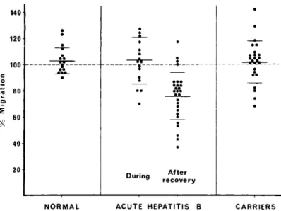
The titers of circulating HBsAg ranged from 1: 1,024 to 1:1 by counterimmunoe1ectrophoresis, and, in two cases, antigen was detectable only by radioimmunoassay (Ausria II; Abbott Laborator-ies, North Chicago, Ill.). The titer was strikingly stable during the observation period and never varied by more than one dilution step. Tests of hepatic function were normal in all of the in-dividuals selected for study. On retesting a few months later, the antigen was still present and the results of hepatic function tests were normal. The mean area of leukocyte migration in the presence of HBs Ag was 101.9<;'0 of that in the control without antigen (SE ± 3.2). Thus, no im-mune response to HBsAg could be demonstrated in these carriers. Only in cases no. 1, 5, 16, and 23 were individual values of <90% demonstra-ble.
Figure 1 clearly shows the lack of leukocyte inhibition by HBs Ag in asymptomatic carriers, compared with individuals who cleared the an-tigen after acute infection with hepatitis B. The difference is significant (t
=
5.35; P < 0.001). Leukocytes from carriers did not behave differ-ently from those from normal individuals (t=
0.34; 0.3 < P < 0.4) or from those from patients with acute hepatitis B infection (t= 0.32; 0.3 < P < 0.4) in the presence of HBsAg.
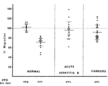
Inhibition of leukocyte migration in the
pres-ence of PPD (figure 2) was demonstrable in 12 carriers only, although all 25 carriers had a posi-tive skin test with PPD. In normal individuals, however, the LMI test in agarose gave a good cor-relation with the PPD skin-tests, as shown in fig-ure 2. The mean area of migration in the pres-ence of PPD was 71.0% (SE ± 2.3) in PPD-posi-tive normal individuals, 95.7% (SE ± 4.4) in PPD-positive patients with acute hepatitis B in-fection, and 91.8% (SE ± 3.2) in PPD-positive carriers. Thus, the leukocytes from these asymp-tomatic carriers behaved, in the presence of PPD, not like those from normal individuals (t
=
4.55; P < 0.001), but in the same manner as those from acutely infected patients (t
=
0.73; 0.2 < P< 0.25). The reproducibility of the LMI test under agarose was satisfactory, as judged by the eight simultaneous measurements in each case; in the four cases with inhibition, all eight values were <90%. The stimulation observed in a few cases may be without significance or may be interpreted as a weak sensitization [18].Results of nonspecific immunological screen-ing were normal. Values obtained for the humor-al parameters were normhumor-al, except for a few iso-lated titers of antibody. This exception can also occur in normal individuals. Only one case had low values for all three antibodies tested (two dilution steps below the lowest limit) and for IgG (550 mg/100 ml). The delayed hypersen-sitivity, as judged by the skin tests, was normal in all patients.
.1 •
---~---JL--- ---~---
}.
Figure 1. Migration, in the presence of purified hepatitis B surface anti-gen (RB s Ag), of leukocytes from normal individuals (left), patients during and after acute hepatitis B infection (center), and asympto-matic carriers of RBs Ag (right).
Points represent the surface of mi-gration expressed as the percentage of the migration in a control pre-paration without antigen. The short horizontal lines are means ± SD. Broken line represents 100% migra-tion. 140 120
-:-
.
..
100 ---*(---c --Ca... 0.
~ 80 ClI ~*'
60 40 20.
---L.
During...
...
...
---A.L-···
~ After recovery .....
508 140 Desaules et al. 120 100 e .~ 80
-
~ Cl ::E 60*
40 20..
---~---
.
--:;-NORMAL·
·
·
---~---...
ACUTE HEPATITIS B-+
________. J _.
----t--..
.:
..
---...
CARRIERSFigure 2. Migration, in the presence of purified protein derivative (PPD), of leukocytes from normal PPD-negative individuals and normal PPD-posi tive individuals(left),
PPD-positive individuals during acute hepatitis B infection (center), and
PPD-positive asymptomatic carriers of hepatitis B surface antigen(right).
The short horizontal lines are means
± SD. Broken line represents 100% migration.
PPD
skin test neg. pes. pes. pes.
Discussion
The inhibition of leukocyte migration by HBs Ag in carriers occurred infrequently (four of 25 carriers). This finding contrasts with the nearly constant inhibition we observed in patients who cleared HBsAg (24 of 28 patients). Consequent-ly, we believe that the specific immune response demonstrated by the LMI test is probably respon-sible for elimination of the antigen, which oc-curs during recovery from hepatitis B infection, and that the lack of this response may explain the carrier state.
Nearly all authors consider the LMI test as an index of cell-mediated immunity. Although this hypothesis has not been proven, the follow-ing findfollow-ings encourage us to support it for the moment. In our experiments with HBsAg as an antigen, migration inhibition did not correlate with antibody production [15, 18]. In our experi-ments with PPD, the correlation with the results of delayed skin tests was excellent (figure 2). Furthermore, Lambert et al.' and Trepo et al. [19] found no relation between the presence of HBs Ag-anti-Hfs, Ag complexes and any of the condi-tions studied here.
1P. H. Lambert, E. Tribollet, A. Celada, K. Madalinski P. C. Frei, P. A. Miescher, "Circulating Immune
Complexe~
I~~ol~ingHBsAg in Patients with Acute and Chronic Hepa-tItIS III Healthy Carriers and in Polyarteritis Nodosa," manuscript in preparation.
. Our observations also contribute to explana-nons of the absence of lesions in the carriers. The lesions of hepatitis cannot, of course, be produced by the virus itself. They might be the consequence of the immune response demon-strated by the LMI test, and the lack of this re-sponse in carriers would explain the absence of cytolysis in the liver. One must then consider the reason that this response could not be demon-strated during the acute phase of hepatitis (fig-ure 1), when the liver is most severely attacked. This lack of response is probably related to a broad, nonspecific anergy during the acute phase. We have shown [16, 18] that the migration of leukocytes from PPD-positive individuals was not inhibited by PPD during hepatitis. Experiments with PPD as antigen (figure2).also showed that leukocytes from some carriers behaved like those from patients with acute hepatitis B infection; the mean surface areas of migration were 91.8% and 95.7%, respectively, without significant dif-ference between the two values. The anergy dem-onstrable in vitro during the acute disease exists, to a lesser extent, in healthy carriers. Thus, it seems that the circulating agent can itself pro-duce a nonspecific anergy, even in the absence of tissue damage. Note that the anergy in these two conditions was not demonstrable by the skin tests. These tests may be more sensitive than the LMI test or may show a slightly different type of response.
The impairment of the immune response dem-onstrable by the LMI test in carriers seems to be an isolated defect, since the results of the non-specific immunological investigation were indis-tinguishable from those one would obtain in nor-mal individuals. Only one carrier had low titers of antibody and a low IgG level. In this respect, our results differ from those of others[4-6]. This difference is probably related to our stricter cri-teria of selection, which excluded individuals with biological signs of hepatic disease.
References
1. Bolin, T. D., Davis, A. E., Liddelow, A. G. Liver dis-ease and cell-mediated immunity in hepatitis-asso-ciated antigen (HAA) carriers. Gut14:365-368, 1973.
2. Reinicke, V., Dybkjaer, E., Poulsen, H., Banke, 0., Lyl-loft', K., Nordenfelt, E. A study of Australia-antigen-positive blood donors and their recipients with spe-cial reference to liver histology. N. Engl.
J.
Med.286: 867-870, 1972.3. Griffin, F., Jr. Hepatitis B antigenemia in apparently healthy blood donors. J.A.M.A.226:753-755, 1973.
4. Giustino, V., Dudley, F.]., Sherlock, S. Thymus-depen-dent lymphocyte function in patients with hepatitis-associated antigen. Lancet2:850-853, 1972.
5. Halikowski, B., Korczowski, R., Zajaczkowski,J.Odczyn skorny na dwunitrochlorobenzen (DNCB) u nosicieli antygenu Australia. Pediatr. Pol.47:1071-1075, 1972.
6. Ortona, L., Pizzigallo, E., Federico, G., Laghi, V. Richerche sui donatori di sangue au positivi: stimola-zione in vitro dei linfociti con PHA. Ann. Sclavo
13:35-48, 1971.
7. Nielsen,J.0., Reinicke, V., Dietrichson, 0., Andersen, V., Thomsen, M., Andersen, E. Immunological studies of Australia antigen carriers with and without liver diseases. Clin. Exp. Immunol.15:9-16,1973.
8. Sutnick, A. I., Bugbee, S. J., London, W. T., Loeb, L. A., Peyretti, F., Litwin, S., Blumberg, B. S. Lymphocyte function in normal people with persistent Australia antigen.J.Lab. Clin. Med.82:79-85, 1973.
9. Yeung Laiwah, A. A. C., Chaudhuri, A. K. R., Ander-son,
J.
R. Lymphocyte transformation andleuco-eyremigration inhibition by Australia antigen. Clin. Exp. Immunol.15:17-34, 1973.
10. Dudley, F.
J.,
Giustino, V., Sherlock, S. Cell-mediated immunity in patients positive for hepatitis-associated antigen. Br. Med. J.4:754-756,1972.11. Vittal, S.B.V., Dourdourekas, D., Shobassy, N., Gerber, M., Telischi, M., Szanto, P. B., Steigmann, F., Clow-dus, B. :F.Asymptomatic hepatic disease in blood donors with hepatitis B antigenemia. Am. J. Clin. Pathol.62:649-654, 1974.
12. Lee, W. M., Reed, W. D., Mitchell, C. J. Eddleston, A. L. W. F., Dymock, I., Williams, R. Cell-mediated immunity to hepatitis B antigen in blood donors with persistent antigenaemia or high titer antibody. Digestion10:362, 1974.
13. Irwin, G. R., Jr., Hierholzer, W.
J.,
jr.,Cimis, R., Mc-Collum, R. W. Delayed hypersensitivity in hepatitis B: clinical correlates of in vitro production of migra-tion inhibimigra-tion factor. J. Infect. Dis. 130:580-587, 1974.14. Koszinowski, D., Thomssen, R., Schober, A. In-vitro-Stimulation der Lymphozyten von H'B-Antikdrper-Tragern durch HB-Antigen (Australia-Antigen). Dtsch. Med, Wochenschr.98:262-267,1973.
15. Frei, P. C., Erard, Ph., Zinkernagel, R. Cell-mediated immunity to hepatitis-associated antigen (HAA) demonstrated by leukocyte migration test during and after acute hepatitis B. Biomedicine [Express] 19: 379-383, 1973.
16. Erard, P., Frei, P. C., Peitrequin, R., Hofstetter, j.-R., Magnenat, P. Etude de I'immunite cellulaire sped-fique de l'antigene Australia. Resultats preliminaires dans differentes formes d'hepatite B. Schweiz. Med. Wochenschr.104:1882-1885, 1974.
17. Clausen,J.E. Tuberculin-induced migration inhibition of human peripheral lymphocytes in agarose medi-um. Acta Allergol. (Kbh.)26:56-80, 1971.
18. Erard, P. Technical study of the leukocyte migration inhibition test in agarose. Application to PPD and to hepatitis B antigen. Clin. Exp. Immunol. 18:439-448,1974.
19. Trepo, C. G., Zuckerman, A.