The status of Ig loci rearrangements in
single cells from different stages of B cell
development
Edwin ten Boekel, Fritz Melchers and Antonius Rolink
Basel Institute for Immunology, Grenzacherstrasse 487, CH-4005 Basel, Switzerland
Key words: B cell development, Ig gene rearrangement, surrogate L chain, K L A L chain ratio
Abstract
Differential expression of c-kit, CD25 (TAC), surrogate L chain and cytoplasmic |iH chain, and surface expression of IgM and IgD allows the separation of B220 (CD45+) B cell subpopulations. PCR analyses with DNA of single cells developed by others and by us have been used to monitor the conformation of the Ig H and L chain gene loci in these different B lineage subpopulations. The results of these analyses Indicate that B220+/c-kit+/CD25" cells are the precursors of large B220+/CD25+/slgM- which, in turn, are the precursors of small B220+/CD25+/slgM" cells. The majority of B220+/c-kit+/CD25~ cells are DnJH-rearranged, with L chain loci In germ line
configuration and are thus pre-B I cells. More than 90% of all large B220+/CD25+/slgM~ cells have at least one H chain locus VHDHJ H rearranged; half of them have also the second locus VHDHJH rearranged and are thus large pre-B II cells. Rearrangements of at least one allele of the KL chain loci become detectable in 65% of the small B220+/CD25+/slgM~ cells, 67% of the immature B and >75% of the mature B cells. The ratio of KL to IL gene rearrangements in all three subpopulations Is -10:1, Indicating that the K I A L ratio is established as soon as rearrangements are made.
Introduction
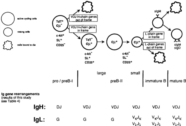
Bone marrow is the major site of B lymphopoiesis during adult life (1-5). Development from early B-lymphocyte-lineage-committed progenitors (pro-B) to precursor (pre-B) and immature B cells in mouse bone marrow and to mature, antigen-sensitive B cells in peripheral lymphoid organs can be dissected into different stages. These cellular stages are characterized by the differential expression of surface-located and intra-cellular markers, and by the differential capacities of cells to proliferate 'in vitro' (Fig. 1) (2,5-8). Hardy and his colleagues (6) have used the differential expression of CD43, heat stable antigen (HSA) and BP-1 to separate precursor B cell subpopulations while our laboratory has employed the analysis of cell size and the expression of c-kit, CD25 (TAC) and surrogate L (SL) chain to do so (7).
Progression along the pathways of B lymphocyte differenti-ation is also marked by successive rearrangements of the Ig gene loci. They begin with DH to JH rearrangements in the H chain gene loci where they appear to involve both alleles within a precursor B cell (9). These so called pro/pre-B I cells express c-kit, but not CD25, and are large, cycling cells when prepared 'ex vivo' (7).
DHJH rearrangements are followed by VH to DHJH
rearrange-ments. Cells with productively VHDHJH-rearranged Ig H chain alleles, i.e. those capable of expressing a |iH chain, appear positively selected over non-productively VHDnJH-rearranged cells (7). This occurs through proliferative expansion of the \iH chain-expressing cells, in all likelihood initiated by the signaling through the nH chain/SL chain pre-B receptor (7). Hence, we expect these cells, again, to be large cycling cells. Large, cytoplasmic nH chain expressing cells in bone marrow are c-kir/CD25+. About 25% of them express the (iH/SL pre-B cell receptor which can appear on the surface when the cells are cultured 'in vitro' (7,10).
The next stage of development is thought to be small, resting c-kir/CD25+ cells expressing cytoplasmic nH chain, but not yet L chains, which are no longer in cell cycle.
L chain expression then leads to deposition of IgM on the surface of so-called immature B cells. These immature B cells are thought to be the precursors of mature, antigen-sensitive, mitogen-reactive slgM+/slgD+ B cells, which are found in part in bone marrow, but are most abundant in peripheral lymphoid organs.
Correspondence to. A. Rolink/F. Melchers
o
o
activ* qrcfing c«Us
resbng c a b
calls bound to die
VDJ H-chaln genes out of frame
pro / preB-l
large small
preB-ll immature B mature B
Ig gene rearrangements
(results of this study see Table 4)
IgH:
,
L.
DJ G VDJ G VDJ G VDJ V KV x
VDJ V K VDJ V KFig. 1. B lymphopoiesis in mouse bone marrow, as analyzed by differential marker expression and status of IgH and L chain gene
rearrangements.
An assay has been developed by Ehlich et al. (5) that allows the characterization of Ig H chain gene rearrangements in single cells. This assay has been previously used to monitor the rearrangement status of the Ig gene loci of B lineage precursors in mouse bone marrow characterized, and thus separable, by the differential expression of CD43, HSA and BP-1 (5). We use this assay in the present study to determine the status of Ig H and L chain gene rearrangements in B lineage precursor cells of mouse bone marrow, as character-ized and thus separable by their differences in cell size, and c-kit, CD25, nH/SL pre-B cell receptor IgM and IgM/lgD B cell receptor expression. The results define the cells in mouse bone marrow in which the majority of either DH-JH. VH-DHJH or VL-JL rearrangements have taken place. The results also allow a comparison with Hardy's bone marrow cell subpopula-tions defined by CD43, HSA and BP-1 expression, and their status of Ig gene rearrangements (6). It appears that separation of B lineage committed cells differentially expressing c-kit, CD25 and SL chain allows a clear distinction and thus purification of precursors before and after productive VHD H JH rearrangements and after VLJL rearrangements.
Methods
Mice
(C57BL/6XDBA/2) F, (BDF,) mice, 6-12 weeks of age, were obtained from the Institut fur Biologische-Medizinische Forschung AG (Fullinsdorf, Switzerland).
FACS staining, sort and DNA preparation
Cells for FACS sorting were prepared from bone marrow and spleen as described (7). Bone marrow cells were stained with the FITC-conjugated mAb RA3 6B2 (anti-CD45R, B220) (PharMingen, San Diego, CA) and double stained with biotin-conjugated mAb ACK-4 (anti-c-kit) (11), LS156 (anti-X5/nH) (10), 7D4 CD25, TAC), M41 nH) (12) or 1.19 (anti-8) (13). Binding of biotin-conjugated mAb was visualized using streptavidin-phycoerythrin (Southern Biotechnology Associates, Birmingham, AL)
Single cells were sorted using the FACStar Plus equipped with an automatic cell deposition unit (Becton Dickinson, Mountain View, CA). Single cells were directly sorted into 96-well PC plates type H (Costar, Cambridge, MA) containing 3 nl of 10 times concentrated PCR buffer, 7 nl H2O and 10 ng tRNA. To prepare DNA, samples were overlaid with PCR oil (Fluka, Buchs, Switzerland) and 2 nl of proteinase K (5 mg/ml; Boehringer, Mannheim, Germany) was added. The samples were digested for 1 h at 55°C and proteinase K was subsequently inactivated for 10 min at 95°C. Plates were then stored at -70°C until use for DNA amplification.
PCR analysis of Ig H and L gene rearrangements
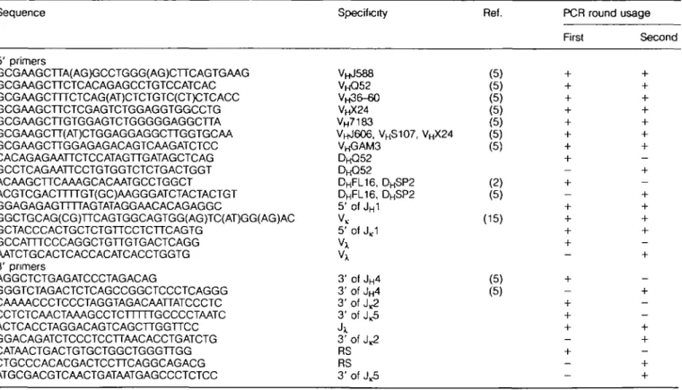
PCR amplification was carried out in two rounds using a Hypaid Omnigene PCR machine (Hybaid Ltd, Middlesex, UK). The first round was done over 28 cycles and contained all 5' and 3' primers listed in Table 1 as first round primers. The PCR amplification conditions were as described by Ehlich
Table 1. Oligonucleotides used for amplification of rearranged Ig H and L genes
Sequence Specificity Ref. PCR round usage
First Second 5' primers GCGAAGCTTA(AG)GCCTGGG(AG)CTTCAGTGAAG GCGAAGCTTCTCACAGAGCCTGTCCATCAC GCGAAGCTTTCTCAG(AT)CTCTGTC(CT)CTCACC GCGAAGCTTCTCGAGTCTGGAGGTGGCCTG GCGAAGCTTGTGGAGTCTGGGGGAGGCTTA GCGAAGCTT(AT)CTGGAGGAGGCTTGGTGCAA GCGAAGCTTGGAGAGACAGTCAAGATCTCC CACAGAGAATTCTCCATAGTTGATAGCTCAG GCCTCAGAATTCCTGTGGTCTCTGACTGGT ACAAGCTTCAAAGCACAATGCCTGGCT ACGTCGACTTTTGT(GC)AAGGGATCTACTACTGT GGAGAGAGTTTTAGTATAGGAACACAGAGGC GGCTGCAG(CG)TTCAGTGGCAGTGG(AG)TC(AT)GG(AG)AC GCTACCCACTGCTCTGTTCCTCTTCAGTG GCCATTTCCCAGGCTGTTGTGACTCAGG AATCTGCACTCACCACATCACCTGGTG 3' primers AGGCTCTGAGATCCCTAGACAG GGGTCTAGACTCTCAGCCGGCTCCCTCAGGG CAAAACCCTCCCTAGGTAGACAATTATCCCTC CCTCTCAACTAAAGCCTCTTTTTGCCCCTAATC ACTCACCTAGGACAGTCAGCTTGGTTCC G GACAG ATCTCCCTCCTTAACACCTG ATCTG CATAACTGACTGTGCTGGCTGGGTTGG CTGCCCACACGACTCCTTCAGGCAGACG ATGCGACGTCAACTGATAATGAGCCCTCTCC V H J 5 8 8 V H Q 5 2 V H 3 6 - 6 0 VHX24 VH7183 VHJ6O6, VHS 1 0 7 , VHX 2 4 V H G A M 3 D H Q 5 2 DHFL16, DHSP2 DHFL16, DHSP2 5' of JH1 VK 5' of JK1 Vx Vx 3' of JH4 3' of JH4 3' of J^2 3' of J ^ Jx 3' of J^2 RS RS 3' of J.5 (5) (5) (5) (5) (5) (5) (5) (2) (5) (15) (5) (5)
et al. (5). For the second PCR round, 1 u.l of the first PCR amplification was reamplified with one specific 5' primer and a specific 3' primer (Table 1, second round PCR primers). The second round was done over 35 cycles (20 s at 95°C, 1 min at 65°C, 2 min at 72°C). All PCR contained dATP, dCTP, dTTP and dGTP (Pharmacia, Uppsala, Sweden) at 200 u.M each and 5 U Taq DNA polymerase (Roche, Basel, Switzerland) in PCR buffer (0.05 mM 2-mercaptoethanol, 50 mM KCI, 10 mM Tris-HCI, pH 8.3, 1.5 mM MgCI2 and 0.001% gelatine). The first round PCR reaction was performed in 30 u.l, the second in 20u.l. Then, 10 u.l of the second round PCR product was analyzed on agarose gels stained with ethidium bromide. The lengths of the different PCR products are summarized in Table 2.
Results and discussion
PCR assay for the analyses of Ig gene rearrangements in single cells
We have used assays for H chain gene rearrangements developed by Ehlich et al. (5) and by Haasner et al. (14), and assays for KL chain gene rearrangements developed by Schlissel and Baltimore (15), and have modified and extended these assays, particularly to include XL chain gene rearrange-ments, as described in Methods. At the single cell level, these assays are based on PCR amplification of gene segments in two steps. In the first, both rearranged Ig loci of a cell were amplified simultaneously using a mixture of 13 5' primers, homologous to VH, VL and DH genes, and upstream of the
Table 2. Approximate PCR product lengths
Rearrangements Germlme Ig H D H JH( S P 2 / F L 1 6 ) DHQ52JH VHDHJH Germline Ig K V^JJ or 2 V ^ J ^ or 5 RSK VxJx Product lengths (bp) 1500 1460 1500 1720 600 650 600 320 318 ( J H 1 ) ( J H 1 ) ( J H D (JK1) (JK4) , 1150 (JH2), 730 , 1190 (JH2), 770 , 1410 (JH2), 990 and 280 (J<2) and 260 (J,(5) (JH3) (JH3) (JH3) and and and 200 (JH4) 240 (JH4) 460 (JH4)
JH1 and JL)C1 segment in combination with five 3' primers binding downstream of J H 4 , JL K2 , JL K5 , J|_X and RSK. The
primers are listed in Table 1. In the second PCR round, the products of the first PCR were analyzed in separate reactions. For VDJ and DJ PCR, each reaction contained respectively a single VH and DH primer together with a nested 3' JH4 primer. The unrearranged state of an Ig H locus was determined by the reaction containing a primer recognizing 5' of the JH1 segment and a nested 3' J H 4 primer. PCR assays for LK chain
rearrangements used a VK consensus primer located at the 5' end of framework 3, as well as two primers in the JK locus; JK1-2 and J,c4-5, located downstream of and J , ^ respectively. In the second PCR step, a nested primer (upstream) of primer JK1-2 and JR4-5 was used. We used two different JK primers in the first PCR, because amplification
of V-JK1 and V - J , ^ rearrangements by using the JK4-5 primer only resulted in a low frequency of JK1 and J,2 rearrangement (results not shown). The X PCR assay utilized a Vx consensus primer and a universal Jx primer in the first PCR round and a nested Vx primer together with the universal Jx primer in the second PCR round. It has been described that expression involving JX1 segments occurs 10 times more frequently than JX2 and 3 whereas expression of JX4 was undetectable (16). Therefore we used a Jx primer which binds to JX1, rather than JX2, 3 and 4 genes. Thus, we cannot exclude that some X rearrangements are not detectable in these assays.
Ig H and L chain gene rearrangements in B cell sub-populations
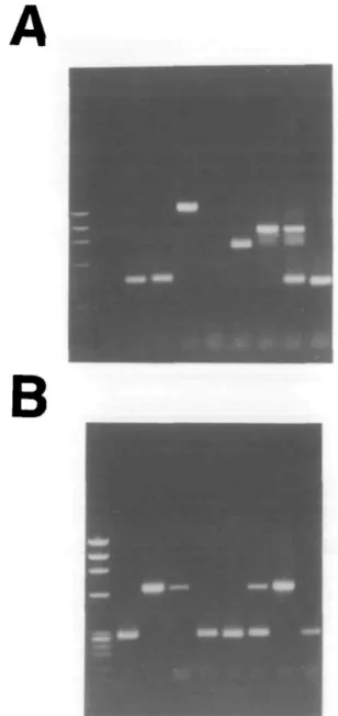
A total of 24 single B220+/c-kit+ cells, 48 single large B220+/ HH/SL+ cells, 48 single large B220+/CD25+/slgM" cells, 48 single small B220+/CD25+/slgM- cells, 48 single immature B cells and 44 single mature B cells were sorted as described in Methods, and analyzed for the status of their two Ig H, KL and XL chain alleles. A representative picture of VnJ558-D|_|JH rearrangements and V ^ I or 2 rearrangments found in single immature B cells is shown in Fig. 2.
H chain alleles. In 42% of the cells, both Ig H alleles were detected and in 36% of the cells only a single PCR product was observed. No amplification was obtained in 22% of the tested cells. However, it should be noted that cells containing certain combinations of rearrangements in Ig H loci might be under-represented Due to the recognition of Dp|_ and D$p segments by the same primer, the present assay does not resolve two DJH joints involving DH elements of these two families rearranged to the same JH genes on both chromo-somes, because these rearrangements will appear as a single PCR band. Moreover, two VHDJH joints involving VH genes of the same family rearranged to the same J H gene on both chromosomes cannot be discriminated on the gel. Further-more, the DSP/H. primer does not bind to the FL16.2 segment Hence, - 7 % of the DJH rearrangements will not be scored, assuming that the usage of the D segments in rearrangements is random, as suggested earlier (17).
The results of the analyses of the H chain alleles in single cells of different stages of B cell development are given in Tables 3 and 4. They show that the majority of all H chain alleles in B220+/c-kit+ cells are DHJH-. but not yet VH
DHJH-rearranged and are thus pre-B I cells. This is in agreement with previous analyses from our laboratory either on B220+/ c-kit+ cell populations obtained 'ex vivo' (8,14) or detected in lines and clones of B220+/c-kit+ cells proliferating on stromal cells in the presence of IL7 (8,14).
VHDHJH-rearranged alleles are detected at equally high frequencies in B 2 2 0+/ J I H / S L+, large and small B220+/CD25+/ slgM", immature B and mature B cells. More than 90% of the PCR positive cells of these showed at least one H chain allele in the VHDnJH-rearranged configuration. This is in agreement with the observation that >90% of all large and small B220+/ CD25+/slgM~ cells express nH chains in their cytoplasm (7). Among the cells in which both alleles were detectable, -50% had both alleles VHDHJH rearranged while the other 50% was VHDHJH/DHJH rearranged. Again, this was true for
A
B
Fig. 2. A representative example of VHJ558-DHJH rearrangments (A) and V ^ I or 2 rearrangements (B) as found in eight single immature B cells (lanes 2-9) Molecular weight markers are shown in lane 1.
B220+/nH/SL+ large and small B220+/CD25+/slgM", imma-ture B and maimma-ture B cells (Table 3). These results agree with and extend the observation that 40% of the peripheral B cells have both H chain alleles in a v^D^n-rearranged configuration (18). Future sequencing of all alleles should answer the question of which alleles are, or are not, product-ively VHDHJH rearranged, and quantitate the reading frame distribution amongst DHJH rearranged H chain alleles (5,14,19). They will also allow an investigation of the usage of V gene segments in the H chain alleles of different B cell subpopulations during development.
L chain alleles. Approximately 40% of V^. genes rearrange by inversion (20), thus retaining previous rearrangements on the same chromosome. In addition, any excised DNA containing
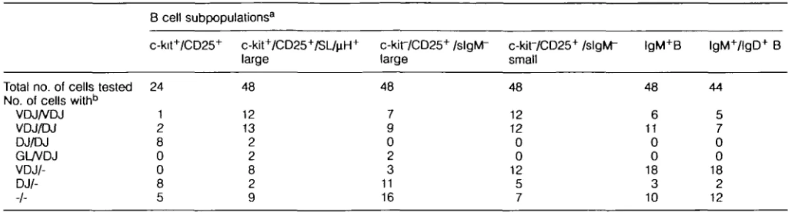
Table 3. Configuration of Ig H loci in pro/pre-B I, pre-B II, immature and mature B cells
Total no. of cells tested No. of cells withb
VDJ/VDJ VDJ/DJ DJ/DJ GUVDJ V DJ/- -/-B cell subpopulations3 c-kit+/CD25+ 24 1 2 8 0 0 8 5 c-kit+/CD25+/SL4iH+ large 48 12 13 2 2 8 2 9 c-kir/CD25+ /slgM" large 48 7 9 0 2 3 11 16 c-kir/CD25+ /slgM" small 48 12 12 0 0 12 5 7 lgM+B 48 6 11 0 0 18 3 10 lgM+/lgD+ B 44 5 7 0 0 18 2 12
aAII subpopulations are B220 (CD45R)+ b-: one or two alleles not detected.
Table 4. The rearrangement status of Ig H and L chain alleles of single cells at different stages of development
B cell subpopulations3
c-kit+/CD25' c-kit7CD25+/SI4iH+ c-kif/CD25+ /slgM" c-kit7CD25+/slgM"
large large small
lgM+B lgM+/lgD+ B lgM+lgD+XL+B No of cells analyzed 24
Configuration of Ig H chain alleles germline 0
DHJH 26
VHD H JH 4
efficiency of detection 63% Configuration of Ig L chain alleles
germline icL ND VA 0 VxJx 0 RS ND 48 48 48 48 44 36 2 19 46 70% ND 1 0 ND 2 20 28 52% ND 1 0 ND 0 17 48 68% 20 47 6 ND 0 14 41 57% 26 50 4 ND 0 9 35 50% 23 47 6 ND NDb ND ND -2 36 36 22
aAII subpopulations are B220 (CD45R)+ bND- not done.
the 'germline fragment' ( J ^ I - J ^ region) or primary V,JK rearrangements may still be present in the cell and thus be available for PCR amplification, since we employed an oligonucleotide (JK1-2) priming inside the JK cluster. When an inverted V,. gene or a non-inverted VK gene joins to J , ^ or 5, the germline fragment is retained in the same chromosome in the former case and is deleted in the latter. Hence, when a secondary (inversional) rearrangement has occurred, more than two PCR bands can be found. The numbers of alleles given in Table 4 as the results of the analyses of the KL chain alleles in different B cell subpopulations make the assumption that only one cell with maximally two KL chain alleles was analyzed in each assay. Hence, in cases where more than two PCR products of the KL chain locus were detectable (in fact in 12 of a total of 164 cells with detectable KL chain loci), they were counted as two alleles.
Of the 24 B22O+/c-kit+ cells, 48 B220+/u.H/SL+ cells and 48 large B220+/CD25+/slgfvr cells, only two V^-rearranged alleles were detectable (Table 4). This shows that the vast majority of precursor B cells rearrange the D H - JH and V H -D|-|JH segment on the H chain loci before they rearrange the
L chain loci. It makes the possible alternate pathway of B cell development improbable, or at least infrequent (21). While the large u.H/SL+ and u.H/SL~ pre-B II cells are both candidates for precursor of small pre-B II cells, it remains to be investi-gated whether both of them, or only one of them, play(s) this role in B cell differentiation.
Rearrangements of at least one allele of the KL chain gene locus became detectable in 65% of the small B220+/CD25+/ slgM~ cells, in 67% of the immature B and in >75% of the mature B cells (data for single cells not shown, but summar-ized as numbers of alleles in Table 4). This indicates that KL chain gene rearrangements are induced when small resting B22O+/CD25+/slgM" cells develop and suggests that large B220+/CD25+/slgM" cells are the precursors of their small counterparts. Collectively, these results define c-kit+ CD25" cells as mostly DHJn-''earranged pre-B I cells and wtiich are the precursors of large c-kir/CD25+/u.H/SL+/" VH DHJH-rearranged preB II cells, which are the precursors of the small c-kit7CD25+/u,H/SL~ VHDHJH/ViA-r e a ( T a n9e d pre-B II cells, which, in turn, are the precursors of the slgM+ immature B cells.
Rearrangements in KL versus XL gene loci
The majority of the L chain rearrangements are found in the K loci. The ratio of rearrangements in the KL versus XL chain is 10:1 (Table 3). This ratio is stable throughout the different B lineage populations with increasing maturity, i.e. from small B220+/CD25+/slgM- to immature B to mature B cells. The majority of V^-rearranged small pre-B II, immature B and mature B cells do not have XL chain gene rearrangements. However, 12 of the 16 cells with XL gene rearrangements have VKJK rearrangements using the most 3' J ^ segment.
Furthermore, mature XL chain-producing B cells from spleen were analyzed for the configurations of their L chain alleles (Table 4, last column). A rearranged XL locus was detected in all X-producing B cells Although we have not yet ruled out by sequencing that all 36 cells analyzed (Table 4) produced X^L chain, we take this as a strong indication that the X PCR assay is very efficient and can probably detect rearrange-ments in all the XL chain loci. Moreover, all XL-producing cells showed rearrangements in their KL chain loci Twenty-nine of the 36 cells analyzed, again, had V ^ rearrangements using J ^ .
These findings are consistent with previous reports (22,23). However, XL chain gene-rearranged B cells in which no KL chain gene rearrangements were detectable have been described (24). While this might imply that some XL chain-expressing cells have their KL chain loci in germline configura-tion, the more frequently observed VxJ^-rearranged pre-B II cells (five of six), immature B cells (two of four) or mature B cells (seven of eight), is one in which td_ chain gene loci are rearranged to downstream JK segments or even deleted via RS. In fact, deletion was observed in 22 of the 36 XL chain+slg+ B cells (Table 4, last column). In addition, due to the complexity of the KL chain gene locus introduced by opposite polarities of V* segments, it is not even certain that the XL chain-rearranged cells, in which no KL chain rearrangements were observed, have, in fact, these KL chain loci in germline configuration.
All these results suggest that the K L A L chain ratio of the mature peripheral B cell pool, which also is of the order of 10:1, is established as soon as L chain gene rearrangements are induced, i.e. in the pre-B II cells. It will require the analysis of a much larger number of cells from the different B lineage compartments in bone marrow and the sequencing of the rearranged Ig L chain loci to determine whether this ratio is exactly the same in all stages of B cell development with rearranged L chain loci or whether selective forces on a small number of the total cells influence this ratio with increasing maturity. It is also too early to conclude from our data that all XL chain rearrangements obligatorily follow KL chain rearrangements, although the majority of them appear to do so.
Finally, since small pre-B II cells do not deposit IgM on the surface and since L chains so far have not been detectable in the cytoplasm by immunofluorescence with specific anti-bodies (while u.H chains are easily detectable), it might well be that KL chain gene rearrangements and the expression of the rearranged L chain loci as proteins are separately regu-lated. One possible example of such a cell with rearranged KL chain loci but without expression of the rearranged gene locus is the 70Z/3 preB lymphoma (25).
A comparision of the B-lineage-committed, CD45RA (B220*) subpopulations of bone marrow characterized by different markers in different laboratories
Hardy's (6) and Rajewsky's (5) laboratories have used the differential expression of CD43, HSA and BP-1 to separate CD45RA (B220+) B-hneage-committed precursors into frac-tions B, C, C and D, and into the immature and mature, slgM+/slgD~ and slgM+/slgD+ populations E and F. Analyses of the status of the Ig gene loci in these fractions have shown that fraction B is enriched for DHJH-rearranged H chain loci which have not yet undergone VHD H JH rearrangements. Fraction B is therefore likely to be largely the same as our pro/pre-B I population (Fig. 1). Fraction C was found to be enriched for cells with two non-productively VHDHJH-rearranged Ig H alleles. We have hypothesized (see Fig. 1) but never detected these cells in our assays.
Ehlich et al. (5) have suggested that fractions C and D might have similar IgH rearrangements. They therefore only analyzed fraction D and found all cells to contain one product-ively VHDHJH-rearranged IgH allele. Fractions C and D could therefore be, at least in part, the same as the large, u.H/SL+ and u.H/SL~ pre-B II cells, and the small pre-B II cells In our analyses, 60% of the large pre-B II cells and 30% of the small pre-B II cells express BP-1 (7), indicating that the comparison of fractions C and D with pre-B II cells is complicated. Also, fractions C and D include to a good part the large, cycling cells that should contain the large, u.H/SL+ and u.H/SL~ pre-B II cells. Our scheme of separation on the basis of large, cycling and small, resting cells, as well as the patterns of expression of c-kit, CD25 and SL and the analysis of the rearrangement status of L chain gene loci appears to allow a better resolution of crucial steps in the development of precursor B cells, especially since L chain gene rearrange-ments are almost totally absent in large pre-B II cells and appear in full when they become resting, small pre-B II cells. With all the analyses on marker expression and status of Ig gene loci rearrangements, we should now be in the position to simplify the nomenclature of precursor B cells in bone marrow and to propose protocols for the analysis of precursor B cell compartments that might vary in their contents due to genetic or environmental influences. More importantly, we are now in a position to clearly separate cells with defined stages of Ig gene rearrangements according to their cellular program of differentiation, i.e. marker expression. This will facilitate the generation of differential cDNA libraries in the search for genes and molecules which control these B cell develop-mental steps.
Acknowledgements
The able technical assistance of Andrea Groenenwegen and Marc Dessing is gratefully acknowledged. We thank Drs Klaus Karjalainen and Thomas Winkler for critical reading of our manuscript. The Basel Institute for Immunology was founded and is supported by F. Hoffmann-La Roche Ltd, Basel, Switzerland.
Abbreviations
HSA heat stable antigen
RS recombination signal sequence (up- and downstream ofCtc)
References
1 Rolink, A. and Melchers, F. 1992. Molecular and cellular origins of B lymphocyte diversity. Cell 66:1081.
2 Rolink, A., Haasner, D., Nishikawa, S I. and Melchers, F. 1993. Changes in frequencies of clonable preB cells during life in different lymphoid organs of mice. Blood 812290.
3 Osmond, D G 1990. B cell development in bone marrow. Semin.
Immunol. 2:173.
4 Osmond, D. G., Kim, N , Manoukian, R., Phillips, R A., Rico-vargas, S. A. and Jacobsen, K. 1992. Dynamics and localization of early B-lymphocyte precursor cells (pro-B cells) in the bone marrow of scid mice. Blood 79:1695
5 Ehlich, A., Martin, V, MQIIer, W. and Rajewsky, K. 1994 Analysis of the B-cell progenitor compartment at the level of single cells.
Curr. Biol. 4:573.
6 Hardy, R. R., Carmack, C E., Shmton, S. A., Kemp, J. D. and Hayakawa, K. 1991. Resolution and characterization of proB and pre-pro B cell stages in normal mouse bone marrow. J. Exp.
Med. 173:1213.
7 Rolink, A., Grawunder, U., Winkler, T. H., Karasuyama, H. and Melchers, F 1994 IL-2 receptor a chain (CD25, TAC) expression defines a crucial stage in preB cell development Int Immunol 6.1257.
8 Rolink, A., Kudo, A., Karasuyama, H., Kikuchi, Y. and Melchers, F. 1991. Long-term proliferating early preB cell lines and clones with the potential to develop to surface-lg positive mitogen-reactive B cells 'in vitro' and 'in vivo' EMBO J 10327.
9 Tonegawa, S. 1983. Somatic generation of antibody diversity.
Nature 302:575
10 Winkler, T. H., Rolink, A. G., Melchers, F and Karasuyama, H. 1995. Precursor B cells of mouse bone marrow express two different complexes with the surrogate light chain on the surface.
Eur. J. Immunol, in press
11 Ogawa, M., Matzusaki, Y, Nishikawa, S , Hayashi, S. I., Kunisada, T., Sudo, T., Kina, T., Nakauchi, H. and Nishikawa, S. I. 1991. Expression and function of c-kit in hemopoietic progenitor cells
J. Exp Med. 174.63.
12 Leptin, M. 1985 MonoclonalantibodiesspecrficformurinelgM.il. Activation of B lymphocytes by monoclonal antibodies specific
for the four constant domains of IgM. Eur. J. Immunol. 15:131. 13 Parkhouse, R. M. E., Preece, G., Sutton, R , Cordell, J. L and
Mason, D. Y. 1992. Relative expression of surface IgM, IgD and the Ig-associating a (mb-1) and p (B-29) polypeptide chains.
Immunology 76:535
14 Haasner, D., Rolink, A. and Melchers, F. 1994. Influence of surrogate L chain on DHJH-reading frame 2 suppression in mouse precursor B cells Int. Immunol 6:21
15 Schlissel, M. S. and Baltimore, D. 1989. Activation of immunolglobulin K gene rearrangement correlates with induction of germline K gene transcription. Cell 58:1001.
16 Miller, J., Seisin, E. and Storb, U. 1982. Structural alterations in j regions of mouse immunoglobulin X genes are associated with differential gene expression. Nature 295.428.
17 Bangs, L. A., Sanz, I. E. and Teale, J. M 1991 Comparison of D, JH and junctional diversity in the fetal, adult, and aged B cell repertoires. J. Immunol. 146:1996
18 Alt, F W., Blackwell, T. K., Depinho, R. A., Reth, M. G. and Yancopoulos, G D. 1986 Regulation of genome rearrangement events during lymphocyte differentiation. Immunol. Rev 89:5. 19 Gu, H., Kitamura, D. and Rajewsky, K 1991. B cell development
regulated by gene rearrangement: arrest of maturation by membrane-bound D,, protein and selection of DH element reading
frames. Cell 65:47.
20 Shapiro, M A. and Weigert, M 1987. How immunoglobulin VK
genes rearrange. J. Immunol. 139:3834.
21 Kubagawa, H., Cooper, M. D., Carroll, A. J and Burrows, P. D. 1989. Light-chain gene expression before heavy-chain gene rearrangement in preB cells transformed by Epstein-Barr virus.
Proc NatlAcad Sci. USA 86.2356.
22 Coleclough, C 1990 A critique of the Cohn-Langman protecton
theory Immunol. Rev. 115:173.
23 Hieter, P. A., Korsmeyer, S J., Waldmann, T. A. and Leder, P 1981. Human immunoglobulin K light chain genes are deleted or rearranged in X producing B cells. Nature 290:368.
24 Berg, J , McDowell, M., Jack, H. M. and Wabl, M. 1990. Immunoglobulin X gene rearrangement can precede K gene rearrangement Dev. Immunol. 1:53
25 Paige, C , Kincade, P. and Ralph, P. 1978. Murine B cell leukemia line with inducible surface immunoglobulin expression. J.