HAL Id: hal-01635382
https://hal.archives-ouvertes.fr/hal-01635382
Submitted on 27 Nov 2017
HAL is a multi-disciplinary open access
archive for the deposit and dissemination of
sci-entific research documents, whether they are
pub-lished or not. The documents may come from
teaching and research institutions in France or
abroad, or from public or private research centers.
L’archive ouverte pluridisciplinaire HAL, est
destinée au dépôt et à la diffusion de documents
scientifiques de niveau recherche, publiés ou non,
émanant des établissements d’enseignement et de
recherche français ou étrangers, des laboratoires
publics ou privés.
Distributed under a Creative Commons Attribution - NonCommercial| 4.0 International
Mutual interference of Cu and Zn ions in Alzheimer’s
disease: perspectives at the molecular level
Elena Atrián-Blasco, Amandine Conte-Daban, Christelle Hureau
To cite this version:
Elena Atrián-Blasco, Amandine Conte-Daban, Christelle Hureau. Mutual interference of Cu and Zn
ions in Alzheimer’s disease: perspectives at the molecular level. Dalton Transactions, Royal Society
of Chemistry, 2017, 46 (38), pp.12750 - 12759. �10.1039/C7DT01344B�. �hal-01635382�
Dalton
Transactions
An international journal of inorganic chemistry
rsc.li/dalton
ISSN 1477-9226
PERSPECTIVE Christelle Hureau et al.
Dalton
Transactions
PERSPECTIVE
Cite this:Dalton Trans., 2017, 46, 12750
Received 13th April 2017, Accepted 22nd June 2017 DOI: 10.1039/c7dt01344b rsc.li/dalton
Mutual interference of Cu and Zn ions in
Alzheimer
’s disease: perspectives at the molecular
level
Elena Atrián-Blasco,
†
a,bAmandine Conte-Daban
†
a,band Christelle Hureau
*
a,bWhile metal ions such as copper and zinc are essential in biology, they are also linked to several amyloid-related diseases, including Alzheimer’s disease (AD). Zinc and copper can indeed modify the aggregation pathways of the amyloid-β (Aβ) peptide, the key component encountered in AD. In addition, the redox active copper ions do produce Reactive Oxygen Species (ROS) when bound to the Aβ peptide. While Cu(I) or Cu(II) or Zn(II) coordination to the Aβ has been extensively studied in the last ten years, characterization
of hetero-bimetallic Aβ complexes is still scarce. This is also true for the metal induced Aβ aggregation and ROS production, for which studies on the mutual influence of the copper and zinc ions are currently appearing. Last but not least, zinc can strongly interfere in therapeutic approaches relying on copper detoxification. This will be exemplified with a biological lead, namely metallothioneins, and with synthetic ligands.
Introduction
Alzheimer’s disease (AD) is the most common neurodegenera-tive disease, with a prevalence of around 35 million patients
worldwide.1This number is expected to triple within the next 35 years, making AD a current major global public health problem. In the brain, one of the hallmarks of the disease is the extracellular accumulation of Amyloid-β (Aβ) peptides into senile plaques. The amyloid cascade hypothesis describes this process: Aβ is present in healthy brains in soluble and mono-meric forms. In contrast, in AD brains, these peptides aggre-gate into oligomers and then fibrils which assemble them-selves into so-called senile plaques.2,3 In the last few years,
Elena Atrián-Blasco (right), Amandine Conte-Daban (left) and Christelle Hureau (middle)
Elena Atrián-Blasco (right) was born in Zaragoza (Spain) in 1988, where she obtained her BSc degree in 2011 after an Erasmus stay at Queen’s University Belfast (UK) in 2009–2010, and her MSc in
Chemistry in 2012. After obtaining her PhD diploma at the ISQCH (Univ. Zaragoza-CSIC) under the supervision of Prof. Mariano Laguna and Dr Elena Cerrada, she joined Christelle’s team in Toulouse for her postdoctoral stay. Amandine Conte-Daban (left) was born in Tarasteix (France) in 1991 and obtained her BSc degree in Physical Chemistry at Université Paris-Sud, and obtained her MSc degree in Medicinal Chemistry from the Université Paul Sabatier in Toulouse in 2014. She is currently per-forming her PhD in the “Alzheimer and amyloids” team with Christelle. Christelle Hureau (middle) was born in Charleville-Mézières (France) in 1976. She has a background in physical in-organic chemistry with a focus on EPR and electrochemistry. She is interested in chemistry in link with the biological world. In 2015, she became the group leader of the “Alzheimer and amy-loids” team at the Laboratoire de Chimie de Coordination (UPR 8241– CNRS). There, she investigates the structural and dynamic aspects of metal ion coordination to the amyloid-β peptide and new chelating concepts against Alzheimer’s disease.
†These authors contributed equally.
aCNRS, LCC (Laboratoire de Chimie de Coordination), 205 route de Narbonne,
BP 44099 31077 Toulouse Cedex 4, France. E-mail: christelle.hureau@lcc-toulouse.fr
bUniversity of Toulouse, UPS, INPT, 31077 Toulouse Cedex 4, France
Open Access Article. Published on 22 September 2017. Downloaded on 27/11/2017 13:32:30.
This article is licensed under a
Creative Commons Attribution-NonCommercial 3.0 Unported Licence.
View Article Online
therapies based on inhibiting the aggregation of the Aβ pep-tides have failed clinical trials and, as a consequence, atten-tion has been paid to other factors.4 Indeed, AD has been linked to the dyshomeostasis of both Cu(I/II) and Zn(II) ions.5
Regarding Zn(II) ions, both increased and decreased
concen-trations have been measured in AD brains compared to healthy brains.6 Similarly, Cu ions are present in a 2-fold higher concentration in the CNS6and in higher concentrations in the hippocampus while intra-neuronal Cu(I) deficiency has
been reported to be a key factor in AD.7,8Important levels of these metal ions have also been found in the senile plaques: 0.4 mM of Cu and 1 mM of Zn(II).9,10 Consequently, these
metal ions are supposed to play a key role in the aggregation of the Aβ peptides in vivo and thus in the associated amyloid cascade process.11,12 In addition, the literature reveals the impact of metal ions on the Aβ aggregation in vitro, although contradictory effects have been reported.11,12They can modify
either the kinetics of aggregation or the morphology of the aggregates. While there are many divergent reports on the impact of metal ions, two main consensual features are described: (i) the importance of the ratio between metal ions and peptides,11–13and (ii) the different effects produced by Cu and Zn ions.11Metal-induced aggregation is also described for other neurodegenerative diseases such as Parkinson’s and Prion diseases, all characterized by misfolding of amyloido-genic proteins and metal ion dyshomeostasis.6,14
In AD, another important consequence of this dyshomeos-tasis is the production of Reactive Oxygen Species (ROS) by the Cu–Aβ complex, which catalyses the reduction of O2, in the
presence of a reductant such as ascorbate.9,15 Indeed, it has been proposed that superoxide, hydrogen peroxide and hydroxyl radicals, which are the resulting species of the incom-plete reduction of O2, attack the surrounding biomolecules,
thus participating in the global oxidative stress observed in AD.16
Within this context, metal chelation (using chelators)‡ or metal redistribution (using metallophores)‡ therapeutic approaches against AD have been under focus during the past few years.8,17 In general, Cu is considered as the most perti-nent target due to the ROS related toxicity of this redox compe-tent ion.8 The first generations of ligands‡ were capable of removing Cu(II) from Aβ peptides, stopping ROS production
and favouring the disaggregation of Aβ senile plaques.8 Clioquinol and PBT2 have shown to be the best candidates and have gone under clinical trials, however, both have failed in phase II.18To explain such unsuccessful results, it has first been proposed that these ligands cannot differentiate between the toxic Cu bound to Aβ and the Cu bound to essential metal-loproteins. A new hypothesis relies on a too weak
discrimi-nation between the Cu(II) target and the Zn ions.19,20In fact,
the significance of the selectivity in the development of ligands in the wide context of the chelation therapy has already been reviewed.21,22Particularly, the need for selectivity between the targeted toxic metal ion and other biological metal ions has been discussed for actinide decorporation,23 and the need for Cu/Zn selectivity in the case of Wilson’s disease.24
Therefore, different approaches are under focus to find new generations of ligands:17,25 (i) prochelators,26 which can be activated by H2O227 or β-secretase28 directly in the brain;
(ii) multi-target compounds, e.g. those with an Aβ recognition moiety and a chelating moiety;29,30(iii) Cu(II)-chelators with a
glucose moiety31 or nanoparticles32 as transporters through the Blood Brain Barrier.
It seems clear that the brain is a highly complex system, which also makes AD an intricate illness. There are many factors that could be implicated in the development of the disease, such as the different metal ions present in the synap-tic cleft altogether with a wide range of biomolecules. The interaction of all of them could be relevant for the etiology, but trying to understand the basis requires starting by the sim-plest systems. For example, from a bio-inorganic chemistry approach of the disease, the effort was put on monometallic systems i.e. Cu–Aβ,10,33or Zn–Aβ.34–36Nevertheless, this is par-tially representative of the reality. Hence, a first step toward a more realistic picture would be to study the mutual influence of both metal ions in their interaction with the peptide at the molecular level, in order to understand the fundamental aspects of AD and to improve the development of ligands.
In this review, we report first the coordination chemistry of Aβ peptides with Cu(II), Cu(I) or Zn(II) and also of
hetero-bi-metallic species. The impact of the co-presence of Cu and Zn ions on the metal-induced Aβ aggregation and/or on the ROS production by the Cu–Aβ complex is then described. Finally, the importance of considering Zn in Cu-based chelation therapy is discussed. Personal points of view are also given about future research lines with respect to these three aspects.
Coordination chemistry of Cu and Zn
to A
β
Monometallic complexes
The amyloid-β (Aβ) peptide is a 40–42 amino-acid residue long peptide, of which the more hydrophobic region, the C-terminal, is prone to aggregation. The high affinity metal binding site of Aβ peptides is found at residues 1–16. For this reason, Aβ1–16 is proposed as a model for the coordination and redox properties of the full-length peptides. Coordination of Cu(II), Cu(I) and Zn(II) to the amyloid-β peptides has been
extensively studied and probed by many different techniques. The most accepted coordination spheres for these metal ions, as well as their corresponding affinities, are summarized in Fig. 1. In the case of Cu(II), two different binding modes can
be found at physiological pH, known as components I and II.
‡Chelators: molecules capable of binding Cu ions with a high affinity constant, which then cannot release them rapidly. Metallophores: molecules capable of binding Cu ions and releasing them in another place. Ligands: molecules capable of binding Cu ions, with no precision on their releasing ability (a ligand can keep the metal ions or can release them, i.e. a ligand can either be a chelator or a metallophore).
Open Access Article. Published on 22 September 2017. Downloaded on 27/11/2017 13:32:30.
This article is licensed under a
They both show a distorted square-planar geometry and share the terminal amine as a ligand. Component I, which is pre-dominant at lower pH, binds to Cu(II) through the N-terminal
amine, the carbonyl from the amide bond of Asp1–Ala2, and the imidazole nitrogen (Nim) from two His residues, His6 and
His13 or His14 in equilibrium. In component II, predominant at higher pH values, the nitrogen atom from the Asp1–Ala2 amide bond is deprotonated and binds to the Cu(II) ion,
together with the N-terminal amine, the CO from the Ala2– Glu3 peptide bond and one imidazole group from a His residue.10,37A second site has been described for the Aβ1–28 peptide but its low affinity would make it a biologically irrele-vant coordination site.38Cu(I) is bound in a linear fashion by
two among the three possible Nimof His6, His13 and His14 in
an equilibrium, in which the His13–His14 pair seems to be the preferred ligands.39–41 The first 16 amino acids are also involved in the coordination of Zn(II). Previous studies stated
the involvement of the three histidine residues in the coordi-nation site.42–48The N-terminal amine and the Glu11 residue (among others) would be also involved in the coordination.34–36 A more recent study proposes a different coordination sphere deduced from the study by1H-NMR and
X-ray absorption spectroscopy of Aβ1–16 and a range of modi-fied peptides.34The Zn(II) ion has a tetrahedral binding to two
histidine residues (His6 and His13 or His14), and two carboxylate residues (Glu11 and Asp1 or Glu3 or Asp7, with a preference for Asp1). In this case, the N-terminal amine does not coordinate to Zn(II) at physiological pH. This proposition
is in line with previously reported affinity studies on the very same series of modified peptides.49
Hetero-bimetallic complexes
A seminal competition study observed that Cu(II) displaced
more than 95% of Zn(II) bound to Aβ.50 The first research
regarding the simultaneous binding of Cu(II) and Zn(II) to Aβ
peptides was performed by Damante et al.51where they used
the polyethylene glycol (PEG) conjugated peptide, which has enhanced water solubility and could aid in the spectroscopic studies. By using potentiometric measurements altogether with UV-Vis, CD and EPR spectroscopy studies, they shed light on the mutual interference of the metal ions. At pH >7, a hetero-bimetallic complex of Cu(II), Zn(II) and Aβ at a ratio of
1 : 1 : 1 is formed. Addition of up to 4-fold equivalents of Zn(II)
to a Cu(II)–Aβ solution shifts the copper binding mode from
Fig. 1 Proposed coordination modes of Cu(II),10Cu(I),10Zn(II)34and the mixed metal Cu(II),Zn(II) or Cu(I),Zn(II) complexes52,53based on experimental
results and bibliographic data. Affinity constant values (Ka) at pH 7.4 for Cu(II),57Cu(I)58,59and Zn(II)49are given in M−1(bold, purple).
Perspective Dalton Transactions
Open Access Article. Published on 22 September 2017. Downloaded on 27/11/2017 13:32:30.
This article is licensed under a
Creative Commons Attribution-NonCommercial 3.0 Unported Licence.
component I to component II (Fig. 1), while His13 and His14 would be involved in Zn(II) binding. The addition of Zn(II)
would not release Cu(II), probably due to the high affinity of
the peptide for Cu(II), but would modify its distribution in the
available binding sites. Spectroscopic data obtained by Alies et al.52support this hypothesis. Furthermore, Silva et al.53have also corroborated the shift of components for Cu(II) bound to
Aβ upon addition of increasing equivalents of Zn(II) (see
Table 1). Contrary to the study of Damante et al.,51they also probed a more important contribution of His13 and His14 as ligands for Cu(II) in the presence of Zn(II).53In a further study,
they proposed that Zn(II) and Cu(II) do compete for one
binding site.54 However, this is in disagreement with the respective affinity values of Cu(II) and Zn(II) for the Aβ peptide
that differ by at least three orders of magnitude (Fig. 1). The most likely explanation for such unexpected observation is linked to the experimental procedure that may force precipi-tation of either Zn(II)–Aβ or Cu(II)–Aβ and modify the
associ-ated thermodynamic equilibria.54 Indeed, with other sample preparation, only the formation of the hetero-bimetallic complex was observed.52Alies et al.52have investigated as well the Cu(I)/Zn(II) system which could be highly relevant in vivo.
They used NMR spectroscopy and XANES to study the co-pres-ence of Cu(I) and Zn(II). Indeed, XANES allows to distinguish
the different oxidation states of the Cu ion and to observe the environments of Cu and Zn simultaneously since Cu and Zn K-edges are close enough to be recorded on the very same sample. While Zn(II) only slightly impacts the Cu(II) binding
site (inducing a weak shift from component I to component II), both Cu(I) and Cu(II) influence the coordination sphere of
Zn(II). Notably, in the case of the mixed Cu(I)/Zn(II) complex,
Cu(I) remains coordinated to two histidine residues, leaving
only one available for Zn(II) binding (Fig. 1).
Kinetics of metal binding by Aβ peptides
Another important parameter to take into account would be the kinetics of the interaction between metal ions and Aβ pep-tides. A pioneering study by Pedersen et al.55investigated the rate of the binding of Cu(II) to Aβ, and its impact on the
aggre-gation. In the same context, a recent study by Branch et al.56 has measured the rate of Cu(II)–Aβ complex formation. The
component I binding mode is very rapidly formed, while com-ponent II forms through comcom-ponent I as an intermediate. The association rates of Cu(II) and Aβ reported vary between 1 and
100 ms. As a consequence, the association between Aβ and Cu(II) is fast and is thus of biological relevance.
Concluding remarks and prospective
All the studies (summarized in Table 2) agree on the mutual influence of Zn(II) and Cu(II) on their coordination. Zn(II) impacts the binding mode of Cu(II) at physiological pH as observed by the shift from component I to component II, whereas Cu(I) maintains its bis-His coordination sphere in the presence of Zn(II). Moreover, Zn(II) coordination is affected by
the presence of both Cu(I) and Cu(II).
Further studies would be needed to evaluate how the affinities of Cu(I/II) and Zn(II) are influenced by the co-presence of the metal ions and to determine the exact nature of the coordination spheres of Zn(II) in the Cu(I/II),Zn(II) hetero-bi-metallic complexes. In addition, probing hetero-bihetero-bi-metallic species of other biologically relevant Aβ peptides (truncated peptides, murine Aβ and Familial Alzheimer’s Disease mutants) is also of interest as well as the kinetics of inter-actions of Aβ with Cu(I), Aβ with Zn(II), and the mutual influ-ence of Cu and Zn.
Aggregation and ROS production
AggregationAβ aggregation is the catalytic auto-assembly of the monomeric peptide (mathematically described by a sigmoid-shape curve).11 Metal-induced aggregation has been widely studied: different pH conditions, temperature, concentration, metal : peptide ratio, etc. seem to have an influence on the aggrega-tion rate and species formed. The influence of Cu(II) on the Aβ
aggregation seems to be mainly dependent on its ratio (see Fig. 2, left),61and may lead to the production of highly cyto-toxic oligomers.55,62,63 In the case of aggregation in the pres-ence of Zn(II), even small sub-stoichiometric quantities of the
metal ion have an important influence on the aggregation of the Aβ peptides (see Fig. 2, right).60,63–68
Intra and inter-molecular Zn(II) binding promotes a fast
aggregation of species different from apo-fibrils. In the pio-neering work by Mayes et al.69, incubation of Aβ1–42 with equi-molar quantities of Cu(II) and Zn(II) showed the coexistence of
both small amorphous (non-fibrillar) aggregates and fibrils. Later, Matheou et al.60showed that Zn(II) has a greater
influ-ence on the aggregation as, at even a quarter less of Zn(II) than
Cu(II), the ThT aggregation curve was strongly affected and
typical Zn(II)-induced aggregates could be observed by TEM
(see Fig. 2, bottom). At the suprastoichiometric ratio of Cu(II) Table 1 Approximate proportions of the Cu(II) binding modes
(com-ponent I/com(com-ponent II) in the mixed Cu,Zn–Aβ complexes formed at different Cu : Zn : Aβ ratios, at pH 7.4
Ref. 1 : 0 : 1 1 : 1 : 1 1 : 2 : 1 1 : 4 : 1 1 :≥8 : 1
52 ∼80/20 55/45 40/60 — —
53 65/35 53/47 40/60 35/65 31/61
Table 2 Summary of the studies on the coordination of mixed Cu,Zn– Aβ complexes: used techniques and peptides
Ref. Techniques Peptides
51 Potentiometry; CD, UV-Vis, EPR; LC-ESI-MS Aβ1–16-PEG; Aβ1–6; Ac-Aβ8–16-(Y10A) 52 XANES, EPR, NMR Aβ1–16 53 CW-ESR, ESEEM Aβ1–16; 15N His-enriched Aβ1–40
54 XANES, EXAFS, CW-ESR Aβ1–16
Open Access Article. Published on 22 September 2017. Downloaded on 27/11/2017 13:32:30.
This article is licensed under a
and/or Zn(II), Attanasio et al.70reported the formation of non-fibrillar aggregates.
ROS production by monomeric species
Since Zn(II) and Cu(I/II) have a clear mutual influence on their
coordination to the Aβ peptides and on the metal-induced aggregation, Zn(II) could also have an impact on the ROS
pro-duction by the Cu–Aβ complex. In this sense, a few studies have taken into account the co-presence of these metal ions and some of them have even indicated a protective role of Zn(II).71 The first studies on the protective role of Zn(II) in
Cu(II)–Aβ-induced ROS production were carried out by Bush
and co-workers.71 They found that the co-presence of Zn(II)
improved the survival of cells incubated with Cu(II) and Aβ1–42
altogether with a decrease of the level of H2O2 production.
They proposed a shift of the Cu ion in the peptide apart from its redox-active binding site as the origin of the redox-silencing properties associated with Zn(II). As it has been reported later,
Zn(II) effectively displaces Cu(II) to its component II binding
mode (see the previous paragraph); however, Alies et al.52 showed that this has no impact on the ROS production as monitored by ascorbate consumption and H2O2 production
(two of the most used methods to probe the production of ROS by Cu–peptide systems, see Fig. 3). In other words, while Zn(II)
impacts the coordination of Cu(II) to Aβ, its addition at the
stoichiometric ratio has no effect on the Cu–Aβ-induced ROS production and associated cellular toxicity (Fig. 4A). This is in line with a recent study proposing that, in contrast to what can be expected based on the redox potential of Cu(II) in
com-ponent I or II,75the coordination mode of Cu(II) is not the
pre-dominant factor in ROS production.76 Other factors such as the metal ion ratio or aggregation states might influence the Fig. 2 This figure resumes the aggregation with different
Cu(II) : Zn(II) : Aβ ratios.60 Zn is the ion that dominates the Aβ metal-induced assembly even in the presence of Cu. For all of these aggre-gation experiments, metal ions have been added at the beginning, i.e. these are metal-induced aggregations.
Fig. 3 ROS production from Cu–Aβ complexes and the classical assays for the detection of ROS produced15(O
2•−,72H2O2,52HO•,73) and indirect
monitoring of Asc consumption.74
Fig. 4 Panel A. This part represents the identical ROS production of a monomeric monometallic Cu–Aβ complex and of a monomeric hetero-bi-metallic Cu,Zn–Aβ complex.52Panel B. This part represents the ROS production by different Cu(
II) : Aβ ratios and by different states of aggregation of
the peptide:fibrils produce less ROS than monomers, for the 0.5 : 1 ratio.52,78Panel C. This last part compares the ROS production by amorphous
aggregates of Cu(II)–Aβ to the ROS production of aggregates in the presence of Zn(II).69Note that for all of the aggregation experiments, metal ions
have been added at the beginning,i.e. these are metal-induced aggregations.
Perspective Dalton Transactions
Open Access Article. Published on 22 September 2017. Downloaded on 27/11/2017 13:32:30.
This article is licensed under a
Creative Commons Attribution-NonCommercial 3.0 Unported Licence.
ROS production by mixed metal complexes and it may be of interest to further investigate them as well.
ROS-production by aggregated species
The relationship between the aggregation states of the Cu–Aβ complexes and their ROS production in vitro has been described in the literature. Cu–Aβ aggregated species are able to produce ROS,69but at a lower level than the Cu–Aβ mono-meric complex (see Fig. 4B).77,78 Mayes et al.69 have also incorporated into their research the ROS production of mixed metal Cu,Zn–Aβ aggregates (see Fig. 4C). They studied the degradation of H2O2 and the consequent HO• production by
different Cu–Aβ and Cu,Zn–Aβ aggregated species. They observed that Aβ aggregates are capable of degrading H2O2
both when they have been formed in the presence of Cu(II) and
when the metal has been added after fibril formation. In addition, increasing the ratio of Zn(II) over Cu(II) : Aβ
dimin-ished the quantity and the rate at which H2O2was degraded.
They also corroborate the“antioxidant” effect associated with Zn(II) with less intense oxidation of the peptides, followed by
carbonyl group formation indicative of peptide oxidation. Concluding remarks
In this part, studies have shown that metal-induced aggrega-tion and/or ROS producaggrega-tion are already complex in mono-metallic systems for which there are many studies reported (see reviews:11,12,79). There are still not enough reported data on hetero-bimetallic systems to reach any kind of general con-clusion about the mutual influence of Cu and Zn ions on aggregation and/or ROS production. Future studies should address this issue and include more detailed investigations on the impact of different factors such as Cu to Zn ratios, metal to peptide ratios, pH, mixture of Aβ peptides (Aβ1–40, Aβ4–40, Aβ11–40, etc.)… In this context, pioneering aggregation studies performed in CSF mimicking media70 or in CSF from AD patients are worth noting.80
Besides, kinetic aspects may also be extremely important for aggregation. This includes determining how the metal (Zn(II) or Cu(II)) addition during the aggregation matters both
for the kinetics of the aggregation process and for the morphology of the aggregates. Knowing the rate of Zn and/or Cu-induced structural rearrangement to aggregation prone species would also be of significance.
Metal ion-based therapy
Due to the important toxicity shown by Cu ions within the context of AD, many Cu(II) ligands have been developed in the
past few years.17,25They all can remove Cu(II) from Aβ peptides,
and reduce its associated toxicity. One of the requirements for the design of ligands is their affinity towards Cu(II) compared
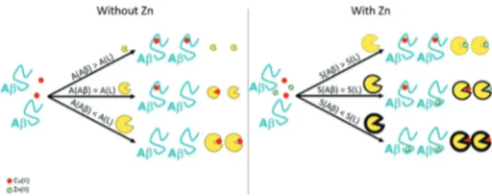
to Aβ: it should be higher than that of the peptide (see Fig. 5, left). Nevertheless, their affinity should be lower than that of important metalloproteins, in order to avoid deleterious side effects. Furthermore, as previously mentioned, Zn(II) is present
in high concentrations in the synaptic cleft and in the senile plaques and has an important impact on the Cu(II)
coordi-nation and the associated ROS production and Aβ aggregation. For this reason, some researchers have studied the impact of Zn(II) on the Cu(II) chelation therapy.
Metallothioneins: a biological model
Vašák and co-workers have studied the impact of metallothio-nein 3 (Zn7-MT-3) on the toxicity of the Cu(II)–Aβ complex.81,82
There is a swap of metallic ions between the Aβ peptides and Zn7-MT-3 (see Fig. 6, left). The mechanism proposed includes
the dissociation of the Cu(II)–Aβ complex and the chelation of
the free metal ion by the metallothionein.83 Also, the for-mation of two disulphide bonds triggers the reduction of Cu(II)
into Cu(I), forming an air-stable Cu(I)4-thiolate cluster inside
the Cu(I)4Zn4MT-3 protein. The removal of Cu ions from the
Aβ peptides and its consequent binding as an air-stable Cu(I
)-complex silence the redox capability of Cu. Therefore, this swap has a protective effect against Cu(II)–Aβ toxicity.82
Another study by West’s group84focused on the metallothio-nein 2A (MT-2A) which is the major human-expressed subtype of metallothionein and, under stressful situations, it can be secreted near the synaptic cleft where aggregation of Aβ pep-Fig. 5 Left panel. Representation of the importance of the affinity con-stant (written A) of the chelator in order to remove Cu from the Aβ peptide. Higher the affinity constant of the ligand is, higher is its efficiency in the removal of the metal ion from the peptide. Right panel. Representation of the importance of the selectivity (written S) of the ligand compared to the peptide one. Higher the selectivity of the ligand is compared to the peptide one, higher is its efficiency in the removal of Cu(II) ion from the peptide in the presence of Zn(II).
Fig. 6 Representation of the swap of metallic ions (Cu(II) in red and
Zn(II) in green) between Aβ peptide and Zn7-MT-3 (left) or a synthetic
chelator, preloaded with Zn(II) (right). The impact on the ROS production
is also illustrated.
Open Access Article. Published on 22 September 2017. Downloaded on 27/11/2017 13:32:30.
This article is licensed under a
tides occurs. West and his colleagues have demonstrated that under physiological conditions, Zn7-MT-2A can also remove
Cu ions from Aβ, stop the ROS production and prevent changes in the metallic balance within neurons. When the same experiments were performed with the apo metallothio-nein, no impact on the Cu removal or on the diminution of the neurotoxicity was detected. Recently, Bal and co-workers have also studied the metal swap between Zn7-MT-3 and the
truncated peptide Aβ4–16. In this case, the high affinity of the peptide prevents the sequestration of Cu(II) by the
metallothio-nein.85 Besides, the impact of the Zn7-MT-3 has also been
studied for the α-synuclein and the Prion proteins and the swap of metallic ions is observed. Cu(II) is sequestered by the
metallothionein, silencing its redox activity and preventing the metal-induced aggregation of the protein.86,87
Synthetic ligands and selectivity issue
Later, our group has highlighted the impact of the presence of Zn(II) within the Cu(II)-chelation context.19Two synthetic
chela-tors were tested, and while both are able to retrieve Cu(II) from
Aβ, only one removes Cu(II) from Aβ in the presence of Zn(II).
For the latter one and as in the case of Zn7-MT-3, there is a
swap of metallic ions between the peptide and the chelator, and the resulting species cannot produce ROS (see Fig. 6, right). We propose the following explanation (based on thermodynamics only): a good chelator needs not only a high affinity constant for Cu(II) (see Fig. 5, left) but also high Cu(II)
over Zn(II) selectivity, that corresponds to the ratio between its
affinity constant for Cu(II) and for Zn(II) (see Fig. 5, right).
Actually, the Cu(II) over Zn(II) selectivity of the chelator should
overcome that of the Aβ peptides themselves, which is high (about 4 orders of magnitude, see Fig. 1).
There are several studies reported in the literature that address the removal of Cu(II) or Zn(II) from Aβ,88–91 and some
introduce the importance of having a high Cu(II) over Zn(II)
selectivity for the ligand.92–94 However, to the best of our knowledge, the importance of overcoming the Cu(II) over Zn(II)
selectivity of Aβ was not taken into account. In other words, the removal of Cu(II) from Aβ by a chelator in the presence of
Zn(II) was not thoroughly described except in ref. 19.
Nevertheless, Zn(II) interference in Cu(II) removal from Aβ
should be taken into account as it can either promote protec-tive effects when the metal swap occurs19,82,84or be detrimen-tal if not.19
Prospective
Importance of the selectivity issue for other metal-based therapeutic approaches. All of these different studies prove that within the chelation therapy context, not only the toxic Cu(II) ion has to be taken into account but also other metal
ions in the environment of the peptides such as Zn(II). The
same kind of study should be applied to the removal of Cu(I)
in the presence of Zn(II). Indeed, the oxidation state of Cu in
the synaptic cleft is not well defined. It would also be interest-ing to design Cu(I) chelators within the AD context, as recently
reported in the literature.95,96 However, there are no studies
about Cu(I) chelation against AD in a Zn(II)-rich environment.
The same selectivity issue may also apply for metallophores, such as clioquinol, PBT2,18 and gtsm,97and for multi-target ligands as well.17
Selectivity issues regarding other peptides. The selectivity of the ligands may take into account the intrinsic selectivity of the peptide of interest as there are many forms of Aβ peptides. In the case of Aβ4–40 and Aβ11–40 peptides, which are N-terminally truncated, an extremely high Cu(II) over Zn(II)
selectivity is expected based on the formation of the high Cu(II)-affinity ATCUN motif,98while Zn(II) affinity may remain
mostly unchanged. This will imply having ligands with even higher Cu(II) over Zn(II) selectivity than the one required in the
case of Aβ1–40. The same would also be true for the murine peptide for which higher Cu(II) affinity has been reported,99as
well as weaker Zn(II) affinity.100 This might be important as
murine peptides are co-secreted along with human peptides in AD model mice.
Influence of other biological components. As probed here for Zn(II) and Cu(I/II), interference of other biological
com-ponents might also be of importance. Iron, for example, is also present in concentrations that would legitimate this kind of investigation. Indeed, iron is also responsible for ROS pro-duction, and might modulate the aggregation of the Aβ pep-tides.10,101,102The removal of this metal ion is likewise impor-tant and some groups are working on specific iron chelators for AD (for review, see ref. 101). Recently, Youdim’s group has studied the ability of new Fe chelators to improve memory loss in rats.103 However, in contrast to Zn(II), much effort is
required to decipher the Fe(II/III) binding ability to Aβ, since
only a very preliminary study has been reported until now.104 Moreover, other metal ions, despite having a lower affinity constant for the peptides, can also interfere since they are highly concentrated. Thus, it would be important to investigate different metal chelation/redistribution therapeutic approaches in the presence of all the biologically relevant ions (Mg(II), Ca(II), Na(I), K(I)…).
Kinetic issues. Another key parameter is the rate of Cu ion removal from the Aβ peptides,56,105including in the presence of Zn(II). In other words, the kinetic aspect has to be taken
into account as well as the thermodynamic one, as previously detailed.
Conclusions
In this review, we have focused on the mutual influence of Cu and Zn regarding their coordination to Aβ peptides, as well as the resulting impact on the aggregation and ROS production. We have also highlighted the importance of the co-presence of both Cu(II) and Zn(II) for the metal ion chelation/redistribution
therapeutic approaches.
As detailed through the present manuscript, future chemi-cal work will undoubtedly include studies of the interaction of both metal ions with the peptide in the presence of other bio-logically relevant components, such as glutamate,
acetyl-Perspective Dalton Transactions
Open Access Article. Published on 22 September 2017. Downloaded on 27/11/2017 13:32:30.
This article is licensed under a
Creative Commons Attribution-NonCommercial 3.0 Unported Licence.
choline, other metal ions, metallothioneins… This will bridge the gap between in vitro and in vivo studies, probing metal ion–peptide interactions under more complex conditions but still at the molecular level.
Abbreviations
Aβn–m Amyloid-β starting from the amino acid residue number n to the number m.
AD Alzheimer’s disease Ala Alanine
Asc Ascorbate Asp Aspartic acid
ATCUN Amino-terminal copper and nickel CCA Coumarin-3-carboxylic acid CD Circular dichroism CNS Central nervous system
CW-ESR Continuous-wave electron spin resonance Cyt c Cytochrome c
EPR Electronic paramagnetic resonance ESEEM Electron spin echo envelope modulation EXAFS Extended X-ray absorption fine structure Glu Glutamic acid
His Histidine
LC-ESI-MS Liquid chromatography–electro spray ionization-mass spectrometry
MT-2A Metallothionein-2A MT-3 Metallothionein-3
Nim Nitrogen from the imidazole ring
NMR Nuclear magnetic resonance
PBT2 5,7-Dichloro-2-[(dimethylamino)methyl]quinolin-8-ol
PEG Polyethylene glycol ROS Reactive oxygen species
TEM Transmission electron microscopy ThT Thioflavin T
UV-Vis UV-Visible
XANES X-ray absorption near edge structure
Acknowledgements
The ERC aLzINK grant (ERC-StG-638712) is acknowledged for financial support. We thank warmly Prof. Peter Faller for inspiring discussions.
Notes and references
1 C. Ising, M. Stanley and D. M. Holtzman, Clin. Pharmacol. Ther., 2015, 98, 469–471.
2 R. Riek and D. S. Eisenberg, Nature, 2016, 539, 227–235. 3 R. Roychaudhuri, M. Yang, M. M. Hoshi and D. B. Teplow,
J. Biol. Chem., 2009, 284, 4749–4753.
4 D. A. Drachman, Alzheimers Dement., 2014, 10, 372–380.
5 K. J. Barnham and A. I. Bush, Curr. Opin. Chem. Biol., 2008, 12, 222–228.
6 H. Kozłowski, M. Luczkowski, M. Remelli and D. Valensin, Coord. Chem. Rev., 2012, 256, 2129–2141.
7 P. S. Donnelly, Z. Xiao and A. G. Wedd, Curr. Opin. Chem. Biol., 2007, 11, 128–133.
8 K. J. Barnham and A. I. Bush, Chem. Soc. Rev., 2014, 43, 6727–6749.
9 A. S. Pithadia and M. H. Lim, Curr. Opin. Chem. Biol., 2012, 16, 67–73.
10 C. Hureau, Coord. Chem. Rev., 2012, 256, 2164–2174. 11 P. Faller, C. Hureau and O. Berthoumieu, Inorg. Chem.,
2013, 52, 12193–12206.
12 J. H. Viles, Coord. Chem. Rev., 2012, 256, 2271–2284. 13 V. Togu, A. Tiiman and P. Palumaa, Metallomics, 2011, 3,
250–261.
14 M. Rowińska-Żyrek, M. Salerno and H. Kozłowski, Coord. Chem. Rev., 2015, 284, 298–312.
15 S. Chassaing, F. Collin, P. Dorlet, J. Gout, C. Hureau and P. Faller, Curr. Top. Med. Chem., 2012, 12, 2573–2595. 16 H. W. Querfurth and F. M. LaFerla, N. Engl. J. Med., 2010,
362, 329–344.
17 M. A. Santos, K. Chand and S. Chaves, Coord. Chem. Rev., 2016, 327–328, 287–303.
18 A. Robert, Y. Liu, M. Nguyen and B. Meunier, Acc. Chem. Res., 2015, 48, 1332–1339.
19 A. Conte-Daban, A. Day, P. Faller and C. Hureau, Dalton Trans., 2016, 45, 15671–15678.
20 M. Nguyen, L. Vendier, J.-L. Stigliani, B. Meunier and A. Robert, Eur. J. Inorg. Chem., 2017, 600–608.
21 O. Andersen, Chem. Rev., 1999, 99, 2683–2710.
22 L. E. Scott and C. Orvig, Chem. Rev., 2009, 109, 4885– 4910.
23 A. E. V. Gorden, J. Xu, K. N. Raymond and P. Durbin, Chem. Rev., 2003, 103, 4207–4282.
24 P. Delangle and E. Mintz, Dalton Trans., 2012, 41, 6335– 6370.
25 N. Xia and L. Liu, Mini-Rev. Med. Chem., 2014, 14, 271–281. 26 Q. Wang and K. J. Franz, Acc. Chem. Res., 2016, 49, 2468–
2477.
27 M. G. Dickens and K. J. Franz, ChemBioChem, 2010, 11, 59–62.
28 D. S. Folk and K. J. Franz, J. Am. Chem. Soc., 2010, 132, 4994–4995.
29 C. Rodríguez-Rodríguez, M. Telpoukhovskaia and C. Orvig, Coord. Chem. Rev., 2012, 256, 2308–2332. 30 S. Noël, S. Cadet, E. Gras and C. Hureau, Chem. Soc. Rev.,
2013, 42, 7747–7762.
31 T. Storr, L. E. Scott, M. L. Bowen, D. E. Green, K. H. Thompson, H. J. Schugar and C. Orvig, Dalton Trans., 2009, 3034–3043.
32 Z. Cui, P. R. Lockman, C. S. Atwood, C. H. Hsu, A. Gupte, D. D. Allen and R. J. Mumper, Eur. J. Pharm. Biopharm., 2005, 59, 263–272.
33 S. C. Drew and K. J. Barnham, Acc. Chem. Res., 2011, 44, 1146–1155.
Open Access Article. Published on 22 September 2017. Downloaded on 27/11/2017 13:32:30.
This article is licensed under a
34 B. Alies, A. Conte-Daban, S. Sayen, F. Collin, I. Kieffer, E. Guillon, P. Faller and C. Hureau, Inorg. Chem., 2016, 55, 10499–10509.
35 V. Tõugu and P. Palumaa, Coord. Chem. Rev., 2012, 256, 2219–2224.
36 C. Migliorini, E. Porciatti, M. Luczkowski and D. Valensin, Coord. Chem. Rev., 2012, 256, 352–368.
37 C. Hureau and P. Dorlet, Coord. Chem. Rev., 2012, 256, 2175–2187.
38 L. Guilloreau, L. Damian, Y. Coppel, H. Mazarguil, M. Winterhalter and P. Faller, J. Biol. Inorg. Chem., 2006, 11, 1024–1038.
39 J. Shearer and V. A. Szalai, J. Am. Chem. Soc., 2008, 130, 17826–17835.
40 C. Hureau, V. Balland, Y. Coppel, P.-L. Solari, E. Fonda and P. Faller, J. Biol. Inorg. Chem., 2009, 14, 995–1000. 41 R. A. Himes, G. Y. Park, G. S. Siluvai, N. J. Blackburn and
K. D. Karlin, Angew. Chem., Int. Ed., 2008, 47, 9084–9087. 42 Y. Mekmouche, Y. Coppel, K. Hochgräfe, L. Guilloreau,
C. Talmard, H. Mazarguil and P. Faller, ChemBioChem, 2005, 6, 1663–1671.
43 C. D. Syme and J. H. Viles, Biochim. Biophys. Acta, Proteins Proteomics, 2006, 1764, 246–256.
44 S. Zirah, S. A. Kozin, A. K. Mazur, A. Blond, M. Cheminant, I. Segalas-Milazzo, P. Debey and S. Rebuffat, J. Biol. Chem., 2006, 281, 2151–2161.
45 J. Danielsson, R. Pierattelli, L. Banci and A. Gräslund, FEBS J., 2007, 274, 46–59.
46 E. Gaggelli, A. Janicka-Klos, E. Jankowska, H. Kozłowski, C. Migliorini, E. Molteni, D. Valensin, G. Valensin and E. Wieczerzak, J. Phys. Chem. B, 2008, 112, 100–109. 47 C. A. Damante, K.Ősz, Z. Nagy, G. Pappalardo, G. Grasso,
G. Impellizzeri, E. Rizzarelli and I. Sóvágó, Inorg. Chem., 2009, 48, 10405–10415.
48 P. O. Tsvetkov, A. A. Kulikova, A. V. Golovin, Y. V. Tkachev, A. I. Archakov, S. A. Kozin and A. A. Makarov, Biophys. J., 2010, 99, L84–L86.
49 S. Noël, S. Bustos Rodriguez, S. Sayen, E. Guillon, P. Faller and C. Hureau, Metallomics, 2014, 6, 1220–1222.
50 A. Clements, D. Allsop, D. M. Walsh and C. H. Williams, J. Neurochem., 1996, 66, 740–747.
51 C. A. Damante, K. Ösz, Z. Nagy, G. Grasso, G. Pappalardo, E. Rizzarelli and I. Sóvágó, Inorg. Chem., 2011, 50, 5342– 5350.
52 B. Alies, I. Sasaki, O. Proux, S. Sayen, E. Guillon, P. Faller and C. Hureau, Chem. Commun., 2013, 49, 1214.
53 K. I. Silva and S. Saxena, J. Phys. Chem. B, 2013, 117, 9386– 9394.
54 E. De Santis, V. Minicozzi, O. Proux, G. C. Rossi, K. I. Silva, M. J. Lawless, F. Stellato, S. Saxena and S. Morante, J. Phys. Chem. B, 2015, 119, 15813–15820. 55 J. T. Pedersen, K. Teilum, N. H. H. Heegaard,
J. Østergaard, H.-W. W. Adolph and L. Hemmingsen, Angew. Chem., Int. Ed., 2011, 50, 2532–2535.
56 T. Branch, P. Girvan, M. Barahona and L. Ying, Angew. Chem., Int. Ed., 2015, 54, 1227–1230.
57 B. Alies, E. Renaglia, M. Rózga, W. Bal, P. Faller and C. Hureau, Anal. Chem., 2013, 85, 1501–1508.
58 B. Alies, B. Badei, P. Faller and C. Hureau, Chem.– Eur. J., 2012, 18, 1161–1167.
59 T. R. Young, A. Kirchner, A. G. Wedd and Z. Xiao, Metallomics, 2014, 6, 505–517.
60 C. J. Matheou, N. D. Younan and J. H. Viles, J. Mol. Biol., 2016, 428, 2832–2846.
61 S. Jun and S. Saxena, Angew. Chem., Int. Ed., 2007, 46, 3959–3961.
62 C. J. Matheou, N. D. Younan and J. H. Viles, Biochem. J., 2015, 466, 233–242.
63 A. K. Sharma, S. T. Pavlova, J. Kim, J. Kim and L. M. Mirica, Metallomics, 2013, 5, 1529–1536.
64 L. Pan and J. C. Patterson, PLoS One, 2013, 8, e70681. 65 P. Giannozzi, K. Jansen, G. La Penna, V. Minicozzi,
S. Morante, G. C. Rossi and F. Stellato, Metallomics, 2012, 4, 156–165.
66 B. Alies, P.-L. Solari, C. Hureau and P. Faller, Inorg. Chem., 2012, 51, 701–708.
67 Y. Miller, B. Ma and R. Nussinov, Proc. Natl. Acad. Sci. U. S. A., 2010, 107, 9490–9495.
68 A. I. Bush, W. H. Pettingell, G. Multhaup, M. D. Paradis, J.-P. Vonsattel, J. F. Gusella, K. Beyreuther, C. L. Masters and R. E. Tanzi, Science, 1994, 265, 1464–1467.
69 J. Mayes, C. Tinker-Mill, O. Kolosov, H. Zhang, B. J. Tabner and D. Allsop, J. Biol. Chem., 2014, 289, 12052–12062.
70 F. Attanasio, P. De Bona, S. Cataldo, M. F. M. Sciacca, D. Milardi, B. Pignataro and G. Pappalardo, New J. Chem., 2013, 37, 1206–1215.
71 M. P. Cuajungco, L. E. Goldstein, A. Nunomura, M. A. Smith, J. T. Lim, C. S. Atwood, X. Huang, Y. W. Farrag, G. Perry and A. I. Bush, J. Biol. Chem., 2000, 275, 19439–19442.
72 K. Reybier, S. Ayala, B. Alies, J. V. Rodrigues, S. Bustos Rodriguez, G. La Penna, F. Collin, C. M. Gomes, C. Hureau and P. Faller, Angew. Chem., Int. Ed., 2016, 55, 1085– 1089.
73 C. Cheignon, F. Collin, P. Faller and C. Hureau, Dalton Trans., 2016, 45, 12627–12631.
74 C. Cheignon, P. Faller, D. Testemale, C. Hureau and F. Collin, Metallomics, 2016, 8, 1081–1089.
75 L. G. Trujano-Ortiz, F. J. González and L. Quintanar, Inorg. Chem., 2015, 54, 4–6.
76 C. Cheignon, M. Jones, E. Atrián-Blasco, I. Kieffer, P. Faller, F. Collin and C. Hureau, Chem. Sci., 2017, 8, 5107–5118.
77 R. C. Nadal, S. E. J. Rigby and J. H. Viles, Biochemistry, 2008, 47, 11653–11664.
78 J. T. Pedersen, S. W. Chen, C. B. Borg, S. Ness, J. M. Bahl, N. H. H. Heegaard, C. M. Dobson, L. Hemmingsen, N. Cremades and K. Teilum, J. Am. Chem. Soc., 2016, 138, 3966–3969.
79 P. Faller and C. Hureau, Chem.– Eur. J., 2012, 18, 15910– 15920.
Perspective Dalton Transactions
Open Access Article. Published on 22 September 2017. Downloaded on 27/11/2017 13:32:30.
This article is licensed under a
Creative Commons Attribution-NonCommercial 3.0 Unported Licence.
80 E. R. Padayachee, H. Zetterberg, E. Portelius, J. Borén, J. L. Molinuevo, N. Andreasen, R. Cukalevski, S. Linse, K. Blennow and U. Andreasson, Brain Res., 2016, 1651, 11– 16.
81 G. Meloni, P. Faller and M. Vašák, J. Biol. Chem., 2007, 282, 16068–16078.
82 G. Meloni, V. Sonois, T. Delaine, L. Guilloreau, A. Gillet, J. Teissie, P. Faller and M. Vašák, Nat. Chem. Biol., 2008, 4, 366–372.
83 J. T. Pedersen, C. Hureau, L. Hemmingsen, N. H. H. Heegaard, J. Østergaard, M. Vašák and P. Faller, Biochemistry, 2012, 51, 1697–1706.
84 R. S. Chung, C. Howells, E. D. Eaton, L. Shabala, K. Zovo, P. Palumaa, R. Sillard, A. Woodhouse, W. R. Bennett, S. Ray, J. C. Vickers and A. K. West, PLoS One, 2010, 5, e12030.
85 N. E. Wezynfeld, E. Stefaniak, K. Stachucy, A. Drozd, D. Płonka, S. C. Drew, A. Krężel and W. Bal, Angew. Chem., Int. Ed., 2016, 55, 8235–8238.
86 G. Meloni and M. Vašák, Free Radical Biol. Med., 2011, 50, 1471–1479.
87 G. Meloni, A. Crameri, G. Fritz, P. Davies, D. R. Brown, P. M. H. Kroneck and M. Vašák, ChemBioChem, 2012, 13, 1261–1265.
88 A. K. Sharma, S. T. Pavlova, J. Kim, D. Finkelstein, N. J. Hawco, N. P. Rath, J. Kim and L. M. Mirica, J. Am. Chem. Soc., 2012, 134, 6625–6636.
89 A. Lakatos, É. Zsigó, D. Hollender, N. V. Nagy, L. Fülöp, D. Simon, Z. Bozsó and T. Kiss, Dalton Trans., 2010, 39, 1302–1315.
90 C. Deraeve, C. Boldron, A. Maraval, H. Mazarguil, H. Gornitzka, L. Vendier, M. Pitié and B. Meunier, Chem. – Eur. J., 2008, 14, 682–696.
91 T. Chen, X. Wang, Y. He, C. Zhang, Z. Wu, K. Liao, J. Wang and Z. Guo, Inorg. Chem., 2009, 48, 5801–5809. 92 T. Storr, M. Merkel, G. X. Song-Zhao, L. E. Scott,
D. E. Green, M. L. Bowen, K. H. Thompson, B. O. Patrick, H. J. Schugar and C. Orvig, J. Am. Chem. Soc., 2007, 129, 7453–7463.
93 S. Lee, X. Zheng, J. Krishnamoorthy, M. G. Savelieff, H. M. Park, J. R. Brender, J. H. Kim, J. S. Derrick, A. Kochi, H. J. Lee, C. Kim, A. Ramamoorthy, M. T. Bowers and M. H. Lim, J. Am. Chem. Soc., 2014, 136, 299–310. 94 J.-S. Choi, J. J. Braymer, R. P. R. Nanga, A. Ramamoorthy
and M. H. Lim, Proc. Natl. Acad. Sci. U. S. A., 2010, 107, 21990–21995.
95 E. Atrián-Blasco, E. Cerrada, A. Conte-Daban, D. Testemale, P. Faller, M. Laguna and C. Hureau, Metallomics, 2015, 7, 1229–1232.
96 G. R. Walke, D. S. Ranade, S. N. Ramteke, S. Rapole, C. Satriano, E. Rizzarelli, G. A. Tomaselli, G. Trusso Sfrazzetto and P. P. Kulkarni, Inorg. Chem., 2017, 56, 3729–3732.
97 P. J. Crouch, L. W. Hung, P. A. Adlard, M. Cortes, V. Lal, G. Filiz, K. A. Perez, M. Nurjono, A. Caragounis, T. Du, K. Laughton, I. Volitakis, A. I. Bush, Q.-X. Li, C. L. Masters, R. Cappai, R. A. Cherny, P. S. Donnelly, A. R. White and K. J. Barnham, Proc. Natl. Acad. Sci. U. S. A., 2009, 106, 381–386.
98 W. Bal, M. Sokołowska, E. Kurowska and P. Faller, Biochim. Biophys. Acta, Gen. Subj., 2013, 1830, 5444–5455. 99 H. Eury, C. Bijani, P. Faller and C. Hureau, Angew. Chem.,
Int. Ed., 2011, 50, 901–905.
100 B. Alies, V. Borghesani, S. Sayen, I. Kieffer, E. Guillon, P. Faller and C. Hureau, manuscript in preparation. 101 A. A. Belaidi and A. I. Bush, J. Neurochem., 2016, 139, 179–
197.
102 N. Singh, S. Haldar, A. K. Tripathi, K. Horback, J. Wong, D. Sharma, A. Beserra, S. Suda, C. Anbalagan, S. Dev, C. K. Mukhopadhyay and A. Singh, Antioxid. Redox Signaling, 2014, 20, 1324–1363.
103 M. Salkovic-Petrisic, A. Knezovic, J. Osmanovic-Barilar, U. Smailovic, V. Trkulja, P. Riederer, T. Amit, S. Mandel and M. B. H. Youdim, Life Sci., 2015, 136, 108–119. 104 F. Bousejra-ElGarah, C. Bijani, Y. Coppel, P. Faller and
C. Hureau, Inorg. Chem., 2011, 50, 9024–9030.
105 P. Girvan, T. Miyake, X. Teng, T. Branch and L. Ying, ChemBioChem, 2016, 17, 1732–1737.
Open Access Article. Published on 22 September 2017. Downloaded on 27/11/2017 13:32:30.
This article is licensed under a