HAL Id: hal-01307135
https://hal-amu.archives-ouvertes.fr/hal-01307135
Submitted on 26 Apr 2016HAL is a multi-disciplinary open access archive for the deposit and dissemination of sci-entific research documents, whether they are pub-lished or not. The documents may come from teaching and research institutions in France or abroad, or from public or private research centers.
L’archive ouverte pluridisciplinaire HAL, est destinée au dépôt et à la diffusion de documents scientifiques de niveau recherche, publiés ou non, émanant des établissements d’enseignement et de recherche français ou étrangers, des laboratoires publics ou privés.
Distributed under a Creative Commons Attribution - NonCommercial - NoDerivatives| 4.0 International License
Long-term safety and efficacy of Gamma Knife surgery
in classical trigeminal neuralgia: a 497-patient historical
cohort study
Jean Régis, Constantin Tuleasca, Noémie Resseguier, Romain Carron, Anne
Donnet, Jean Gaudart, Marc Levivier
To cite this version:
Jean Régis, Constantin Tuleasca, Noémie Resseguier, Romain Carron, Anne Donnet, et al.. Long-term safety and efficacy of Gamma Knife surgery in classical trigeminal neuralgia: a 497-patient historical cohort study. Journal of Neurosurgical Anesthesiology, Lippincott, Williams & Wilkins, 2016, �10.3171/2015.2.JNS142144�. �hal-01307135�
J Neurosurg 124:1079–1087, 2016
aBBreViatiONS BNI = Barrow Neurological Institute; CTN = classical trigeminal neuralgia; GKS = Gamma Knife surgery; MVD = microvascular decompression; TN =
trigeminal neuralgia.
SuBmitted September 17, 2014. accepted February 3, 2015.
iNclude wheN citiNg Published online September 4, 2015; DOI: 10.3171/2015.2.JNS142144.
* Drs. Régis and Tuleasca contributed equally to this work.
Long-term safety and efficacy of Gamma Knife surgery
in classical trigeminal neuralgia: a 497-patient historical
cohort study
*Jean régis, md,1 constantin tuleasca, md,1,2,3,6,7 Noémie resseguier, md, mSc,4 romain carron, md, phd,1 anne donnet, md,5 Jean gaudart, md, phd,4 and marc levivier, md, phd6,7 1Functional and Stereotactic Neurosurgery Service and Gamma Knife Unit, Centre Hospitalier Universitaire La Timone Assistance Publique-Hopitaux de Marseille, Université de la Méditerranée, Marseille, France; 2Signal Processing Laboratory (LTS-5), Swiss Federal Institute of Technology, Lausanne, Switzerland; 3Medical Image Analysis Laboratory, Centre Hospitalier Universitaire Vaudois, Lausanne, Switzerland; 4Department of Public Health and Medical Information, Centre Hospitalier Universitaire La Timone, Assistance Publique-Hopitaux de Marseille, France; 5Department of Neurology, Clinical Neuroscience Federation, Centre Hospitalier Universitaire La Timone Assistance Publique-Hopitaux de Marseille, France; 6Department of Clinical Neurosciences, Neurosurgery Service and Gamma Knife Center, Centre Hospitalier Universitaire Vaudois, Lausanne, Switzerland; and 7Faculty of Biology and Medicine, University of Lausanne, Switzerland OBJectiVe Gamma Knife surgery (GKS) is one of the surgical alternatives for the treatment of drug-resistant trigemi-nal neuralgia (TN). This study aims to evaluate the safety and efficacy of GKS in a large population of patients with TN with very long-term clinical follow-up. methOdS Between July 1992 and November 2010, 737 patients presenting with TN were treated using GKS. Data were collected prospectively and were further retrospectively evaluated at Timone University Hospital. The frequency and severity of pain, as well as trigeminal nerve function, were evaluated before GKS and regularly thereafter. Radiosur-gery using the Gamma Knife (model B, C, 4C, or Perfexion) was performed with the help of both MR and CT targeting. A single 4-mm isocenter was positioned in the cisternal portion of the trigeminal nerve at a median distance of 7.6 mm (range 4–14 mm) anterior to the emergence of the nerve (retrogasserian target). A median maximum dose of 85 Gy (range 70–90 Gy) was prescribed. reSultS The safety and efficacy are reported for 497 patients with medically refractory classical TN who were never previously treated by GKS and had a follow-up of at least 1 year. The median age in this series was 68.3 years (range 28.1–93.2 years). The median follow-up period was 43.8 months (range 12–174.4 months). Overall, 456 patients (91.75%) were initially pain free in a median time of 10 days (range 1–180 days). Their actuarial probabilities of remaining pain free without medication at 3, 5, 7, and 10 years were 71.8%, 64.9%, 59.7%, and 45.3%, respectively. One hundred fifty-seven patients (34.4%) who were initially pain free experienced at least 1 recurrence, with a median delay of onset of 24 months (range 0.6–150.1 months). However, the actuarial rate of maintaining pain relief without further surgery was 67.8% at 10 years. The hypesthesia actuarial rate at 5 years was 20.4% and at 7 years reached 21.1%, but remained stable until 14 years with a median delay of onset of 12 months (range 1–65 months). Very bothersome facial hypesthe-sia was reported in only 3 patients (0.6%). cONcluSiONS Retrogasserian GKS proved to be safe and effective in the long term and in a very large number of patients. Even if the probability of long-lasting effects may be modest compared with microvascular decompression, the rarity of complications prompts discussion of using GKS as the pragmatic surgical first- or second-intention alternative for classical TN. However, a randomized trial, or at least a case-matched control study, would be required to compare with microvascular decompression. http://thejns.org/doi/abs/10.3171/2015.2.JNS142144
J. régis et al.
T
rigeminal neuralgia (TN), also known as “tic dou-loureux,” is a serious health problem with a preva-lence of 12.6 per 100,000 people.44 Patients typi-cally describe a severe and sudden pain in the face like an electric shock. While the etiology remains unclear, there is growing evidence supporting the fact that in most patients one of the main causal factors resides in the compression of the trigeminal nerve root, close to its entry into the pons by an aberrant arterial or venous loop.19According to the most recent classification of the
Inter-national Headache Society,10 classical trigeminal neuralgia (CTN) must be distinguished from symptomatic TN. CTN includes all cases without an established etiology, i.e., the idiopathic cases, as well as cases with potential vascular compression of cranial nerve V.10 Drug therapy is the first line of treatment and offers adequate pain relief in many patients.41 Carbamazepine (highest level of evidence) and oxcarbazepine (best tolerance) are the most commonly prescribed drugs for the treatment of TN.5 A minority of patients have hypersensitivity reactions, with some forms being associated with substantial morbidity and mortality.
In Northern Europeans, the HLA-A*3101 allele is
associ-ated with carbamazepine-induced hypersensitivity reac-tions.20 Patients who do not respond to medical therapy or have intolerable adverse effects are suitable candidates for surgery.41 Surgical treatments include microvascular decompression (MVD), percutaneous ablative procedures that produce a partial lesion of the nerve (thermocoagula-tion, microcompression, and glycerol injection), and radio-surgery.21 MVD tackles the presumed cause by separating the offensive vessel loop from the trigeminal nerve and is currently considered as the gold-standard surgical treat-ment for drug-resistant TN.1
Radiosurgery is a minimally invasive neurosurgical
approach. The concept of radiosurgery was first
intro-duced by Lars Leksell in 1951 when he treated a patient suffering from essential TN using a prototype guiding device linked to a dental x-ray machine.16 Later, Leksell conceived the Gamma Knife, a tool dedicated to radio-surgery that uses multiple focusing beams from cobalt-60 sources.15 Several retrospective studies6,13 and a few pro-spective studies32 have reported good short-term and
mid-term safety and efficacy of Gamma Knife surgery (GKS)
for TN. GKS is known to be the least invasive neurosurgi-cal approach for medineurosurgi-cally refractory TN.5,9 However, the long-term outcomes have not been well documented.6,13,43
methods
type of Study
The study was designed as an open, self-controlled, noncomparative study.30 A case report form was created
and was completed prospectively when the first patient
was treated at Timone University Hospital. Clinical
ex-aminations and MRI were performed (the later to exclude
secondary cases). Data were retrospectively analyzed. Permission from the ethics committee was obtained for this historical cohort study.
patients
From July 1992 through November 2010, 737 patients
presenting with intractable TN were prospectively se-lected and treated with radiosurgery at Timone Univer-sity Hospital in Marseille, France. A total of 497 patients had more than 1 year of follow-up. We excluded from
our final analysis patients with TN secondary to multiple
sclerosis,37 megadolichobasilar artery compression,39 or a second GKS treatment,40 which are reputed to have more variable responses to radiosurgery and were beyond the scope of this study.
Basic demographic data
The median patient age was 68.3 years (range 28.1–93.2 years); 225 patients (45.3%) were men and 272 (54.7%) were women. Pain was on the right side in 267 patients (53.7%) and on the left side in 230 patients (46.3%). Only 19 patients (3.8%) had bilateral pain, but never simulta-neously. Pain was predominantly distributed in the V2 territory of the trigeminal nerve (29.4%), followed by V2 and V3 (24.5%), V3 (19.5%), V1 and V2 (13.9%), V1, V2, and V3 (6.8%), V1 (5.4%), and V1 and V3 (0.02%) ter-ritories (Table 1). All patients presented with typical pain according to further described criteria (i.e., TN1; please see Diagnostic Criteria Using the International
Head-ache Society Definitions). The median time between pain
onset and radiosurgery treatment was 68.3 months (range
6–531 months). Preoperative MRI revealed the presence
of a vascular compression in 278 cases (55.9%). Twenty-six patients (5.2%) died but were not excluded from the study because they had at least 1 year of follow-up, as did the other patients enrolled in the study.
details of previous treatments
One hundred seventy-three (34.8%) patients had prior surgical procedures, of which 102 (20.5%) patients had only 1 previous intervention, 41 (8.2%) patients had 2 previous surgeries, and 30 (6%) had 3 or more previous surgeries.
Previous surgeries consisted of radiofrequency ablation in 99 (19.9%) patients, balloon microcompression in 64 (12.9%) patients, MVD in 45 (9.1%) patients, and glycerol rhizotomy in 6 (1.2%) patients.
Before GKS, 107 (21.5%) patients had sensory distur-bance in relation to a previous surgical procedure, which consisted of slight hypesthesia in 99 (19.9%) and severe
hypesthesia in 8 (1.6%) patients. GKS was the first
sur-gical procedure in 324 patients (65.2%). All patients had drug-resistant TN or major intolerance to all therapies. Two hundred sixty-three patients (52.9%) reported sub-stantial side effects to drug therapy at the time of radio-surgery.
diagnostic criteria using the international headache Society Definition
All patients fulfilled the criteria of the International
Headache Society.10 Evaluation of the type of TN was
made according to the classifications proposed by Eller
et al.7 and comprised idiopathic TN1 and TN2. TN1 is de-scribed as typically sharp, shooting, electrical shock like, with pain-free intervals between attacks that is present for more than 50% of the time; TN2 is described as an
ach-J Neurosurg Volume 124 • April 2016 1080
ing, throbbing, or burning pain that is present for more than 50% of the time and is constant in nature (constant
background pain being the most significant attribute). Only patients fulfilling the criteria for the TN1 type were included. The preoperative MRI protocol included 3D T1-weighted images, with and without contrast, and T2 CISS
(constructive interference in steady state) without contrast.
Brief description of the Operative technique
All patients underwent GKS. After application of the
Leksell model G stereotactic frame (Elekta Instruments
AB) under local anesthesia, all patients underwent
ste-reotactic MRI and CT for target definition. The MRI
se-quences used to identify the trigeminal nerve were
T2-type semi-millimetric CISS (Siemens) without contrast
and contrast-enhanced T1-weighted images. Bone CT routinely supplements the neuroradiological investigation
to correct any distortion errors on the MRI images.32,38 Between July 1992 and November 2010, models B, C, 4C, or Prefexion of the Gamma Knife were successively
used (Elekta Instruments AB).
A single 4-mm isocenter was used in all patients and positioned in the anterior cisternal portion of the trigemi-nal nerve at a median distance of 7.6 mm (range 4–14 mm) anterior to the emergence of the nerve (retrogasserian tar-get). This target has been classically used in our center since the beginning of GKS treatments for TN, as detailed in previous studies.29,30,32,33
The median value of the maximum dose delivered was 85 Gy (70–90 Gy). Furthermore, we initially give a dose of 90 Gy at the 100% isodose. Beam channel blocking is used depending on the maximal dose received by 10 mm3
of the brainstem. If this dose is more than 15 Gy, we
di-minish the dose, and then if still necessary we start beam channel blocking to make it possible for us to avoid the so-called “Flickinger effect” (increasing the mean dose to the nerve also increases toxicity).8 All interventions were performed by the senior neurosurgeon (J.R.).
Follow-up monitoring
Initial follow-up was based on clinical evaluations
per-formed at regular intervals of 3 months, 6 months, and 1 year after the treatment and on a yearly basis thereafter. All patients were seen in person for the proper evaluation
of safety and efficacy, including facial sensory testing, corneal reflex, and jaw motility. For long-term follow-up
updates, telephone interviews were considered acceptable for patients unable to visit us either because of distance or general health-related conditions.
The patients and referring doctor were instructed to continue the medication unchanged for at least 1 month, and then were instructed to diminish the drug doses
pro-gressively in cases of pain freedom. Every clinical
evalu-ation made by our medical team during the follow-up course was prospectively noted in the database so that we had continuous and prospective up-to-date information. The 15 types of essential data, as considered by Zakrze-wska and Thomas48 for articles reporting the outcomes of the surgical treatment for TN, were followed and are pre-sented hereafter. At our center, systematic MR follow-up has never been part of our protocol.
Explicit Definitions of Outcome Measures
Outcome measures included initial pain freedom, on-set of the sensory disturbance, recurrence, and recurrence
without further surgery. Efficacy is reported according to the Barrow Neurological Institute (BNI) scale (Class I, no trigeminal pain and no medication; Class II, occasional pain not requiring medication; Class IIIa, no pain but con-tinued medication; Class IIIb, controlled with medication; Class IV, some pain but not adequately controlled with
medication; Class V, severe pain and no pain relief). A successfully treated patient was pain free without
medica-tion (BNI Class I).
The degree of hypesthesia is reported using the BNI
fa-taBle 1. clinical preoperative and demographic data
Variable Value* Sex Male 225 (45.3) Female 272 (54.7) Median age (yrs) (range) 68.3 (28.1–93.2) Median duration of follow-up (mos) (range) 43.8 (12–174.4) Side of pain Right 267 (53.7) Left 230 (46.3) Pain distribution V2 146 (29.4) V2 & V3 122 (24.5) V3 97 (19.5) V1 & V2 69 (13.9) V1 & V2 & V3 34 (6.8) V1 27 (5.4) V1 & V3 1 (0.02) Preop MRI vascular conflict (other than megadolichobasilar compression) 278 (55.9) No prior surgery 324 (65.2) Prior surgery 173 (34.8) 1 102 (20.5) 2 41 (8.2) ≥3 30 (6) Type of prior surgery Radiofrequency lesion 99 (19.9) Balloon microcompression 64 (12.9) Microvascular decompression 45 (9.1) Glycerol rhizotomy 6 (1.2) Side effects from prior surgery 107 (21.5) Facial sensitivity before GKS Normal 393 (79.1) Slight hypesthesia 96 (19.3) Severe hypesthesia 8 (1.6) Anesthesia 1 (0.02) * Values indicate the number of patients (%) unless otherwise indicated.
J. régis et al.
cial hypesthesia scale (Class I, no facial numbness; Class II, mild facial numbness and not bothersome; Class III, facial numbness and somewhat bothersome; Class IV,
fa-cial numbness and very bothersome).34 The corneal reflex was assessed in all patients. Additionally, the appearance of dysesthesias, allodynias, paresthesias, anesthesia do-lorosa, masseteric weakness, neurological complications outside of the trigeminal nerve territory, systemic compli-cations, and death were carefully noted.
Recurrence was defined as change from Class I to a
low-er outcome class. Thus, the situation of a patient who had
been pain free without medication (Class I) and who then restarted taking specific drugs but who remained pain free on medication (Class II) was considered as a recurrence.
The latency intervals to becoming pain free or devel-oping recurrence or a sensory disturbance, the dates of medication changes, and the dates of further surgical pro-cedures were also carefully monitored.
Definition of Minor and Major Recurrence
A minor recurrence was defined as one that was well
tolerated by the patient (lower pain frequency and inten-sity) that did not require a new surgical therapy. A major
recurrence was defined as requiring a further surgical procedure. We use the term “initial efficacy” when a pa-tient is pain free with or without medication in the first 6
months after the radiosurgery and has no recurrence in the year that follows the procedure.
The probability of maintaining pain relief with further surgery will be separately reported in our clinical study.
patient Satisfaction
Patient satisfaction was evaluated at the last follow-up through a simple questionnaire. The items proposed as an answer in our semistructured questionnaire included: “No
regret, I would have radiosurgery again with no hesita-tion;” “No opinion;” and “I regret performing
radiosur-gery (and would not do it again).”
Statistical analysis
All statistical analyses were performed using R soft-ware (version 2.12.0, R Foundation for Statistical Com-puting). The survival R package was used for the survival analysis. For the evaluation of outcomes such as pain free, hypesthesia, and recurrence, the time-to-event was esti-mated using the Kaplan-Meier method. Bivariate analy-sis was then performed to identify the predictive factors among the collected variables. For qualitative variables, Kaplan-Meier curves were used to graphically represent survival among the different groups and compared using the univariate log-rank test. For all variables, the effects
were estimated and tested by fitting univariate Cox
pro-portional hazards regression models. The propro-portionality of the hazards was assessed graphically by log cumulative hazard plots. For qualitative variables, the chi-square test was performed when valid; otherwise the exact Fischer test was used. For quantitative variables, the Mann-Whit-ney test was performed given the number of patients. All tests were 2-sided, and p values < 0.05 were judged to be
significant.
results
details of Follow-up period
The median follow-up period was 43.8 months (range 12–174.4 months).
initial rate of pain Freedom response
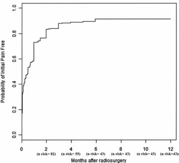
Four hundred fifty-six patients (91.75%) were initially
pain free in a median time of 10 days (range 1–180 days). The initially pain-free actuarial rates at 0.5, 1, 2, 3, 4, 5, and 6 months were 53.52%, 73%, 83.5%, 88.1%, 88.9%,
89.5%, and 91.75%, respectively (Fig. 1; with the flat part
of the curve being practically reached at 6 months). The following characteristics showed a negative and
statistically significant difference: previous surgical inter-vention (p = 0.005, HR 0.24, and 95% CI 0.09–0.65), only
1 previous surgical intervention (p = 0.009, HR 0.16, and
95% CI 0.04–0.64), and a previous history of MVD (p =
0.01, HR 0.64).
Differences in age (p = 0.172), time elapsed until treat-ment onset (p = 0.731), and the sides of pain (p = 0.4) were
not statistically significant.
postoperative Sensory assessment: details of Other postoperative complications
No patient experienced an early complication after GKS. Seventy-two patients (21.1% actuarial rate) later developed sensory dysfunction such as paresthesias or objective facial sensory loss, which occurred especially
during the first 5 years after GKS. The time of onset of
hypesthesia occurred at a median of 12 months (range 1–65). Patients had either mild hypesthesia in 49 (8.3%) cases or severe in 23 (4.6%) cases. We also assessed
hyp-esthesia using the BNI facial hyphyp-esthesia scale: mild
fa-cial numbness in 61 (12.3%) patients; fafa-cial numbness that
Fig. 1. Probability of an initial pain-free onset depending on the time since GKS. The probability of being initially pain free reaches a plateau at 6 months (with a rate of freedom from pain of 91.75%). The initial pain-free actuarial rates at 0.5, 1, 2, 3, 4, 5, and 6 months were 53.52%, 73%, 83.5%, 88.1%, 88.9%, 89.5%, and 91.75%, respectively. J Neurosurg Volume 124 • April 2016 1082
was somewhat bothersome in 8 (1.6%) patients; and facial numbness that was very bothersome in 3 (0.6%) patients.
The 3 patients with very bothersome hypesthesia said that their quality of life was worse and that this dysfunc-tion was not a good tradeoff, whereas the majority of the patients who developed numbness after GKS (69 of 72 patients; 95.8%) considered that their quality of life im-proved after GKS and that the sensory dysfunction was a good tradeoff for pain relief.
The hypesthesia actuarial rates at 0.5, 1, 2, 3, 5, and 7 years were 6.4%, 10.2%, 16.8%, 18.3%, 20.4%, and 21.1%, respectively, and remained stable for 14 years (Fig. 2). No
patients developed a trigeminal motor deficit after GKS or other cranial nerve deficits. There were 0 cases of
anesthe-sia dolorosa or dry-eye syndrome.
management and results of recurrent pain
One hundred fifty-seven (34.4%) of the patients who
were initially pain free (456 patients) experienced at least 1 recurrence after GKS. The median time to recurrent pain was 24 months (range 0.6–150.1 months). Because of recurrent medically refractory pain, 112 (22.5%) patients
required further surgeries. Eighty-five (17.1%) patients
re-quired only 1 further surgery, 21 (4.2%) rere-quired 2 further surgeries, and 6 (1.6%) required 3 or more surgeries.
In our unit, the most common intervention after failed
GKS was balloon microcompression, which was per-formed in 61 (12.3%) patients, followed by thermocoagu-lation in 29 (5.8%) patients, MVD in 21 (4.2%) patients, and glycerol rhizotomy in 1 (0.02%) patient (Table 2).
Usually, in our clinic, if a first-intention MVD
treat-ment was proposed but declined by the patient in the
ab-sence of efficacious GKS, it is usually proposed again and
frequently accepted by the patient.
The actuarial probabilities of maintaining pain relief
without medication at 0.5, 1, 2, 3, 5, 7, 10, 12, and 14 years were 93.4%, 85.9%, 78.6%, 71.8%, 64.9%, 59.7%, 45.3%, 40.7%, and 33.9%, respectively (see Fig. 3). Having three or more previous surgeries was a factor associated with
decreased long-term efficacy (p = 0.0163; HR 1.98; 95% CI
1.13–3.47) in comparison with patients having undergone fewer than 2 prior surgeries. Post-GKS hypesthesia onset was associated with a higher probability of maintaining
pain relief (p = 0.0003; HR 0.27; 95% CI 0.13–0.56).
probability of maintaining pain relief without Further Surgery
The actuarial probabilities of maintaining pain re-lief without further surgery at 0.5, 1, 2, 3, 5, 7, and 10 years were 96.1%, 92.1%, 88.1%, 84.2%, 79.7%, 75.4%, and 67.8%, respectively, and remained stable through 14
years (Fig. 4). We found statistically significant data for 3 or more previous surgeries (p = 0.007; HR 2.64; 95% CI
1.3–5.37) and the presence of post-GKS hypesthesia (p =
0.006; HR 0.06; 95% CI 0.01–0.46).
postoperative patient Satisfaction
The vast majority of patients (93.8%) expressed a high level of satisfaction, did not regret undergoing GKS, and would undergo the procedure again. A minority of the pa-tients had no opinion (4.6%), and 2.3% would not undergo the procedure again.
discussion
There are several neurosurgical therapeutic options for
Fig. 2. Actuarial probability of having new-onset hypesthesia depend-ing on the time since GKS. The hypesthesia actuarial rates at 0.5, 1, 2, 3, 5, and 7 years were 6.4%, 10.2%, 16.8%, 18.3%, 20.4%, and 21.1%, respectively, and remained stable for 14 years.
taBle 2. postoperative assessment
Variable Value* Initially pain free 456 (91.75) Post-GKS sensory dysfunction 72 (14.5) Mild 49 (9.8) Severe 23 (4.6) BNI facial hypesthesia scale No facial numbness 425 (85.5) Mild facial numbness 61 (12.3) Facial numbness, somewhat bothersome 8 (1.6) Facial numbness, very bothersome 3 (0.6) Recurrence of pain 157 (34.4) Median time to pain recurrence in mos (range) 24 (0.6–150.1) Additional treatment after GKS 112 (22.5) No. of treatments 1 85 (17.1) 2 21 (4.2) ≥3 6 (1.2) Type of treatment Balloon microcompression 61 (12.3) Radiofrequency lesion 29 (5.8) Microvascular decompression 21 (4.2) Glycerol rhizotomy 1 (0.02) * Values indicate the number of patients (%) unless otherwise indicated.
J. régis et al.
drug-resistant CTN. Radiosurgery was first used for TN
treatment in the 1960s by Lars Leksell.16 Its safety and
ef-ficacy started to be evaluated in the early 1990s, including
by our team.28,31 A cornerstone paper was the multicentric study of Kondziolka et al., which appeared in 199612 and generated a profound paradigm shift in radiosurgical prac-tice.33 The reappraisal of radiosurgery for TN in the 1990s was made possible by the development of high-resolution
MRI, which enabled the proper visualization of the
cister-nal portion of cranial nerve V.30,33 Radiosurgery is current-ly regarded as the least invasive neurosurgical approach for TN.5,9,44 A trigeminal nerve deficit usually appears within
the first 2 years after radiosurgery, but has also been
re-ported as late as 5 years after treatment.13 The only side effect reported in our long-term study is trigeminal nerve sensory disturbance. Only 21.5% of the patients reported this side effect, and the vast majority did not consider it as bothersome or disabling. The mechanisms of action of radiosurgery for TN are not known. Several authors have reported a higher rate of pain relief in patients experienc-ing hypesthesia.25 However, contrary to the percutaneous techniques, it is noteworthy to point out that the majority of the patients experiencing long-term pain freedom in our study did not report hypesthesia. Consequently, we do not
consider hypesthesia necessary for the efficacy of radio-surgery. In the meta-analysis by Gronseth et al.,9 the rate of hypesthesia reported after radiosurgery is similar to the rate of hypesthesia after MVD and much lower than the rate of hypesthesia reported after percutaneous ablative procedures. This observation suggests that radiosurgery may involve a mechanism of action that is more subtle than a purely destructive one.
The technical nuances of GKS have a major impact on the clinical outcome of radiosurgery for TN.33 Pain control increases according to the dose prescription, but a larger
volume of nerve treated has been reported to dramatically increase the toxicity (i.e., the risk of bothersome hypes-thesia) without increasing the rate of pain relief.8 Also, a target placed close to the brainstem at the level of the root entry zone26 seems to be associated with a higher risk of numbness and higher risk of bothersome hypesthesia. This
fact is confirmed by the recent paper by Sheehan et al.42 who found more numbness in a group of patients where the target used was the so-called dorsal root entry zone in comparison with the more anterior cisternal target that the authors used later on in their series. The major impact of these technical nuances may explain the large variability
in the safety and efficacy reported in the literature. Of note,
the remarkable high response rate in our series (initial pain freedom rate of 91.3% at 6 months) and low hypesthesia rate on a long-term basis are likely due to the use of a high maximum prescription dose (median 85 Gy) and use of the “anterior retrogasserian target.”
There are only 3 studies reporting long-term outcome after GKS.6,13,43 Dhople et al.6 described the outcomes for 102 patients, with a median follow-up of 5.6 years (range 13–115 months). The target was the dorsal root entry zone with a median maximal dose of 75 Gy (range 70–80 Gy). Regarding the classical outcomes, the initial pain freedom rate was 81%, bothersome hypesthesia rate was 6% (the global rate not reported), and the probabilities of maintain-ing pain relief at 3, 5, and 7 years were 41%, 34%, and 22%, respectively.
Kondziolka et al.13 reported the outcomes for 503 pa-tients, of whom 107 had more than 5 years of follow-up. The target was placed at “3–8 mm anterior from the junc-tion of the trigeminal nerve and pons.” The maximal dose delivered was 80 Gy. Regarding the classical outcomes: the initial pain freedom rate was 89%; sensory dysfunction
Fig. 3. Actuarial probability of maintaining pain relief without medication. The actuarial probabilities of maintaining pain relief at 0.5, 1, 2, 3, 5, 7, 10, 12, and 14 years were 93.4%, 85.9%, 78.6%, 71.8%, 64.9%, 59.7%, 45.3%, 40.7%, and 33.9%, respectively. Fig. 4. Actuarial probability of maintaining pain relief without new surgery depending on the time since pain cessation. The actuarial prob-abilities of maintaining pain relief without further surgery at 0.5, 1, 2, 3, 5, 7, and 10 years were 96.1%, 92.1%, 88.1%, 84.2%, 79.7%, 75.4%, and 67.8%, respectively, and remained stable for 14 years. J Neurosurg Volume 124 • April 2016 1084
appeared in 10.5% of patients with 1 case of deafferenta-tion pain (in a patient who already had decreased facial sensation after previous MVD); and the probabilities of maintaining pain relief at 3, 5, and 10 years were 71%, 46%, and 30%, respectively.
Young et al.43 reported outcomes for 315 patients with a mean duration of follow-up of 68.9 ± 41.8 months. The target was placed “on the trigeminal nerve, with the 20% isodose line tangential to the pontine surface.” All patients were treated with a maximal dose of 90 Gy. Regarding the classical outcomes, initial pain-freedom rate was found in 85.6% of cases, and hypesthesia was found in 32.9% (and very bothersome in 4.5%) of cases. Furthermore, dry-eye syndrome was encountered in 22.4% and jaw weakness in 11.2% of patients.
The gold-standard neurosurgical procedure is MVD. Although no prospective randomized trial exists, MVD seems to be the approach that provides the highest chance of maintaining long-term pain relief.17,27 MVD is not ex-pected to damage the nerve, but presumably acts by al-leviating the pathophysiological cause of TN, namely, compression of the trigeminal nerve by a vascular loop.
MVD carries a small but definite risk of major, including fatal, complications. In 1996, Barker et al.1 reported the retrospective evaluation of a large cohort of 1155 patients treated with MVD and followed up for at least 1 year (me-dian follow-up 6.2 years). Thirty percent of the patients experienced recurrence of pain. Due to severe recurrence, 11% of patients underwent a second operation. The major complications reported by the authors included 2 post-operative deaths (0.2%), 1 brainstem infarction (0.1%), 4 intracerebral hematomas, 4 cerebellar edemas, 2 cases of hydrocephalus, 12 cases of facial palsy (2 permanent), 15 cases of extraocular muscle palsy (2 permanent), 16 cases of ipsilateral hearing loss, 22 cases of severe facial numb-ness, 20 cases of CSF leakage, 4 cases of pseudomeningo-cele, 5 cases of bacterial meningitis, 225 cases of chemi-cal meningitis, 2 cases of pneumonia, and 1 case each of septicemia, myocardial infarction, transverse sinus throm-bosis, pulmonary embolus, and permanent contralateral hearing loss.1 Few other reports2,24,47,48 used independent outcome assessments after MVD.5 These case series
con-firm the results of Barker et al., with a 75% chance of
maintaining pain relief at 3 years and a risk of operative mortality of 0.2% (rising to 0.5% in other reports).1 Ma-jor problems such as CSF leakage, infarcts, or hematomas are reported in 4% of the patients, and aseptic meningitis
in 11%; diplopia due to injury to cranial nerves IV and VI (most frequently transient) and facial palsy are rare.2,47
Ipsilateral hearing loss is a major long-term complication
that has been reported in as many as 10% of patients.2,47 Sensory loss is observed in 7% of patients.
Percutaneous techniques (thermocoagulation, balloon microcompression, and glycerol injection) share an ab-lative mechanism of action. All of these procedures re-quire brief general anesthesia, the penetration of a probe
through the foramen ovale under fluoroscopic control or
navigation, and a physical action (thermal, mechanical,
or chemical) on the fibers of cranial nerve V at the level
of the gasserian ganglion. Generating a certain level of hypesthesia is classically necessary for the complete and
prolonged efficacy of these techniques. Thus, all these
percutaneous techniques are associated with a high rate of
more or less disabling trigeminal nerve dysfunction. In the
main series of the literature (see Lopez et al.18), the 3-year actuarial rate of complete pain relief has been reported as 58% to 64% for radiofrequency thermocoagulation,18,45 53% to 54% for glycerol rhizolysis,23,36 and 69% for bal-loon microcompression.3 Masticatory weakness has been reported in 12% of patients after thermocoagulation14,18,45 and in 3% after glycerol rhizolysis.23,35,36 Troublesome dys-esthesia has been reported in 4% of patients after thermo-coagulation,11,14,45 in 8.5% after glycerol rhizolysis,4,23 and in 10% after balloon microcompression.22 Anesthesia do-lorosa has been reported in 1.5% of patients after thermo-coagulation,11,14,45 2.5% after glycerol rhizolysis23,35,36 and has not been reported after balloon microcompression.22 Keratitis is observed was 1.5% of patients after thermo-coagulation11,14,45 and 2% after glycerol rhizolysis23,35,36 but was not reported after balloon microcompression.22 It is important to note that with all these neurosurgical pro-cedures, postoperative morbidity is lower in high-volume units.
Very few Level I evidence papers concerning the
evalu-ation of the different surgical techniques for the treatment of TN are available.44 The quality of reporting evaluations of surgical treatments for TN rarely follow the recommen-dations published in 2003 by Zakrzewska and Lopez.46
conclusions
The present study is unique due to the fact that the data were collected in a prospective fashion, the cohort is very large, and a long-term follow-up was conducted; however, this study is still limited by the absence of randomization. This series represents, to date, the study with the largest case series available and has the advantage of a very long-term follow-up. Additionally, this study brings to light the fact that the percentage of bothersome hypesthesia is low (0.3%) using a range of doses between 70 and 90 Gy with the retrogasserian target. This study provides reasonable
long-term evidence of the very high safety and efficacy of GKS in CTN. Radiosurgery is a rational first-line
neu-rosurgical option for TN. The spectrum of complications clearly differs between different neurosurgical options and must be taken into account during the decision-making process. The rarity of the complications and the impor-tant probability of long-lasting effects prompt us to regard
GKS as a pragmatic surgical first- and/or second-intention
alternative for CTN. However, MVD remains as the ref-erence technique, and further prospective randomized
studies are still needed to compare the long-term efficacy
of radiosurgery with MVD. We expect these studies to clarify the potential role of each approach. Neurosurgical techniques offer the highest chance of improving quality of life in patients with medically refractory TN. However, surgery is not indicated for all these patients; they should be informed about the full range of choices, and they must
integrate the benefits and risks of each alternative in the
decision-making process. The centers able to provide pa-tients with all these techniques are better placed to
J. régis et al.
acknowledgment
This study was funded by Timone University Hospital, Assistance-Publique, Hopitaux de Marseille, France.
references
1. Barker FG II, Jannetta PJ, Bissonette DJ, Larkins MV, Jho HD:
The long-term outcome of microvascular decompression for trigeminal neuralgia. N Engl J Med 334:1077–1083, 1996
2. Broggi G, Ferroli P, Franzini A, Servello D, Dones I:
Micro-vascular decompression for trigeminal neuralgia: comments on a series of 250 cases, including 10 patients with multiple sclerosis. J Neurol Neurosurg Psychiatry 68:59–64, 2000 3. Brown JA, McDaniel MD, Weaver MT: Percutaneous
trigem-inal nerve compression for treatment of trigemtrigem-inal neuralgia: results in 50 patients. Neurosurgery 32:570–573, 1993 4. Burchiel KJ: Percutaneous retrogasserian glycerol rhizolysis
in the management of trigeminal neuralgia. J Neurosurg
69:361–366, 1988
5. Cruccu G, Gronseth G, Alksne J, Argoff C, Brainin M,
Burchiel K, et al: AAN-EFNS guidelines on trigeminal
neu-ralgia management. Eur J Neurol 15:1013–1028, 2008 6. Dhople AA, Adams JR, Maggio WW, Naqvi SA, Regine WF,
Kwok Y: Long-term outcomes of Gamma Knife radiosurgery for classic trigeminal neuralgia: implications of treatment and critical review of the literature. Clinical article. J
Neuro-surg 111:351–358, 2009
7. Eller JL, Raslan AM, Burchiel KJ: Trigeminal neuralgia: definition and classification. Neurosurg Focus 18(5):E3,
2005
8. Flickinger JC, Pollock BE, Kondziolka D, Phuong LK, Foote
RL, Stafford SL, et al: Does increased nerve length within the treatment volume improve trigeminal neuralgia radiosur-gery? A prospective double-blind, randomized study. Int J
Radiat Oncol Biol Phys 51:449–454, 2001
9. Gronseth G, Cruccu G, Alksne J, Argoff C, Brainin M, Burchiel K, et al: Practice parameter: the diagnostic evalu-ation and treatment of trigeminal neuralgia (an evidence-based review): report of the Quality Standards Subcommittee
of the American Academy of Neurology and the European
Federation of Neurological Societies. Neurology 71:1183– 1190, 2008
10. International Headache Society: The International
Classifica-tion of Headache Disorders. Cephalalgia 24 (Suppl 1):1–151, 2004
11. Kanpolat Y, Savas A, Bekar A, Berk C: Percutaneous con-trolled radiofrequency trigeminal rhizotomy for the treat-ment of idiopathic trigeminal neuralgia: 25-year experience with 1,600 patients. Neurosurgery 48:524–534, 2001 12. Kondziolka D, Lunsford LD, Flickinger JC, Young RF,
Vermeulen S, Duma CM, et al: Stereotactic radiosurgery for trigeminal neuralgia: a multiinstitutional study using the gamma unit. J Neurosurg 84:940–945, 1996
13. Kondziolka D, Zorro O, Lobato-Polo J, Kano H, Flannery TJ, Flickinger JC, et al: Gamma Knife stereotactic radio-surgery for idiopathic trigeminal neuralgia. J Neurosurg
112:758–765, 2010
14. Latchaw JP Jr, Hardy RW Jr, Forsythe SB, Cook AF: Tri-geminal neuralgia treated by radiofrequency coagulation. J
Neurosurg 59:479–484, 1983
15. Leksell L: Cerebral radiosurgery. I. Gammathalanotomy in
two cases of intractable pain. Acta Chir Scand 134:585– 595, 1968
16. Leksell L: Sterotaxic radiosurgery in trigeminal neuralgia.
Acta Chir Scand 137:311–314, 1971
17. Linskey ME, Ratanatharathorn V, Peñagaricano J: A
pro-spective cohort study of microvascular decompression and Gamma Knife surgery in patients with trigeminal neuralgia.
J Neurosurg 109 Suppl:160–172, 2008
18. Lopez BC, Hamlyn PJ, Zakrzewska JM: Systematic review of ablative neurosurgical techniques for the treatment of trigeminal neuralgia. Neurosurgery 54:973–983, 2004 19. Love S, Coakham HB: Trigeminal neuralgia: pathology and
pathogenesis. Brain 124:2347–2360, 2001
20. McCormack M, Alfirevic A, Bourgeois S, Farrell JJ, Kasperavičiūtė D, Carrington M, et al: HLA-A*3101 and carbamazepine-induced hypersensitivity reactions in
Europe-ans. N Engl J Med 364:1134–1143, 2011
21. Merrison AF, Fuller G: Treatment options for trigeminal neuralgia. BMJ 327:1360–1361, 2003
22. Mullan S, Lichtor T: Percutaneous microcompression of the trigeminal ganglion for trigeminal neuralgia. J Neurosurg
59:1007–1012, 1983
23. North RB, Kidd DH, Piantadosi S, Carson BS: Percutaneous retrogasserian glycerol rhizotomy. Predictors of success and failure in treatment of trigeminal neuralgia. J Neurosurg
72:851–856, 1990
24. Piatt JH Jr, Wilkins RH: Treatment of tic douloureux and hemifacial spasm by posterior fossa exploration: therapeutic implications of various neurovascular relationships.
Neuro-surgery 14:462–471, 1984
25. Pollock BE: Radiosurgery for trigeminal neuralgia: is
sen-sory disturbance required for pain relief? J Neurosurg 105
Suppl:103–106, 2006
26. Pollock BE, Phuong LK, Foote RL, Stafford SL, Gorman
DA: High-dose trigeminal neuralgia radiosurgery associated with increased risk of trigeminal nerve dysfunction.
Neuro-surgery 49:58–64, 2001
27. Pollock BE, Schoeberl KA: Prospective comparison of
pos-terior fossa exploration and stereotactic radiosurgery dorsal root entry zone target as primary surgery for patients with idiopathic trigeminal neuralgia. Neurosurgery 67:633–639, 2010
28. Rand RW, Jacques DB, Melbye RW, Copcutt BG, Levenick MN, Fisher MR: Leksell Gamma Knife treatment of tic dou-loureux. Stereotact Funct Neurosurg 61 (Suppl 1):93–102, 1993
29. Régis J, Arkha Y, Yomo S, Murata N, Roussel P, Donnet A, et al: [Radiosurgery in trigeminal neuralgia: long-term
results and influence of operative nuances]. Neurochirurgie 55:213–222, 2009 (Fr)
30. Régis J, Carron R, Tuleasca C, Donnet A: Distal radiosurgi-cal targeting for trigeminal neuralgia, in Sheehan JP, Gerszten PC (eds): Controversies in Stereotactic
Radio-surgery: Best Evidence Recommendations. New York:
Thieme, 2014, pp 120–130
31. Régis J, Manera L, Dufour H, Porcheron D, Sedan R, Peragut
JC: Effect of the Gamma Knife on trigeminal neuralgia. Stereotact Funct Neurosurg 64 (Suppl 1):182–192, 1995
32. Régis J, Metellus P, Hayashi M, Roussel P, Donnet A, Bille-Turc F: Prospective controlled trial of gamma knife surgery for essential trigeminal neuralgia. J Neurosurg 104:913– 924, 2006
33. Régis J, Tuleasca C: Fifteen years of Gamma Knife surgery for trigeminal neuralgia in the Journal of Neurosurgery: his-tory of a revolution in functional neurosurgery. J Neurosurg
115 Suppl:2–7, 2011
34. Rogers CL, Shetter AG, Fiedler JA, Smith KA, Han PP, Speiser BL: Gamma knife radiosurgery for trigeminal neuralgia: the initial experience of The Barrow Neurological
Institute. Int J Radiat Oncol Biol Phys 47:1013–1019, 2000
35. Saini SS: Reterogasserian anhydrous glycerol injection therapy in trigeminal neuralgia: observations in 552 patients.
J Neurol Neurosurg Psychiatry 50:1536–1538, 1987
36. Slettebø H, Hirschberg H, Lindegaard KF: Long-term results after percutaneous retrogasserian glycerol rhizotomy in patients with trigeminal neuralgia. Acta Neurochir (Wien)
122:231–235, 1993
J Neurosurg Volume 124 • April 2016 1086
37. Tuleasca C, Carron R, Resseguier N, Donnet A, Roussel P, Gaudart J, et al: Multiple sclerosis-related trigeminal neuralgia: a prospective series of 43 patients treated with gamma knife surgery with more than one year of follow-up.
Stereotact Funct Neurosurg 92:203–210, 2014
38. Tuleasca C, Carron R, Resseguier N, Donnet A, Roussel P, Gaudart J, et al: Patterns of pain-free response in 497 cases of classic trigeminal neuralgia treated with Gamma Knife surgery and followed up for least 1 year. J Neurosurg 117
Suppl:181–188, 2012
39. Tuleasca C, Carron R, Resseguier N, Donnet A, Roussel P, Gaudart J, et al: Trigeminal neuralgia related to mega-dolichobasilar artery compression: a prospective series of twenty-nine patients treated with gamma knife surgery, with more than one year of follow-up. Stereotact Funct
Neuro-surg 92:170–177, 2014
40. Tuleasca C, Carron R, Resseguier N, Donnet A, Roussel P, Gaudart J, et al: Repeat Gamma Knife surgery for recurrent trigeminal neuralgia: long-term outcomes and systematic review. J Neurosurg 121 Suppl:210–221, 2014
41. Vandertop WP, Lagerwaard FJ: Case 21-2006: a man with left-sided facial pain. N Engl J Med 355: 2375–2376, 2006 42. Xu Z, Schlesinger D, Moldovan K, Przybylowski C, Sun X,
Lee CC, et al: Impact of target location on the response of
trigeminal neuralgia to stereotactic radiosurgery. J
Neuro-surg 120:716–724, 2014
43. Young B, Shivazad A, Kryscio RJ, St Clair W, Bush HM: Long-term outcome of high-dose γ knife surgery in treatment of trigeminal neuralgia. J Neurosurg 119:1166–1175, 2013 44. Zakrzewska JM, Akram H: Neurosurgical interventions for
the treatment of classical trigeminal neuralgia. Cochrane
Database Syst Rev 9:CD007312, 2011
45. Zakrzewska JM, Jassim S, Bulman JS: A prospective, longi-tudinal study on patients with trigeminal neuralgia who un-derwent radiofrequency thermocoagulation of the Gasserian ganglion. Pain 79:51–58, 1999
46. Zakrzewska JM, Lopez BC: Quality of reporting in evalua-tions of surgical treatment of trigeminal neuralgia: recom-mendations for future reports. Neurosurgery 53:110–122, 2003
47. Zakrzewska JM, Lopez BC, Kim SE, Coakham HB: Patient
reports of satisfaction after microvascular decompression and partial sensory rhizotomy for trigeminal neuralgia.
Neu-rosurgery 56:1304–1312, 2005
48. Zakrzewska JM, Thomas DG: Patient’s assessment of out-come after three surgical procedures for the management of trigeminal neuralgia. Acta Neurochir (Wien) 122:225–230, 1993
disclosure
Dr. Régis has a financial relationship with Accuray, Brainlab,
Elektra, and Radionic and receives non–study-related clinical or research support from Elektra and Medtronic.
author contributions
Conception and design: Régis, Tuleasca, Levivier. Acquisition of data: Régis, Tuleasca, Donnet. Analysis and interpretation of data: Régis, Tuleasca, Levivier. Drafting the article: Régis, Tuleasca, Levivier. Critically revising the article: Régis, Tuleasca, Resseguier, Carron, Donnet, Levivier. Reviewed submitted ver-sion of manuscript: Régis, Tuleasca, Resseguier, Carron, Donnet, Levivier. Approved the final version of the manuscript on behalf of all authors: Régis. Statistical analysis: Resseguier, Gaudart.
Administrative/technical/material support: Régis, Donnet,
Gaudart, Levivier. Study supervision: Régis, Gaudart, Levivier.
correspondence
Jean Régis, Functional and Stereotactic Neurosurgery Service and Gamma Knife Unit, 264, Rue Saint Pierre, Marseille 13385, France. email: jregis@ap-hm.fr.