HAL Id: inserm-00187319
https://www.hal.inserm.fr/inserm-00187319
Submitted on 14 Nov 2007
HAL is a multi-disciplinary open access
archive for the deposit and dissemination of sci-entific research documents, whether they are pub-lished or not. The documents may come from teaching and research institutions in France or abroad, or from public or private research centers.
L’archive ouverte pluridisciplinaire HAL, est destinée au dépôt et à la diffusion de documents scientifiques de niveau recherche, publiés ou non, émanant des établissements d’enseignement et de recherche français ou étrangers, des laboratoires publics ou privés.
Intra subject 3D/3D Kidney Registration using Local
Mutual Information Maximization
Hui Tang, Jean-Louis Dillenseger, Limin Luo
To cite this version:
Hui Tang, Jean-Louis Dillenseger, Limin Luo. Intra subject 3D/3D Kidney Registration using Local Mutual Information Maximization. 29th Annual International Conference of the IEEE Engineer-ing in Medicine and Biology Society, 2007. EMBS 2007., Aug 2007, Lyon, France. pp.6379-6382, �10.1109/IEMBS.2007.4353815�. �inserm-00187319�
Abstract— One of the goal of the Nephron-Sparing Surgery
properative planning is to delineate as exactly as possible the renal carcinoma and to specify its relations to the renal arterial, venous and collecting system anatomies. The classical preoperative imaging system is the Spiral CT Urography, which gives sucessive 3D acquisitions of complementary information The integration of this information within the a patient spacific anatomical referential can be achieved by intra-patient registration techniques. A local MI maximization registration method is proposed in this paper. The kidneys are extracted from the abdomen volumes and then the registration between the extracted kidneys is implemented by maximizing the MI between them. The experimental results demonstrates that this method is effective.
I. INTRODUCTION
ENAL cancer represents 2~3% of whole cancers and is the third most frequent in urologic cancer. Today, renal tumors are detected more and more precociously. Such tumors are usually less than 4 cm, so a nephron sparing surgery can be considered instead of a total nephrectomy.
In order to practice this preserving technique, the surgeon needs a precise preoperative planning to delineate as exactly as possible the renal carcinoma and to specify its relations with the renal arterial, venous and collecting system anatomy.
The CT uroscan is the classical preoperative examination. It consists of four time spaced 3D acquisitions, which give complementary information about the kidney anatomy. The first acquisition is realized without injection of contrast agent and informs the surgeon about intern morphology of the patient. The second one, taken just after a contrast medium injection, reveals the renal arterial system. Obtained just a time later, the third acquisition presents the venous and renal parenchyma vascularization. These two acquisitions give also information about the nature and the location of the renal carcinoma. About ten minutes later on the last acquisition the collecting system is enhanced.
Since information from these acquisitions is of a complementary nature, it is useful for the surgeon to integrate this information within a unique spatial volume. The first step in this integration process is to bring the different acquisitions
This work was preformed at CRIBS - Inserm, international laboratory, Université Rennes I, France – SouthEast University, Nanjing, China.
H. Tang and L. M. Luo are with LIST, Southeast University, 210096, Nan Jing, China (phone: +86(0)2583794249; fax: +86(0)2583792698; e-mail: [email protected]; [email protected])
J.-L. Dillenseger and H. Tang are with Inserm, U642, Rennes, F-35000, France; Université de Rennes 1, LTSI, Rennes, F-35000, France. (e-mail: [email protected]).
into spatial alignment. This procedure is referred as registration.
The nature of the CT acquisitions leads us to decide for a 3D-3D, monomodal, intra subject registration technique [1]. We supposed that the kidney shape is not deformed during the acquisition, even during the respiratory movements. This hypothesis leads us to choose a rigid kidney-centered registration technique.
We do not dispose of extrinsic landmarks, and the selection of intrinsic landmarks is not easy and too much operator dependant. We choose to base our registration on the object spatial information. Maximization of Mutual Information (MI) has been recommended as a powerful intrinsic criterion for medical image registration. The method applies the concept of MI to measure the statistical dependence between the image intensities of corresponding voxel in both images, which is assumed to be maximal if the images are geometrically aligned.
Because the registration subject is the kidney instead of the whole abdomen this article will introduce a local MI maximum registration method.
II. MUTUAL INFORMATION
The MI between two discrete random variables, A and B, is defined as [2]:
∑∑
∈ ∈ = A B S i i S A B AB AB j p i p j i p j i p B A I ) ( ) ( ) , ( log ) , ( ) , ( (1)where SA denotes the symbol set for A, SB denotes the
symbol set for B, pAB(i,j) denotes the joint probability of A and
B, pA(i) denotes the marginal probability of A, and pB(j)
denotes the marginal probability of B.
As to a discrete volume, the joint probability can be expressed by the joint histogram:
∑
= j i AB j i h j i h j i p , ) , ( ) , ( ) , ( (2) where h(i,j) denotes the joint histogram of two volumes. The marginal probability can be calculated by the joint probability: =∑
j AB A i p i j p ( ) ( , ), =∑
i AB B j p i j p ( ) ( , ) (3) Because of the rigid-body registration, transformation between two volumes can be expressed by six parameters:α=
{
tx,ty,tz,rx,ry,rz}
(4)Intra subject 3D/3D Kidney Registration using Local Mutual
Information Maximization
Hui Tang, Jean-Louis Dillenseger, and Li Min Luo
R
This material is presented to ensure timely dissemination of scholarly and technical work. Copyright and all rights therein are retained by authors or by other copyright holders.
All persons copying this information are expected to adhere to the terms and constraints invoked by each author's copyright. In most cases, these works may not be reposted without the explicit permission of the copyright holder.
HAL author manuscript inserm-00187319, version 1
where tx, ty and tz denote the translation in x, y and z direction respectively; rx, ry and rz denote the rotation around the x, y and z axis respectively.
Although the two kidney volumes are acquired at different moment and at different situation, they come from the same body and include the same anatomical information. So when the two volumes are spatial registered, the MI between the two kidneys reaches its maximum.
III. REGISTRATION METHOD A. Registration framework
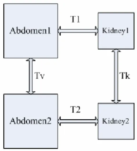
In order to realize the local MI maximum registration, we make use of the following framework (see Fig. 1):
1. Rough semi-automatic extraction of the kidney from the abdominal volume. During extraction, we keep a matrix (denoted respectively T1 and T2 for volume 1 and volume 2) to express the spatial relationship between the kidney volume and the corresponding abdominal volume.
2. Registration of the two extracted kidneys by maximizing the MI. This registration gives the transform matrix Tk.
3. The transformation matrix Tv between the two abdominal volumes can be estimated with the help of
T1, T2 and Tk.
So the transformation between abdominal volumes can be achieved though a kidney-centered registration.
Fig 1: Transformation between volumes B. Volume extraction
Automatically extracting the kidney volume from the abdomen volume is difficult and time consuming. We develop a semi-automatic snake-based segmentation method to broadly extract the kidney volume in a reasonable time. As the input data is a series of CT slices, we first segment the kidney from each slice and then reconstruct the kidney volume from the segmented kidney slice.
The extraction steps are as follows:
1) Use 2D snake [3] to segment the external kidney contour in one slice. The result on one slice is propagated to the
neighboring slice. This propagated snake is then automatically adjusted to the new data. Sometimes manual corrections can be performed. In this way, the kidney external contour is extracted on each slice.
2) Interpolate contours.
3) Fill contours to form a binary volume.
4) Do 3D dilatation to the binary kidney volume to make sure that the kidneys are inside.
5) Intersect the binary volume with the original grey volume.
After the five steps, the kidney volume is achieved. We can make sure that the kidneys are inside and so that the registration between kidney volumes can be performed.
The total amount of time of this semi-automatic volume extraction is about three minutes for a non trained user.
C. Kidney Registration
As MI is used as matching metric, registration can be performed by optimizing this similarity criterion. They are two critical points in all the optimization methods: the choice of the initial parameters set (tx0, ty0, tz0, rx0, ry0, rz0) and the
choice of the optimization method itself.
1) Initial parameters
In order to analyze the effect of the initial parameters set (tx0, ty0, tz0, rx0, ry0, rz0), we displayed the MI variation within
the parametric search space in order to estimate the presence or not of local extrema.
For this, we formed a synthetic kidney volume by translating and rotating a real volume by known parameters (txT, tyT, tzT, rxT, ryT, rzT). From the initial parameters set (tx0,
ty0, tz0, rx0, ry0, rz0), we sampled the parametric search space
and measure the MI for each (tx, ty, tz, rx, ry, rz).
In order to present some graspable results, we fixed constant the value of ty=ty0, tz=tz0, ry=ry0, rz=rz0. Only tx and
rx are varying and the MI variation can be seen as a surface as
shown in Fig 2.
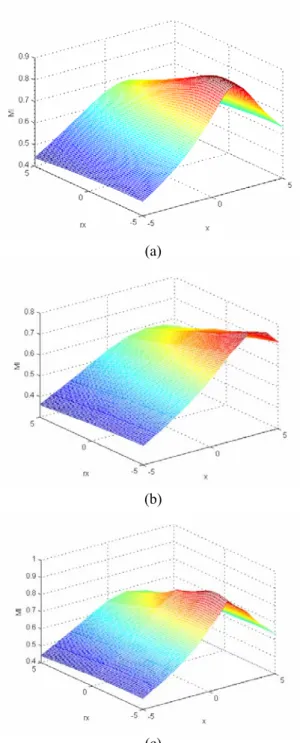
Fig 2(a) illustrates the MI surface in the situation that the parameters set is initialized by the real value: ty0=tyT, tz0=tzT,
ry0=ryT, rz0=rzT. We can see that in this situation, the MI
surface is smooth and the global extremum is obvious. The extremum can be achieved by any optimization method easily. Fig 2(b) sets ty0=0, tz0=0, ry0=0, rz0=0 and it can be
seen that many local extrema appear and the global extremum is inconspicuous. Fig 2(c) is the situation that ty0=tyT=0,
tz0=tzT=0, ry0=ryT=0, rz0=rzT=0. We can see that although
there are still many local extrema, the global extremum is obvious and easy to achieve.
From the analysis above we can get the conclusion that the more initial parameters close to their real values the smoother MI surface will be. According to the experiment result, we utilize the characteristics of the image geometry moment to initialize the parameters instead of initialization by zero or random values.
(a)
(b)
(c)
Fig 2: tx-rx search space MI surface for different parameter initializations. (a): ty, ry, tz, rz are initialized on the real values when the real values are not zero; (b): ty, ry, tz, rz are initialized to zero when the real values are not zero; (c): ty, ry, tz, rz are all initialized to zero when the real values are zero;
For a three dimensional discrete image f(i,j,k), its geometrical moments are defined as:
=
∑∑∑
i j k w v u uvw f i j k i j k m ( , , ) (5)The first order geometrical moments denotes the volume center of gravity. The translation between the centers of gravity can be the initial value of (tx0, ty0, tz0). The second
order moments can determine the 2 volumes main direction axes. A rotation matrix can be estimated from these main
axes. Then the three initial parameters (rx0, ry0, rz0) can be
achieved from the matrix [4].
2) Optimization method
The registration process is a multi-variable optimization problem. There are many existing optimization methods [5]. Among these methods, the downhill simplex method is one the most used. Although compared to simulated annealing methods it has more probability to meet local extrema, simplex method is accurate enough after MI surface smoothing and it is faster than other methods.
IV. RESULTS
The results on synthetic and real data are presented in this section.
To evaluate the registration process, an experiment is done on synthetic data. A synthetic volume is build by translating and rotating the real kidney volume. On each experiment, we set a group of translation and rotation parameters to build up a synthetic volume and then do registration between the original volume and the synthetic one. Several experiments have been performed and the errors between the estimated parameters and the real ones are estimated. From a set of 20 experiments, the maximal translation error is less than 0.08 mm and the maximum rotation angle error is less than 0.3°.
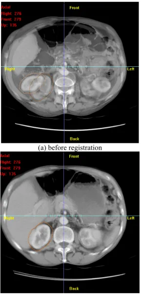
Fig 3 presents the registration results on real data. Here we take the registration of the arterial and the venous phase acquisition for example. Two axial slices taken from the same position of the two volumes are merged in Fig 3. We can see that after registration, although the two volumes are not fitted exactly, the kidneys seem to be well register together.
Using the same registration framework, the kidneys present on all the 3 or 4 CT uroscans can now be merged within the same patient-specific referential. The next step on our problematic will consist in performing refined segmentation methods on this his multivariate volume in order to extract the several kidneys anatomical components: carcinoma, arterial, venous and collecting system.
V. CONCLUSION
In this paper we presented an intra-subject registration method by local MI maximization. Kidney volumes were extracted and the registration was performed between the extracted kidneys instead of the whole volumes. The experimental results illustrated that effective initial parameters can improve the accuracy of the registration. The final results demonstrate the effectiveness of the registration method.
(a) before registration
(b) after registration
Fig 3: Kidney-centered registration result.
REFERENCES
[1] J. B. Maintz and M. A. Viergever, "A survey of medical image registration," Med Image Anal, vol. 2, pp. 1-36., 1998.
[2] T. M. Cover and J. A. Thomas, Elements of information theory. New York: Wiley, 1991.
[3] S. Lobregt and M. A. Viergever, "A discrete dynamic contour model," IEEE Trans Med Imaging, vol. 14, pp. 12-24, 1995. [4] T. L. Faber, "Orientation of 3D structures in medical images,"
IEEE trans. on PAMI, vol. 10, pp. 626-633, 1988.
[5] W. H. Press, S. A. Teukolsky, W. T. Vetterling, and B. P. Flannery, Numerical Recipes in C, 2nd ed. Cambridge: Cambridge University Press, 1992.