Phosphorylation, Kinase Upregulation, and Neurite Outgrowth
The MIT Faculty has made this article openly available. Please share
how this access benefits you. Your story matters.
Citation
Zukerberg, Lawrence R, Gentry N Patrick, Margareta Nikolic,
Sandrine Humbert, Chin-Lee Wu, Lorene M Lanier, Frank B Gertler,
Marc Vidal, Richard A Van Etten, and Li-Huei Tsai. “Cables Links
Cdk5 and c-Abl and Facilitates Cdk5 Tyrosine Phosphorylation,
Kinase Upregulation, and Neurite Outgrowth.” Neuron 26, no. 3
(June 2000): 633-646. Copyright © 2000 Cell Press
As Published
http://dx.doi.org/10.1016/S0896-6273(00)81200-3
Publisher
Elsevier
Version
Final published version
Citable link
http://hdl.handle.net/1721.1/83489
Terms of Use
Article is made available in accordance with the publisher's
policy and may be subject to US copyright law. Please refer to the
publisher's site for terms of use.
Cdk5 Tyrosine Phosphorylation, Kinase
Upregulation, and Neurite Outgrowth
Cdks are involved in events unrelated to cell division. A function of Cdk5 has been demonstrated only in the development of the nervous system. Introduction of a dominant-negative Cdk5 mutant inhibited neurite out-growth in cultured primary cortical neurons (Nikolic et Lawrence R. Zukerberg,*# Gentry N. Patrick,*
Margareta Nikolic,* Sandrine Humbert,*
Chin-Lee Wu,#§Lorene M. Lanier,‡Frank B. Gertler,‡
Marc Vidal,§Richard A. Van Etten,†and Li-Huei Tsai*k * Howard Hughes Medical Institute
al., 1996). A mouse strain lacking Cdk5 exhibited a late Department of Pathology
embryonic to perinatal lethality with obvious defects in
†Center for Blood Research
the developing CNS (Ohshima et al., 1996). Indeed, Cdk5 Department of Genetics
protein is expressed at the highest levels in the nervous Harvard Medical School
system, and its associated kinase activity has only been Boston, Massachusetts 02115
detected in brain lysates (Lew et al., 1992; Shetty et al.,
‡Department of Biology
1993; Tsai et al., 1993; reviewed by Lew and Wang, Massachusetts Institute of Technology
1995). This temporal and spatial specificity of Cdk5 ki-Cambridge, Massachusetts 02129
nase activity is due to the restricted expression of its
§Massachusetts General Hospital Cancer Center
regulatory subunit p35 (Ishiguro et al., 1994; Lew et al., Boston, Massachusetts 02139
1994; Tsai et al., 1994). p35 is a neuronal specific protein that is important in normal neuronal migration (Chae et al., 1997). The amino acid sequence of p35 does not Summary
resemble that of the cyclins, although it was suggested that the predicted ternary structure of p35 is similar to Cyclin-dependent kinase 5 (Cdk5) is a small serine/
that of cyclin A (Tang et al., 1997). Activation of Cdk5 threonine kinase that plays a pivotal role during
devel-kinase activity can be achieved by mixing purified bacte-opment of the CNS. Cables, a novel protein, interacts
rially expressed p35 and Cdk5 proteins in vitro, indicat-with Cdk5 in brain lysates. Cables also binds to and
ing that posttranslational modification is not required is a substrate of the c-Abl tyrosine kinase. Active c-Abl
for Cdk5 activation (Lew et al., 1994; Tsai et al., 1994). kinase leads to Cdk5 tyrosine phosphorylation, and
This is supported by the finding that CAK does not phos-this phosphorylation is enhanced by Cables.
Phos-phorylate Cdk5 on the serine 159 residue (equivalent to phorylation of Cdk5 by c-Abl occurs on tyrosine 15
T160 of Cdk2; Poon et al., 1997), and a serine to alanine (Y15), which is stimulatory for p35/Cdk5 kinase
activ-mutation of Cdk5 on the 159 residue does not affect the ity. Expression of antisense Cables in primary cortical
extent of Cdk5 activation by p35 (Y. Ramos and L.-H. T., neurons inhibited neurite outgrowth. Furthermore,
ex-unpublished data). A kinase activity has been identified pression of active Abl resulted in lengthening of
neu-that phosphorylates Cdk5 on the threonine 14 residue rites. The data provide evidence for a Cables-mediated and inhibits the p35/Cdk5 kinase (Matsuura and Wang, interplay between the Cdk5 and c-Abl signaling
path-1996), despite the fact that T14 phosphorylation of Cdk5 ways in the developing nervous system. has not been demonstrated in vivo. Finally, tyrosine phosphorylation of Cdk5 was reported in lysates
pre-Introduction pared from developing rat cerebellum (Lazaro et al.,
1996), although the identity of the phosphorylated tyro-Cyclin-dependent kinases (Cdks) are a family of small sine residue and the consequences of tyrosine phos-serine/threonine kinases that require association with phorylation on Cdk5 kinase activity are not known. regulatory subunits known as cyclins for activation. In First identified as the cellular homolog of the trans-addition to binding to cyclins, posttranslational phos- forming gene of Abelson murine leukemia virus, c-abl phorylation and dephosphorylation events regulate Cdk encodes a nonreceptor tyrosine kinase related to the activity (reviewed by Morgan, 1995). Phosphorylation of Src family. c-Abl is found in both the nuclear and cyto-the threonine residue in cyto-the T loop (T160 on Cdk2 or plasmic compartments, including the plasma membrane T161 on Cdc2) by Cdk-activating kinase (CAK) is an and actin cytoskeleton. Thus, c-Abl is likely to have obligatory step in kinase activation, and a threonine to multiple functions or integrate multiple signaling events. alanine mutation of this residue renders the Cdk inactive. Mice with homozygous disruption of the c-abl gene dis-On the other hand, phosphorylation of the threonine 14 play pleiotropic defects, including increased perinatal and tyrosine 15 (Y15) residues by the Wee1 family of lethality, runtedness, lymphopenia, and abnormal head dual specificity kinases is inhibitory for the Cdks, and and eye development (Schwartzberg et al., 1991; Tybu-dephosphorylation of these residues by the Cdc25 fam- lewicz et al., 1991). Mice lacking both Abl and Arg (the Abl-related gene) nonreceptor tyrosine kinases show ily of phosphatases coincides with Cdk activation.
delayed closure of the neural tube, and the neuroepithel-Initially identified as regulators of the cell division
cy-ium buckles into the lumen of the neural tube (Koleske cle, there is emerging evidence to suggest that some
et al., 1998). In Drosophila, Abl is abundantly localized to the axons of the CNS (Gertler et al., 1993). While kTo whom correspondence should be addressed (e-mail: li-huei_tsai@
mutations in Drosophila abl have no detectable effect hms.harvard.edu).
on the embryonic CNS, animals with heterozygous mu-# Present address: Department of Pathology, Massachusetts
background are embryonic lethal and display defects in binding, and two tyrosine-based sorting motifs (YXXLE), which have been implicated in axonal growth cone sort-organization and fasciculation of axons (Gertler et al.,
1989). A mammalian homolog of Dab, mDab1, was iso- ing (Kamiguchi and Lemmon, 1998). It contains three serine proline/threonine proline minimal Cdk phosphor-lated by virtue of its interaction with Src (Howell et al.,
1997a). mDab1 undergoes tyrosine phosphorylation in ylation sites and at least one potential c-Abl phosphory-lation site (YXXP; Songyang et al., 1995). A single 4 a developmental stage–specific manner, and it binds to
phosphotyrosine-containing proteins. Strikingly, mouse kb band was detected on Northern blots (Figure 1D), confirming that a full-length cDNA was identified. strains with naturally occurring mutations in mDab1
(scrambler) or homozygous transgenic deletion of mDab1 Among adult tissues, Cables mRNA is most highly ex-pressed in the brain (Figure 1D). High levels of mRNA display cortical lamination defects (Howell et al., 1997b;
Sheldon et al., 1997; Ware et al., 1997), indicating a role are also present in kidney, liver, and lung. During mouse embryonic development, Cables can be detected as of mDab1 in neuronal migration. These observations
support a function of the c-Abl-mediated signal trans- early as E7, and its expression increases as develop-ment proceeds (Figure 1D).
duction pathway in the development of the nervous
system. A specific antibody was raised against a histidine6
-tagged Cables (His6-Cables). The affinity-purified
anti-We identified a Cdk5 binding protein that also
inter-acts with c-Abl. This protein is named Cables because body recognizes a protein doublet of about 68 kDa in cortical lysates on SDS–PAGE that comigrates with Ca-it is a Cdk5 and Abl enzyme substrate. Cables links
Cdk5 and c-Abl, and enhances Y15 phosphorylation of bles synthesized in rabbit reticulocyte lysates in vitro and migrates slightly faster than Myc-tagged Cables Cdk5 by c-Abl tyrosine kinase. While Y15
phosphoryla-tion on Cdc2 and Cdk2 is inhibitory, Y15 phosphorylaphosphoryla-tion transfected into COS7 cells (Figure 2A). Thus, the size of Cables estimated from mobility on gel electrophoresis of Cdk5 by Cables and c-Abl increases the kinase
activ-ity of the p35/Cdk5 complex in developing neurons. Ex- is larger than the predicted size. Treatment of Cables immunoprecipitates with acid phosphatase increased pression of antisense Cables in primary neurons causes
axonal shortening, similar to expression of a dominant- the mobility of the lower band, while the upper band remained relatively unaltered, indicating that at least negative Cdk5, while expression of active Abl results in
axonal lengthening. The data suggest that Cables and one component of the doublet is phosphorylated (Figure 2A). The nature of the doublet is currently under investi-Cdk5 tyrosine phosphorylation are involved in axon
growth regulation. gation. During cortical development, the appearance of
Cables undergoes dynamic changes (Figure 2B). The lower band first appears at E15, peaks around birth, and Results
gradually declines. In contrast, the abundance of the slower mobility species is first detectable by E17 and Cables Is a Novel Cdk5-Interacting Protein
increases at the time of maturation of the cortex. To identify potential substrates of Cdk5, a yeast
two-hybrid screen was performed using a kinase-inactive Cdk5 mutant (Cdk5N144; van den Heuvel and Harlow,
Association of Cables with Cdk5 In Vivo 1994) and a cDNA library made from embryonic day 14.5
Association of Cables with Cdk5 was verified by cotrans-(E14.5) whole mouse embryos (Vidal et al., 1996; Hu et
fection in COS7 cells and in cortical lysates. Upon co-al., 1997). A total of 120 clones were isolated that scored
transfection, association between Cdk5 and Cables positively on at least two of three reporter genes (ura3,
could be readily demonstrated (Figure 2C). However,
his3, and lacZ; see Experimental Procedures) and of
anti-Cables immunoprecipitates from a triple transfec-which 80 encode complete or partial open reading
tion, including p35, Cdk5, and Cables, did not contain frames of cyclins D1, D2, and D3. The D-type cyclins
p35 (data not shown). Similarly, Cables was not present have been shown previously to associate with Cdk5
in anti-p35 immunoprecipitates from the triple transfec-(Xiong et al., 1992). The other clones represent seven
tion (Figure 2C), suggesting that p35 and Cables do not different cDNAs which encode proteins that associate
coexist stably in a complex with each other. However, with Cdk5 in yeast and in vitro binding assays. When
the efficacy of the reagents may preclude our ability to tested for binding specificity to Cdk5, 6 of the clones
detect the presence of p35 in the Cables compex or displayed unique affinity for Cdk5. The seventh clone
vice versa. Complex formation between endogenous (Cables) bound to Cdk5 with high affinity but also bound
Cables and Cdk5 could also be demonstrated in mouse with much reduced affinity to Cdk2 and Cdk3 (Figure
cortical lysates, as Cdk5 was present in Cables immuno-1A). A 4 kb cDNA of the seventh clone was isolated from
precipitates, and Cables in Cdk5 immunoprecipitates a mouse neonatal brain cDNA library using the partial
(Figure 2D). p35 immunoprecipitates contain Cdk5 but cDNA as a probe. This cDNA encodes a putative protein
not Cables (Figure 2D), and Cables immunoprecipitates product of 568 residues (Figure 1B) with a predicted
contain Cdk5 without p35 (data not shown). These re-molecular size of 63 kDa. Cables displays little sequence
sults demonstrate an in vivo association of Cdk5 and homology to other known proteins in the databases. It
Cables and no evidence of p35 in the complex. Because does, however, show weak homology to cyclin A (Figure
of the limited but significant homology between Cables 1C) and weaker homology to cyclin C over anⵑ200
and cyclin A, we tested whether Cables could activate amino acid stretch in the C-terminal third of the protein
Cdk5. No Cdk5-associated histone H1 kinase activity that may be the Cdk-interacting region. Cables also
con-was detected when Cdk5 and Cables were cotrans-tains six PXXP motifs (Cicchetti et al., 1992; Ren et al.,
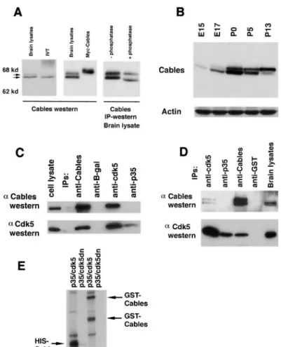
Figure 1. Cables Is a Novel Protein with Limited Homology to Cyclin A
(A) Cables interacts strongly with Cdk5 and weakly with Cdk2 and Cdk3. No interaction is seen with Cdk4 and Cdk6. Yeast strain MV101, containing Cables fused to the GAL4 transactivation domain, was transformed with different Cdk baits, and-gal activity was detected by incubation of filters with X-gal.
(B) Nucleotide and amino acid sequence of Cables. The arrow indicates the position of the point of fusion of the cDNA recovered in the yeast two-hybrid screen. Potential serine/threonine and tyrosine phosphorylation sites are in bold type. PXXP domains are underlined. The axonal sorting motifs are bracketed. The region of homology to cyclin A is marked with a dashed underline.
(C) Region of homology between Cables and cyclin A using a gapped BLAST search (NCBI).
Figure 2. Cables Associates with Cdk5 in Brain Lysates
(A) Affinity-purified anti-Cables antisera rec-ognize Cables in brain lysates as a doublet of 68 kDa that migrates with Cables synthe-sized in rabbit reticulocyte lysate (IVT), slightly faster than with Myc-tagged Cables transfected into COS7 cells. Cables immuno-precipitated from brain lysate was treated with or without phosphatase prior to an anti-Cables Western blot.
(B) A Western blot of brain lysates (100g) from different stages of mouse development was probed with anti-Cables antisera and re-probed with antibodies against actin as a loading control.
(C) COS7 cells were transfected with Cables, Cdk5, and p35. Immunoprecipitations with antibodies to Cables, Cdk5, p35, and-gal were performed followed by anti-Cables and anti-Cdk5 Western blots.
(D) Immunoprecipitations with antibodies to Cables, Cdk5, p35, and GST were performed from mouse brain lysate. Western blots show that Cables and Cdk5 coimmunoprecipitate. (E) In vitro32P␥-ATP kinase assays with
trans-fected p35/Cdk5 (wild-type kinase) or p35/ Cdk5dn (inactive kinase) were performed us-ing bacterially produced His6-Cables and
GST-Cables as substrate. A parallel Western blot showed that the indicated bands reacted with anti-Cables antisera (arrows). GST-Cables prepared in E. coli contains several breakdown products.
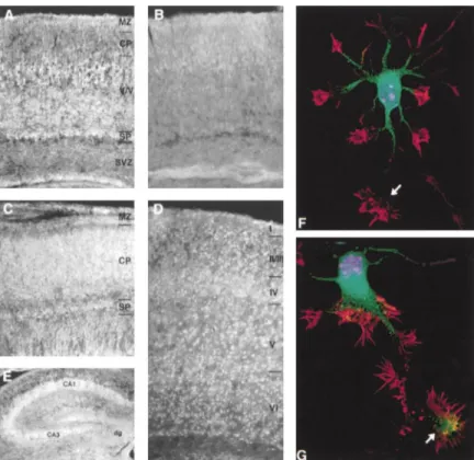
Cables Is a Cdk5 Substrate not shown). In postnatal day 1 (P1) sections, staining is again seen in the subplate and marginal zone, as well In light of the three potential Cdk phosphorylation sites
present in Cables, we tested if Cables could be phos- as in the deep layers of the cortex (V/VI), but less staining is present in the more superficial cortical layers (Figure phorylated by the p35/Cdk5 kinase. Figure 2E shows
that both GST-Cables and His6-Cables fusion proteins 3A). In P7 brain sections, strong staining is seen in all
layers of the cerebral cortex with prominent staining in could be readily phosphorylated by the p35/Cdk5 kinase
in vitro. The extent of Cables phosphorylation by the the cell body (Figure 3D). In situ hybridization was also performed on sections of various ages, which is consis-p35/Cdk5 kinase is similar to that of histone H1, a
well-known substrate of Cdk5, when identical amounts of tent with the data obtained from immunohistochemistry. Collectively, these results indicate that Cables is present protein were used in the kinase reactions (data not
shown). Phosphatase treatment downshifted Cables on in postmitotic neurons. High levels of Cables were also seen in the CA1 and CA3 regions of the hippocampus, SDS–PAGE (Figure 2A), suggesting that Cables is a
phosphoprotein in vivo. and less staining was seen in the dentate gyrus (Figure 3E). Hippocampal neurons prepared from E16 mice show that in immature neurons without a dominant pro-Cables Is Expressed in Postmitotic Neurons
cess, Cables is present in the cell body and proximal As it was shown previously that the p35/Cdk5 kinase is
region of the developing axonal shaft, as revealed by expressed in postmitotic neurons of the cerebral cortex,
Deltavision deconvolution microscopy (Figure 3F). In we asked if Cables was also present in postmitotic
neu-more mature neurons with one dominant process, Ca-rons. To examine protein expression, Cables
immuno-bles is also present in the axonal growth cone but not histochemistry was performed on coronal and sagittal
in the distal part of the axon shaft or in dendritic growth sections of mouse brain of different developmental
cones (Figure 3G), suggesting a specific function of Ca-stages using affinity-purified anti-Cables antisera
(Fig-bles in the axonal growth cones. ure 3). Preabsorbed antiserum was used as a negative
control to show specificity (Figure 3B). Strong staining
is seen in neurons of the subplate, cortical plate, and Association of Cables and c-Abl
In addition to the potential Cdk phosphorylation sites, marginal zone of E18 mouse embryos (Figure 3C).
Ca-bles staining is also present in the subventricular zone. Cables contains six minimal SH3 domain binding motifs (PXXP; Cicchetti et al., 1992; Ren et al., 1993), two of No detectable staining was seen in neurons of the
Figure 3. Cables Is Present in Cortical and Hippocampal Neurons
In coronal sections of E18 mouse brain, strong Cables immunostaining is seen in the neurons of the subplate (SP), cortical plate (CP), and marginal zone (MZ) (C). In P1 sec-tions (A and B), staining is seen in the sub-plate, marginal zone, and early layers of the cortex (V/VI). No staining is seen with preab-sorbed antisera (B). In P7 sections (D and E), strong staining is seen in all layers of the cerebral cortex and in the hippocampus (CA1 and CA3) with less staining of the dentate gyrus (dg). Magnification is 100⫻ for (A) through (D), and 200⫻ for (E). Embryonic hip-pocampal neurons were labeled with DAPI (blue), Texas red phalloidin (red), and antise-rum to Cables followed by FITC-labeled sec-ondary antibody (green). Texas red phalloidin labels both axonal and dendritic growth cones. At early stages, Cables is detected in the cell body and proximal region of the developing axon shaft but is not detected in the growth cone (arrow, [F]). As the axon elon-gates, Cables is detected in the axonal growth cone (arrow, [G]) but is not detected in the distal part of the axon shaft or in the dendritic growth cones. Magnification is 600⫻.
PXPP) with good potential for binding to the SH3 domain that a region different from the SH3 and SH2 domains and the C terminus was also involved in binding (data of the c-Abl tyrosine kinase (Cicchetti et al., 1992; Dai
and Pendergast, 1995; Shi et al., 1995). To test if Cables not shown).
As mentioned, Cables contains six minimal SH3 do-binds to SH3 domain–containing proteins, Cables
tran-scribed and translated in vitro was mixed with glutathi- main binding motifs (PXXP); 5 of the 6 PXXP sites pres-ent in Cables are located in the N-terminal one-third of one S-transferase (GST) fusion proteins containing
vari-ous SH3 domains. Cables bound to GST-c-Src and the protein. The yeast clone is missing these sites and showed little association with c-Abl (Figure 4F), sug-GST-c-Abl SH3 domains but not GST-NCK or GST alone
(Figure 4A). Compared with the 10% input in lane 1, gesting that this region is important in c-Abl binding. nearly all of the available Cables bound to GST-SrcSH3
and GST-AblSH3, suggesting a high affinity interaction.
Cables Is a Substrate of c-Abl We further verified these interactions in transfected
In light of the association between Cables and c-Abl cells. While a robust association was readily observed
and the presence of a consensus tyrosine phosphoryla-between Cables and c-Abl when coexpressed in COS7
tion site for c-Abl in Cables, we determined if Cables cells, we failed to detect a similar association between
could be phosphorylated by c-Abl. Strong tyrosine Cables and c-Src, including the F527 mutant (Figures
phosphorylation of Cables was evident upon coexpres-4B and 4E). As the SH3 domain of c-Abl associated with
sion of active Abl with Cables (Figure 4D) and, to a lesser Cables in vitro, an activated version of c-Abl with the
extent, upon coexpression of Cables with active Src SH3 domain deleted (⌬XB; Jackson and Baltimore, 1989)
(SrcY527F). As no in vivo interaction of Cables and c-Src and a catalytically inactive mutant of c-Abl (K290M; Van
or the 527 mutant was detected (Figure 4E), the possible Etten et al., 1994) were tested for binding to Cables in
phosphorylation of Cables by c-Src remains to be deter-transfection assays. Surprisingly, the⌬XB mutant bound
mined. No tyrosine phosphorylation was seen when Ca-to Cables, albeit Ca-to a lesser extent than did wild-type
bles was transfected alone. In cortical lysates of E15 c-Abl (Figure 4C). Conversely, the K290M mutant
dis-mice, anti-phosphotyrosine antibody recognizes Ca-played a more stable interaction with Cables. The
bles, indicating that in vivo Cables exists as a tyrosine-Abl⌬XB/K290M double mutant also associated with
Ca-phosphorylated protein (Figure 5E). Thus, Cables is bles (data not shown). These results suggest that both
likely to be an in vivo substrate of c-Abl. the SH3 domain and a region outside the SH2 and SH3
domains of c-Abl are involved in association with
Ca-bles. A dot blot of overlapping 15 amino acid c-Abl Phosphorylation of Cdk5Y15 by c-Abl
The observation that Cables interacts with a serine/thre-peptides was probed with Cables, and multiple binding
regions were identified, many of which are in the Abl C onine kinase and a tyrosine kinase raised the possibility of cross-regulation of the two kinases via Cables. We terminus (data not shown). However, an Abl deletion
mutant missing both the SH3 domain and C terminus failed to detect a difference in the level of c-Abl tyrosine kinase activity in the presence of increasing amounts maintained strong binding to Cables in vivo, suggesting
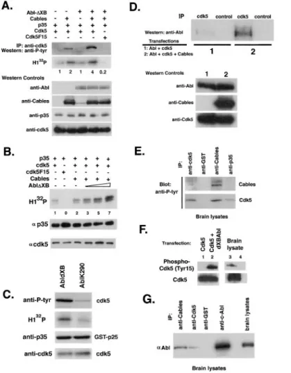
Figure 4. Cables Binds to and Is Tyrosine Phosphorylated by c-Abl
(A) In vitro translated (IVT)35S-labeled Cables
was mixed with GSH beads bound to GST, GST-NCK, GST-SrcSH3, and GST-AblSH3. The bottom panel shows a Coomassie blue– stained gel of the proteins bound to the GSH agarose.
(B) COS7 cells were transfected with Cables and c-Abl or c-Src, and cell lysates were sub-jected to Cables or control (-gal) immuno-precipitations followed by anti-Abl or anti-Src Western blots.
(C) Lysates of COS7 cells transfected with c-Abl, SH3-deleted Abl (⌬XB), or kinase-inac-tive Abl (K290M) with or without GST-Cables were subjected to a GSH pull-down and Abl Western blot. The bottom panel shows an Abl Western blot of the cell lysates.
(D) COS7 cells were transfected with Cables and active Src (SrcY527F) or active Abl (Abl⌬XB), and cell lysates were subjected to Cables immunoprecipitations followed by an anti-phosphotyrosine Western blot. The bot-tom panels show Cables, Src, and Abl in the cell lysate.
(E) COS7 cells were transfected with Cables and c-Abl, c-Src, or Src527 mutant, and cell lysates were subjected to Cables precipita-tions followed by anti-Abl or anti-Src Western blots.
(F) COS7 cells were transfected with Cdk5, c-Abl, and full-length or partial (yeast clone) Cables, and lysates were subjected to Cables precipitations followed by Abl, Cables, and Cdk5 Western blots.
of p35/Cdk5, as indicated by c-Abl autophosphoryla- tyrosine phosphorylated in vivo. In addition, a phospho-tion in an in vitro kinase assay (data not shown). Interest- Cdc2 (Tyr15) antibody that is specific for Y15-phosphor-ingly, Cdk5 became tyrosine phosphorylated when Cables ylated Cdc2 and Cdk2 reacted only with tyrosine-phos-or Abl⌬XB was overexpressed, and the level of tyro- phorylated Cdk5, as evidenced by transfection of Cdk5 sine phosphorylation increased when both Cables and with or without active Abl (Figure 5F). The antibody re-Abl⌬XB were coexpressed (Figure 5A). Phosphorylation acted with Cdk5 from brain lysates, further suggesting of the Y15 residue is a known regulatory event for the that Cdk5Y15 is phosphorylated in vivo.
cyclin-dependent kinases (Morgan, 1995). This residue is conserved in Cdk5. A Y15 to phenylalanine Cdk5
mu-tant (F15) was no longer phosphorylated by Cables and Y15 Phosphorylation of Cdk5 by c-Abl Enhances Cdk5 Kinase Activity
Abl⌬XB (Figure 5A), demonstrating that tyrosine
phos-phorylation of Cdk5 occurs on the highly conserved Y15 Y15 phosphorylation of Cdc2 and Cdk2 by the Wee1 family kinases is inhibitory and must be relieved by the residue. The F15 Cdk5 mutant is able to interact with
p35 and Cables to a wild-type extent (data not shown). Cdc25 family of phosphatases for kinase activation. Sur-prisingly, we found that Cdk5F15 is much less active To further verify the phosphorylation of Cdk5 by c-Abl,
bacterially produced 6⫻His-Cdk5 fusion protein was (but does have some kinase activity) than is the wild-type kinase (Figures 5A and 5B), yet it binds to p35 as used as an in vitro substrate for c-Abl. An
anti-phospho-tyrosine blot and32P␥-ATP incorporation showed that well as or better than wild-type Cdk5. This result
sug-gests that Y15 phosphorylation is stimulatory for Cdk5 Cdk5 is a direct substrate of the c-Abl kinase (data not
shown). To determine if Cdk5 is tyrosine phosphorylated kinase activity. To test this hypothesis directly, kinase activity of p35/Cdk5 was compared in the absence or in vivo, an anti-phosphotyrosine Western blot was
per-formed on Cdk5 immunoprecipitates from E15 mouse presence of Cables and c-Abl. Higher levels of p35/ Cdk5 kinase activity were consistently observed in the brain lysates. As demonstrated in Figure 5E, that Cdk5 is
Figure 5. Cables Links Cdk5 and c-Abl, Facil-itating Cdk5 Tyrosine Phosphorylation and Stimulation of Kinase Activity
(A) COS7 cells were transfected with p35, Cdk5, or Cdk5F15 and Cables, Abl⌬XB, or both, and cell lysates were subjected to Cdk5 immunoprecipitation followed by an anti-phosphotyrosine Western blot. Cdk5 activity was assessed by histone H1 kinase assay. In the lower panel, the lysates were reprobed with antibodies to c-Abl, Cables, p35, and Cdk5 to assess protein levels.
(B) COS7 cells were transfected with p35, Cdk5 or Cdk5F15, Cables, and increasing amounts of Abl⌬XB. Cell lysates were sub-jected to p35 immunoprecipitation followed by in vitro32P␥-ATP kinase assays using
his-tone H1 as a substrate. The lower panels show a parallel p35 immunoprecipitation fol-lowed by p35 and Cdk5 Western blots. (C) Baculovirus-produced and -purified GST-p25 and Cdk5 were mixed with Abl immunopre-cipitates from cells transfected with active Abl (Abl⌬XB) or inactive Abl (AblK290M) in the presence of cold ATP and kinase buffer fol-lowed by a Western blot for anti-phosphoty-rosine. Similarly, the baculovirus proteins were mixed with32P␥-ATP and histone H1 in
a kinase assay. When active Abl is mixed with histone H1, no kinase activity is seen. The lower panel shows that the levels of p25 and Cdk5 were the same in both reactions. (D) COS7 cells were transfected with Cdk5 and c-Abl in the presence or absence of Ca-bles, and cell lysates were subjected to Cdk5 or control immunoprecipitations followed by anti-Abl Western blot.
(E) Immunoprecipitations with antibodies to Cables, Cdk5, p35, and GST were performed from E15 mouse brain lysate and followed by anti-phosphotyrosine Western blot. (F) Immunoprecitipations of Cdk5 (lanes 1–3) or control antibody (lane 4) from transfected cell lysates and E15 mouse brain lysates were probed with an antibody specific for Y15-phosphorylated Cdk5 and anti-Cdk5.
(G) Immunoprecipitations with antibodies to Cables, Cdk5, GST, and c-Abl were performed from E15 mouse brain lysates followed by anti-Abl Western blot.
presence of Cables and c-Abl or Abl⌬XB measured ei- Together, these results strongly suggest that Y15 phos-phorylation of Cdk5 by c-Abl upregulates its kinase ac-ther by histone H1 phosphorylation (Figures 5A and 5B)
or p35 autophosphorylation. The p35/Cdk5 kinase activ- tivity. p35 immunoprecipitates from transfected cells or E15 brain lysates contain tyrosine-phosphorylated Cdk5 ity increased with increasing amounts of Abl⌬XB, up to
7-fold higher than did basal p35/Cdk5 kinase activity (Figure 5E), which is consistent with the notion that Y15-phosphorylated Cdk5 is more active when associated (Figure 5B), and correlated with Cdk5 tyrosine
phos-phorylation (Figure 5A). Further support for these obser- with p35 than is unphosphorylated Cdk5. vations was gained using purified components. p25/
Cdk5 kinase was produced in insect cells after baculovi- Cdk5, Cables, and c-Abl Trimolecular Complex rus infection, incubated with either Abl⌬XB or AblK290M Formation In Vivo
immunoprecipitated from transfected COS7 cells, and As tyrosine phosphorylation of Cdk5 by c-Abl appears tyrosine phosphorylation, as well as kinase activity of to be enhanced by Cables, it is possible that Cables is Cdk5, were evaluated. Like the Cdk5 produced in E. necessary to recruit c-Abl to Cdk5. To examine this
coli, Cdk5 synthesized in insect cell lysates was strongly possibility, Cdk5 immunoprecipitations followed by
phosphorylated by Abl⌬XB compared with AblK290M c-Abl Western blots were performed from cells trans-(Figure 5C). Histone H1 phosphorylation assays indi- fected with Cdk5 and c-Abl with or without Cables. Fig-cated that Cdk5 kinase activity correlated with the in- ure 5D shows that while only a marginal interaction was crease in tyrosine phosphorylation and was at least observed in the absence of Cables, a strong interaction 3-fold higher in the presence of Abl⌬XB than in the between Cdk5 and c-Abl was observed when Cables presence of AblK290M (Figure 5C). No phosphorylation was expressed. In addition, we do not have evidence for a direct interaction between Cdk5 and c-Abl in vitro. of histone H1 by Abl⌬XB was seen (data not shown).
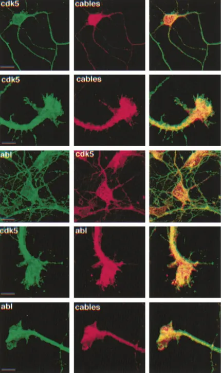
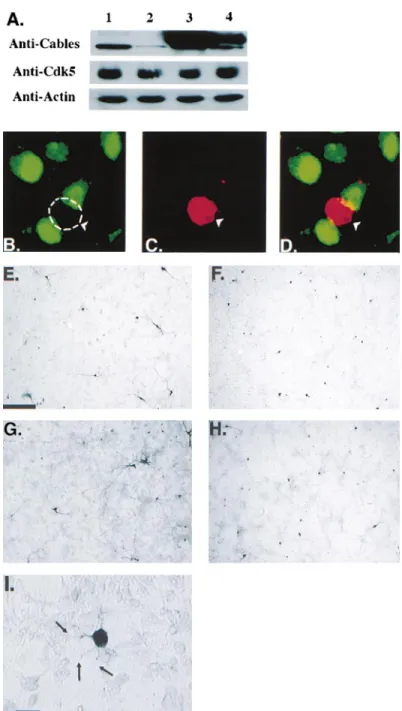
Figure 6. Cdk5, Cables, and c-Abl Colocalize in Primary Cortical Neurons
Immunostaining of Cdk5, c-Abl, and Cables was performed on cultured primary cortical neurons using confocal microscopy. As seen in Figure 3, intense Cables signal was present in the soma of the cultured neurons. In growth cones, Cdk5, c-Abl, and Cables colocalized in the central regions of the growth cones, while Cdk5 extended further to the periphery. Preabsorption of the Cables antibody with cognate antigen eliminated the staining sig-nal, demonstrating the specificity of staining (data not shown). Scale bars, 10m.
Thus, it is suggestive that Cables facilitates the interac- the soma of cultured cortical neurons (Figure 6). Cables staining was evident in axonal growth cones in which tion between Cdk5 and c-Abl. To examine if a
trimolecu-lar complex was present in vivo, Cdk5 and Cables immu- Cdk5 is also enriched (Figure 6; Nikolic et al., 1996), and the two proteins colocalized. Cables seemed to be noprecipitations were performed using E17 cortical
lysates followed by c-Abl Western blots. Figure 5G present more centrally in the growth cone, while Cdk5 extended to the peripheral lamelipodia. c-Abl appears shows that c-Abl is present in both Cdk5 and Cables
immunoprecipitates. This result corroborates data shown to be highly enriched in neurites, in agreement with a previous report on c-Abl distribution in the nervous sys-in Figure 5D and sys-indicates the existence of trimolecular
complexes containing Cdk5, c-Abl, and Cables in vivo. tem (Koleske et al., 1998). In axonal growth cones, Cdk5, c-Abl, and Cables fully colocalize in central regions (Fig-Therefore, Cables may act as an adaptor or cable
be-tween Cdk5 and c-Abl. To examine the extent of the ure 6). Together, the immunostaining results suggest that association between Cables, c-Abl, and Cdk5 takes subcellular colocalization of these proteins,
immuno-staining of Cdk5, c-Abl, and Cables was performed on place in the neuronal peripheries, such as axonal growth cones in which dynamic actin cytoskeleton rearrange-cultured primary cortical neurons followed by confocal
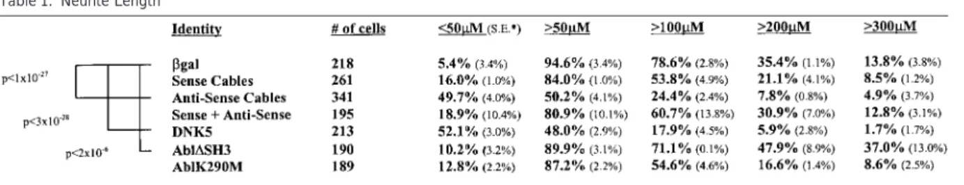
Figure 7. Expression of Antisense Cables in Primary Cortical Neurons Inhibits Neurite Outgrowth
Western blot of HeLa cells transfected with empty vector (lane 1), antisense Cables (lane 2), sense (Myc-tagged) Cables (lane 3), or sense⫹ antisense Cables (lane 4) and probed with antibodies to Cables, Cdk5, and-actin (A). Immunostaining is shown of endogenous Cables (green) and-gal (red) in cultured cor-tical neurons transfected with-gal and anti-sense Cables. Antianti-sense Cables–expressing neuron is devoid of Cables compared with nontransfected neurons (B–D). Cultured cor-tical neurons were transfected with the fol-lowing CMV expression constructs: -gal alone (E),-gal in combination with Cables antisense (F),-gal in combination with active Abl (Abl⌬SH3) (G), and -gal in combination with Cdk5dn (H). High-magnification Nomar-ski image of antisense Cables–transfected neuron is shown in (I). Scale bar, 200M (E–H) and 20M (I).
Cables Is Essential for Neurite Outgrowth when overexpressed in HeLa cells (Figure 7A). In addi-tion, the antisense Cables construct decreased Cables Cortical neurons obtained from E17–E18 rat embryos
mature morphologically during the course of culturing protein levels in cortical neurons, as evidenced by immu-nostaining (Figures 7B–7D). Expression of the antisense and can be made to express ectopic protein by
transfec-tion using a calcium phosphate method (Nikolic et al., Cables construct in cortical neurons caused extensive neurite shortening comparable to that of Cdk5N144
(Fig-1996; Dudek et al., 1997). This approach has been used
previously to assess the effect of Cdk5 and p35 on ures 7E, 7F, 7H, and 7I; Table 1). This phenotype was reversed by coexpression of sense and antisense Ca-neurite outgrowth (Nikolic et al., 1996). It was shown that
dominant-negative Cdk5 (Cdk5dn, Cdk5N144) markedly bles. Rare neurons transfected with antisense Cables
had long axons, possibly from expression of only the inhibited neurite outgrowth. To evaluate the role of
Ca-bles in the axonal growth cones, we analyzed neurite marker protein LacZ. Expression of sense Cables pro-duced a minor inhibitory effect that might be due to a length after transfection of sense and antisense Cables
constructs in primary cortical neurons. The antisense dominant-negative effect of expressing nonphysiologi-cal levels of Cables. In addition, to examine if neurite Cables construct was demonstrated to reduce
Table 1. Neurite Length
Asterisk denotes standard error. Significant ANOVA p values (compared with-gal) are indicated.
kinase, active Abl was expressed in the primary neurons. of growing axons. Based on the expression pattern, colocalization, and strong association in brain lysates, An increase in neurite length was found with expression
of active Abl but not inactive Abl (Figures 7E and 7G; it is likely that the association of Cdk5 and Cables is part of the signaling pathway that operates during brain Table 1).
To quantitate the differences between neurons with development.
-galactosidase (-gal), Cdk5dn, Cables sense and
anti-sense constructs, and Abl constructs, the neurite Cables Contains Sequence Motifs for Cdk lengths of transfected cells were examined using a and SH3 Binding
cooled charge-coupled device (CCD) camera and Meta- Cables is a 568 amino acid protein that exhibits little morph image analysis software (Universal Imaging). For homology to other proteins in the databases. The C-ter-each transfection, 200–300 positive cells were exam- minal portion of Cables binds to Cdk5, as indicated ined, as described in the Experimental Procedures. All by the partial clone isolated from the two-hybrid yeast measurements were performed in a “blind” manner, and screen. A 200 amino acid stretch in the C terminus of each transfection was analyzed by at least three observ- Cables is 25% identical and 45% similar to cyclin A in ers. The longest neurite emanating from each cell was a region that coincides with the cyclin box, which is a measured, and only if it was longer than the diameter key element of the cyclin–Cdk interface. Cables contains of the cell body. Analysis of the obtained data revealed the conserved alanine residues that are characteristic that 50% and 24% of the cells transfected with antisense of the cyclin fold and also the lysine and glutamic acid Cables elaborated neurites longer than 50 and 100m, residues that contact the backbone of Cdk2 in the cyclin respectively, in contrast to 95% and 79% of mock trans- molecules (Jeffrey et al., 1995). This region interacts fected neurons (Table 1). Similarly, less neurons trans- with the PSTAIRE helix of the Cdks. Cdk5 contains the fected with the antisense construct showed neurites PSSALRE sequences in place of PSTAIRE, and this re-longer than 200m compared with the other transfec- gion is necessary for p35 binding to Cdk5 (Y. Ramos tions (Table 1). This effect was reversed when coex- and L.-H. T., unpublished data). Thus, it appears that pressed with sense Cables. In contrast, 37% of cells both Cables and p35 bind to regions of Cdk5 that include transfected with active Abl elaborated neurites longer the PSSALRE sequence. Therefore, stable binding of than 300m compared with 14% of mock transfected both Cables and p35 to Cdk5 at the same time may be cells or cells expressing inactive Abl. Statistical analysis difficult, which fits with our experimental data. using ANOVA (Table 1) showed that compared with Cables also contains six PXXP sites, which is the
-gal, transfection of Cdk5dn (p ⬍ 3.2 ⫻ 10⫺28) and
minimal requirement for SH3 domain binding (Cicchetti antisense Cables (p⬍ 1.0 ⫻ 10⫺27) resulted in
signifi-et al., 1992; Ren signifi-et al., 1993). Based on the PXXP sites, cantly shorter neurites, which was reversed when sense we tested and found that Cables binds strongly to the and antisense Cables were coexpressed (p⫽ 0.069). SH3 domains of c-Src and c-Abl, but not other SH3-containing proteins, like Nck, in vitro. Furthermore, we found that Cables bound to full-length c-Abl, but not Discussion
c-Src or the 527 mutant, upon cotransfection. The c-Abl binding was not dependent on the SH3 domain of c-Abl, Cdk5 plays an important role in neuronal migration and
neurite outgrowth. Mice lacking Cdk5 exhibit late embry- as an SH3 deletion mutant bound almost as strongly as did wild-type c-Abl. A dot blot of overlapping 15 amino onic to perinatal lethality and show many CNS
abnormal-ities, including lamination defects of the cerebral cortex, acid c-Abl peptides showed that Cables bound to multi-ple overlapping peptides of c-Abl, many in the C termi-abnormal hippocampal formation, and cerebellar
defoli-ation (Ohshima et al., 1996). To date, only a few Cdk5- nus. A deletion mutant of c-Abl lacking both the SH3 domain and C terminus bound Cables, suggesting that interacting molecules are known, including the Cdk5
activators p35 and p39 (Lew et al., 1994; Tsai et al., Cables can interact with c-Abl at many sites. The multi-ple binding sites of Cables to c-Abl in regions with no 1994; Tang et al., 1995). In this paper, we describe a
novel protein, Cables, that binds directly to Cdk5 and Src homology would support our finding that Cables does not stably bind c-Src in vivo. Other c-Abl inter-associates with Cdk5 in brain lysates. Cables is
ex-pressed at the highest levels in the brain, where it is actors, such as the recently described Abi-2 protein, interact with at least two domains in c-Abl, the SH3 and present in postmitotic neurons of the cerebral cortex.
Cables exists as a doublet ofⵑ68 kDa on Western blots of brain lysates using two different affinity-purified antisera. The reason for the doublet is not clear at this time, but both forms associate with Cdk5. Although strong binding of Cables and Cdk5 can be demonstrated using either Cdk5 or Cables antibodies, no association of Cables and p35 has been detected in transfected cells or brain lysates. This suggests that Cdk5 stably exists in at least two separate complexes, one with p35 and the other with Cables. It is possible that Cdk5 trans-locates from Cables to p35 while remaining Y15 phos-phorylated, such that there is a transient but unstable interaction among Cdk5, p35, and Cables. The presence of such a complex would also allow the p35/Cdk5 kinase to phosphorylate Cables. Alternatively, heterologous p35/Cdk5 complexes may phosphorylate Cables.
Cables Is a Substrate of Cdk5 and c-Abl
Cables exists in vivo as a phosphoprotein; treatment with phosphatase shifts at least the faster migrating species but does not condense the doublet. In addition to Cdk5 binding, Cables may be an in vivo substrate of Cdk5, since it contains three potential Cdk5 phosphory-lation sites (serine proline/threonine proline), one of
Figure 8. Model Illustrating Possible Cdk5/Cables/c-Abl Pathway which is followed by basic residues and is a preferred Involved in Neuronal Development
Cdk5 phosphorylation site (Beaudette et al., 1993;
Song-c-Abl is activated in response to local stimuli and phosphorylates yang et al., 1996). Cables is readily phosphorylated by the Y15 residue of Cdk5, which is enhanced by Cables. p35 binds the p35/Cdk5 kinase in in vitro kinase assays. Cotrans- to Y15-phosphorylated Cdk5, and the Cdk5/Cables/c-Abl complex dissociates. c-Abl and p35/Cdk5 may both phosphorylate Cables. fection of Cables with active Abl resulted in Cables
tyro-sine phosphorylation. Thus, Cables interacts with and can be phosphorylated by c-Abl, suggesting that it is
Y15 Phosphorylation Augments p35/Cdk5 an in vivo substrate. Phosphorylation of Cables by Cdk5
Kinase Activity and c-Abl may cause rapid dissociation of Cables from
Evaluation of Cdk5 tyrosine phosphorylation, using an this complex and allow free p35/Cdk5 to phosphorylate
F15 mutant of Cdk5, showed that tyrosine phosphoryla-substrates involved in neurite outgrowth and neuronal
tion occurred on Y15, an important Cdk regulatory site. migration.
Despite the sequence homology to Cdk2 and Cdc2, it was previously shown that Cdk5 is not activated by CAK and is neither phosphorylated nor inhibited by the Wee1 Cables Enhances Y15 Phosphorylation of Cdk5
kinase (Poon et al., 1997). There is a report of in vivo by c-Abl Tyrosine Kinase
Cdk5 tyrosine phosphorylation in rat cerebellum, but the Cables associates with both the Cdk5 serine/threonine
site of phosphorylation and the kinase responsible were kinase and c-Abl nonreceptor tyrosine kinase. We found
not known (Lazaro et al., 1996). Our data implicate the that Cdk5 became tyrosine phosphorylated in the
pres-nonreceptor tyrosine kinases, especially c-Abl, as Cdk5 ence of c-Abl, and this was enhanced by inclusion of tyrosine kinases. The Y15 to F15 mutant of Cdk5 had Cables. In transfection experiments, expression of either dramatically less kinase activity, despite binding to p35 c-Abl or Cables resulted in Cdk5 tyrosine phosphoryla- and Cables as well as or better than wild-type Cdk5. tion. Endogenous c-Abl and Cables are detected in the The data suggest that Cdk5 Y15 phosphorylation is a cell lysates, which may explain why c-Abl alone phos- stimulatory posttranslational modification that upregu-phorylates Cdk5. When transfected together, there was lates Cdk5 kinase activity. Evidence for an inhibitory an increased level of Cdk5 tyrosine phosphorylation. It kinase activity distinct from Wee1 and Myt1 (Matsuura is not clear why transfection of Cables itself leads to and Wang, 1996) was found to affect threonine 14 of Cdk5 tyrosine phosphorylation. Endogenous c-Abl is Cdk5. Cdk5Y15 phosphorylation may also affect sub-thought to be inhibited by cellular inhibitors that bind strate specificity and/or localization of the kinase. Fur-to the SH3 domain of c-Abl (Pendergast et al., 1991; thermore, an effect on Cdk5 activity imposed by tyrosine Wen and Van Etten, 1997). It is possible that Cables acts phosphorylation of p35 cannot be dismissed.
not only as an adaptor molecule but also as a c-Abl
activator. Cables binds to the SH3 domain of c-Abl, Cables, c-Abl, and Cdk5 Colocalize and perhaps this binding displaces inhibitor molecules, at the Neuronal Periphery
leading to Cdk5 tyrosine phosphorylation. Mutants of In the developing cerebral wall, Cables is expressed in Cables that no longer bind to the SH3 domain of c-Abl postmitotic neurons but is absent in neuronal precur-sors. Cables is discontinuously expressed in the growth will be useful for examining this hypothesis.
(pPC86, Trp1). Two million primary library transformants were ampli-cones of developing axons, where it colocalizes with
fied and screened on Trp-Leu-His containing 10 mM 3-aminotri-Cdk5 and c-Abl. Similar expression is not seen in the
azole. Transformants (120) were selected as meeting the following growing tips of dendrites. We examined Cdk5 kinase
criteria: (1) they grew on Ura-His⫹ Trp-Leu plates or (2) they grew activity in abl⫺/⫺mice but could not detect a difference on Ura⫹ His-Trp-Leu plates and turned blue when incubated with (data not shown). If Cdk5 activity is only transiently X-gal. Plasmids from these interacting clones were isolated by
trans-formation of E. coli strain DH5␣. upregulated in the axonal growth cones in developing
neurons, a Cdk5 in vitro kinase assay may not be
sensi-Cell Culture and Transfection tive enough to detect a difference. Alternatively,
c-Abl-COS7 cells were propagated in Dulbecco’s modified Eagle’s me-related genes, such as arg (Kruh et al., 1990), may be
dium with 4.5 g/l of glucose, 10% calf serum, and penicillin/strepto-redundant in phosphorylation of Cdk5. This is supported mycin. Neuronal cortical cell cultures were prepared as described by the finding that the c-abl knockout mice lack a neu- (Nikolic et al., 1996). Similarly, cultures of embryonic hippocampal neurons were prepared from E16 mouse hippocampus as previously ronal phenotype, while the c-abl/arg double knockout
described for E18 rat (Goslin and Banker, 1989). Transient transfec-mice die at E9.5 with neural tube defects (Koleske et
tions in COS7 cells were performed using the calcium phosphate al., 1998). The early lethality of the double mutant makes
method with 10–20g of total DNA (2.5 of g CMV-Cdk5, 5 g of it difficult to assess the profile of Cdk5 phosphorylation
CMV-p35, 5g of Cables, 5 g of Abl, and 5 g of CMV-in these animals. Src). Transfections of primary cortical cultures were carried out on 3-day-old cortical cultures as described previously (Nikolic et al., 1996).
A Role for Cables in Axon Growth
DNA Constructs In light of the preferential distribution of Cables in the
Full-length Cables was obtained from a mouse neonatal brain cDNA growing tips of axons rather than dendrites, a possible
library (Clontech) and sequenced. For production of tagged mam-role of Cables in axon growth was evaluated by
overex-malian constructs and bacterial fusion proteins, the Cables cDNA pression of sense and antisense Cables constructs in was subcloned in-frame into the eukaryotic GST expression vector primary cortical neurons. Transfection using the calcium PEBG (gift of B. Mayer, Children’s Hospital, Boston, MA) at the BamHI site, PCDNA3.1/Myc-His B (Invitrogen) at the BamHI site, phosphate method has been shown to be efficient in
PCDNA3.1 (Invitrogen) at the BamHI site, pGEX-4T-2 (Pharmacia) ectopic gene expression in postmitotic neurons (Nikolic
at the BamHI site, and PET15b (Novagen) at the BamHI site. For et al., 1996; Dudek et al., 1997) and for assessing neurite
production of the antisense Cables construct, Cables was cloned outgrowth following overexpression of various
mole-in reverse orientation mole-into PCDNA3.1 (Invitrogen) at the BamHI site. cules (Nikolic et al., 1996). Expression of the antisense The mammalian Abl expression vector pcDNA-c4 Abl and its vari-Cables construct caused neurite shortening similar to ous internal deletion mutations (c4⌬SH3 ⫽ Abl⌬XB, c4⌬SH3⌬Bcl) and point mutants (c4K290M) have been described previously (Jack-that seen for dominant-negative Cdk5. This effect could
son and Baltimore, 1989; Van Etten et al., 1994). The Src and be rescued using sense Cables, suggesting that the
SrcY527F constructs were gifts of D. Morgan (University of Califor-phenotype was specific. In contrast, expression of
ac-nia, San Francisco). GST-SrcSH3, GST-AblSH3, and GST-NCK con-tive Abl produced remarkably longer neurites than did
structs and protein were a gift of B. Mayer, Children’s Hospital. inactive Abl or the control-gal transfection, consistent To make Y15F Cdk5, human Cdk5 cDNA was cloned into the with genetic data implicating Abl in axonal development BamHI site of pAlter (Promega) and was mutagenized using the Promega Altered Sites II kit according to the manufacturer’s instruc-and organization (Gertler et al., 1989, 1993).
tions. The following oligonucleotide was used for mutagenesis: When taken together, our data suggest that Cables
5⬘-GAAGGCACCTTCGGAACTGTGTTC-3⬘. The mutant was con-serves as an adaptor molecule, facilitating Cdk5 tyrosine
firmed by sequencing. The insert was cloned into the BamHI site phosphorylation and regulation by c-Abl (Figure 8). of pcDNA3 (Invitrogen).
Phosphorylation of key substrates involved in actin and
microtubule dynamics by active Cdk5 is likely to contrib- Protein Analysis
Transfected cell or brain lysate was produced in E1A lysis buffer ute to its role in neuronal migration and neurite
out-(50 mM Tris-HCl [pH 7.5], 250 mM NaCl, 5 mM EDTA [pH 8.0], 0.1% growth. Furthermore, Cdk5 was shown to downregulate
Nonidet P-40, 5 mM DTT, 10 mM NaF, 1 mM phenylmethylsulfonyl N-cadherin-mediated cell adhesion (Kwon et al., 2000).
fluoride [PMSF], 1g/ml aprotinin, 1 g/ml leupeptin, and 1 g/ml Data presented in this communication suggest that Ca- Na
3VO4) and RIPA lysis buffer (50 mM Tris-HCl [pH 7.5], 150 mM
bles mediates an interaction between c-Abl and Cdk5, NaCl, 1% Nonidet P-40, 0.5% DOC, 0.1% SDS, 5 mM DTT, 10 mM and may positively affect brain development and neurite NaF, 1 mM PMSF, 1g/ml aprotinin, 1 g/ml leupeptin, and 1 g/ ml Na3VO4). Proteins were analyzed by direct Western blotting (50
outgrowth by enhancing Cdk5 tyrosine phosphorylation
g/lane) or blotting after immunoprecipitation (300–1000
g/immu-and upregulation of kinase activity. Cables may also
noprecipitation). COS7 cell extracts were immunoprecipitated with mediate an interaction between Cdk5 and mDab1 by
glutathione-Sepharose (GSH) beads, anti-Cdk5 (pAb C-8, Santa binding to both Cdk5 and c-Abl. Cruz), anti-p35 (pAb C-19, Santa Cruz), and anti-Cables (pAb 64, see above). Immunoprecipitates were collected by binding to protein A–Sepharose or protein G–Sepharose. Western blots were probed Experimental Procedures with anti-Cdk5 (mAb DC17), anti-p35 (pAb C-19), anti-Cables (pAb 64), anti-Abl (mAb 8E9, Pharmigen), anti-phosphotyrosine (mAb Yeast Two-Hybrid Screen 4G10, Upstate Biotechnology), anti-Src (mAb 327, gift of J. Brugge, A genetic screen using the yeast interaction trap was performed as Harvard Medical School, Boston, MA), anti-phospho-Cdc2 (Tyr15; described (Vidal et al., 1996). The bait plasmid (pPC97, Leu2) Cdk5dn pAb, New England Biolabs).
contained full-length Cdk5 with a point mutation at the aspartic acid
144 position (D144 to N144) fused to a yeast transcription factor Production and Purification of GST-Tagged GAL4 DNA binding domain. It was transformed into yeast strain or His-Tagged Proteins
MV101, which contained three reporter genes, ura3, his3, and lacZ. GST and His fusion proteins were expressed in E. coli and purified This strain in turn was transformed with an embryonic mouse E14 with GSH beads or nickel-charged beads according to the
manufac-turer’s instructions (Novagen). library fused to the GAL4 activation domain in the reporter plasmid
Immunohistochemistry scholar, and a recipient of an Esther A. and Joseph Klingenstein Foundation Fund.
Primary neuronal cultures were fixed for 10 min in 4% paraformalde-hyde and 4% sucrose in 1⫻ phosphate-buffered saline (PBS)
pre-warmed to 37⬚C for 10 min. Fixed cells were washed three times in Received December 8, 1998; revised April 4, 2000. PBS containing 0.1% Triton X-100 and 10 mM glycine, once in PBS
containing 0.1% Triton X-100, and incubated with 10% goat serum,
References 3% bovine serum albumin (BSA), and 2% Tween-20 in PBS for 1 hr
at room temperature to block nonspecific binding of antibodies.
Beaudette, K., Lew, J., and Wang, J.H. (1993). Substrate specificity Embryonic brains were fixed in 4% paraformaldehyde in PBS for
characterization of a cdc2-like protein kinase purified from bovine 10–12 hr. After fixation, brains were equilibrated in a 20% sucrose
brain. J. Biol. Chem. 268, 20825–20830. solution and frozen in OCT compound (VWR, Boston, MA). Sections
Chae, T., Kwon, Y.T., Bronson, R., Dikkes, P., Li, E., and Tsai, L.-H. were thawed at room temperature, equalized in PBS, and blocked
(1997). Mice lacking p35, a neuronal specific activator of cdk5, dis-with 10% goat serum and 2% Triton X-100 in PBS for 1 hr at room
play cortical lamination defects, seizures and adult lethality. Neuron temperature. The indicated primary antibodies diluted in 3% BSA
18, 29–42.
and 0.2% Tween-20 in PBS were incubated with coverslips/slides
for 16 hr at 4⬚C. Subsequently, the coverslips/slides were washed Cicchetti, P., Mayer, B.J., Thiel, G., and Baltimore, D. (1992). Identifi-in PBS and exposed to FITC or Texas red–conjugated secondary cation of a protein that binds to the SH3 region of Abl and is similar antibodies for 1 hr at room temperature. After washing extensively to Bcr and GAP-rho. Science 257, 803–806.
in PBS, they were mounted in nonfade (Molecular Probes). Dai, Z., and Pendergast, A.M. (1995). Abi-2, a novel SH3-containing The following antibodies were used for immunohistochemistry at protein interacts with the c-Abl tyrosine kinase and modulates c-Abl the indicated dilutions. Primary antibodies were Cdk5 (mAb DC39, transforming activity. Genes Dev. 9, 2569–2582.
1:5), c-Abl (mAb K-12, Santa Cruz, 1:10), -gal (mAb, Promega,
Dudek, H., Datta, S.R., Franke, T.F., Birnbaum, M.J., Yao, R., Cooper, 1:300; pAb, 1:500), and Cables (pAb 64, raised against whole protein,
G.M., Segal, R.A., Kaplan, D.R., and Greenberg, M.E. (1997). Regula-affinity purified, and concentrated against protein A, 1:25; specificity
tion of neuronal survivial by the serine–threonine protein kinase Akt. was evaluated by [1] using preabsorbed antisera and [2] staining
Science 275, 661–665. transfected versus untransfected Swiss-3T3 cells, which contain
Gertler, F.B., Bennett, R.L., Clark, M.J., and Hoffmann, F.M. (1989). undetectable protein by Western blot). Secondary antibodies were
Drosophila abl tyrosine kinase in embryonic CNS axons: a role in fluorescein- (FITC-) conjugated, affinity-purified goat anti-mouse
axonogenesis is revealed through dosage-sensitive interactions IgG, 1:150 (Cappel); fluorescein- (FITC-) conjugated, affinity-purified
with disabled. Cell 58, 103–113. goat anti-rabbit IgG, 1:150 (Sigma); Texas red–conjugated,
affinity-purified goat anti-rabbit IgG, 1:150 (Cappel); Texas red–conjugated, Gertler, F.B., Hill, K.K., Clark, M.J., and Hoffmann, F.M. (1993). Dos-age-sensitive modifiers of Drosophila abl tyrosine kinase function: affinity-purified F(ab⬘)2sheep anti-mouse IgG, 1:150 (Cappel); and
biotinylated, affinity-purified goat anti-rabbit IgG, 1:100 (Vector Lab- prospero, a regulator of axonal outgrowth, and disabled, a novel tyrosine kinase substrate. Genes Dev. 7, 441–453.
oratories).
Goslin, K., and Banker, G. (1989). Experimental observations on the development of polarity by hippocampal neurons in culture. J. Cell Neurite Measurement
Biol. 108, 1507–1516. Cells were analyzed using a Zeiss LSM 410 confocal scanning
micro-Howell, B.W., Gertler, F.B., and Cooper, J.A. (1997a). Mouse dis-scope and a Deltavision deconvolution microdis-scope, and sections,
abled (mDab1): a Src binding protein implicated in neuronal develop-using a Nikon TE300 immunofluorescent microscope. Neurite length
ment. EMBO J. 16, 121–132. was analyzed using a Nikon TE300 microscope, a CCD camera, and
Metamorph image analysis software (Universal Imaging) as de- Howell, B.W., Hawkes, R., Soriano, P., and Cooper, J.A. (1997b). scribed previously (Nikolic et al., 1996). Transfected neurons and Neuronal position in the developing brain is regulated by mouse their neurites were distinguished by immunodetection of-gal as a disabled-1. Nature 389, 733–737.
marker protein. The axons and dendrites were measured for each Hu, G., Zhang, S., Vidal, M., Baer, J.L., Xu, T., and Fearon, E.R. neuron; however, only one longest neurite per cell was used for (1997). Mammalian homologs of seven in absentia regulate DCC via comparative purposes with other neurons. Lengths were determined the ubiquitin–proteasome pathway. Genes Dev. 11, 2701–2714. as the distance between the edge of the cell body and the tip of
Ishiguro, K., Kobayashi, S., Omore, A., Takamatsu, M., Yonekura, the growth cone.
S., Anzai, K., Imahori, K., and Uchida, T. (1994). Identification of the 23 kDa subunit of tau protein kinase II as a putative activator of cdk5 in bovine brain. FEBS Lett. 342, 203–208.
In Vitro Kinase Assay
Kinase assays were performed by washing immunoprecipitates Jackson, P., and Baltimore, D. (1989). N-terminal mutations activate three times with lysis buffer and once with kinase buffer (50 mM the leukemogenic potential of the myristoylated form of c-abl. EMBO HEPES [pH 7.0], 10 mM MgCl2, and 1 mM DTT). p35 levels, which J. 8, 449–456.
have been shown to be limiting (Patrick et al., 1998), were equalized
Jeffrey, P.D., Russo, A.A., Polyak, K., Gibbs, E., Hurwitz, J., Mas-by Western blotting prior to immunoprecipitation. Subsequently, the
sague, J., and Pavletich, N.P. (1995). Mechanism of cdk activation beads were incubated with kinase buffer containing 2g of
sub-revealed by the structure of a cyclin A–cdk2 complex. Nature 376, strate and 5Ci of32P␥-ATP in a final volume of 50 l at room
313–320. temperature for 30 min. Substrates added included histone H1,
GST-Kamiguchi, H., and Lemmon, V. (1998). A neuronal form of the cell Cables, and His6-Cables; for autophosphorylation of p35, no
sub-adhesion molecule L1 contains a tyrosine-based signal required for strate was added.
sorting to the axonal growth cone. J. Neurosci. 18, 3749–3756. Koleske, A.J., Gifford, A.M., Scott, M.L., Nee, M., Bronson, R.T., Acknowledgments Miczek, K.A., and Baltimore, D. (1998). Essential roles for the Abl
and Arg tyrosine kinases in neurulation. Neuron 6, 1259–1272. The authors would like to thank Yolande Ramos and Shih-Te Wen
Kruh, G.D., Perego, R., Miki, T., and Aaronson, S.A. (1990). The for DNA constructs and technical support; Ming Sum Lee, Vivien
complete coding sequence of arg defines the Abelson subfamily of Tannoch, Young Kwon, Deanna Smith, and Elena Porro for help in
cytoplasmic tyrosine kinases. Proc. Natl. Acad. Sci. USA 87, 5802– preparation of this manuscript; and members of the Tsai laboratory
5806. for helpful discussions. L. Lanier is supported by the Anna Fuller
Kwon, Y.T., Gupta, A., Zhou, Y., Nikolic, M., and Tsai, L.-H. (2000). Fund, and F. Gertler is supported by Merck and Company. This
Regulation of the N-cadherin-mediated adhesion by the p35/Cdk5 work was supported in part by National Institutes of Health grant
kinase. Curr. Biol. 10, 363–372. RO1-NS37007 (to L.-H. T.). L.-H. T. is an assistant investigator of