The C. elegans heterochronic pathway controls the timing of
NAB/EGR-mediated terminal differentiation and the onset of adulthood
by
David T. Harris
B.S., Genetics
University of California at Davis, 2000
Submitted to the Department of Biology in Partial Fulfillment of the Requirements for the Degree of MASSACHUSETTS INSTITE. OF TECHNOLOGY DOCTOR OF PHILOSOPHY
JAN 2 6 2011
at theLIBRARIES
MASSACHUSETTS INSTITUTE OF TECHNOLOGY
-ARCHIVES
February 2011© 2011 David T. Harris. All rights reserved
hereby grants to MIT permission to reproduce and to distribute publicly paper and electronic copies of this thesis document in whole or in part.
Signature of author David T. Harris February 2011 H. Robert Horvitz Professor of Biology Thesis Supervisor Accepted by Stephen P. Bell Chairman of the Graduate Committee Department of Biology The author
Certified
The C. elegans heterochronic pathway controls the timing of
NAB/EGR-mediated terminal differentiation and the onset of adulthood
by
David T. Harris
Submitted to the Department of Biology in Partial Fulfillment of the Requirements For the Degree of DOCTOR OF PHILOSOPHY
Abstract
Most animals pass through a series of juvenile stages on their way from embryo to adult. These stages represent periods of time in which stage-specific developmental processes occur. At the end of development, the organism transitions from a juvenile state to a mature adult. The failure to undergo a specific developmental stage, or the failure to transition to an adult can lead to specific developmental deficits or the retention of juvenile characteristics throughout the remainder of the organism's life.
This thesis focuses on the study of the mechanisms controlling the larval-to-adult
transition in C. elegans. In Chapter two, I perform a screen to identify mutants that are defective in a specific aspect of the larval-to-adult transition, exit from the molting cycle. From this screen
I identify new alleles of the previously identified gene mab-10. I then clone mab-10, showing
that it encodes the only C. elegans NAB family transcriptional cofactor. I go on to show that MAB- 10 acts with LIN-29 to control the larval-to-adult transition and that LIN-29 shares homology with mammalian EGR proteins. These observations highlight the similarity between the mechanisms controlling the onset of adulthood in C. elegans and mammals as LIN-29 is required for the larval-to-adult transition in C elegans and EGRI is required for the onset of puberty in mice.
Chapter three is a further analysis of both mab-10 and lin-29, focusing on their
expression patterns and roles in the terminal differentiation of tissues outside of the hypoderm. We use a mab-10 translational reporter to show that MAB-10 accumulates precociously in precocious heterochronic mutants and that mab-10 transcription does not require lin-29,
suggesting that the heterochronic pathway controls the timing of mab-10 transcription in parallel to lin-29. We use single molecule fluorescence in situ hybridization to establish the wild-type transcription patterns of mab-10 and lin-29 and show that both undergo dynamic changes in expression throughout development. We show that mab-10 is required for terminal
differentiation in at least one other tissue outside of the hypoderm and that mab-10 prevents seam cell overproliferation by antagonizing the activities of the C. elegans Runx and CBF-Beta homologs rnt-1 and bro-1.
Thesis Advisor: H. Robert Horvitz Title: Professor of Biology
Acknowledgments
There are many people I would like to thank for helping me to complete the work presented here. First, I would like to thank Ezequiel Alvaarez-Savedra, Daniel Denning, and Nicolas Paquin for reading drafts of each of my thesis chapters. Secondly, I would like to thank Na An, Beth Castor, Rita Droste, Elissa Murphy, Erika Hartwieg, and Victoria Hatch for technical support including strain maintenance, electron microscopy, and DNA sequencing.
I would like to thank all of the current members of the Horvitz lab, including several that
have moved on to exciting new ventures. I would especially like to thank my roommates: Brendan Galvin, Takashi Hirose, Dengke Ma, and Ala Berdichevsky for creating an excellent working environment and for many fun and interesting conversations. It was Brendan that originally suggested mab-10 to me as a potential project and for that I am extremely grateful.
I would also like to thank the many members of the Anna's Taqueria crew: Melissa
Harrison, Johanna Varner, Mike Hurwitz, Dan Omura, Nicolas Paquin, and Adam Saffer. Our lunchtime trek was always a welcome distraction and often a time for great discussion.
I would like to thank the members of my thesis committee who provided me with helpful
advice over the years. The members of my committee were Chris Kaiser, Jianzhu Chen, and Dennis Kim. I would also like to thank Hazel Sive, Troy Littleton, and Victor Ambros for joining my committee in the eleventh hour.
I would like to thank my advisor Bob Horvitz for creating the type of open environment
that was exactly what I was looking for in my graduate studies. He provided the freedom for me to explore my own ideas to success and failure, but was always there to help me see my way through to the end.
Finally, I would like to thank my wife, Heather, and my family for their support. Without Heather to keep my mind focused on the task at hand, none of this would have been possible.
Table of Contents
Title Page... . -.. ---...-- 1
Abstract ... ---... 2
Acknowledgm ents ... 3
Table of Contents... ... --...--- 4
CHAPTER ONE Introduction...--11
Introduction ... . -.. ---... 12
Developm ental tim ing...12
Life stages... 12
Heterochrony ... 13
Heterochrony in evolution...14
Developm ental tim ing in C. elegans... 15
The power of C. elegans... 15
The seam cell lineage ... 16
Heterochronic m utants ... ... 17
The early developm ental tim er ... 18
lin-4 controls the progression from L I to L2...18
lin-14 is required for the execution of L 1 developmental programs...19
lin-28 is required for the execution of L2 developmental programs...19
The late developm ental tim er ... 20
let- 7 m utants undergo an extra larval stage ... 21
lin-41 m utants becom e adult prem aturely ... 21
lin-4 and let-7 encode sm all non-coding RNAs... 22
The m iRN A revolution ... 22
LIN -28 and LIN -41 likely regulate m iRNA function... 23
lin-28 likely has m ultiple functions... 23
TRIM-NHL proteins can promote or inhibit miRNA activity...24
Developm ental patterns are generated by protein gradients ... 25
Spatial protein gradients drive anterior-posterior patterning in Drosophila ... 26
Temporal patterning in C. elegans vs. Spatial patterning in Drosophila...27
The hunchback homolog hbl-1 controls developmental timing in C. elegans ... 27
rnt-i promotes seam cell divisions ... 28
Environmental cues drive developmental progression in C. elegans ... 30
DAF-12 integrates signals from the environment to control development...30
DAF- 12 promotes the expression of the let- 7 family of miRNAs in favorable environments. ... 3 1 LIN-29 is the master regulator of the larval-to-adult transition ... 31
Multiple pathways control the timing of LIN-29 activity ... 32
Circadian rhythm components control LIN-29 activity in C elegans ... 32
A Cullin-RING complex prevents precocious LIN-29 activity...33
LIN-29 likely has multiple target genes...34
LIN-29 is expressed in multiple cell types throughout development ... 35
LIN-29 controls male linker cell death ... 35
LIN-29 acts redundantly with DRE-1 to control distal tip cell migration ... 36
LIN-29 homologs in other organisms ... 37
The juvenile-to-adult transition in humans ... 38
Humans undergo a metamorphosis during puberty ... 38
The pulsatile secretion of GnRH controls the onset of puberty... 39
Heterochronic syndromes in humans?... . ... ... ... ... ... . . . 39
C on clu sio n ... 4 0 F igure L egends ...---- 4 1 Figure 1. C. elegans is a soil nematode with a well-characterized life cycle...43
Figure 2. The seam cell lineage is an excellent indicator of developmental stage and h eterochrony ...-- 4 4 Figure 3. Multiple mechanisms converge at LIN-29 to control the timing of the larval-to-adult tran sitio n ... 4 5 A cknow ledgm ents ... 46 R eferen ces ...- 4 7
CHAPTER TWO The C. elegans heterochronic pathway controls NAB/EGR-mediated
term inal differentiation...56
Sum m ary ... 57
Introduction ... 58
Results ... 59
A screen to identify m ales that undergo an extra m olt...59
mab-10 mutants make a normal adult cuticle, but undergo an extra molt ... 60
Seam cells of mab-10 mutants fuse normally, but fail to exit the cell cycle...60
mab-10 acts downstream of or parallel to lin-28 and lin-41 ... 61
mab-10 encodes the only C. elegans NAB transcriptional cofactor... 61
MAB-10 interacts with an evolutionarily conserved domain within LIN-29 ... 63
The LIN -29 C-term inus is required for term inal differentiation ... 64
Discussion ... 65
M aterials and M ethods...66
Strains and genetics...66
Screen for m ales with an extra cuticle ... 66
Phenotypic analyses ... 66 Plasm ids...67 Oligos ... 67 Transgenics...68 GST pull-down Assay ... 69 Figure Legends ... 70
Figure 1: mab-10 prom otes hypoderm al term inal differentiation ... 75
Figure 2: mab-10 encodes the only C. elegans member of the NAB family of transcriptional co-factors...76
Fgure 3: mab-10 and lin-29 are co-expressed in vivo and physically interact in vitro ... 77
Figure 4: Animals lacking the LIN-29 C-terminus generate a normal adult cuticle but undergo an extra m olt...78
Supplementary Table 1: mab-10 is required for animals to exit the molting cycle and for seam cell exit from the cell cycle ... 79
Supplementary Figure 2: Removal of the LIN-29 C-terminus disrupts MAB- 10 binding ... 81
Supplementary Figure 3: The MAB-10 interaction domain is conserved among distantly related nem atodes...-...---... . ---... 82
A cknow ledgm ents ...---. . ---... 83
R eferences ...---... 84
CHAPTER THREE mab-10 and lin-29 are expressed dynamically throughout development and control multiple terminal differentiation events...88
Sum m ary ...---... 89
Introdu ction ...---..--.... 90
R esults ...----... . ---.. ---... 92
The timing of MAB-10::GFP accumulation is controlled by hbl-1 and lin-41...92
MAB-10::GFP nuclear localization in the seam cells requires lin-29 ... 92
mab-10 transcription is not dependent on lin-29 ... 93
lin-29 expression oscillates in the hypoderm ... 94
MAB-10 promotes male linker cell death...96
Loss of rnt-1 or bro-1 suppresses the mab-10 seam cell defect ... 97
D iscu ssion ... . --... ---9 8 MAB-10::GFP accumulates precociously in hbl-1 and lin-41 mutants ... 98
Seam cell localization of MAB-10::GFP requires LIN-29 ... 99
LIN-29::mCherry localization does not require MAB-10...99
mab-10 transcription does not require lin-29 ... 100
lin-29 is expressed dynamically throughout development...101
MAB- 10 promotes a non-apoptotic programmed cell death...102
MAB-10 prevents RNT-1/BRO-1 mediated overproliferation ... 102
EGR and NAB proteins have conserved roles in the terminal differentiation of cell lineages and the onset of adulthood...103
M aterials and M ethods...105
Strains and genetics...105
Single M olecule FISH ... 105
Probes...-.. ---... 106
m a b-10 prob es ... 106
lin-29 probes... .. ---... 109
Phenotypic analyses ...-... ... 111
Figure L egends ...---.. . ---. ...112
Figure 1: MAB-10::GFP expression is controlled by the heterochronic pathway ... 115
Figure 2: mab-10 expression pattern in wild-type and lin-29(n836) mutant backgrounds...116
Figure 3: lin-29 expression oscillates in the hypoderm during development...117
Figure 4: mab-10 promotes male linker cell death ... 118
Figure 5: mab-10 prevents rnt-1/bro-1-mediated seam cell overproliferation...119
A cknow ledgm ents ... 120
R eferences ...---...---.. . - ---... 121
CHAPTER FOUR Future Directions ... 125
Introduction ... . ---...--- 126
Identifying regulators of MAB- 10 and LIN-29 expression ... 126
Screens to identify mutants with altered MAB-10::GFP or LIN-29::mCherry expression .... 127
Does the heterochronic pathway control expression of mab-10 and lin-29?... ... ...127
Identifying M AB-10 interactors ... 128
Screens to identify factors that control seam cell exit from the cell cycle...128
A candidate gene approach...129
Biochemical purification of MAB- 10 and LIN-29 interactors...129
The identification of MAB-10 and LIN-29 target genes ... 130
Screens for enhancement or suppression of mab-10(e]248) mutant phenotypes...130
Chromatin immunoprecipitation to identify target genes ... 131
A cknow ledgm ents ... 132
R eferences ...-- ... . ---... 133
APPENDIX I A screen to identify negative regulators of programmed cell death in C. eleg an s ...-.. . ---... 135
Introduction ... 137
Program m ed cell death in C. elegans...137
Program m ed cell death is evolutionarily conserved ... 138
Negative regulators of PCD ... 139
Results...- .---... 140
A screen to identify negative regulators of program m ed cell death...140
Loss of ced-9 function causes the death of touch neurons carrying nIs1 74...141
Pilot Screening...--141
Discussion and Future Directions...142
M aterials and M ethods...143
Strains and Genetics...143
Plasm id construction ... 143
Transgenics...-144
Quantitation of touch neuron death...144
Screen for mutants without touch neurons...144
Figure Legends ... 144
Figure 1: A diagram showing the expression of GFP and ced-3 driven by the mec-3 and mec-7 prom oters, respectively ... 147
Figure 2: Diagram for the F2 non-clonal screen to identify hermaphrodites that are missing the touch neurons...148
Table 1: The transgene nIs] 74 kills touch neurons in ced-9(lf) animals but not in ced-9(+) anim als ... 149
Figure 3: The dying ALM and PLM neurons become refractile corpses ... 150
Table 2: Results of a pilot screen for mutants defective in the control of programmed cell death ... 1 5 1 Table 3: Three isolates have increased touch neuron death without increased GFP fluorescence ... 1 5 1 Acknowledgm ents ... 152
References ... 153
Introduction, Result, and Discussion ... 157
M aterials and M ethods... ... 158
Strains and Genetics...158
egi-1 non-com plem entation screen ... 159
Determ ination of m utant allele sequences...159
Figure Legends ...--..---.... 160
Figure 1: The lin-Il::gfp reporter is an excellent marker for programmed cell death in the ventral nerve cord ...----..--- 161
Figure 2: The egl-1(n4629) mutation is identical to the egl-](n4045) mutation...162
Acknowledgm ents ...---..-.... 163
References ... ... 164
Chapter One
Introduction
The development of a multicellular organism is no small task. The mechanisms
controlling the fate decisions of specific cell lineages, from stem cell to differentiated state, are
of central importance to stem cell biology and metazoan biology in general. Together with the
question of which specific fate a cell should take, is the question of when a cell should take that
fate. Proper cell-fate decisions require that tissues throughout the organism develop within tight
temporal constraints. Altering the timing of these precise developmental events can have
dramatic consequences ranging from inviability to speciation. The work presented here is aimed
at providing a better understanding of the mechanisms that control developmental timing.
Developmental timing
Life stages
When discussing the developmental timing of an organism, we often refer to its life
cycle. The life cycle can be said to begin at fertilization when the organism is a zygote. The
zygote proceeds through embryogenesis, birth, postembryonic development, maturation to adult
(when the cycle can be re-initiated), senescence and finally death.
While embryogenesis proceeds as a more-or-less continuous process from the first cell
division to the completion of organogenesis, post-embryonic development in most organisms
proceeds through a series of larval/juvenile stages. In arthropods like Drosophila melanogaster,
these larval stages represent discrete developmental programs that are demarcated by a molting
cycle. In this case, the transition from the juvenile stage to the adult stage is accompanied by a
By contrast, in humans, post-embryonic development lacks the obvious demarcation that
a molting cycle provides, and the transition from juvenile to adult appears superficially to be a
continuous process. However, humans do undergo a juvenile-to-adult transition during puberty
that involves sexual maturation, the development of adult physical characteristics, and the
adoption of adult behaviors. While it is clear that this transition is controlled by genetic and
environmental factors, it is unclear if the mechanisms controlling the timing of this transition are
conserved across the animal kingdom.
Heterochrony
What is heterochrony?
The term "heterochrony," which comes from latin for "different time," can be defined as
a change in the timing of one developmental event relative to another. Historically, the term
heterochrony was first used when comparing a particular developmental process between two
closely related species. If the onset time or offset time of the process differs, or if the rate of the
process differs between the two species, then the process is heterochronic. More recently,
heterochrony has been used to describe a change in the timing of one developmental process
with respect to another developmental process within the same organism.
A classic example of heterochrony comes from the Mexican salamander, the axolotl
(Ambystoma mexicanum), and the closely related tiger salamander (Ambystoma tigrinum).
Salamanders typically undergo metamorphosis from egg to larva (an aquatic tadpole form) and
then again from larva to a terrestrial adult form. The axolotl displays a specific form of
adult lives. Instead of transforming from a larval form to an adult form, the larval axolotl grows
in size, sexually matures, and remains aquatic for the duration of its life. This life cycle is in
contrast to the life cycle of the closely related tiger salamander, which proceeds from its aquatic
larval state to a terrestrial adult. Molecularly, this adaptation is caused by the failure to release
thyrotropin, the thyroid stimulating hormone-3
Heterochrony in evolution
Heterochrony, neoteny specifically, has been proposed to be a driving force in human
evolution. Early comparisons between humans and chimpanzees revealed that infant
chimpanzees and infant humans have a very similar facial bone structure. Juvenile chimpanzees
appear to have a bone structure that resembles the human adult bone structure. As juvenile
chimpanzees age, their facial bones continue to grow and as adults are morphologically distinct
from the adult human. These observations drove the idea that humans may have evolved in a
neotenic fashion'. More specifically, humans evolved to retain the juvenile form of the
chimpanzee-human common ancestor. As a result, adult humans have retained the physical
attributes and cognitive plasticity found in juvenile chimpanzees. By prolonging the juvenile
state, humans have more time to develop and learn.
Similar observations have been made with respect to the domestication of dogs.
Domesticated dogs are descendants of the Gray wolf (Canis lupus)6. The gray wolf is a predatory
canine that was commonly found throughout Eurasia and North America. Present day
domesticated dogs are the result of multiple generations of selective breeding for traits that were
selected for a juvenile temperament that is more compatible with human-canine relationships.
This selection strategy has had the interesting side-effect of also selecting for the retention of
other juvenile wolf characteristics. Therefore, current domesticated dogs more closely resemble
the juvenile wolf with respect to behavior, temperament, and bone structure than they do adult
wolves7'8.
These observations have been mirrored in a more directed experiment involving the
domestication of the silver fox (Vulpes vulpes). After over 40 generations of breeding and
selection for "tameness," the domesticated fox has lost many of its wild traits and has in both
behavior and physical appearance, retained many of its juvenile qualities.
The grouping of these juvenile traits via selection suggests that there are specific loci that
can affect multiple aspects of developmental timing4. Understanding the mechanisms that govern
developmental timing will give us a better understanding of our own development and might
give us insight into our own evolution.
Developmental timing in C. elegans
The power of C. elegans
The nematode C. elegans is an excellent organism with which to study the genetic control
of developmental timing. Adult C. elegans are approximately 1 mm in length and exist as either
self-fertilizing hermaphrodites or males (Figure 1 a). C. elegans has an extremely well
characterized life cycle in which the animal begins as an embryo and proceeds through four
larval stages (L1-L4) before becoming a sexually mature adult (Figure 1b). Each stage of
morphogenesis and is demarcated by a period of molting during which the animal generates a
new cuticle and sheds its old cuticle'0. C. elegans is atypical in that its cell lineage is essentially
invariant and has been fully described". As such, it is the only organism in which the timing and
position of every cell division and cell death from zygote to adult is known. This knowledge
makes it possible to study alterations of specific cell lineages at single-cell resolution at specific
times during development.
Cell lineages that continue to develop post-embryonically are particularly well suited for
the study of developmental timing. Whereas embryonic development can be thought of as a
continuous period from zygote to hatching, post-embryonic development is naturally punctuated
by the molting cycle. Therefore, events that occur within a specific larval stage act as indicators
of the developmental state of the animal. For example, in a wild-type animal, the vulva begins to
develop during the L3 stage". Therefore the initiation of vulval development is a proxy for the
L3 stage. One specific set of hypodermal cell lineages, the seam cell lineages, has been used
extensively for the study of developmental timing.
The seam cell lineage
The seam cells are a stem-cell-like population of hypodermal cells that undergo a
synchronized pattern of cell division during each larval stage' '(Figure 2a). In a wild-type animal,
the seam cells divide once at the beginning of the first, third and fourth larval stages to give rise
to two daughter cells, one that fuses with the syncytial hypodermis and one (seam cell) that will
divide again during the next larval stage. During the second larval stage, the seam cells undergo
both of which divide again asymmetrically in the pattern described for the other three larval
stages. This symmetric division during the second larval stage results in the doubling of the
number of seam cells and acts as an indicator of the L2 stage. During the final larval stage (L4)
the seam cells undergo a process of terminal differentiation that indicates the end of development
and the onset of adulthood.
Heterochronic mutants
Part of the fun of performing a genetic screen comes from the fact that you rarely know
what you are going to find, and you usually find something you do not expect. This is especially
true when you are working with an organism that is relatively unexplored. Early screens in C.
elegans yielded several mutants with aberrant cell lineages. For example, in these mutants,
specific cells would fail to divide or reiterate the division pattern of their parental or
grandparental cells2. Upon closer inspection (and some keen insight) it was discovered that
some of these mutants were reiterating the division pattern of a specific larval stage rather than
of their parent or grandparent. These mutants were altered with respect to multiple aspects of
developmental timing and were named "heterochronic" mutants.
The heterochronic mutants typically are categorized as "precocious mutants," which
display late developmental traits too early, and "retarded mutants," which reiterate early
developmental traits at later stages or retain juvenile characteristics. For example, precocious
mutants might generate adult specific traits such as an adult cuticle (as evidenced by the presence
of lateral alae) at the end of the L3 stage instead of the L4 stage. They might also show late-stage
patterns of cell division and gene expression at earlier stages (Figure 2b). By contrast, retarded
cell division associated with earlier stages (Figure 2c). Double mutant analyses with precocious
and retarded mutants allowed the construction of a genetic pathway that has led to important
insights into the regulation of developmental timing as well as to landmark discoveries
concerning novel methods of post-transcriptional regulation with the discovery of microRNAs.
The early developmental timer
lin-4 controls the progression from Li to L2
Early studies of developmental timing grew from the analysis of mutants with altered,
and often reiterated, cell lineages'3. In these mutants, abnormal lineages typically underwent one
of three types of cell lineage change: a parental reiteration, a grandparental reiteration, or a
proliferation. In a parental reiteration, one of the two daughter cells repeats the division pattern
of its parent. In a grandparental reiteration, a cell repeats the division pattern of its grandparent.
In a proliferative alteration, both daughter cells adopt the parental fate and reiterate the parental
division. Interestingly, one mutant, lin-4 (abnormal cell LINeage), caused not just the reiteration
of cell divisions but also the reiteration of a general stage-specific developmental program. For
example, while wild-type animals develop through four larval stages before becoming an adult,
specific tissues within lin-4 animals reiterate an LI pattern of cell division and never progress to
the L2 stage. Therefore, it was concluded that lin-4 is required for the animal to progress from
the LI to the L2 stage. Because lin-4 animals reiterate a developmental program, lin-4 is
lin-14 is required for the execution of Li developmental programs
A second mutant, lin-14, had the opposite phenotype to lin-414'15. These animals appeared
to skip LI-specific cell divisions and directly executed L2-specific developmental programs after
hatching. Because lin-14 animals skip a developmental stage and initiate a later developmental
program prematurely, they are considered precocious. The absence of an LI stage also resulted
in the premature initiation of the larval-to-adult transition. It was concluded that lin-14 is
required for the animal to execute the LI developmental program. A gain-of-function allele of
lin-14 was isolated that dominantly conferred lineage alterations that were identical to those
conferred by the lin-4 loss-of-function mutation discussed above. Double mutant analyses
between lin-4 and lin-14 loss-of-function mutations suggested that lin-4 acts upstream of lin-14
to negatively regulate its activity'6. It was proposed that the gain-of-function allele of lin-14 uncoupled lin-14 from lin-4 regulation.
lin-28 is required for the execution of L2 developmental programs
Loss-of-function mutations in lin-28 behave similarly to lin-14 loss-of-function
mutations, with one significant difference. While LI stage developmental programs are skipped
in lin-14(lf) mutants, L2 stage developmental programs are skipped in lin-280f) mutants5. For example, the L2 proliferative seam cell division does not occur, and therefore adult lin-28
animals have roughly half the number of seam cells as wild-type animals. Furthermore, lin-28
animals become adult one and sometimes two stages prematurely. lin-28 was proposed to specify
the L2 developmental program and to prevent the premature initiation of the L3 developmental
16
While lin-4, lin-14, and lin-28 were found to control early developmental events, a fourth
heterochronic gene, lin-29, was found to control late developmental events. In lin-29 mutants,
early development proceeds normally but animals fail to transition from larva to adult because
they reiterate a late-stage developmental program5. Double mutant analyses demonstrated that 29 was required for the precocious larval-to-adult transition that occurs in the absence of
lin-16
14 and lin-28, making lin-29 the furthest downstream component of the heterochronic pathway1
The late developmental timer
When we discuss the larval-to-adult transition, we are referring to a set of events that
occur during the L4 stage. Cells stop dividing and undergo a process of terminal differentiation.
In the hypoderm, the larval-to-adult transition comprises four events. The stem cell-like
population of seam cells stop dividing and fuse and form a syncytium. The hypoderm generates
an adult cuticle and the organism exits from the molting cycle. lin-29 mutants are defective in all
four of these processes, and therefore LIN-29 is thought of as a master regulator of the
larval-to-adult transition in the hypoderm" .
While lin-4, lin-14, and lin-28 control the timing of the larval-to-adult transition by
regulating early developmental events, another set of heterochronic genes more directly regulates
late developmental events. Animals with mutations in these late-acting genes undergo early
development normally but either reiterate a late larval stage, as is the case with let-7 (LEThal)",
let-7 mutants undergo an extra larval stage
let- 7 mutants were originally identified based on late-stage lethality as a result of bursting
from the vulva21. A subsequent screen looking for suppressors of a synthetic sterility caused by
the lin-14 gain-of-function mutation and egl-35 (EGg Laying defective) identified a second allele
of let-7 and suggested that it might be a heterochronic gene17. Upon closer examination, let-7
mutants were found to undergo an extra larval stage. Instead of undergoing the larval-to-adult
transition at the end of the L4 stage, let-7 animals became adult after an inappropriate "L5" stage.
It was observed that mutations that cause a precocious heterochronic defect could suppress the
lethality associated with let- 7, and this suppression of let- 7 lethality provided a powerful means
for the identification of new precocious heterochronic mutants.
lin-41 mutants become adult prematurely
lin-41 mutants were isolated from a screen to identify mutations that suppress the
lethality and the heterochronic defects conferred by let- 718. Like let-7 mutants, early
development in lin-41 mutants proceeds normally. However, many lin-41 hypodermal cells
undergo the larval-to-adult transition one stage prematurely. This precocious differentiation
suggested that lin-41 acts to prevent premature LIN-29 activity. Because lin-41 could suppress
the heterochronic defects of let- 7, lin-41 was thought to act downstream of let- 7 to negatively
regulate lin-29. The relationship between let-7 and lin-41 was similar to the relationship of lin-4
and lin-14 in that let-7 and lin-4 mutants both undergo extra larval stages and mutants that
lin-4 and let-7 encode small non-coding RNAs
lin-4 and let- 7 were cloned and found to encode small non-coding RNAs termed miRNAs
(MIcroRNAs)17,2 2. Strikingly, the 3' UTRs of lin-14 and lin-28 contained several sites partially
complementary to the lin-4 miRNA (up to seven in lin-14 and one in lin-28), and the lin-41 3'
UTR contained six sites complementary to the let-7 miRNA17,2 2. These observations provided
the first hints into the mechanism of miRNA action. The genetic data suggested that lin-4 acted
to negatively regulate lin-14 and lin-28, and the sequence complementarity suggested that lin-4
might act by directly binding the 3' UTRs of the lin-14 and lin-28 mRNA to inhibit their
translation. The finding that the gain-of-function allele of lin-14 contained a deletion within the
3' UTR that removed several of the lin-4 complementary sites corroborated this idea23-25. This
seminal work with lin-4 and let- 7 established miRNAs as a novel means of post-transcriptional
regulation and as important regulators of developmental timing and differentiation.
The miRNA revolution
It is strange to think now that the cloning of lin-4 was not the catalyst that started the
miRNA revolution. Seven years passed between the cloning of lin-422 and the cloning of let-7.126. Prior to the cloning of let-7, the lin-4 miRNA existed as an oddity in the biological world.
No obvious homologs were known to exist in other organisms, and it was the only example of a
small RNA's acting via the 3' UTR of a target gene to control gene expression.
The cloning of let- 7 transformed miRNAs from a C. elegans oddity to a newly
across a wide range of metazoans, including humans. While lin-4 and let-7 were identified from
genetic screens, researchers quickly turned to biochemical approaches to identify candidate
miRNAs in different organisms 27-29. These approaches relied on the cloning of small endogenous
RNAs, and they have proven incredibly fruitful for the identification of miRNAs in a
high-throughput fashion. At the same time, computational biologists began creating algorithms to
predict miRNAs and possible miRNA targets based on the known interactions involving lin-4
and let- 73032
To date, the characterization of miRNA/miRNA target interactions has had limited
success. This limitation might be in part because of redundancy within miRNA families. In C.
elegans for example, there are 23 families of miRNAs based on sequence homology within the
seed region (nucleotides 2-6 of the mature miRNA). It is likely that these families share target
genes, and the deletion of a single member of the family has no obvious effect on the phenotype
of the animal33. Surprisingly, even the removal of entire miRNA families in many cases has no
or little effect. It is not clear at this point if the majority of miRNA families have very specific
functions that when removed do not affect the gross development of the animal, if there is
redundancy between miRNA families or with other non-coding or protein-coding genes, or if
most of the miRNA families are simply not involved in development.
LIN-28 and LIN-41 likely regulate miRNA function
lin-28 likely has multiple functions
C. elegans lin-28 was found to encode a highly conserved cytosolic protein with two
35
Humans have two LIN-28 homologs, LIN-28A and LIN-28B36. Recent work has demonstrated
that mammalian LIN-28A is one of a combination of four factors that can promote
reprogramming of differentiated human somatic cells into induced pluripotent stem cells".
Furthermore, LIN-28A has been shown to inhibit the activity of the let-7 family of miRNAs by
binding to the let- 7 pre-miRNA to prevent miRNA processing3 8. Evidence from studies of C.
elegans suggests that LIN-28 might act to shuttle the primary let- 7 transcript to P-bodies for
storage or degradation3 9. Taken together, these data suggest that LIN-28 homologs act in
mammals to promote pluripotency by repressing the activity of the let-7 family of miRNAs.
Interestingly, LIN-28A has also been suggested to increase the translation of cell
cycle-related mRNAs by associating with their 3' UTRs40. It is possible then that LIN-28 might play
multiple roles in the promotion of stemness, some that involve miRNA processing and others
that involve the direct control of mRNA translation.
TRIM-NHL proteins can promote or inhibit miRNA activity
lin-41 mutants were originally identified based on their ability to suppress the lethality
associated with mutations in let-718. Strong lin-41 loss-of-function mutations cause sterility and
precocious hypodermal terminal differentiation. lin-41 was cloned and found to encode a
TRIM-NHL (TRIpartite Motif- Nl- 1, H2A, Lin-4 1) family member8. Recently, the mammalian
TRIM-NHL protein TRIM32 has been shown to act as a ubiquitin ligase for the argonaute
protein Ago 141. In this respect TRIM32 acts to inhibit miRNA activity by targeting the miRNA
processing argonaute protein for degradation. Interestingly, the C. elegans TRIM-NHL protein
Silencing Complex) and promoting translational repression 2. It appears then that some
TRIM-NHL proteins act to inhibit miRNA activity, while others act to promote it. It is not yet known if
LIN-41 promotes miRNA activity, inhibits miRNA activity, or functions independently of
miRNAs to control the timing of LIN-29 activity.
Developmental patterns are generated by protein gradients
lin-4 and let-7 create temporal protein gradients
While miRNAs have been implicated in the control of several developmental processes in
multiple organisms, their influence is best understood in the control of developmental timing in
C. elegans. lin-4 and let- 7 act on their target genes to create temporal protein gradients over the
course of development'i, 23 ,24.lin-4 is weakly expressed during the mid-LI stage, increases
expression to its highest point in the L2 stage and continues throughout development. LIN- 14
protein levels are high early in the LI stage when lin-4 miRNA levels are low. As lin-4 miRNA
becomes more abundant, the level of LIN-14 decreases until it is undetectable by the L2 stage.
Similarly, LIN-41 protein levels are high while let-7 miRNA levels are low, until the L4 stage
when let- 7 levels are elevated and LIN-41 levels dramatically decline. The generation of a
temporal protein gradient is thought to act as a switch in the transition from one developmental
stage to the next. The LIN-14 and LIN-28 gradients specifiy the transition from LI to L2 and L3
Spatial protein gradients drive anterior-posterior patterning in Drosophila
The use of protein gradients to specify temporal developmental fates in C. elegans is an
interesting parallel to the use of protein gradients to specify spatial developmental cell fates
during Drosophila embryogenesis. hunchback, bicoid, nanos, and caudal mRNAs are maternally
loaded into the oocyte during oogenesis. bicoid mRNA forms a gradient from anterior to
posterior, nanos mRNA forms a gradient from posterior to anterior, and hunchback and caudal
mRNA are evenly dispersed. Upon fertilization, the mRNAs are translated and Hunchback and
Caudal protein gradients are formed. Bicoid in the anterior binds to the 5' cap of caudal mRNA
and inhibits its translation4 3. Nanos in the posterior acts with another protein, Pumilio, to inhibit
the translation of hunchback mRNA by binding to specific sequences, Nanos Response
Elements, within the hunchback 3' UTR44. Therefore, Hunchback protein levels are high at the anterior end of the embryo and decline towards the embryo midline. Conversely, Caudal protein
levels are high at the posterior end of the embryo and decline towards the embryo midline. Both
the Hunchback and Caudal gradients are generated by post-transcriptional regulation. The
Bicoid, Nanos, Caudal, and Hunchback gradients establish the anterior-posterior axis and lay the
foundation for the spatially segmented body plan of the Drosophila embryo.
The anterior-to-posterior gradient of Hunchback promotes the expression of downstream
Gap genes, including hunchback and kruppel45'46, the expression of which roughly subdivides the anterior half of the embryo. High levels of Hunchback are found in the anterior of the embryo,46
and a high level of Kruppel is found in a single band around the middle of the embryo47. The gap
genes work together in a combinatorial fashion to promote and repress the expression of the
further subdivide the embryo. These stripes represent the rudimentary segmentation of the
embryo into a series of distinct spatial domains.
Temporal patterning in C. elegans vs. Spatial patterning in Drosophila
The hunchback homolog hbl-1 controls developmental timing in C. elegans
The parallel between C elegans temporal and Drosophila spatial developmental
patterning became more apparent when the C. elegans heterochronic gene lin-57 was shown to
encode a homolog of Drosophila Hunchback19,2 0. Loss-of-function mutations in lin-57/hbl-1 (HunchBack-Like) cause a precocious heterochronic defect in which a small number of L2
proliferative seam cell divisions are skipped and the animal transitions from larva to adult
prematurely. This premature larval-to-adult transition is the result of precocious activity of the
Kruppel-type zinc finger protein LIN-29. While mutations in hunchback cause an anterior shift
of Kruppel activity in Drosophila48, mutations in hbl-1 cause a precocious shift in the activity of
LIN-29
Like lin-41, hbl-] is also a target of the let-7 family of microRNAs19' 2 0
,4 9. hbl-]
expression is high during embryogenesis and the LI stage, and declines over the course of
development. By the L4 and adult stages the levels of hbl-1 are undetectable50. HBL-1 protein
forms a temporal gradient. This temporal gradient is generated by the combined activities of
mir-48, mir-84, mir-241, and let- 7, collectively known as the let- 7 family of miRNAs49. Loss of the
functions of mir-48, mir-84, and mir-241 causes a re-iteration of the L2 seam cell developmental
program and increased levels of HBL- 1 late in development. The loss of hbl-1 function from the
hbl-1 late in development can cause the reiteration of a previous developmental stage. The relief of
hbl-1 repression by the removal of mir-48, mir-84 and mir-241 is analogous to the relief of
Hunchback repression by the removal of nanos in Drosophila. nanos mutant embryos develop
normal anterior segments (acron and head) where Hunchback levels are normally high. However,
the posterior abdominal segments of nanos embryos are replaced with an enlarged anterior
thoracic region51. In C. elegans, an earlier developmental program replaces a later program, and
in Drosophila an anterior program replaces a posterior program. Thus, miRNAs can generate a
temporal gradient of a transcription factor like hbl-1 within a cell lineage (seam cells) to control
cell-fate specification.
rnt-1 promotes seam cell divisions
During Drosophila embryogenesis, the spatial gradient of Hunchback protein establishes
the anterior-posterior segmentation plan of the early embryo. The gap genes, giant, huckebein,
tailless5 2, hunchback, knirps and Kruppel, act together to drive the expression of the pair-rule
genes in a striped pattern of seven bands perpendicular to the anterior-posterior axis. Once the
seven bands have formed, the syncytial blastoderm cellularizes and the cells within an individual
stripe develop in a fashion unique to each stripe. One of the pair-rule genes expressed in this
pattern of seven stripes is runt5 3.
The RUNX (Runt X) family of proteins is a class of transcription factors that share a
common evolutionarily conserved Runt domain. C. elegans has one RUNX family member,
rnt-154, while more complex organisms typically have three or four. RUNX proteins can function as
protein in Drosophila is crucial for the early patterning of the embryo, studies of C. elegans and
vertebrate systems have shown important roles for RUNX proteins in a variety of developmental
contexts, including the maintenance and proliferation of stem cells.
Runx genes are thought to act primarily at the junction of cell differentiation and
proliferation. In some processes, such as hematopoiesis in Drosophila, Runx proteins act to
promote differentiation5 5; in other contexts, such as during sea urchin embryogenesis, Runx
proteins act to promote proliferation5 6. Recent work has demonstrated that Runx1 promotes the
transition of hair follicle stem cells from a quiescent state (telogen) into a proliferative state
(anagen). In C. elegans, rnt-1 is required for the proliferation of the stem-cell-like seam cells
during each larval stage. In the absence of rnt-1, seam cells fail to divide, and when rnt-1 is
overexpressed, the seam cells undergo ectopic divisions57'58.
In Drosophila, the spatial gradient of Hunchback protein in combination with the other
gap genes establishes the seven-stripe expression pattern of RUNT protein during
embryogenesis. In C. elegans, the temporal gradient of HBL- 1 controls the RNT- 1-mediated
division of the seam cells. A comparison of the gradients used to pattern anterior-posterior
patterning in Drosophila with the gradients used to determine temporal identity in C. elegans
suggests that each of these processes uses a conserved set of components and that the graded
expression of lin-4 and let- 7 family miRNAs acts to convert a spatial patterning program into a
Environmental cues drive developmental progression in C. elegans
DAF-12 integrates signals from the environment to control development
If the activities of lin-4 and let- 7 family miRNAs act to create a temporal gradient of HBL- 1, what then controls the activity of these miRNAs? Loss-of-function mutations in the genedaf-12 (abnormal DAuer Formation) cause a reiteration of the L2 seam cell divisions, similar to
what is observed in the mir-48, mir-84, mir-241 triple mutant5 9. daf-12 encodes a nuclear hormone receptor that is similar to the mammalian Vitamin D and Liver X receptors60. DAF- 12
acts downstream of the TGF-p and insulin-like signaling pathway in response to environmental
input". In favorable environmental conditions, such as the presence of food, C. elegans achieves
reproductive competence as rapidly as possible. DAF- 12 binds its ligand (dafachronic acid)62 and
promotes the transcription of downstream target genes that promote continuous development. In
unfavorable conditions, the amount of DAF- 12 ligand is greatly reduced and DAF- 12 is bound
instead to its corepressor DIN-1 63. Under these conditions DAF- 12 acts as a transcriptional
repressor and promotes entry into the dauer stage, an alternative third larval stage specialized for
long-term survival. The organism-wide coordination of this transition from a developmental state
DAF-12 promotes the expression of the let-7 family of miRNAs in favorable environments
Recent work has demonstrated that DAF- 12 promotes the expression of the let- 7 family
of miRNAs under favorable conditions and inhibits their expression in unfavorable conditions4.
In good conditions DAF- 12 binds its ligand and promotes the expression of the let- 7 miRNAs
that downregulate hbl-1, initiating the transition from the L2 to L3 stage. Under unfavorable
conditions, DAF- 12 inhibits miRNA expression and promotes either the L2 program or larval
arrest. In daf-12 null mutants, the let-7 family of miRNAs is not upregulated and therefore hbl-]
levels remain elevated during the L3 stage, leading to the reiteration of the L2 pattern of seam
cell divisions. In this way, DAF- 12 acts to integrate extrinsic signals from the environment to
affect the intrinsic components that regulate developmental timing.
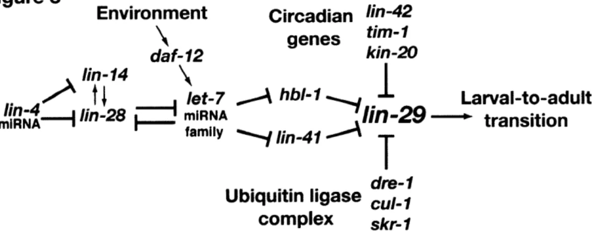
LIN-29 is the master regulator of the larval-to-adult transition
LIN-29 encodes a Kruppel type C2H2 zinc finger transcription factor that acts at several
times during development but is most notably required for animals to take on adult
characteristics at the end of development'5 ,65.lin-29 mutants were originally isolated from a screen for animals that were egg laying defective66. While these mutants had a very obvious
protruding vulva defect, they also failed to generate an adult-specific cuticle and underwent extra
molts. The seam cells, which normally exit the cell cycle during the L4 stage, fail to do so in a
lin-29 mutant. Based on these observations it was concluded that lin-29 mutants were defective
mutants placed lin-29 as the furthest downstream component of the pathway. Subsequent work
has demonstrated that the timing of LIN-29 activity is regulated by many different sets of genes.
Multiple pathways control the timing of LIN-29 activity
Circadian rhythm components control LIN-29 activity in C. elegans
Many organisms undergo physical or behavioral changes that occur in a 24-hour cycle.
These traits or behaviors are said to act with a circadian rhythm. The first observations of
67
circadian rhythms came from studies of the plant Mimosa pudica (Sensitive plant) in the 1700s
The leaves of Mimosa pudica undergo changes in their orientation in coordination with the
light-dark cycle. However, it was observed that these plants successfully changed their leaf orientation
even in complete darkness, indicating that while the change in orientation coincided with the
light/dark cycle, the light-dark cycle was not required.
lin-42 encodes the only C. elegans member of the PERIOD (PER) family of circadian
rhythm proteins68. In several organisms, including flies and humans, PER proteins control
circadian rhythms. Mutations that disrupt period homologs disrupt period length or cause
arrhythmia with respect to circadian behaviors. For example, Drosophila has two peaks of
increased locomotory behavior within a single circadian cycle; one peak occurs around dawn,
and the second occurs around dusk. per mutants in which the circadian period is shortened still
display two peaks of activity, but the second peak occurs earlier in the day6 9.
While C. elegans has several of the core components that make up the circadian
molecular clock, including lin-42 (per), tim-1 (timeless), and kin-20 (doubletime), circadian
adult-specific circadian locomotory behavior7", but it is not clear if the canonical circadian rhythm
regulators control this behavior. Interestingly, in organisms that display circadian behaviors the
mRNA levels of many of the molecular clock components, including per, rise and fall with the
circadian cycle72 73. In C. elegans, where there do not appear to be obvious circadian behaviors,
the levels of lin-42, tim-1 and kin-20 rise and fall with the molting cycle. It was recently
proposed that the period just prior to molting, lethargus, represents a sleep-like state in C.
elegans and that the oscillations observed in lin-42, tim-1 and kin-20 mRNA levels might be the
correlate of the oscillations of mammalian clock mRNA levels during the light-dark cycle.
These observations suggest that in C. elegans the components of the molecular clock might play
a central role in driving the timers that control the molting cycle and the progression from one
developmental stage to the next.
In accordance with this hypothesis, lin-42 mutants have developmental timing defects. In
lin-42 loss-of-function animals, seam cell development appears normal from the LI to the L3
stage, but during the L3 molt, the seam cells precociously exit the cell cycle, fuse, and generate
adult cuticle7 5. lin-42, like lin-41 and hbl-1, acts to prevent the precocious activity of LIN-29.
A Cullin-RING complex prevents precocious LIN-29 activity
Studies of daf-12 mutants, particularly with respect to gonad migration, suggested that
there were likely factors that functioned in parallel to daf-12 to control developmental timing. A
screen to identify these parallel factors yielded dre-] (Daf- 12 Redundant)76. dre-1 encodes an F
box protein that functions with skr-1 (SKpl Related ubiquitin ligase component) and cul-i
ubiquitin ligases and target specific proteins for degradation. One hypothesis is that the DRE- 1
complex targets LIN-29 for degradation, thus preventing LIN-29 activity prior to the L4 stage.
However, an in vitro interaction between LIN-29 and DRE- 1 has not been observed, suggesting
that DRE-1 might act through another target protein. While dre-] acts as a negative regulator of
lin-29 with respect to the larval-to-adult transition, dre-1 and lin-29 mutants have a highly
penetrant synthetic gonad migration defect. LIN-29 expression had been observed in the distal
tip cells, but no significant gonad migration defect had been observed previously.
LIN-29 activity is controlled directly via HBL- 1 and LIN-4 1, the circadian rhythym
homologs LIN-42, TIM-1, and KIN-20, and an E3 ubiquitin ligase complex comprising DRE- 1,
SKR-1 and CUL-1 (Figure 3). It is not clear if these mechanisms represent one large connected
pathway or if multiple pathways converge to ensure the proper timing of LIN-29 protein
accumulation. Furthermore, it is not clear if these pathways regulate lin-29 transcriptionally or
post-transcriptionally.
LIN-29 likely has multiple target genes
LIN-29 is believed to promote the larval-to-adult transition by acting on many different
target genes. In some cases LIN-29 might act to activate the expression of target genes that
promote adulthood, and in other cases LIN-29 might act to repress genes required for larval
development. The LIN-29-dependent activation and repression of collagen genes is a good
example. During larval development, the C. elegans hypoderm expresses col-17 77. When the
animal transitions to adulthood, col-1 7 is repressed and col-19 is expressed. In lin-29 mutants,
col-19 is never expressed and col-i 7 continues to be expressed in all stages after L477. When
wild-type seam cells permanently exit the cell cycle during the L4 stage, they express an elevated
expression is abolished in lin-29 mutants as the seam cells continue to undergo extra divisions78.
Recently, it was shown that the seam cells fail to fuse during the larval-to-adult transition in
animals that are lacking the fusogen aff-179. Therefore, it seems likely that aff-1 is a downstream
target of LIN-29 with respect to seam cell fusion.
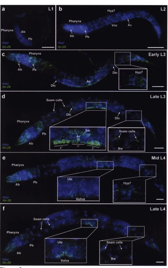
LIN-29 is expressed in multiple cell types throughout development
Expression analysis using an antibody that recognizes the LIN-29 C-terminus revealed
that LIN-29 might also have developmental roles outside of the larval-to-adult transition7 5. For example, during the LI stage, LIN-29 was detected primarily in the pharynx. During the L3
stage, LIN-29 accumulates in the vulval precursor cells, the descendants of the sex myoblasts,
and the distal tip cells. It is not until the L4 stage that LIN-29 is seen in the seam cells and
hypodermis. Interestingly, a lacZ transcriptional reporter driven by the lin-29 promoter showed
expression in the hypoderm as early as the L2 stage. This result might be an artifact of the
promoter region used, or it might suggest that LIN-29 is regulated post-transcriptionally in the
hypoderm.
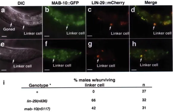
LIN-29 controls male linker cell death
While lin-29 has primarily been studied in the context of the terminal differentiation of
the hypodermis, it is known to function in other tissues and at other times during development. It
is not surprising that LIN-29 controls other processes given its expression pattern. Interestingly,
lin-29 has been shown to function in the gonad of both males and hermaphrodites. In males and
hermaphrodites, the gonad starts as a small collection of somatic and germline cells. Over the
course of development, the gonad elongates, driven by the migration of specific leading cells80'.8 1 In males there is one leading cell, the linker cell, and in hermaphrodites there are two leading
cells, the distal tip cells. The linker cell undergoes a characteristic pattern of migration that
begins at the animal's midbody and ends at the tail near the cloaca. Late in the L4 stage the linker
cell undergoes a non-canonical programmed cell death and is engulfed by a neighboring cell.
The removal of the linker cell fuses the lumen of the vas deferens to the cloacal tube and creates
a passage for the transfer of sperm during mating2,8. LIN-29 appears to be involved in the timing of linker cell death, because in lin-29 deficient animals the linker cell inappropriately
survives well into adulthood84.
The abnormal linker cell death shows that LIN-29 action during the larval-to-adult
transition is not confined to the hypoderm and that different target genes are likely regulated in
different tissues. It is not known if the same mechanisms that control the timing of LIN-29
activity in the hypoderm control the timing of LIN-29 activity in the linker cell.
LIN-29
acts
redundantly with DRE-1 to control distal tip cell migration
In hermaphrodites, gonad elongation is controlled by the migration of the two distal tip
cells80'81. These cells also migrate in a characteristic pattern, each making two specific turns at
distinct points in development. During the L2 and L3 stages, the two arms of the gonad migrate
along the ventral side of the animal from the midbody towards the anterior and posterior ends.
During mid-L3 stage, the distal tip cells change their direction and migrate in a ventral-to-dorsal
direction. Finally, near the beginning of the L4 stage, the distal tip cells change direction once
more and migrate along the dorsal side of the animal back towards the midbody. This process
generates two U-shaped gonad arms at the end of development.
While lin-29 animals do not have obvious gonad migration defects on their own
(although a mild defect has been reported), in combination with a daf-12 null or a dre-1
these doubly deficient animals, distal tip cell migration can be defective at either of the two
gonad turns76. Therefore lin-29 likely acts at multiple times within the distal tip cell to control
gonad migration.
LIN-29 homologs in other organisms
LIN-29 is a Kruppel-type C2H2-zinc finger transcription factor. In C. elegans there are
somewhere between 150 and 200 members of this class of protein. There are over 300
C2H2-zinc finger containing proteins in both Drosophila and humans, in the latter case accounting for
1-2% of the human genome . Members of this group are defined by the presence of a C2H2 zinc
finger motif that is highly conserved across metazoa. While the core sequence within the C2H2
domain is highly conserved, the number of C2H2 domains within an individual protein can vary
dramatically.
In Drosophila, the closest homologs to LIN-29 are Rotund and Squeeze. Rotund is
required for male and female fertility and for the normal development of many adult body
structures8 6. Squeeze acts primarily in the central nervous system and controls the temporal
specification of specific neuroblast lineages87.
In mammals, the closest predicted homologs of LIN-29 based on conservation within the
C2H2 zinc finger domains are CIZ (Castor-Interacting Zinc finger protein) and ZNF362 (ZiNc
Finger protein 362)65. CIZ is thought to be a nucleocytoplasmic shuttling transcription factor
involved in bone growth8 8, and ZNF362 has not been extensively studied. Neither CIZ nor ZNF362 has been implicated in the control of developmental timing or terminal differentiation in
mammals, and it is unclear if they share LIN-29-like function. There is little to no conservation