Publisher’s version / Version de l'éditeur:
Analytical Chemistry, 75, 23, pp. 6708-6717, 2003-12
READ THESE TERMS AND CONDITIONS CAREFULLY BEFORE USING THIS WEBSITE. https://nrc-publications.canada.ca/eng/copyright
Vous avez des questions? Nous pouvons vous aider. Pour communiquer directement avec un auteur, consultez la première page de la revue dans laquelle son article a été publié afin de trouver ses coordonnées. Si vous n’arrivez pas à les repérer, communiquez avec nous à PublicationsArchive-ArchivesPublications@nrc-cnrc.gc.ca.
Questions? Contact the NRC Publications Archive team at
PublicationsArchive-ArchivesPublications@nrc-cnrc.gc.ca. If you wish to email the authors directly, please see the first page of the publication for their contact information.
NRC Publications Archive
Archives des publications du CNRC
This publication could be one of several versions: author’s original, accepted manuscript or the publisher’s version. / La version de cette publication peut être l’une des suivantes : la version prépublication de l’auteur, la version acceptée du manuscrit ou la version de l’éditeur.
For the publisher’s version, please access the DOI link below./ Pour consulter la version de l’éditeur, utilisez le lien DOI ci-dessous.
https://doi.org/10.1021/ac034664d
Access and use of this website and the material on it are subject to the Terms and Conditions set forth at
Multilabeling biomolecules at a single site. 1. Synthesis and
characterization of a dendritic label for electrochemiluminescence
assays
Zhou, Ming; Roovers, Jacques; Robertson, Gilles P.; Grover, Chander P.
https://publications-cnrc.canada.ca/fra/droits
L’accès à ce site Web et l’utilisation de son contenu sont assujettis aux conditions présentées dans le site LISEZ CES CONDITIONS ATTENTIVEMENT AVANT D’UTILISER CE SITE WEB.
NRC Publications Record / Notice d'Archives des publications de CNRC:
https://nrc-publications.canada.ca/eng/view/object/?id=d795fddf-c5ee-4f0a-a89f-f0f12ff122f8
https://publications-cnrc.canada.ca/fra/voir/objet/?id=d795fddf-c5ee-4f0a-a89f-f0f12ff122f8
M ult ila be ling Biom ole c ule s a t a Single Sit e . 1 .
Synt he sis a nd Cha ra c t e riza t ion of a De ndrit ic
La be l for Ele c t roc he m ilum ine sc e nc e Assa ys
Ming Zhou,*
,†Jacques Roovers,
‡Gilles P. Robertson,
‡and Chander P. Grover
†Institute for National Measurement Standards, Institute for Chemical Process and Environmental Technology,
National Research Council of Canada, 1200 Montreal Road, Ottawa, ON K1A 0R6, Canada
Ultrasensitive bioanalytical assays are of great value for early detection of human diseases and pathogens. The sensitivities of immunoassays and DNA probing can be enhanced by multilabeling the biorecognition partner used for affinity-based assays. However, the bioreactivity of biomolecules is affected by a high degree of multilabeling at multiple functional sites. It is proposed that dendritic scaffoldings be used to link multiple signal-generating units to a single site with potentially minimum impact on the bioaffinity. A prototype label, a zeroth-generation dendron, bearing three [Ru(bpy)3]2+ units for
electro-chemiluminescence (ECL) assays was synthesized and characterized preliminarily by spectroscopic, electro-chemical, and ECL methods. No evidence of interaction between the neighboring [Ru(bpy)3]2+units in the label
molecule was found from these characterizations. Both the photoluminescence and ECL of the prototype label have features very similar to those of mononuclear [Ru-(bpy)3]2+compounds. Labeling a model protein with a
triad of [Ru(bpy)3]2+ at one NH2 position was
demon-strated. The results reported here provide support to applying the proposed multilabeling strategy to affinity-based bioanalytical assays.
Affinity-based bioanalytical assays, such as immunoassay and DNA probing, rely largely on the labeling technique, by which signal-generating units are linked to some functional groups of biomolecules that selectively bind to the analytes. For a high signal level in immunoassay, multilabeling at multiple accessible sites (e.g., NH2) of a protein molecule is normally practiced. However, a high degree of multilabeling may result in the loss of biological activity of the biomolecules,1thus high nonspecific binding of the antianalytes and eventually the low signal-to-noise ratio. For some monoclonal antibodies, multilabeling may even lead to the precipitation of proteins.1 One approach to introduce a large number of label molecules at as few sites as possible is to use carrier proteins.2,3However, this approach involves complicated
biochemical processes and the carriers themselves are big in size and mass. Here we show, through the synthesis of a prototype label, a concept of multilabeling biomolecules at a single site (Chart 1) using a dendritic platform bearing a plurality of signal-generating units. It is expected that the single-site multilabeling strategy enhances signal intensity with potentially minimum perturbation of the biobinding affinity of the labeled biomolecules and thus helps reduce nonspecific binding in affinity-based bioanalytical assays.
Use of dendrimer4technology for various signal amplification processes in genomic research and clinical diagnostics has been demonstrated by several authors5-9and was reviewed recently.10 In our proof-of-concept research, a zeroth-generation dendron, succinimidyl-functionalized trinuclear ruthenium(II)
tris(bipyri-* Corresponding author. E-mail: ming.zhou@ nrc-cnrc.gc.ca. †Institute for National Measurement Standards.
‡Institute for Chemical Process and Environmental Technology.
(1) ORIGEN TAG-NHS ester: A simple labeling procedure for proteins; Technical Sheet from IGEN International, Inc., 1997.
(2) Diamandis, E. P. Clin. Chem. 1 9 9 1 , 37/ 9, 1486-1491.
(3) (a) Scorilas, A.; Diamandis, E. P. Clin. Biochem. 2 0 0 0 , 33, 345-350. (b) Qin, Q.-P.; Lo¨vgren, T.; Pettersson, K. Anal. Chem. 2 0 0 1 , 73, 1521-1529. (4) (a) Tomalia, D. A.; Naylor, A. M.; Goddard, W. A., III. Angew. Chem., Int.
Ed. Engl.1 9 9 0, 29, 138-175. (b) Grayson, S. M.; Fre´chet, J. M. J. Chem.
Rev.2 0 0 1, 101, 3819-3867.
(5) Wang, J.; M. Jiang, M. J. Am. Chem. Soc. 1 9 9 8 , 120, 8281-8282. (6) Bryant, L. H., Jr.; Brechbiel, M. W.; Wu, C.; Bulte, J. W. M.; Herynek, V.;
Frank, J. A. J. Magn. Reson. Imaging 1 9 9 9 , 9, 348-352.
(7) Stears, R. L.; Getts, R. C.; Gullans, S. R. Physiol. Genomics 2 0 0 0 , 3, 93-99. (8) Ong, K. K.; Jenkins, A. L.; Cheng, R.; Tomalia, D. A.; Durst, H. D.; Jensen, J. L.; Emanuel, P. A.; Swim, C. R.; Yin, R. Anal. Chim. Acta 2 0 0 1 , 444, 143-148.
(9) Yordanov, A. T.; Lodder, A. L.; Woller, E. K.; Cloninger, M. J.; Patronas, N.; Milenic, D.; Brechbiel, M. W. Nano Lett. 2 0 0 2 , 2, 595-599.
(10) Stiriba, S.-E.; Frey, H.; Haag, R. Angew. Chem., Int. Ed. 2 0 0 2 , 41, 1329
-1334.
Cha rt 1 . M ult ila be ling a Biom ole c ule a t a Single Sit e w it h a De ndrit ic La be l Be a ring M ult iple Signa l-Ge ne ra t ing U nit s
Anal. Chem.2003,75,6708-6717
6708 Analytical Chemistry, Vol. 75, No. 23, December 1, 2003 10.1021/ac034664d CCC: $25.00 Published 2003 Am. Chem. Soc. Published on Web 10/10/2003
dine) complex (4 in Scheme 1), was synthesized. The reason for choosing [Ru(bpy)3]2+as a signal-generating unit of the prototype multilabeling reagent is 3-fold. First, [Ru(bpy)3]2+derivatives are used not only as valuable probes11-14in biological systems such as nucleic acids, peptides, and proteins but also as labels for
various bioanalytical assays such as fluorescence polarization15 and electrochemiluminescence (ECL) immunoassays and DNA
(11) Peek, B. M.; Ross, G. T.; Edwards, S. W.; Meyer, G. J.; Meyer, T. J.; Erickson, B. W. Int. J. Pept. Protein Res. 1 9 9 1 , 38, 114-123.
(12) Terpetschnig, E.; Szmacinski, H.; Malak, H.; Lakowicz, J. R. Biophys. J. 1 9 9 5 ,
68, 342-350.
(13) (a) Durharm, B.; Millett, F. J. Chem. Educ. 1 9 9 7 , 74, 636-640. (b) Erkkila, K. E.; Odom, D. T.; Barton, J. K. Chem. Rev. 1 9 9 9 , 99, 2777-2795. (14) Shreder, K.; Harriman, A.; Iverson, B. L. J. Am. Chem. Soc. 1 9 9 6 , 118,
3192-3201.
probing.16,17 In particular, [Ru(bpy)
3]2+-based ECL has been developed into a mature, highly sensitive bioanalytical technology widely employed in basic research laboratories, pharmaceutical industry, clinical settings, and homeland security.16-20For an ECL immunoassay, typically, [4-(N-succimidyloxycarbonylpropyl)-4′ -methyl-2,2′-bipyridine] bis-(2,2′-bipyridine)ruthenium (II) dihexa-fluorophosphate16a serves as a protein labeling reagent, which, upon electrochemical oxidation and follow-up chemical reduction by the deprotonated tripropylamine radical, generates lumines-cence through its metal-to-ligand charge-transfer (MLCT) excited state.21-24The trinuclear prototype label with similar [Ru(bpy)
3]2+ units and a succinimidyl linking group provides a direct access to the well-formulated assays developed for the commercially available instruments. Second, [Ru(bpy)3]2+, its derivatives, and its other N-N chelating analogues have played a central role in areas of photochemistry, photophysics, photoelectrochemistry, and photoinduced electron and energy transfer for the last two decades.25,26Their multiple, reversible redox states and long-lived photoemission are very useful for us to characterize the prototype assembly and compare it with the available database of the [Ru(bpy)3]2+family. Third, in addition to ECL, the electrochemical and photoluminescent signals can be utilized as supplementary data to evaluate the multilabeling effect in our research even though these signals are not currently exploited for practical assays.
Although previous studies have demonstrated that, unlike some di- and polynuclear systems with electronically delocalized bridging ligands,26,27 the appended [Ru(bpy)
3]2+ units at the peripheries of star scaffoldings are noninteracting and retain
their redox and photophysical properties,28,29a concern about the feasibility of the prototype label is still associated with the possible interaction among the very closely packed [Ru(bpy)3]2+ units. In this preliminary account, we report the synthesis, characterization, and ECL of the prototype label and demonstrate multilabeling a model protein with the prototype label at a single NH2position.
EXPERIMENTAL SECTION
Chemicals.For synthesis, reagent grade solvents and reac-tants were used as received unless otherwise specified. For characterization, Ru(bpy)3Cl2‚6H2O (Aldrich), tetrabutylammo-nium hexafluorophosphate (TBAPF6, Fluka, electrochemical grade), tri-n-propylamine (TPA, 99+%, Aldrich), bovine serum albumin (BSA, lyophilized powder, Sigma), anhydrous acetonitrile (Ald-rich), phosphate buffered saline (PBS, in the form of tablet for preparing solution of pH 7.4, Sigma), and deionized water (18 MΩ) were used as received.
Synthesis. Synthesis of 4 -Chloro-2 ,2′-bipyridine and cis-Ru(bpy)2Cl2‚2 H2O.The procedures were described previously.29
Synthesis of 1 . A 7.5-g (5.51×10⁻²mol) aliquot of penta-erythritol and 3 g of KOH were stirred in 15 mL of DMSO for 15 min. A 1.5-g (5.66×10⁻³mol) aliquot of 11-bromoundecanoic acid was dissolved in 5 mL of DMSO and added in eight portions to the pentaerythritol/ KOH mixture in a period of 8 h (1 portion/ h). The reaction mixture was kept stirred under argon at room temperature for 14 h (total 22 h). The oil-like liquid was poured into 150 mL of water, and the solution was acidified with 1 N HCl to pH 1-2. The precipitate was filtered, washed, and dried to afford 1.38 g of white powder (yield 76%): 1H NMR (400 MHz, acetone-d6)δ10.4 (b, 1 H), 3.62 (s, 6 H, 3 CH2O), 3.46 (s, 2 H, CH2O), 3.40 (t, 2 H, OCH2), 2.28 (t, 2 H, CH2), 1.59 (q, 2 H, CH2), 1.54 (q, 2 H, CH2), 1.32 (b, 12 H, 6 CH2).
Synthesis of 2 .A 0.303-g (9.45×10⁻⁴mol) aliquot of 1 and 2 g of KOH were stirred in 10 mL of DMSO for 10 min. A 0.645-g (3.38×10⁻³mol) aliquot of 4-chloro-2,2′-bipyridine was added. The reaction mixture was kept stirred under argon at 50°C for 22 h. After reaction, the mixture was poured into 30 mL of water. Extraction with 100 mL of CH2Cl2 was tried when the solution was highly alkaline, but it was found difficult to separate the two phases. After evaporation of CH2Cl2, the oil was purified by chromatography (silica gel treated with 20% triethylamine in hexane, elution 5-10% methanol in CH2Cl2, and pure methanol) and vacuum-dried to afford a sticky transparent product. This was dissolved in methanol and precipitated in acidified water to afford 52 mg of white powder. The remaining water phase was adjusted to pH 8 with NH3‚H2O. The solution was further extracted with CH2Cl2until no more bipyridine derivatives could be detected by TLC. After evaporation of CH2Cl2, the oil was purified by chro-matography (silica gel treated with 20% triethylamine in hexane, elution 5-10%methanol in CH2Cl2, and pure methanol), vacuum-dried, and precipitated in acidified water to afford 223 mg of product.
(15) Terpetschnig, E.; Szmacinski, H.; Lakowicz, J. R. Anal. Biochem. 1 9 9 5 , 227, 140-147.
(16) (a) Blackburn, G. F.; Shah, H. P.; Kenten, J. H.; Leland, J.; Kamin, R. A.; Link, J.; Peterman, J.; Powell, M. J.; Shah, A.; Talley, D. B.; Tyagi, S. K.; Wilkins, E.; Wu, T.-G.; Massey, R. J. Clin. Chem. 1 9 9 1 , 37/ 9, 1534-1539. (b) Kenten, J. H.; Casadei, J.; Link, J.; Lupold, S.; Willey, J.; Powell, M.; Rees, A.; Massey, R. J. Clin. Chem. 1 9 9 1 , 37/ 9, 1626-1632. (c) Deaver, D. R. Nature 1 9 9 5 , 377, 758-760.
(17) (a) Luppa, P. B.; Reutemann, S.; Huber, U.; Hoermann, R.; Poertl, S.; Kraiss, S.; von Bu¨ low, S.; Neumeier, D. Clin. Chem. Lab. Med. 1 9 9 8 , 36, 789-796. (b) Erler, K. Wien. Klin. Wochenschr. 1 9 9 8 , 110, 5-10. (c) Namba, Y.; Usami, M.; Suzuki, O. Anal. Sci. 1 9 9 9 , 15, 1087-1093.
(18) Jameison, F.; Sanchez, R. I.; Dong, L.; Leland, J. K.; Yost, D.; Martin, M. T.
Anal. Chem.1 9 9 6, 68, 1298-1302.
(19) Bruno, J. G.; Kiel, J. L. Biosens. Bioelectron. 1 9 9 9 , 14, 457-464. (20) Yu, H.; Raymonda, J. W.; McMahon, T. M.; Campagnari, A. A. Biosens.
Bioelectron.2 0 0 0, 14, 829-840.
(21) Leland, J. K.; Powell, M. J. J. Electrochem. Soc. 1 9 9 0 , 137, 3127-3131. (22) (a) Zu, Y.; Bard, A. J. Anal. Chem. 2 0 0 0 , 72, 3223-3232. (b) Kanoufi, F.;
Zu, Y.; Bard, A. J. J. Phys. Chem. B 2 0 0 1 , 105, 210-216.
(23) Miao, W.; Choi, J. P.; Bard, A. J. J. Am. Chem. Soc. 2 0 0 2 , 124, 14478
-14485.
(24) Gross, E. M.; Pastore, P.; Wightman, R. W. J. Phys. Chem. B 2 0 0 1 , 105, 8732-8738.
(25) Meyer, T. J. Acc. Chem. Res. 1 9 8 9 , 22, 163-170.
(26) (a) Juris, A.; Balzani, V.; Barigelletti, F.; Campagna, S.; Belser, P.; von Zelewsky, A. Coord. Chem. Rev. 1 9 8 8 , 84, 85-277. (b) Balzani, V.; Juris, A.; Venturi, M. Chem. Rev. 1 9 9 6 , 96, 759-833.
(27) (a) Schmehl, R. H.; Auerbach, R. A.; Wacholtz, W. F. J. Phys. Chem. 1 9 8 8 ,
92, 6202-6206. (b) Baba, A. I.; Ensley, H. E.; Schmehl, R. H. Inorg. Chem.
1 9 9 5, 34, 1198-1207. (c) Liang, Y. Y.; Baba, A. I.; Kim, W. Y.; Atherton, S. J.; Schmehl, R. H. J. Phys. Chem. 1 9 9 6 , 100, 18408-18414. (d) Richter, M. M.; Bard, A. J.; Kim, W.; Schmehl, R. H. Anal. Chem. 1 9 9 8 , 70, 310-318. (e) Boyde, S.; Strouse, G. F.; Jones, W. E.; Meyer, T. J. J. Am. Chem. Soc.
1 9 9 0, 112, 7395-7396. (f) Strouse, G. F.; Schoonover, J. R.; Duesing, R.; Boyde, S.; Jones, W. E.; Meyer, T. J. Inorg. Chem. 1 9 9 5 , 34, 473-487. (g) Treadway, J. A.; Loeb, B.; Lopez, R.; Anderson, P. A.; Keene, F. R.; Meyer, T. J. Inorg. Chem. 1 9 9 6 , 35, 2242-2246.
(28) (a) Lehn, J.-M.; Ziessel, R. J. Chem. Soc., Chem. Commun. 1 9 8 7 , 1292
-1294. (b) Marvaud, V.; Astruc, D. Chem. Commun. 1 9 9 7 , 773-774. (c) Marvaud, V.; Astruc, D.; Leize, E.; Dorsselaer, A. V.; Guittard, J.; Blais, J.-C. New J. Chem. 1 9 9 7 , 21, 1309-1319. (d) Storrier, G. D.; Takada, K.; Abrun˜ a, H. D. Langmuir 1 9 9 9 , 15, 872-884.
(29) Zhou, M.; Roovers, J. Macromolecules 2 0 0 1 , 34, 244-252.
The yield for the combined product is 37%: 1H NMR (400 MHz, CDCl3)δ8.63 (d, 3 H), 8.45 (d, 3 H), 8.32 (d, 3 H), 7.4-8.2 (b, 4 H, NH4), 7.97 (d, 3 H), 7.76 (t, 3 H), 7.26 (t, 3 H), 6.84 (dd, 3 H), 4.39 (s, 6 H, 3 CH2O), 3.72 (s, 2 H, CH2O), 3.38 (t, 2 H, OCH2), 2.20 (t, 2 H, CH2), 1.53 (q, 2 H, CH2), 1.45 (q, 2 H, CH2), 1.0-1.2 (b, 12 H, 6 CH2).
Synthesis of 3 . 2 (0.102 g, 1.275× 10⁻⁴mol) and cis-Ru-(bpy)2Cl2‚2H2O (0.252 g, 4.843×10⁻⁴mol) were mixed with 10 mL of methanol and 3 mL of water and refluxed under nitrogen for 24 h. After being cooled to room temperature, the solution was rotoevaporated. The remaining solid was dissolved in 10 mL of water and filtered to remove unreactedcis-Ru(bpy)2Cl2. The filtrate was rotoevaporated and redissolved in 20 mL of water. Three drops of concentrated HCl were added, and the solution was left overnight. The water was rotoevaporated, and the acidification process was repeated with three drops of concen-trated HCl in 5 mL of water. The solution was again filtered, rotoevaporated, and dried to afford 0.262 g of dark brown solid 3-Cl (yield 92%).
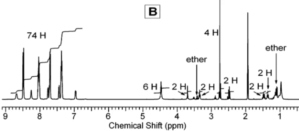
The remaining small amount of unreactedcis-Ru(bpy)2Cl2was further washed out by CH2Cl2. 3 -PF6was prepared by adding a large excess of saturated NH4PF6/ water solution to 3 -Cl water solution. The orange precipitate was filtered, washed with water, and dried. The dried solid was redissolved in acetonitrile and treated with 60% HPF6 aqueous solution (Caution. HPF6 is corrosive! Incompatible with glass! Avoid breathing fumes!) and then precipitated in dry diethyl ether. After centrifugal separation and vacuum-drying, very pure 3 -PF6was obtained: 1H NMR (400 MHz, acetonitrile-d3)δ8.59 (d, 3 H), 8.49 (d, 12 H), 8.16 (s, 3 H), 8.04 (m, 15 H), 7.77 (d, 3 H), 7.72 (m, 12 H), 7.46 (d, 3 H), 7.38 (m, 15 H), 6.98 (d, 3 H), 4.46 (s, 6 H, 3 CH2O), 3.71 (s, 2 H, CH2O), 3.35 (t, 2 H, OCH2), 2.13 (t, 2 H, CH2), 1.35 (m, 4 H, 2 CH2), 0.95-1.15 (b m, 12 H, 6 CH2).
Pure 3 -Cl was prepared by replacing PF6- with Cl-. The preparation was carried out by adding an excess of tetrabutylam-monium chloride saturated in acetone to the acetone solution of 3-PF6, followed by acidification with hydrochloric acid, filtration, and vacuum-drying: 1H NMR (400 MHz, acetonitrile-d
3) δ9.19 (d, 3 H), 8.80 (m, 3 H), 8.62 (d m, 12 H), 8.05 (m, 15 H), 7.78 (m, 3 H), 7.70 (m, 12 H), 7.45 (d, 3 H), 7.38 (m 15 H), 7.05 (d, 3 H), 4.62 (s, 6 H, 3 CH2O), 3.71 (s, 2 H, CH2O), 3.40 (t, 2 H, OCH2), 2.19 (t, 2 H, CH2), 1.34 (m, 2 H, CH2), 1.28 (m, 2 H, CH2), 0.90 -1.10 (b m, 12 H, 6 CH2);1H NMR (400 MHz, D2O)δ8.52 (m, 15 H), 8.25 (m, 3 H), 7.98 (m, 15 H), 7.53-7.80 (m, 18 H), 7.15-7.40 (m, 15 H), 7.06 (m, 3 H), 4.50 (m, 6 H, 3 CH2O), 3.70 (m, 2 H, CH2O), 3.39 (t, 2 H, OCH2), 1.90 (t, 2 H, CH2), 1.29 (b, 2 H, CH2), 0.82 (b, 4 H, 2 CH2), 0.71 (b, 2 H, CH2), 0.52 (b, 4 H, 2 CH2), 0.38 (b, 2 H, CH2), 0.23 (b, 2 H, CH2). Synthesis of 4 -PF6. N,N-Dicyclohexylcarbodiimide (DCC,
2.31 mg, 1.10×10⁻⁵mol) andN-hydroxysuccinimide (NHS, 1.36 mg, 1.15×10⁻⁵mol) were mixed with 3 -PF6(16.1 mg, 5.56× 10⁻⁶mol) in 0.4 mL of acetonitrile and stirred overnight at room temperature. The reaction mixture was injected into 10 mL of dry diethyl ether through a 0.2-µm syringe filter. The orange precipi-tate was collected by centrifuging and vacuum-dried to afford 11.2 mg of product (yield 67%): 1H NMR (400 MHz, acetonitrile-d
3)δ 8.68 (d, 3 H), 8.48 (d, 12 H), 8.25 (s, 3 H), 8.04 (m, 15 H), 7.77 (d, 3 H), 7.72 (m, 12 H), 7.45 (d, 3 H), 7.37 (m, 15 H), 6.96 (d, 3 H),
4.47 (s, 6 H, 3 CH2O), 3.70 (s, 2 H, CH2O), 3.35 (t, 2 H, OCH2), 2.76 (s, 4 H), 2.48 (t, 2 H, CH2), 1.46 (q, 2 H, CH2), 1.35 (q, 2H CH2), 0.9-1.2 (b m, 12 H, 6 CH2).
Synthesis of 5 . Pentaerythritol (0.07 g, 5.2×10⁻⁴mol) and 1 g of KOH were stirred in 5 mL of freshly dried DMSO. The mixture was evacuated for 10 min and saturated with argon. 4-Chloro-2, 2′-bipyridine (0.49 g, 2.57×10⁻³mol, 1.24 equiv/ OH) was added, and the heterogeneous mixture was stirred at 40°C for 60 h. The resulting suspension was poured into 50 mL of water and neutralized with 1 N HCl. Filtering, washing with pentane, and vacuum-drying afford 0.33 g of white powder 5 (84% yield): 1H NMR (400 MHz, CDCl
3)δ8.66 (d, 4 H), 8.50 (d, 4 H), 8.46 (d, 4 H), 8.07 (d, 4 H), 7.83 (t, 4 H), 7.33 (m, 4 H), 6.92 (m, 4 H), 4.61 (s, 8 H, CH2O).
Synthesis of 6 . 5 (0.11 g, 1.46 × 10⁻⁴ mol) and cis-Ru-(bpy)2Cl2‚2H2O (0.413 g, 7.94×10⁻⁴mol) were refluxed in 20 mL of absolute ethanol for 44 h. After being cooled to room temperature the solution was filtered. The filtrate was evaporated and redissolved in a small amount of ethanol. The ethanol solution was dropped into dry ether. Centrifuging and drying at 58°C for 6 h afforded 0.377 g of product 6 -Cl (95% yield): 1H NMR (400 MHz, methanol-d4)δ8.86 (d, 4 H), 8.70 (m, 16 H), 8.51 (d, 4 H), 8.11 (m, 20 H), 7.88 (d, 4 H), 7.79 (m, 16 H), 7.54 (d, 4 H), 7.47 (m, 20 H), 7.24 (dd, 4H), 4.77 (s, 8 H, CH2O).
6-PF6was prepared by adding a large excess of saturated NH4 -PF6-water solution to 3 -Cl water solution. The orange precipitate was filtered, washed with water and vacuum-dried at 58 °C overnight.1H NMR (400 MHz, acetone-d
6)δ8.78 (m, 16 H), 8.68 (d, 4 H), 8.32 (d, 4 H), 8.18 (m, 16 H), 7.96-8.1 (m, 24 H), 7.71 (d, 4 H), 7.54 (m, 20 H), 7.13 (dd, 4 H), 4.80 (s, 8 H, CH2O).
Labeling of Protein. 4-PF6was dissolved in anhydrous DMF (Caution. DMF is toxic and flammable!) and then mixed with BSA in a PBS with a pH of 7.4 at room temperature for 4 h under stirring. The resulting mixture was then transferred to a dialysis tube with cutoff molecular weight of 10 000 for dialysis against PBS solution. After 3 days of dialysis at 277 K, the attachment of the ruthenium assembly to BSA, via a stable amide linkage, was readily visualized by the remaining color in the dialysis tube and further evidenced by the strong orange fluorescence of BSA solution under UV. A comparison dialysis experiment with only 4-PF6in PBS showed that there were essentially no ruthenium compounds left in the dialysis tube after 3 days of dialysis against PBS.
Characterization.1H NMR spectra were obtained on a Varian Inova (400 MHz) spectrometer. UV-visible spectra were recorded on a Hewlett-Packard 8453 spectrophotometer. Emission spectra were obtained with a Fluorolog spectrofluorometer. Both absorp-tion and emission measurements were carried out in argon-saturated acetonitrile or PBS solutions in a quartz cell with a 1-cm optical path at room temperature. Cyclic voltammetry was per-formed in a three-electrode configuration in a specially designed electrochemical cell.30A Pt disk (diameter 1 mm, area 0.785 mm2) sealed in a soft glass rod was employed as the working electrode. It was polished with diamond polishing paste (0.25µm), rinsed thoroughly with acetone and acetonitrile, and dried by a warm air flow. Pt and Ag wires were used as counter and quasi-reference
(30) (a) Kiesele, H. Anal. Chem. 1 9 8 1 , 53, 1952-1954. (b) Zhou, M.; Heinze, J.
electrodes, respectively. Potentials versus the Ag quasi-reference electrode were then calibrated with the ferrocene/ ferrocenium (Fc/ Fc+) redox couple by taking E°
Fc/ Fc+)0.35 V versus Ag/ AgCl (4 M KCl saturated with AgCl). A Parstat 2263-2 Advanced Electrochemical System with PowerSUITE software was used for electrochemical measurement and control. For ECL measurement, a pair of parallel platinum foils (6×24 mm) and Ag/ AgCl were employed as working, counter, and reference electrodes in a quartz cell with the working electrode facing the photomultiplier tube of the Fluorolog spectrofluorometer. The TPA-saturated PBS solution for ECL measurement was prepared by adding TPA (nominal concentration 0.2 M) to PBS (pH 7.4) followed by vigorous shaking. The resulting solution has a pH value of∼9.0. Matrix-assisted laser desorption/ ionization time-of-flight (MALDI-TOF) mass spectra were recorded on a Perseptive Biochemistry Voyager-DE STR MALDI spectrometer operated in linear mode using sinapinic acid as the matrix.
RESULTS AND DISCUSSION
Synthesis.In the past, we attempted to introduce a free arm in a carbosilane dendrimer system in order to build a bioconju-gatable assembly with multiple [Ru(bpy)3]2+at the periphery of a second-generation dendritic scaffolding.29This was unsuccessful due to multiple protective steps required by the synthesis procedures. The present simpler supramolecular assembly with three [Ru(bpy)3]2+units (4 in Scheme 1) is based on a penta-erythritol platform.31First, a symmetric, nonbioconjugatable tetra-nuclear [Ru(bpy)3]2+assembly 6 was synthesized following the procedure described by Astruc et al.28b,c,29The succinimidyl group introduced in the trinuclear assembly 4 is capable of linking the three luminophores to NH2 sites of proteins through peptide formation under mild conditions. The long C11 spacer arm is designed for alleviating steric hindrance between the bulky molecule and protein molecules.
1 was prepared by the reaction of 11-bromoundecanoic acid with a large excess of pentaerythritol in DMSO in the presence of KOH, followed by precipitation in water under acidic conditions. The structure of 1 was confirmed by1H NMR, which shows the characteristic features of the two singlets of pentaerythritol-derived methylene groups in a ratio of 3:1 atδ3.62 and 3.46 ppm in CDCl3. These structural features remain in compounds 2-4 and are detected in their NMR spectra. The ligand 2 and trinuclear assembly 3 were synthesized by the same approaches as for 5 and 6 , respectively. However, due to the amphipathy of 2 , which possesses both basic and acidic groups, the separation of 2 from the reaction mixture was difficult. By adjusting pH, we were able to extract and chromatographically purify 2 with a yield of 37% (compared to 84% yield of 5 ). Since the cationic assembly of 3 was synthesized in a methanol/ water mixture, care had to be taken to ensure that the carboxylic acid group of 3 remains after the formation of metal complex. Hydrochloric acid was added to the aqueous solution of the chloride form (3 -Cl). For the same reason, acidification of 3 -PF6, with hexafluorophosphoric acid (Caution. HPF6 is corrosive! Incompatible with glass! Avoid breathing fumes!) was carried out after 3 -PF6was precipitated by ammonium hexafluorophosphate from the aqueous solution of 3 -Cl.
1H NMR of 3 -PF
6in deuterated acetonitrile is displayed in Figure 1A. The integration and position of aromatic and aliphatic peaks match the structure perfectly. The assignment of the individual aromatic peaks is difficult due to the existence of diastereoisomers as a result of the three chiral octahedral ruthenium centers.32
The quantitative high-yield conversion of the carboxylic acid group into an NHS ester is usually performed in DMF in the presence of DCC at low temperatures. We have found that this conditionsin DMF, at 0°C for more than 20 hsresulted in yields lower than 75%according to the NMR spectrum analysis. Because of the difficulty of separating 4 from 3 , a higher conversion rate is required so that further characterization and application can be unambiguously carried out.
The incomplete conversion of the carboxylic acid group into the NHS ester is due probably to the bulky [Ru(bpy)3]2+triad, which limits the mobility of the reactive carboxyl group at a lower temperature. To gain a better understanding of the reaction, we performed a kinetic study of the esterification at room temperature in acetonitrile-d3by monitoring NMR spectral changes in situ. As can be seen in the NMR spectra provided in the Supporting Information, shifts of two peaks are significant during the reaction, i.e., the singlet peak of CH2CH2in the succinimidyl group shifts from 2.53 (NHS) to 2.76 ppm (ester) and the triplet peak of CH2 adjacent to the carboxyl group shifts from 2.13 (acid) to 2.48 ppm (ester). The NMR kinetic study demonstrated that the reaction started immediately after mixing 3 -PF6with NHS and DCC (both in excess). In∼95 min, half of 3 -PF6was converted into 4 -PF6, and in 5 h,∼80% of 3 -PF6was converted. After 20 h, complete conversion was evidenced by the disappearance of the triplet at 2.13 ppm and by the equal split of the multiplet at 1.35 ppm (see Figure 1B for the NMR of the isolated 4 -PF6). To isolate product 4-PF6, we dropped the reaction mixture through a 0.2-µm syringe filter into anhydrous diethyl ether. The urea byproduct was filtered out and the unreacted DCC and NHS dissolved in ether, while 4-PF6 precipitated in ether and was separated by centrifuging. Although some small peaks of impurities can still be found in the NMR spectrum of the isolated 4 -PF6shown in Figure 1B, these impurity peaks are easily traceable, as they can be found also in the NMR spectra of the kinetic study. As a result of the precipitation in diethyl ether, a small amount of ether, as indicated in Figure 1B, remains in the precipitated solid even after 2 days of vacuum-drying at room temperature.29
Absorption and Emission.The absorption spectra of 4 -PF6 and Ru(bpy)3(PF6)2in acetonitrile are compared in Figure 2. Like the nonbioconjugatable dendrimer analogues, such as hexa- and octanuclear [Ru(bpy)3]2+assemblies with spherical architecture,28c,29 a strong MLCT absorption band was found. The absorption maximum of 4 -PF6is slightly red-shifted, with respect to Ru(bpy)3 -(PF6)2, from 451 to 456 nm and the extinction coefficient based on Ru unit concentration (ǫ456)13 900 M-1cm-1) is slightly lower than that of Ru(bpy)3(PF6)2(ǫ451)14 720 M-1cm-1) but the same as that of the octanuclear [Ru(bpy)3]2+dendritic supramolecule.29 As confirmed previously in the octanuclear [Ru(bpy)3]2+dendritic systems, the shifts of spectra are due to the tuning effect of the substitution of the bipyridine ligands at the 4-position. The emission spectrum, with a maximum also slightly red-shifted to
(31) Constable, E. C.; Housecroft, C. E.; Cattalini, M.; Phillips, D. New J. Chem.
1 9 9 8, 193-200. (32) Keene, F. R. Chem. Soc. Rev. 1 9 9 8 , 27, 185-193.
λmax)613 nm from 605 nm for Ru(bpy)3(PF6)2, is also shown in Figure 2. The intensity at 613 nm is∼75%of the peak intensity of Ru(bpy)3(PF6)2. This value is the same as those obtained from the octanuclear [Ru(bpy)3]2+assembly and a model monomer with a similar 4-position substitution reported previously.29Based on the extinction and relative emission intensity, the emission efficiency of the Ru unit in 4 -PF6is therefore close to 90%of that of Ru(bpy)3(PF6)2.33The good wavelength separation between the MLCT adsorption and emission of [Ru(bpy)3]2+prevents largely the occurrence of fluorescence quenching, which is often the case for fluorescent dyes with a small Stokes’ shift. The 613-nm emission lifetime is 870 ns, comparable with the published data,
which are in the range from 850 to 1100 ns26afor Ru(bpy) 3(PF6)2 under various experimental conditions. As a reference compound closely related to the prototype label molecule, 6 -PF6has absorp-tion and emission spectra completely identical to those of 4 -PF6 at the same concentration of Ru unit.
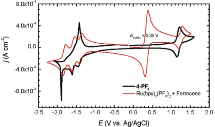
Electrochemistry. The cyclic voltammogram (Figure 3) of 4-PF6recorded in acetonitrile exhibits a fully reversible metal-centered oxidation wave of process [Ru(bpy)3]2+f[Ru(bpy)3]3+ withE1/ 2at 1.185 V (∆Ep)61 mV) and three successive reduction waves with Epcat-1.450,-1.610, and-1.890 V (vs Ag/ AgCl) corresponding to the process of [Ru(bpy)3]2+f[Ru(bpy)3]+, [Ru-(bpy)3]+f[Ru(bpy)3]0, and [Ru(bpy)3]0f[Ru(bpy)3]-, respec-tively. As previously observed in the octanuclear dendritic system, the reduction process of [Ru(bpy)3]0to [Ru(bpy)3]-shows a very sharp peak as a result of the accumulation34of the neutral product on the electrode. After the Ru units undergo the third reduction, the [Ru(bpy)3]-units in the compound are, upon potential scan
(33) For practical ECL-based immunoassay and DNA probing, a derivative of [Ru(bpy)3]2+, such as [4-(N-succimidyloxycarbonylpropyl)-4′-methyl-2,2′ -bipyridine]bis-(2,2′-bipyridine)ruthenium(II) dihexafluorophosphate, is used to label a biomolecule that recognizes the analyte. Since no comparison between Ru(bpy)3(PF6)2and the commercial labels is readily available, the comparative data related to Ru(bpy)3(PF6)2in this paper are meant to provide information only and not intended to give the impression that the prototype label will produce 2.7-fold stronger luminescence in any case. Due to the effect of the substitution in one of the bipyridine rings, meaningful intensity comparison for the absorption and emission can only be made between the [Ru(bpy)3]2+triad and a mononuclear [Ru(bpy)3]2+with an OR group at the 4-position of a bipyridine ring. This has been discussed and done in ref 29.
(34) Flanagan, J. B.; Margel, S.; Bard, A. J.; Anson, F. C. J. Am. Chem. Soc. 1 9 7 8 ,
100, 4248-4253.
Figure 2 . UV-visible absorption and emission spectra (455 nm
excitation) of 4-PF6and Ru(bpy)3(PF6)2recorded in acetonitrile at 293
K. The Ru unit concentration is 40 µM for both compounds.
Figure 1 . 1H NMR in acetonitrile-d
reversal, reoxidized in an overall three-electron process to [Ru-(bpy)3]2+showing a wave with the peak potential at-1.420 V. However, if the potential scan is reversed immediately after the first reduction wave, no electroprecipitation but a normal reoxi-dation peak is observed. If the potential is reversed immediately after the second reduction, a combined two-electron oxidation process leading to [Ru(bpy)3]2+ is observed also at the peak potential of-1.420 V.
The tetranuclear [Ru(bpy)3]2+assembly 6 displays very similar voltammetrical behavior with a small difference when the potential is reversed after the second reduction wave leading to the accumulation of the redox product bearing four neutral [Ru-(bpy)3]0units. Instead of a combined two-electron wave found in the case of 4 -PF6, a sharp peak ([Ru(bpy)3]0f[Ru(bpy)3]+) and a separated normal wave ([Ru(bpy)3]+ f[Ru(bpy)3]2+) can be seen. As an example, the voltammograms of 6 -Cl are shown in Figure 4, in which the redox wave next to the [Ru(bpy)3]2+/ 3+ wave is associated with Cl-/ 0,35 which does not exist in the compounds with PF6-as a counterion.
The redox potentials available are compared in Table 1. These values and the discussed features are very much the same, within experimental error, as those reported for the octanuclear dendritic system.29
The spectroscopic and electrochemical studies of 4 -PF6 re-ported here demonstrate that the independence of [Ru(bpy)3]2+ units, as investigated before with several symmetrical systems,28,29 holds also for the asymmetric assembly with three closely packed [Ru(bpy)3]2+units. This noninteracting nature is the foundation for the single-site multilabeling strategy introduced in the begin-ning of the paper.
Electrochemiluminescence. The ECL of [Ru(bpy)3]2+and its analogues can be generated in both organic27d,29and aqueous16-24 electrolyte solutions in the presence of TPA following the mech-anisms proposed for TPA/ [Ru(bpy)3]2+coreactant systems.21-24 Among polynuclear metal complexes, dinuclear [Ru(bpy)3]2+ systems with electronically delocalized bridging ligands27dand an octanuclear dendritic system29with appended [Ru(bpy)
3]2+units have been reported to be able to generate ECL in TPA/ acetonitrile electrolyte solution. For the sake of utilization in the practical bioanalytical settings, the ECL of the trinuclear assembly 3 -Cl36 and tetranuclear assembly 6 -Cl was first examined by using buffer solution (ProCell, pH 6.8) specially designed for commercial ECL immunoassay systems. The ECL spectra of both 3 -Cl and 6 -Cl were found to be the same as their photoluminescence spectra. However, due to the presence of some undisclosed additives in the commercial ECL assay solutions, for the purpose of primary characterization, we prefer to report here the similar ECL measurement results in TPA saturated PBS solutions.37
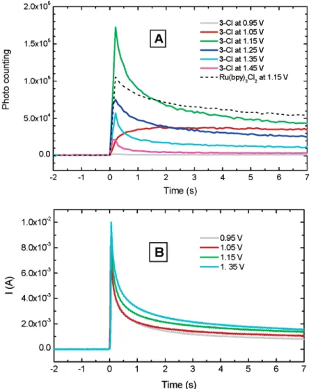
The potential step method was employed to generate the ECL. The typical waveform is set from initial potential-0.05 V for 5 s to a series of predemintermined potentials for 7 s and then to -0.3 V for 5 s. It was found that applying the negative potentials after and before the potential step at the predemintermined potentials regenerates the electrode surface state and keeps it constant from experiment to experiment. The values of-0.05 and -0.3 V were determined from the voltammogram of the solution prepared for ECL (see Supporting Information). Figure 5A demonstrates the ECL intensities as a function of time when different potential steps are applied to the working electrode. In
(35) (a) Tokel, N. E.; Bard, A. J. J. Am. Chem. Soc. 1 9 7 2 , 94, 2862-2863. (b) In our work, the chloride ion wave was further confirmed by the voltammogram of tetrabutylammonium chloride in acetonitrile.
(36) In consideration of both the solubility and the hydrolysis stability of NHS ester, 3 -Cl, instead of 4 -Cl and 4 -PF6, was used for the measurement in this work.
(37) (a) As one reviewer of this paper pointed out, the generation of the ECL from this solutions with pH∼9.0 may also follow a mechanism, in addition to those discussed in ref 23, involving the reduction of [Ru(bpy)3]3+to [Ru-(bpy)3]2+* by hydroxyl ion. See ref 38b, in which [Ru(bpy)3]3+in 9 N sulfuric acid was allowed to react with 9 N NaOH and chemiluminescence was observed. (b) Hercules, D. M.; Lytle, F. E. J. Am. Chem. Soc. 1 9 6 6 , 88, 4745-4746.
Figure 3 . Cyclic voltammogram of 4-PF6(0.40 mM 1.2 mM Ru unit)
in comparison with Ru(bpy)3(PF6)2(1.0 mM, with added ferrocene
as a reference) in acetonitrile at 293 K. Supporting electrolyte, 0.1 M TBAPF6. Scan rate, 100 mV s-1.
Figure 4 . Cyclic voltammograms of 6-Cl (0.2 mM or 0.8 mM Ru
unit) in acetonitrile at 293 K. Supporting electrolyte, 0.1 M TBAPF6.
Scan rate, 50 mV s-1.
T a ble 1 . Re dox Pot e nt ia ls vs Ag/AgCl in Ac e t onit rile a t 2 9 3 Ka
Epc,re
compound E1/ 2,ox first second third
4-PF6 1.185 -1.450 -1.610 -1.890 (0.061) 6-Cl 1.185 -1.395 -1.545 -1.915 (0.051) Ru(bpy)3(PF6)2 1.248 -1.380 -1.570 -1.815 (0.082) (0.058) (0.058) (0.066)
aThe numbers in parentheses are the separation of the peak
potentials (∆Ep) for the completely reversible waves. All the potential values for Ru(bpy)3(PF6)2are taken asE1/ 2.
most cases (E>1.05 V in Figure 5A), the ECL reaches the highest level once the potentials are stepped. It decays with time, due possibly to electrode oxidation38and the depletion of reactants.39 The current responses recorded for the potential step experiments are given in Figure 5B. By comparing Figure 5A and Figure 5B, one can easily see that although both ECL intensities and currents decay with time, the decay patterns are not the same for all the potential steps and an optimal potential (∼ 1.15 V in the experiment of Figure 5A) for a maximum ECL emission exists. At the lowest applied potential (0.95 V in Figure 5A), a slightly higher than background photon counting remains constant over the measurement period. A similar nondecaying behavior was also found for the measurement at 1.05 V. Since the complication of many potential-dependent ECL reaction pathways23,37 and Pt electrode surface change38in aqueous solutions, the time transient profile and the nonmonotonic potential dependence reported here
cannot be thoroughly explained without more comprehensive investigation. It is very likely that different ECL mechanisms23,37 exist at different potentials. However, discussion on these issues is beyond the scope of this paper. The results reported here just confirm the ECL activity of the triad.
Before performing the ECL experiments, it was expected that Ru(bpy)3Cl2might have a higher level of ECL emission than that of 3 -Clswithout considering any other factorssimply because of the presumed higher diffusion coefficient of Ru(bpy)3Cl2due to the size difference. However, after repetitive experiments, it was reproducibly confirmed that there is no significant difference in ECL intensities of 3 -Cl and Ru(bpy)3Cl2at the same concentration of Ru unit, no matter whether the ECL experiments were performed in the TPA-saturated PBS or in the commercial ECL assay buffers with unknown ingredients. As comparison, the ECL of Ru(bpy)3Cl2at a potential of 1. 15 V is given in Figure 5A. Note that the concentration of Ru(bpy)3Cl2 in Figure 5A is slightly higher than the Ru unit concentration of 3 -Cl, and so is the emission intensity. We have to recognize again that the course of generating ECL with TPA involvement is a fairly complicated one and there are many factors governing the cascade of electro-chemical/ chemical reactions and photophysical process,21-24,37
(38) (a) Anson, F. C.; Lingane, J. J. J. Am. Chem. Soc. 1 9 5 7 , 79, 4901-4904. (b) Angerstein-Kozlowska, H.; Conway, B. E.; Sharp, W. B. A. J. Electroanal.
Chem.1 9 7 3, 43, 9-36.
(39) Although [Ru(bpy)3]2+undergoes a circulation, [Ru(bpy)3]2+f[Ru(bpy)3]3+
f[Ru(bpy)3]2+*f[Ru(bpy)3]2+, the coreactant TPA is consumed in a series of irrevervible electrochemical and chemical reactions. For the details of the mechanism, see refs 21-24 and references therein.
Figure 5 . ECL generated by potential step method. (A) Intensity of ECL emission maximum as a function of time and applied potential and
(B) chronoamperometric response of 3-Cl. Solutions: 3-Cl (0.275 mM or 0.825 mM Ru unit) and Ru(bpy)3Cl2(0.865 mM) in TPA saturated PBS
which lead finally to the luminescence. The surfactant-like structure of 3 -Cl may also have some favorable effects40on its ECL.
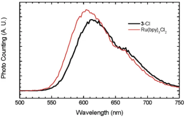
A 5-s scan was required to collect ECL emission spectra from 500 to 800 nm in our experimental setting. Therefore, the ECL spectra were recorded when the ECL intensities became relatively steady, normally 4 s after the potentials were stepped to the predetermined levels (see Figure 5A). As expected, within experimental error, the ECL spectra obtained for 3 -Cl at different electrode potentials have the same features as the photolumines-cence spectra of 4 -PF6in acetonitrile (Figure 2), 3 -Cl, and Ru-labeled BSA in the same PBS solutions (Supporting Information), because the emission is the result of the decay of the same MLCT excited state [Ru(bpy)3]2+*, generated by either photo- or elec-trochemical excitation. In addition, these spectra are also the same as that of 6 with the same four [Ru(bpy)3]2+units. Figure 6 shows the ECL spectrum of 3 -Cl compared with that of Ru(bpy)3Cl2 generated at the potential of 1.15 V.
Labeling of Protein. Protein labeling experiments were carried out by using BSA as a model protein, which is commonly employed as a protein standard in bioanalytical assays and as a molecular weight standard (66 4319) for gel permeation chroma-tography. BSA contains 59 lysines, and 30-35 of these are primary amines capable of reacting with the succinimidyl conjugation group.41It should be noted that the chlorides of 3 , 4 , and 6 are very soluble in water. However, due to the generally possible slow hydrolysis of NHS ester in aqueous solutions, we used 4 -PF6, instead of the water-soluble 4 -Cl, to prepare stock solution for the labeling experiment. Like other hexafluorophosphate salts, 4-PF6is very soluble in polar organic solvents such as acetone, acetonitrile, methanol, DMF, and DMSO, but insoluble in water. The UV-visible absorption of the labeled BSA in PBS solution has the ligand-centered transition absorption at 286 nm and the MLCT absorption at 458 nm, which is slightly red-shifted with respect to its MLCT absorption band in acetonitrile. The average number of [Ru(bpy)3]2+units attached to a BSA molecule was determined by the absorbance peaks at 286 and 458 nm, assuming the extinction coefficients for the free and BSA-bound trinuclear
assemblies are the same. 3 -Cl (extinction coefficients in PBS based on Ru unit areǫ456)11 500 M-1cm-1andǫ286)57 400 M-1cm-1) was used as a reference in PBS. In one labeling experiment with the initial molar ratio of 4 -PF6to BSA set as 5.1:1, we found that on average four triads, i.e., 12 [Ru(bpy)3]2+units were bound to a BSA molecule.
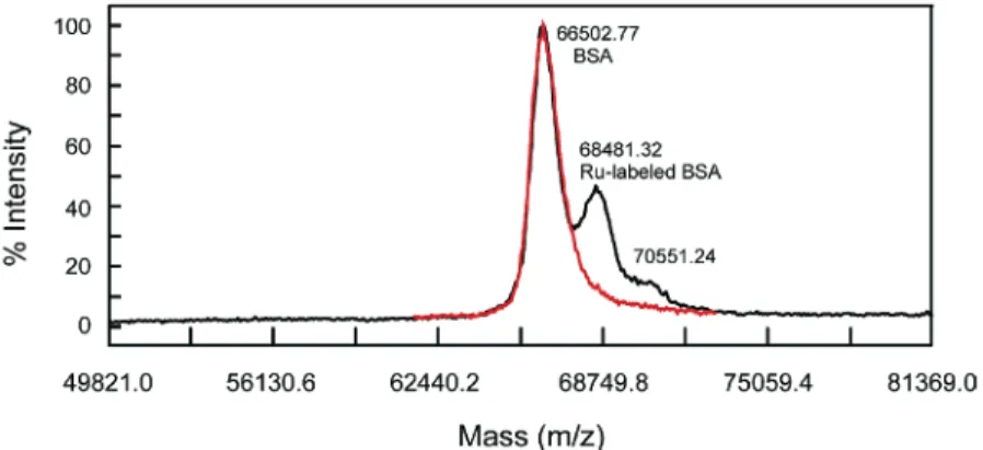
The binding of the prototype label to the BSA and the number of bound [Ru(bpy)3]2+units were further confirmed by MALDI-TOF mass spectrum. The mass spectra in Figure 7 demonstrates the BSA triply labeled with [Ru(bpy)3]2+at a single site. Compared to the measured BSA mass of 66 503 Da, the peak withm/zat 68 481 Da indicates the labeled BSA has a mass increase of∼1978 Da, whichsassuming that all six PF6-moieties were lost during the ionization processsis in excellent agreement with the calcu-lated value of 2005.25 Da within the general mass error of 0.5% for protein MALDI-TOF mass spectra. For the purpose of internal reference in Figure 7, the BSA used for labeling was in excess (the molar ratio of BSA to 4 -PF6was 1.2:1). However, a shoulder at ∼70 551 Da (4048 Da shift from 66 503 Da) is apparent, indicative of a small amount of BSA labeled with two [Ru(bpy)3]2+ triads, i.e., six [Ru(bpy)3]2+units (calculated mass increase 4010.50 Da, assuming 12 PF6-moieties lost). The mass spectrum of the pristine BSA is also exhibited in Figure 7, showing a single peak at 66 503 Da and ruling out any concern about the existence of impurities in the displayed mass scale. The MALDI-TOF mass spectra in Figure 7 represent direct and clear evidence for the successful multilabeling with [Ru(bpy)3]2+triad at a single site of a protein molecule.
ECL was observed also from the Ru-labeled BSA in a TPA/ PBS solution, indicating that the [Ru(bpy)3]2+attached to protein remain ECL active. Although we have not conducted a compara-tive ECL immunoassay with a multireporter label and a similar monoreporter label (see ref 33 for the concern of a meaningful comparison), which will need much more effort, including establishing well-defined labeling procedures for controlled label-ing levels and careful characterization of the labeled antianalytes, we hope that using a dendritic platform bearing multiple signal-generating units for multilabeling biomolecules will enhance signal intensity by increased number of reporters or reduce nonspecific binding by using fewer functional groups of biomolecules while maintaining the same lebeling level.
CONCLUSIONS
For the purpose of multilabeling biomolecules at a single site in bioanalytical science, a dendritic prototype label with three [Ru-(bpy)3]2+linked to a succinimidyl group was synthesized and characterized by structural, photophysical, and electrochemical methods. The confirmed independence of each [Ru(bpy)3]2+unit, the covalent attachment of the trinuclear [Ru(bpy)3]2+assembly to BSA in PBS, and the generation of ECL in tripropylamine-containing aqueous buffer solution substantiate the applicability of the novel multilabeling strategy to the established ECL assays. Although the prototype label is demonstrated only for ECL-based assays in this preliminary work, it is likely applicable to other analytical techniques based on [Ru(bpy)3]2+ labeling such as fluorescence polarization immunoassay and fluorescence lifetime imaging. Moreover, the concept of single-site multilabeling of biologically active substances for enhanced signal with minimum impact on bioreactivities could be generally utilized for other
(40) Workman, S.; Richter, M. M. Anal. Chem. 2 0 0 0 , 72, 5556-5561. (41) Hermanson, G. T. Bioconjugate Techniques; Academic Press: San Diego,
1996; p 423.
Figure 6 . ECL spectra of 3-Cl (0.275 mM or 0.825 mM Ru unit)
and Ru(bpy)3Cl2 (0.865 mM) in TPA-saturated PBS (pH ∼9).
Recorded after the emission became steady at the potential of 1.15 V vs Ag/AgCl.
labeling-dependent technologies, which use radioactive isotopes, fluorescent dyes, or other redox-active or luminescent metal complexes as signal-generating units. The increasing interests in dendritic and supramolecular systems have resulted in many synthetic strategies for assembling more signal-generating units into suitable molecular platforms. Based on the availability of the triad label and the preliminary characterization, further research work is currently under way in our laboratories.
ACKNOWLEDGMENT
We thank the Bioanalysis Group of NRC’s IBS for MALDI-TOF mass spectra, L. Johnston (NRC’s SIMS) for fluorescence lifetime measurement, Y. Tao (NRC’s IMS) for providing access
to the spectrofluorometer, and Y. Li and J. Ding (NRC’s ICPET) for useful discussion on the synthesis.
SUPPORTING INFORMATION AVAILABLE
NMR kinetic data, absorption and emission spectra of other related compounds, and cyclic voltammogram of ECL solution. This material is available free of charge via the Internet at http:/ / pubs.acs.org.
Received for review June 19, 2003. Accepted September 3, 2003.
AC034664D
Figure 7 . MALDI-TOF mass spectra of Ru-labeled BSA mixed with excess BSA (black) and pristine BSA (red). The labeled BSA mixture used