Characterization of Synaptotagmin 7 function in
neurotransmission and its subcellular localization at synapses
By
Mónica C. Quiñones-Frías
B.S. Cellular and Molecular Biology
University of Puerto Rico Río Piedras Campus, 2014
SUBMITTED TO THE DEPARTMENT OF BIOLOGY IN PARTIAL
FULFILLMENT OF THE REQUIREMENTS FOR THE DEGREE OF
DOCTOR OF PHILOSOPHY IN BIOLOGY
AT THE
MASSACHUSETTS INSTITUTE OF TECHNOLOGY
MAY 2020
© 2020 Mónica C. Quiñones-Frías. All Rights Reserved.
The author hereby grants to MIT permission to reproduce and to distribute publicly
paper and electronic copies of this thesis document in whole or in part in any
medium now known or hereafter created.
Signature of Author: __________________________________________________
Department of Biology
May 4, 2020
Certified by: ________________________________________________________
Dr. J. Troy Littleton
Menicon Professor of Biology and The Picower Institute for Learning and Memory
Thesis Supervisor
Accepted by: ________________________________________________________
Dr. Stephen P. Bell Professor
Biology Chairman of the Graduate Committee
Acknowledgements
During my time at MIT I’ve come across many people that have become my lifetime friends and have shaped my experience for the best. First, I want to thank my advisor, Troy, for giving me the opportunity to join his lab and giving me this project. I will forever be indebted to him because he helped me pave the way to achieve my dream of being a scientist. I also want to thank Yulia, without her mentorship every step of the way I would have not been able to do half of the experiments in this thesis. I want to thank Dina for her tireless efforts to keep our lab in the best shape and deeply caring for every single one of us. Dina is the best lab manager in the world! I also want to thank Shirley for being my mentor and friend. To the rest of the lab, I want to thank you for all the good times we’ve had throughout the years.
Above all, I want to thank my family and friends for their love and support because when times were hard they were always there for me. I want to thank my parents and sister for always given me unconditional support. They’ve always believed in my potential and encouraged me to never give up to achieve my dreams. I want to give a special thanks to my friends, Gal and Shaul, for countless hours of backgammon and eating udon. Finally, I want to thank my fiancé Josh. He’s my best friend and I love him with all my heart. After a long day in lab, the best part was that I got to hang out with you.
Characterization of Synaptotagmin 7 function in neurotransmission
and its subcellular localization at synapses
By Mónica C. Quiñones-Frías
Synaptic vesicle (SV) fusion is dependent on proteins that can sense Ca2+ and trigger fusion with
the plasma membrane. Neurotransmitter release occurs during a rapid synchronized phase of SV fusion mediated by the Ca2+ sensor Synaptotagmin 1 (SYT1). A slower SYT1-independent
asynchronous phase is also present at many synapses and has been hypothesized to be mediated by another Synaptotagmin, SYT7. To determine if SYT7 plays an evolutionarily conserved role as an asynchronous Ca2+ sensor, we used the CRISPR-Cas9 system to generate mutations in the Syt7 locus and introduced tags to label the endogenous protein in Drosophila. Electrophysiology,
FM1-43 analysis and quantal imaging revealed that release probability is elevated 2-fold at larval neuromuscular junctions (NMJs) in Syt7 mutants. No structural changes were identified that could contribute to the elevated evoked response. Syt1/Syt7 double mutants also display more release than Syt1 mutants alone, indicating SYT7 is not the asynchronous release Ca2+ sensor. Syt7 mutants
display a larger pool of releasable vesicles during high frequency stimulation and a faster recovery of releasable SVs following stimulation, suggesting SYT7 is likely to regulate SV trafficking. Endogenously-tagged SYT7 localizes to a presynaptic membrane compartment called the peri-active zone that has been implicated in SV endocytosis and recycling. SYT7 forms an internally connected presynaptic membrane compartment that surrounds and contacts a host of other intracellular compartments, including endosomes, ER and lysosomes.
In addition to regulating asynchronous release, SYT7 is also known to regulate facilitation and vesicle replenishment. Heterogeneity of SYT7 functions across neurons could arise from posttranslational modification of SYT7 at synapses or differential expression of SYT7 across different neuronal populations. The Drosophila NMJ serves as an ideal model synapse to study how SYT7 regulates SV fusion in different neuronal types because muscle contraction is regulated by two glutamatergic motor neuron populations that exhibit tonic and phasic electrophysiological properties. Preliminary data suggests that SYT7 levels might differentially regulate release probability in tonic and phasic neurons at NMJs. In addition, initial structure function studies of SYT7’s C2 domains suggest they redundantly aid in trafficking SYT7 to nerve terminals, but are also required for normal stability of the protein.
Advisor: J. Troy Littleton, Menicon Professor of Biology and The Picower Institute for Learning and Memory
Table of Contents
Cover page ... 1 Acknowledgments ... 3 Abstract ... 5 Chapter 1: Introduction ... 9 Neurotransmission ... 10 SV fusion ... 13Synaptotagmins are calcium sensors for SV fusion ... 17
The Drosophila Neuromuscular Junction as a model synapse to study the role of SYT7 in neurotransmission ... 24
References ... 26
Figures ... 36
Chapter 2: Drosophila Synaptotagmin 7 negatively regulates synaptic vesicle release and replenishment in a dosage-dependent manner... 43
Introduction ... 44
Results ... 49
Discussion ... 67
Materials and Methods ... 75
References ... 86
Figures ... 97
Chapter 3: Characterization of differential SYT7 expression in tonic versus phasic neurons and the trafficking properties of mutant C2 domain SYT7 proteins. ... 129
Introduction ... 130
Results ... 133
Discussion ... 137
Materials and Methods ... 140
References ... 143
Figures ... 145
Chapter 4: Conclusions and Future Directions. ... 151
Main Conclusion ... 152
Future Directions ... 153
References ... 159
Appendix: Increasing in vivo fluorescence of endogenous SYT7 using the Split-GFP system. ... 161
Results ... 162 References ... 164 Figures ... 165
Chapter 1: Introduction
Mónica C. Quiñones-Frías
I. Neurotransmission
1.1 Synapses are sites of neuronal communication in the nervous system
The nervous system relies on the regulated secretion of neurotransmitter molecules to transfer information across excitable cells through a process known as neurotransmission. This takes place at specialized junctions called synapses were neurotransmitters are released from the axon terminal of the presynaptic neuron to activate the postsynaptic cell (Südhof and Rizo, 2011) (Figure 1). In the presynaptic axon terminal, neurotransmitters are packaged into synaptic vesicles (SVs) that subsequently fuse with the plasma membrane following an action potential (Rizzoli, 2014). SV fusion leads to the release of neurotransmitters into the synaptic cleft that diffuse to the plasma membrane of the postsynaptic compartment to bind ligand-gated receptors that regulate the activity of the downstream neuron (Smart and Paoletti, 2012). Neurons can also form a different kind of junction that allows the passive flow of ions and small molecules to regulate the activity of excitable cells called electrical synapses (Pereda, 2014). The plasma membranes of the presynaptic and postsynaptic compartments at electrical synapses are linked together through gap junctions. The main focus of this thesis is on how SV trafficking and release is regulated in the presynaptic terminal at Drosophila melanogaster neuromuscular junctions (NMJs) by a member of the Synaptotagmin superfamily, Synaptotagmin 7 (SYT7).
1.2 SV pools
Depending on the species and synapse in question, presynaptic terminals can contain from ~100 SVs to many thousands, but only a small fraction of the SV population fuse during an evoked response (Rizzoli and Betz, 2005). SVs that are available to fuse during evoked stimuli form part
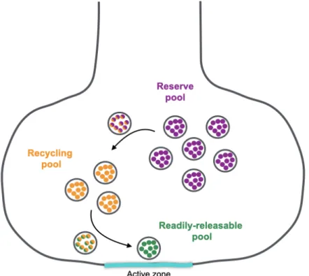
of the readily releasable pool (Rizzoli and Betz, 2005) (Figure 2). SVs in the readily releasable pool are docked and primed at fusion sites called active zones until release is triggered during an evoked response (Kaeser and Regehr, 2017; Sühof, 2012). As nerve stimulation depletes the readily releasable pool, its replenished by SVs in the recycling pool (Rizzoli and Betz, 2005). Together, the readily releasable pool and the recycling pool comprise 10-20% of all SVs at nerve terminals. The other 80-90% of SVs is known as the reserve pool (Rizzoli and Betz, 2005). This pool is recruited for fusion when the recycling pool is depleted during periods of long stimulation (Denker and Rizzoli, 2010; Rizzoli and Betz, 2005). Defining each pool gives the illusion that they are physically separated from each other but increasing evidence suggests that the reserve and recycling pools are intermixed (Alabi and Tsien, 2012; Denker and Rizzoli, 2010).
The recruitment of SVs into each pool relies on interactions with proteins associated with active zones and the actin cytoskeleton. Active zone proteins interact with SVs to molecularly dock and prime them for fusion. In particular, the active zone protein Munc-13 is essential for rendering SVs fusion competent. Munc-13 mutants lack neurotransmission even though SVs are found near the membrane, suggesting priming is impaired in these mutants (Aravamudan et al., 1999; Imig et al., 2014). It is thought that Munc-13 is crucial for establishing the SV population that form the readily releasable pool (Kaeser and Regehr, 2017). In addition to Munc-13, other active zone proteins have been found to affect the size of the readily releasable pool though to a lesser extent (Körber and Kuner, 2016). Recruitment of SVs into the readily releasable pool is dependent on actin polymerization and the phosphorylation state of Synapsin (Körber and Kuner, 2016; Rizzoli, 2014). Synapsin is a small cytosolic protein that tethers SVs to the actin
cytoskeleton and becomes phosphorylated to release SVs to replenish the readily releasable pool and recycling pool during nerve stimulation (Denker and Rizzoli, 2010; Rizzoli, 2014).
1.3 Neurotransmission is modulated by short-term plasticity
Neurons can modulate the number of SVs that fused during stimulation by enhancing or decreasing their synaptic strength through a phenomenon called short-term plasticity. Different patterns of nerve stimulation can induce short-term plasticity that may last from milliseconds to minutes. Facilitation is a type of synaptic enhancement induced by paired stimuli occurring within milliseconds of each other that results in a transient increase of neurotransmitter release that decays after ~100 milliseconds. Sustained nerve stimulation can trigger longer forms of synaptic enhancement known as augmentation and post-tetanic potentiation that decay after 5-10 seconds and 30-60 seconds, respectively. A decrease in synaptic strength can be observed at some synapses after paired stimuli or longer periods of nerve stimulation that collectively is termed depression (Catterall and Few, 2008; Zucker and Regehr, 2002).
Neurons possess intrinsic properties that favor facilitation or depression. This is observed in phasic and tonic neurons of NMJs in crayfish, Drosophila and mammals (Hennig and Lømo, 1985; Kennedy and Takeda, 1965a, 1965b; Kurdyak et al., 1994). Upon train stimulation, phasic neurons depress while tonic neurons facilitate. Phasic neurons depress because SV fusion sites have a high release probability resulting in less SVs available for fusion during the second stimulus response. In contrast, tonic neurons facilitate because they have a low release probability resulting in more SVs available for fusion during the second stimulus (Regehr, 2012; Zucker and Regehr, 2002). Even though synapses typically exhibit tonic or phasic properties, the initial release
probability at most synapses is not fixed and can be adjusted by altering extracellular calcium concentrations to manipulate the initial release probability, allowing the same synapse to exhibit either facilitation or depression (Jorquera et al., 2012; Mallart, 1993; Regehr, 2012).
Synaptic enhancement and depression are primarily regulated by intrinsic properties in the presynaptic compartment that establish the release probability of release sites. Facilitation can result from calcium channel facilitation, saturation of calcium buffers, activation of a calcium sensor that regulates the fusion of a specific SV population and increased residual calcium. In addition to these presynaptic changes, augmentation and post tetanic potentiation can result from an increased readily releasable pool and vesicle to vesicle fusion to increase neurotransmitter release (Catterall and Few, 2008; Fioravante and Regehr, 2011; Jackman and Regehr, 2017; Regehr, 2012; Zucker and Regehr, 2002). In contrast, depression can result from calcium channel inactivation, depletion of the readily releasable pool, active zone inactivation and slow replenishment of SVs (Catterall and Few, 2008; Fioravante and Regehr, 2011; Regehr, 2012; Zucker and Regehr, 2002). In some neurons, synaptic depression has also been associated with postsynaptic changes such as desensitization of ligand-gated receptors (Zucker and Regehr, 2002).
II. SV fusion
2.1 Evoked SV fusion
To trigger SV fusion after an evoked response, the plasma membrane of the presynaptic terminal must become permeable to calcium ions (Katz and Miledi, 1967). Depolarization of the presynaptic terminal opens voltage-gated calcium channels in the plasma membrane to allow
extracellular calcium into the cell (Catterall and Few, 2008; Stanley, 1993). Voltage-gated calcium channels are concentrated at active zones in the plasma membrane. Active zones cluster calcium channels and prime SVs for fusion to ensure fast neurotransmitter release (Sühof, 2012). Once a SV fuses, lipids and proteins from the SV are endocytosed adjacent to the active zone in the peri-active zone to replenish the SV pool and prepare for the next round of fusion (Saheki and De Camilli, 2012).
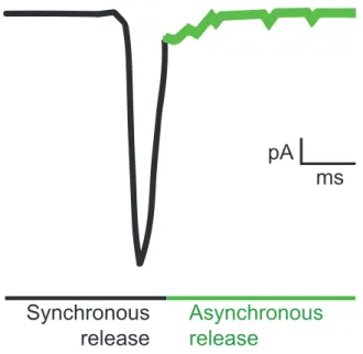
The duration of an evoked response can last up to 100-200 milliseconds that is divided in two kinetic phases known as synchronous and asynchronous release (Kaeser and Regehr, 2014) (Figure 3). The synchronous phase accounts for most of the neurotransmitter released and decays within a few milliseconds. The asynchronous phase (also known as the delayed response) is much slower and decays after 100-200 milliseconds (Goda and Stevens, 1994; Katz and Miledi, 1969). In most neurons, asynchronous release makes up less than 10% of the total neurotransmitter release at low frequency stimulation although in some synapses asynchronous release can account for 80% of total release (Hefft and Jonas, 2005; Kaeser and Regehr, 2014). In synapses where asynchronous release is not predominant, it can be enhanced by periods of high frequency stimulation, temperature and nerve stimulation in the presence of strontium (Atluri and Regehr, 1998; Hubbard, 1963; Huson and Regehr, 2020; Lu and Trussell, 2000; Rahamimoff and Yaari, 1973).
2.2 Spontaneous SV fusion
SVs can also fuse in the absence of nerve stimulation (Fatt and Katz, 1952). Spontaneous events were discovered at the frog neuromuscular junction and were initially dismissed as
electrophysiological noise from tears in the muscle after improper dissection. Further study of spontaneous events led to the proposal of the quantal hypothesis of neurotransmission that suggests spontaneous events represent the fusion of a single “quanta”, while an evoked response is the fusion of many of these quanta at the same time (del Castillo and Katz, 1954; Fatt and Katz, 1952). Later it was discovered that these quanta are neurotransmitter-filled SVs at axon terminals (De Robertis and Bennet, 1955; Palay, 1956). Like evoked release, spontaneous release plays important physiological roles in neurons, such as regulating synapse formation, homeostatic plasticity and activating different forms of synaptic plasticity (Kaeser and Regehr, 2014; Ramirez and Kavalali, 2011). In contrast to evoked release, spontaneous events have been found to be both calcium-dependent and incalcium-dependent (Kaeser and Regehr, 2014). Differences in the spatial segregation and recycling pathways have also been found between SVs that fuse through evoked or spontaneous release (Fredj and Burrone, 2009; Kavalali, 2015; Sabeva et al., 2017; Sara et al., 2005). This suggests that the SVs that fuse during evoked and spontaneous release may be molecularly distinct. More work is required to define the molecular signatures that define SVs that fuse during evoked or spontaneous release.
2.3 The SNARE complex is the molecular machine that drives SV fusion
The SNARE complex is a molecular machine formed by a group of proteins in the plasma membrane and SV membrane that drives bilayer fusion. The SNARE complex is composed of the vesicle (v)-SNARE, Synatobrevin (nSYB), and the target membrane (t)-SNAREs, Syntaxin1a (SYX1a) and SNAP-25 (for “Synaptosomal-Associated Protein, 25kDa”) (Figure 4). They were identified as the cleavage targets that blocked neurotransmitter release after treatments with clostridial botulinum and tetanus toxins (Blasi et al., 1993a, 1993b; Link et al., 1992; Schiavo et
al., 1992). nSYB is tethered to the SV by a single pass transmembrane domain. At the plasma membrane, SYX1a is tethered by a single pass transmembrane domain and SNAP-25 is tethered intracellularly through a palmitoyl side chain (Rizo and Rosenmund, 2008; Südhof and Rizo, 2011). Together they form a stable four helix bundle assembled from a helix provided by nSYB, a helix from SYX1a and two helices from SNAP-25. Formation of this complex is thought to drive full fusion of the SV with the plasma membrane to release their neurotransmitter contents into the synaptic cleft (Rizo and Rosenmund, 2008; Südhof and Rizo, 2011).
Primed SVs are predicted to contain a “partially zippered” trans-SNARE complex prior to fusion that can quickly assemble into the full coiled-coil bundle as release is triggered. During bilayer fusion, the SNARE complex initiates the formation of a fusion pore made from proteins and lipids in the SV and the plasma membrane (Bao et al., 2016). Both membranes are partially merged by the pore to allow diffusion of neurotransmitter molecules from the lumen of the SV into the synaptic cleft (Chang et al., 2017; Rizo and Rosenmund, 2008). If the fusion pore closes rapidly, a form of release known as kiss and run occurs and full collapse of the SV is not observed (Alabi and Tsien, 2013). However, it is thought that the major mode of SV fusion occurs when expansion of the fusion pore leads to the complete fusion and collapse of the SV membrane into the plasma membrane (Chang et al., 2017; Rizo and Rosenmund, 2008). Once fusion is completed the fully zippered cis-SNARE complex is dissociated by an ATPase called NSF (for “N-ethylmaleimide sensitive factor”). Once disassembled, chaperones bind to each dissociated SNARE to prevent re-formation of the cis-SNARE complex until the next round of fusion (Rizo and Rosenmund, 2008; Südhof and Rizo, 2011).
III. Synaptotagmins are calcium sensors for SV fusion
3.1 SYT1 regulates synchronous SV fusion
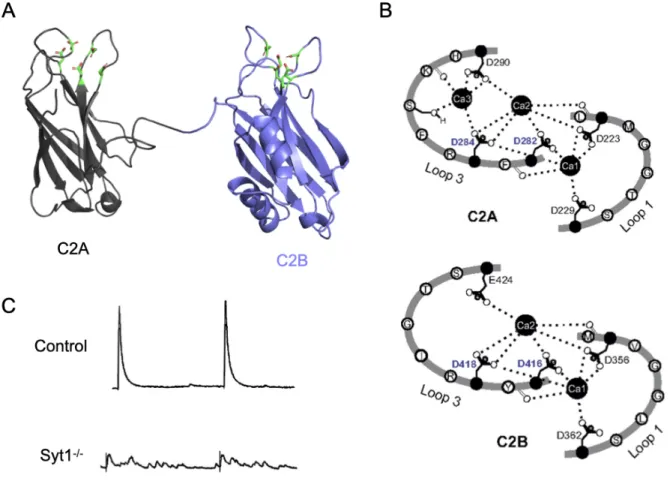
At active zones, calcium builds up to micromolar concentrations to activate calcium sensing proteins to trigger SV fusion on a millisecond timescale (Llinás et al., 1992; Regehr, 2012). The SNARE complex can promote the fusion of membranes in vitro, but the complex itself does not possess intrinsic calcium sensitivity and fusion rates mediated solely by the SNAREs are much slower than those observed in neurons (Weber et al., 1998). Synaptotagmin 1 (SYT1) is the primary calcium sensor activated during an evoked response that couples calcium influx to the zippering of the SNARE complex to drive SV fusion in neurons and some neurosecretory cells (Chapman, 2008) (Figure 4). Neurotransmission is acutely reduced in Syt1 mutants because the synchronous component of neurotransmitter release is abolished (Fernández-Chacón et al., 2001; Geppert et al., 1994; Littleton et al., 1994; Yoshihara and Littleton, 2002) (Figure 5C). Syt1 mutants exhibit higher rates of spontaneous fusion suggesting it also serves as a molecular clamp to prevent fusion of docked SVs in the absence of nerve stimulation and calcium influx (Chicka et al., 2008; Littleton et al., 1993; Xu et al., 2009). However, asynchronous release is elevated in Syt1 mutants suggesting that SYT1 is not the sole calcium sensor that drives SV fusion during an evoked response. SYT1 is enriched in the brain and found on SVs, and is highly conserved across evolution (Adolfsen et al., 2004; Perin et al., 1991b, 1991a, 1990). SYT1 contains a single-pass transmembrane domain, a short linker and two calcium sensing C2 domains, termed C2A and C2B (Perin et al., 1991a). Each C2 domain is composed of a beta barrel formed from 4 antiparallel beta strands with protruding loops that contain 5 negatively charged residues that bind calcium ions (Adolfsen et al., 2004; Chapman, 2008; Shao et al., 1996) (Figure 5A-B). The loops in each C2
domain of SYT1 creates a calcium binding pocket that binds 3 calcium ions in C2A and 2 calcium ions in C2B.
To drive bilayer fusion, SYT1 interacts with the plasma membrane and the SNARE complex in a calcium-dependent manner. In the absence of calcium, SYT1 has low affinity for the plasma membrane but can bind to PIP2 present in the plasma membrane through a polybasic stretch found in C2B, which is predicted to “steer” SYT1 to the correct location and increase its membrane penetration during an evoked response (Bai et al., 2004; Chapman, 2008; Chapman and Davis, 1998; Chapman and Jahn, 1994; Davletov and Sudhof, 1993; Fernandez et al., 2001; Li et al., 1995; Ubach et al., 1998; S. Wang et al., 2016; Zhang et al., 1998). Once SYT1 binds calcium, the calcium binding loops in each C2 domain penetrate the plasma membrane to bring the SV closer for bilayer fusion (Chapman, 2008;Chapman and Davis, 1998; Fernández-Chacón et al., 2001; Pang et al., 2006a; Shin et al., 2009). At the same time, the C2B domain of SYT1 interacts with t-SNAREs, SYX1a and SNAP-25, to aid in formation of the fusion pore and promote its expansion to drive full collapse of the SV (Das et al., 2020; Wang et al., 2001; Zhou et al., 2015). Structure function studies of SYT1 have revealed that C2B is essential for regulating synchronous release while C2A functions to inhibit asynchronous release (Desai et al., 2000; Guan et al., 2017; Mackler et al., 2002; Schupp et al., 2016; Yoshihara et al., 2010; Zhou et al., 2015). SYT1 is a multifunctional protein that regulates other neuronal processes in addition to regulating evoked release. SYT1 interacts with the endocytic machinery to promote SV retrieval after exocytosis (Grass et al., 2004; Li et al., 2017; Yao et al., 2012; Zhang et al., 1994). SYT1 has also been shown to regulate the delivery of AMPA receptors (a ligand-gated receptor) in the postsynaptic compartment of mammals during long-term potentiation (Wu et al., 2017).
3.2 Synaptotagmins are enriched in the nervous system and regulate membrane trafficking
SYTs are a family of membrane-trafficking proteins mostly expressed in the nervous system although expression in non-neuronal tissues has been observed for some isoforms (Hudson and Birnbaum, 1995; Mittelsteadt et al., 2009; Poser Von and Südhof, 2001; Sugita et al., 2001; Xiao et al., 2010). All SYTs possess a single-pass transmembrane domain, a variable linker and two C2 domains, termed C2A and C2B (Adolfsen et al., 2004; Brewer et al., 2015; Fernandez et al., 2001; Sutton et al., 1995; Zhou et al., 2015). There are 16 SYT isoforms in mammals and 7 in Drosophila (Adolfsen et al., 2004; Bhalla et al., 2005; Craxton, 2010, 2004; Gustavsson et al., 2008). Gene duplication of many mammalian SYT homologs is responsible for the discrepancy in the number of isoforms observed between vertebrates and invertebrates (Craxton, 2010, 2004). For example, SYT1 mediates all forms of synchronous release in Drosophila while in mammals SYT1, SYT2 and SYT9 serve this role (Marquhze et al., 1995; Pang et al., 2006b).
SYTs with conserved calcium binding in each C2 domain regulate exocytosis in a calcium-dependent manner. As previously described, SYT1 homologs regulate the synchronous phase during evoked release and dense core vesicle (DCV) fusion in endocrine cells (Chapman, 2008; Fernández-Chacón et al., 2001; Fukuda, 2004; Geppert et al., 1994; Iezzi et al., 2005; Littleton et al., 1994; Xu et al., 2007; Yoshihara and Littleton, 2002). SYT3 is an isoform specific to vertebrates that regulates the internalization of AMPA receptors in a calcium-dependent manner to weaken synaptic strength (Awasthi et al., 2019). In Drosophila, SYT4 regulates retrograde signaling to regulate synaptic strength and structure after strong stimulation (Barber et al., 2009;
Cho et al., 2015; Harris et al., 2016; Korkut et al., 2013). SYT7 regulates SV fusion in neurons, and modulates lysosome and DCV fusion in endocrine cells (Bacaj et al., 2013; Chakrabarti et al., 2003; Chen et al., 2017; Fukuda et al., 2004; Gao et al., 2000; Gustavsson et al., 2008; Jackman et al., 2016; Luo et al., 2015; Luo and Südhof, 2017; Martinez et al., 2000; Rao et al., 2004; Reddy et al., 2001; Wen et al., 2010).
SYTs that don’t bind calcium are less studied but can also play important roles in neuronal physiology. Mammalian SYT4 does not bind to calcium like the Drosophila homolog, suggesting that these isoforms have diverged in how they regulate membrane trafficking (Dai et al., 2004; Wang and Chapman, 2010). Mammalian SYT4 has been shown to control dense core vesicle trafficking and release of the neurotrophic factor BDNF (Bharat et al., 2017; Dean et al., 2009). SYT11 regulates endocytosis and Syt11 mutants have defects in long-term potentiation (Ferguson et al., 2004; Shimojo et al., 2019; C. Wang et al., 2016). A recent study found that SYT13 delays muscle degeneration in mouse models of ALS (Amyotrophic Lateral Sclerosis) and SMA (Spinal Muscle Atrophy) (Nizzardo et al., 2020).
3.3 The many roles of SYT7 in regulating SV fusion
The discovery that SYT1 regulates synchronous release raised the possibility that other members of the SYT family might act as the asynchronous release calcium sensor. Of the SYT isoforms, SYT7 emerged as an intriguing candidate because it has biochemical features resembling the asynchronous release calcium sensor, such as high affinity to calcium and loose coupling to voltage-gated calcium channels at active zones (Atluri and Regehr, 1998; Bhalla et al., 2005; Cummings et al., 1996; Geppert et al., 1994; Goda et al., 1994; Hui et al., 2005; Rahamimoff and
Yaari, 1973; Van der Kloot and Molgo, 1993). Compared to SYT1, SYT7 has 400x higher affinity to calcium and tighter binding to membranes (Bhalla et al., 2005; Hui et al., 2005; Voleti et al., 2017). These features might allow SYT7 to regulate SV fusion further away from active zones and potentially contribute to asynchronous release (Figure 6).
Indeed, several studies suggest that SYT7 regulates asynchronous release while SYT1 regulates synchronous release (Bacaj et al., 2013; Chen et al., 2017; Luo et al., 2015; Luo and Südhof, 2017; Wen et al., 2010). Initial studies in mammals and invertebrates found that SYT7 had no role in asynchronous release (Maximov and Südhof, 2005; Saraswati et al., 2007). The attention was brought back to SYT7 by a study at zebrafish neuromuscular junctions that found asynchronous release was significantly reduced by knocking down SYT7 (Wen et al., 2010). Mammalian SYT7 was later found to regulate asynchronous release at excitatory and inhibitory neurons by knocking down SYT1 and SYT7 (Bacaj et al., 2013). SYT1 deficient neurons have enhanced asynchronous release making it easier to detect changes in this phase of release. SYT7 knockouts have also been shown to selectively impair asynchronous release (Chen et al., 2017; Luo et al., 2015; Luo and Südhof, 2017). However, the role of SYT7 in asynchronous release is still controversial, with several groups finding the kinetics of asynchronous release is altered while the total amount of release remains intact in SYT7 knockouts (Turecek and Regehr, 2019). In addition, the expression of SYT1 and not SYT7 was found to regulate the relative contributions of synchronous and asynchronous release at synapses (Turecek and Regehr, 2019).
Besides asynchronous release, SYT7 has been suggested to play multiple roles in regulating vesicle trafficking in neurons and other cells. SYT7 has been postulated to function as
the facilitation sensor at several mammalian synapses (Chen et al., 2017; Jackman et al., 2016; Luo and Südhof, 2017). Interestingly, it has been suggested that the asynchronous release sensor and the facilitation sensor could be mediated by the same mechanism (Rahamimoff and Yaari, 1973). SYT7 has also been suggested to regulate SV replenishment during high frequency stimulation (Jackman et al., 2016; Liu et al., 2014). The authors argue that the absence of asynchronous release is secondary to reduced replenishment rates observed in Syt7 mutants because less SVs are available for this slower phase of release. This hypothesis has been challenged by others that detected no changes in SV replenishment (Luo et al., 2015). In addition, changes in the replenishment rate of the recycling and reserve pool have been reported that could lead to defects in the replenishment of the readily releasable pool (Durán et al., 2018). SYT7 along with SYT1 have also been suggested to target SVs to distinct endocytic pathways (Li et al., 2017). Besides neurons, SYT7 regulates fusion of lysosomes and DCVs in non-neuronal cells. (Chakrabarti et al., 2003; Fukuda et al., 2004; Gao et al., 2000; Gustavsson et al., 2008; Martinez et al., 2000; Rao et al., 2004; Reddy et al., 2001).
SYT7’s role as the asynchronous release sensor, facilitation sensor and regulator of vesicle replenishment in neurons may not be mutually exclusive. Multiple studies have suggested SYT7 has multiple roles in regulating SV fusion at the same synapse (Chen et al., 2017; Luo and Südhof, 2017). A recent review suggests that different experimental manipulations and intrinsic synaptic properties, such as synapse-specific expression of SYT7 isoforms, could lead to heterogeneity of SYT7 function across synapses (Huson and Regehr, 2020). Although multiple phenotypes have been described, it is poorly understood how SYT7 regulates SV trafficking across synapses and more studies are needed to address this question.
SYT7 localization studies have also given inconsistent results. SYT7 has been suggested to localize to the plasma membrane, dense core vesicles and endo/lysosomal compartments (Adolfsen et al., 2004; Martinez et al., 2000; Monterrat et al., 2007; Reddy et al., 2001; Schonn et al., 2008; Sugita et al., 2001). The precise location of SYT7 at synapses could shed light on how it regulates neurotransmission. For example, it would be unlikely that SYT7 is the asynchronous calcium sensor if SYT7 is exclusively found on endosomes. However, it could regulate SV replenishment from this compartment.
3.4 Similarities and differences between SYT1 and SYT7
SYT1 and SYT7 have distinct roles during evoked neurotransmitter release and endocytosis but appear to be functionally redundant in some mechanisms. SYT1 and SYT7 both serve as fusion clamps to suppress spontaneous fusion of SVs and regulate the size of the readily releasable pool (Bacaj et al., 2015, 2013; Luo and Südhof, 2017). Even though SYT1 and SYT7 are largely presynaptic, a recent study found that exocytosis of AMPA receptors during long-term potentiation is redundantly regulated by both (Wu et al., 2017). These studies suggest that SYT1 and SYT7 likely share partners that regulate aspects of vesicle trafficking through common pathways.
C2 domains in SYT1 and SYT7 do not regulate SV fusion in the same way. In contrast to SYT1, C2A in SYT7 plays a major role in regulating SV release while C2B plays a minor role (Bacaj et al., 2013; Jackman et al., 2016). Tight membrane binding occurs through the C2A domain in SYT7 and the C2B domain in SYT1, suggesting these C2 domain differences may be critical in
how the two proteins regulate neurotransmitter release (Voleti et al., 2017). However, the C2 domains from SYT1 and SYT7 are not interchangeable because SYT1/ SYT7 chimeras cannot rescue the Syt1 mutant phenotype (Xue et al., 2010). Another known difference is that SYT7’s C2A domain binds 2 calcium ions while SYT1’s C2A domain binds to 3 calcium ions (Voleti et al., 2017). These findings suggest that C2 domains in SYT1 and SYT7 contain intrinsic properties shaped through evolution that provide unique features in each homolog to regulate SV fusion.
IV. The Drosophila Neuromuscular Junction as a model synapse to study the role of SYT7 in neurotransmission
My thesis is aimed at elucidating the conserved role of SYT7 in neurotransmission. Here I use the larval Drosophila neuromuscular junction (NMJ) as model synapse. This synapse is created by neuronal contacts that form axonal swellings, called boutons, with the muscle. Boutons contain active zones that appose glutamate receptors in the postsynaptic density at the muscle. The highly stereotyped nature of the NMJ makes this glutamatergic synapse a powerful model to characterize the role of SYT7 in neurotransmission. In addition, the Drosophila NMJ provides an extensive genetic toolkit, amenability to light and electron microscopy, and accessibility to perform electrophysiology recordings.
In Chapter 2, I describe our studies on the role of SYT7 in neurotransmission at the Drosophila neuromuscular junction. We found that SYT7 negatively regulates neurotransmitter release in a dosage dependent manner and resides in the peri-active zone region of the synapse. In addition, we found that Syt7 mutants have enhanced recovery after high frequency stimulation suggesting that SVs are more fusogenic in the absence of SYT7.
In Chapter 3, I describe our studies that indicate the protein levels of SYT7 differ at tonic and phasic synapses at the Drosophila neuromuscular junctions. Our preliminary data suggest SYT7 may be a critical regulator for release probability primarily in tonic neurons. We also observe that mutating the calcium binding sites in the C2A and C2B domains of SYT7 redundantly affects its trafficking to terminals and may be essential for protein stability.
References
1. Adolfsen B, Saraswati S, Yoshihara M, Littleton JT. 2004. Synaptotagmins are trafficked to distinct subcellular domains including the postsynaptic compartment. J Cell Biol 166:249– 60. doi:10.1083/jcb.200312054
2. Alabi AA, Tsien RW. 2013. Perspectives on Kiss-and-Run: Role in Exocytosis, Endocytosis, and Neurotransmission. Annu Rev Physiol 75:393–422.
doi:10.1146/annurev-physiol-020911-153305
3. Alabi ARA, Tsien RW. 2012. Synaptic vesicle pools and dynamics. Cold Spring Harb
Perspect Biol 4. doi:10.1101/cshperspect.a013680
4. Aravamudan B, Fergestad T, Davis WS, Rodesch CK, Broadie K. 1999. Drosophila UNC-13 is essential for synaptic transmission. Nat Neurosci 2:965–971. doi:10.1038/14764
5. Atluri PP, Regehr WG. 1998. Delayed release of neurotransmitter from cerebellar granule cells. J Neurosci 18:8214–8227. doi:10.1523/jneurosci.18-20-08214.1998
6. Awasthi A, Ramachandran B, Ahmed S, Benito E, Shinoda Y, Nitzan N, Heukamp A, Rannio S, Martens H, Barth J, Burk K, Wang YT, Fischer A, Dean C. 2019. Synaptotagmin-3 drives AMPA receptor endocytosis, depression of synapse strength, and forgetting. Science
(80- ) 363. doi:10.1126/science.aav1483
7. Bacaj T, Wu D, Burré J, Malenka RC, Liu X, Südhof TC. 2015. Synaptotagmin-1 and -7 Are Redundantly Essential for Maintaining the Capacity of the Readily-Releasable Pool of Synaptic Vesicles. PLoS Biol 13:1–26. doi:10.1371/journal.pbio.1002267
8. Bacaj T, Wu D, Yang X, Morishita W, Zhou P, Xu W, Malenka RC, Südhof TC. 2013. Synaptotagmin-1 and synaptotagmin-7 trigger synchronous and asynchronous phases of neurotransmitter release. Neuron 80:947–59. doi:10.1016/j.neuron.2013.10.026
9. Bai J, Tucker WC, Chapman ER. 2004. PIP 2 increases the speed of response of
synaptotagmin and steers its membrane-penetration activity toward the plasma membrane.
Nat Struct Mol Biol 11:36–44. doi:10.1038/nsmb709
10. Bao H, Goldschen-Ohm M, Jeggle P, Chanda B, Edwardson JM, Chapman ER. 2016.
Exocytotic fusion pores are composed of both lipids and proteins. Nat Struct Mol Biol 23:67– 73. doi:10.1038/nsmb.3141
11. Barber CF, Jorquera RA, Melom JE, Littleton JT. 2009. Postsynaptic regulation of synaptic plasticity by synaptotagmin 4 requires both C2 domains. J Cell Biol 187:295–310.
doi:10.1083/jcb.200903098
12. Barthet G, Jordà-Siquier T, Rumi-Masante J, Bernadou F, Müller U, Mulle C. 2018. Presenilin-mediated cleavage of APP regulates synaptotagmin-7 and presynaptic plasticity.
Nat Commun 9:4780. doi:10.1038/s41467-018-06813-x
13. Bhalla A, Tucker WC, Chapman ER. 2005. Synaptotagmin Isoforms Couple Distinct Ranges of Ca2+, Ba2+, and Sr2+ Concentration to SNARE-mediated Membrane Fusion. Mol Biol
Cell 16:4755–4764. doi:10.1091/mbc.e05-04-0277
14. Bharat V, Siebrecht M, Burk K, Ahmed S, Reissner C, Kohansal-Nodehi M, Steubler V, Zweckstetter M, Ting JT, Dean C. 2017. Capture of Dense Core Vesicles at Synapses by JNK-Dependent Phosphorylation of Synaptotagmin-4. Cell Rep 21:2118–2133.
doi:10.1016/j.celrep.2017.10.084
15. Blasi J, Chapman ER, Link E, Binz T, Yamasaki S, Camilli P De, Südhof TC, Niemann H, Jahn R. 1993a. Botulinum neurotoxin a selectively cleaves the synaptic protein SNAP-25.
16. Blasi J, Chapman ER, Yamasaki S, Binz T, Niemann H, Jahn R. 1993b. Botulinum
neurotoxin C1 blocks neurotransmitter release by means of cleaving HPC-1/syntaxin. EMBO
J 12:4821–4828. doi:10.1002/j.1460-2075.1993.tb06171.x
17. Brewer KD, Bacaj T, Cavalli A, Camilloni C, Swarbrick JD, Liu J, Zhou A, Zhou P, Barlow N, Xu J, Seven AB, Prinslow EA, Voleti R, Häussinger D, Bonvin AMJJ, Tomchick DR, Vendruscolo M, Graham B, Südhof TC, Rizo J. 2015. Dynamic binding mode of a Synaptotagmin-1-SNARE complex in solution. Nat Struct Mol Biol 22:555–564. doi:10.1038/nsmb.3035
18. Cabantous S, Terwilliger TC, Waldo GS. 2005. Protein tagging and detection with
engineered self-assembling fragments of green fluorescent protein. Nat Biotechnol 23:102– 107. doi:10.1038/nbt1044
19. Catterall WA, Few AP. 2008. Calcium Channel Regulation and Presynaptic Plasticity.
Neuron 59:882–901. doi:10.1016/j.neuron.2008.09.005
20. Chakrabarti S, Kobayashi KS, Flavell RA, Marks CB, Miyake K, Liston DR, Fowler KT, Gorelick FS, Andrews NW. 2003. Impaired membrane resealing and autoimmune myositis in synaptotagmin VII-deficient mice. J Cell Biol 162:543–549. doi:10.1083/jcb.200305131 21. Chang CW, Chiang CW, Jackson MB. 2017. Fusion pores and their control of
neurotransmitter and hormone release. J Gen Physiol 149:301–322. doi:10.1085/jgp.201611724
22. Chapman ER. 2008. How Does Synaptotagmin Trigger Neurotransmitter Release? Annu Rev
Biochem 77:615–641. doi:10.1146/annurev.biochem.77.062005.101135
23. Chapman ER, Davis AF. 1998. Direct interaction of a Ca2+-binding loop of synaptotagmin with lipid bilayers. J Biol Chem 273:13995–14001. doi:10.1074/jbc.273.22.13995
24. Chapman ER, Jahn R. 1994. Calcium-dependent interaction of the cytoplasmic region of synaptotagmin with membranes. Autonomous function of a single C2-homologous domain. J
Biol Chem 269:5735–5741.
25. Chen C, Satterfield R, Young SM, Jonas P. 2017. Triple Function of Synaptotagmin 7 Ensures Efficiency of High-Frequency Transmission at Central GABAergic Synapses. Cell
Rep 21:2082–2089. doi:10.1016/j.celrep.2017.10.122
26. Chicka MC, Hui E, Liu H, Chapman ER. 2008. Synaptotagmin arrests the SNARE complex before triggering fast, efficient membrane fusion in response to Ca 2+. Nat Struct Mol Biol 15. doi:10.1038/nsmb.1463
27. Cho RW, Buhl LK, Volfson D, Tran A, Li F, Akbergenova Y, Littleton JT. 2015. Phosphorylation of Complexin by PKA Regulates Activity-Dependent Spontaneous Neurotransmitter Release and Structural Synaptic Plasticity. Neuron 88:749–761. doi:10.1016/j.neuron.2015.10.011
28. Craxton M. 2010. A manual collection of Syt, Esyt, Rph3a, Rph3al, Doc2, and Dblc2 genes from 46 metazoan genomes - an open access resource for neuroscience and evolutionary biology. BMC Genomics 11:1–21. doi:10.1186/1471-2164-11-37
29. Craxton M. 2004. Synaptotagmin gene content of the sequenced genomes. BMC Genomics 5:1–14. doi:10.1186/1471-2164-5-43
30. Cummings DD, Wilcox KS, Dichter MA. 1996. Calcium-dependent paired-pulse facilitation of miniature EPSC frequency accompanies depression of EPSCs at hippocampal synapses in culture. J Neurosci 16:5312–5323. doi:10.1523/jneurosci.16-17-05312.1996
31. Dai H, Shin O-H, Machius M, Tomchick DR, Südhof TC, Rizo J. 2004. Structural basis for the evolutionary inactivation of Ca2+ binding to synaptotagmin 4. Nat Struct Mol Biol
11:844–849. doi:10.1038/nsmb817
32. Das D, Bao H, Courtney KC, Wu L, Chapman ER. 2020. Resolving kinetic intermediates during the regulated assembly and disassembly of fusion pores. Nat Commun 11:1–12. doi:10.1038/s41467-019-14072-7
33. Davletov BA, Sudhof TC. 1993. A single C2domain from synaptotagmin I is sufficient for high affinity Ca2+/phospholipid binding. J Biol Chem 268:26386–26390.
34. De Robertis ED, Bennet HS. 1955. Some features of the submicroscopic morphology of synapses in frog and earthworm. J Biophys Biochem Cytol 1:47–58. doi:10.1083/jcb.1.1.47 35. Dean C, Liu H, Dunning FM, Chang PY, Jackson MB, Chapman ER. 2009.
Synaptotagmin-IV modulates synaptic function and long-term potentiation by regulating BDNF release. Nat
Neurosci 12:767–776. doi:10.1038/nn.2315
36. del Castillo J, Katz B. 1954. Quantal components of the end-plate potential. J Physiol 124:560–573. doi:10.1113/jphysiol.1954.sp005129
37. Denker A, Rizzoli SO. 2010. Synaptic vesicle pools: An update. Front Synaptic Neurosci 2:1–12. doi:10.3389/fnsyn.2010.00135
38. Desai RC, Vyas B, Earles CA, Littleton JT, Kowalchyck JA, Martin TFJ, Chapman ER. 2000. The C2B domain of synaptotagmin is a Ca2+-sensing module essential for exocytosis.
J Cell Biol 150:1125–1135. doi:10.1083/jcb.150.5.1125
39. Durán E, Montes MÁ, Jemal I, Satterfield R, Young S, Álvarez de Toledo G. 2018. Synaptotagmin-7 controls the size of the reserve and resting pools of synaptic vesicles in hippocampal neurons. Cell Calcium 74:53–60. doi:10.1016/j.ceca.2018.06.004
40. Fatt P, Katz B. 1952. Spontaneous subthreshold activity at motor nerve endings. J Physiol 117:109–128. doi:10.1113/jphysiol.1952.sp004735
41. Ferguson GD, Wang H, Herschman HR, Storm DR. 2004. Altered hippocampal short-term plasticity and associative memory in synaptotagmin IV (- / -) mice. Hippocampus 14:964– 974. doi:10.1002/hipo.20013
42. Fernández-Chacón R, Königstorfer A, Gerber SH, García J, Matos MF, Stevens CF, Brose N, Rizo J, Rosenmund C, Südhof TC. 2001. Synaptotagmin I functions as a calcium regulator of release probability. Nature 410:41–49. doi:10.1038/35065004
43. Fernandez I, Araç D, Ubach J, Gerber SH, Shin OH, Gao Y, Anderson RGW, Südhof TC, Rizo J. 2001. Three-dimensional structure of the synaptotagmin 1 C2B-domain:
Synaptotagmin 1 as a phospholipid binding machine. Neuron 32:1057–1069. doi:10.1016/S0896-6273(01)00548-7
44. Fioravante D, Regehr WG. 2011. Short-term forms of presynaptic plasticity. Curr Opin
Neurobiol 21:269–274. doi:10.1016/j.conb.2011.02.003
45. Fredj N Ben, Burrone J. 2009. A resting pool of vesicles is responsible for spontaneous vesicle fusion at the synapse. Nat Neurosci 12:751–758. doi:10.1038/nn.2317
46. Fukuda M. 2004. RNA interference-mediated silencing of synaptotagmin IX, but not synaptotagmin I, inhibits dense-core vesicle exocytosis in PC12 cells. Biochem J 380:875– 879. doi:10.1042/BJ20040096
47. Fukuda M, Kanno E, Satoh M, Saegusa C, Yamamoto A. 2004. Synaptotagmin VII is targeted to dense-core vesicles and regulates their Ca2+-dependent exocytosis in PC12 cells.
J Biol Chem 279:52677–52684. doi:10.1074/jbc.M409241200
48. Fukuda M, Katayama E, Mikoshiba K. 2002. The calcium-binding loops of the tandem C2 domains of synaptotagmin VII cooperatively mediate calcium-dependent oligomerization. J
49. Fukuda M, Mikoshiba K. 2000. Calcium-Dependent and -Independent Hetero-Oligomerization in the 645:637–645.
50. Gao Z, Reavey-Cantwell J, Young RA, Jegier P, Wolf BA. 2000. Synaptotagmin III/VII isoforms mediate Ca2+-induced insulin secretion in pancreatic islet β-cells. J Biol Chem 275:36079–36085. doi:10.1074/jbc.M004284200
51. Geppert M, Goda Y, Hammer RE, Li C, Rosahl TW, Stevens CF, Südhof TC. 1994. Synaptotagmin I: A major Ca2+ sensor for transmitter release at a central synapse. Cell 79:717–727. doi:10.1016/0092-8674(94)90556-8
52. Goda Y, Stevens CF, Goda, Yukiko and Stevens CF. 1994. Two components of transmitter release at a central synapse. PNAS 91:12942–12946. doi:10.1073/pnas.91.26.12942
53. Grass I, Thiel S, Höning S, Haucke V. 2004. Recognition of a basic AP-2 binding motif within the C2B domain of synaptotagmin is dependent on multimerization. J Biol Chem 279:54872–54880. doi:10.1074/jbc.M409995200
54. Guan Z, Bykhovskaia M, Jorquera RA, Sutton RB, Akbergenova Y, Littleton JT. 2017. A synaptotagmin suppressor screen indicates SNARE binding controls the timing and Ca2+ cooperativity of vesicle fusion. Elife 6:1–30. doi:10.7554/eLife.28409
55. Gustavsson N, Lao Y, Maximov A, Chuang JC, Kostromina E, Repa JJ, Li C, Radda GK, Südhof TC, Han W. 2008. Impaired insulin secretion and glucose intolerance in
synaptotagmin-7 null mutant mice. Proc Natl Acad Sci U S A 105:3992–3997. doi:10.1073/pnas.0711700105
56. Harris, K.P., Littleton, J.T., 2015. Transmission, development, and plasticity of synapses. Genetics 201, 345–375. doi:10.1534/genetics.115.176529
57. Harris KP, Zhang Y V., Piccioli ZD, Perrimon N, Troy Littleton J. 2016. The postsynaptic t-SNARE syntaxin 4 controls traffic of neuroligin 1 and synaptotagmin 4 to regulate retrograde signaling. Elife 5:1–26. doi:10.7554/eLife.13881
58. Hefft S, Jonas P. 2005. Asynchronous GABA release generates long-lasting inhibition at a hippocampal interneuron-principal neuron synapse. Nat Neurosci 8:1319–1328.
doi:10.1038/nn1542
59. Hennig R, Lømo T. 1985. Firing patterns of motor units in normal rats. Nature 314:164–166. doi:10.1038/314164a0
60. Hoang B, Chiba A. 2001. Single-cell analysis of Drosophila larval neuromuscular synapses.
Dev Biol 229:55–70. doi:10.1006/dbio.2000.9983
61. Hubbard JI. 1963. Repetitive stimulation at the mammalian neuromuscular junction, and the mobilization of transmitter. J Physiol 169:641–662. doi:10.1113/jphysiol.1963.sp007286 62. Hudson AW, Birnbaum MJ. 1995. Identification of a nonneuronal isoform of synaptotagmin.
Proc Natl Acad Sci U S A 92:5895–5899. doi:10.1073/pnas.92.13.5895
63. Hui E, Bai J, Wang P, Sugimori M, Llinas RR, Chapman ER. 2005. Three distinct kinetic groupings of the synaptotagmin family: candidate sensors for rapid and delayed exocytosis.
Proc Natl Acad Sci U S A 102:5210–4. doi:10.1073/pnas.0500941102
64. Huson V, Regehr WG. 2020. Diverse roles of Synaptotagmin-7 in regulating vesicle fusion.
Curr Opin Neurobiol 63:42–52. doi:10.1016/j.conb.2020.02.006
65. Iezzi M, Eliasson L, Fukuda M, Wollheim CB. 2005. Adenovirus-mediated silencing of Synaptotagmin 9 inhibits Ca 2+-dependent insulin secretion in islets. FEBS Lett 579:5241– 5246. doi:10.1016/j.febslet.2005.08.047
66. Imig C, Min SW, Krinner S, Arancillo M, Rosenmund C, Südhof TC, Rhee JS, Brose N, Cooper BH. 2014. The Morphological and Molecular Nature of Synaptic Vesicle Priming at
Presynaptic Active Zones. Neuron 84:416–431. doi:10.1016/j.neuron.2014.10.009
67. Jackman SL, Regehr WG. 2017. The Mechanisms and Functions of Synaptic Facilitation.
Neuron 94:447–464. doi:10.1016/j.neuron.2017.02.047
68. Jackman SL, Turecek J, Belinsky JE, Regehr WG. 2016. The calcium sensor synaptotagmin 7 is required for synaptic facilitation. Nature 529:88–91. doi:10.1038/nature16507
69. Jorquera RA, Huntwork-Rodriguez S, Akbergenova Y, Cho RW, Troy Littleton J. 2012. Complexin controls spontaneous and evoked neurotransmitter release by regulating the timing and properties of synaptotagmin activity. J Neurosci 32:18234–18245.
doi:10.1523/JNEUROSCI.3212-12.2012
70. Kaeser PS, Regehr WG. 2017. The readily releasable pool of synaptic vesicles. Curr Opin
Neurobiol 43:63–70. doi:10.1016/j.conb.2016.12.012
71. Kaeser PS, Regehr WG. 2014. Molecular Mechanisms for Synchronous, Asynchronous, and Spontaneous Neurotransmitter Release. Annu Rev Physiol 76:333–363. doi:10.1146/annurev-physiol-021113-170338
72. Kamiyama D, Sekine S, Barsi-Rhyne B, Hu J, Chen B, Gilbert LA, Ishikawa H, Leonetti MD, Marshall WF, Weissman JS, Huang B. 2016. Versatile protein tagging in cells with split fluorescent protein. Nat Commun 7. doi:10.1038/ncomms11046
73. Katz B, Miledi R. 1969. Spontaneous and evoked activity of motor nerve endings in calcium Ringer. J Physiol 203:689–706. doi:10.1113/jphysiol.1969.sp008887
74. Katz B, Miledi R. 1967. A study of synaptic transmission in the absence of nerve impulses. J
Physiol 192:407–436. doi:10.1113/jphysiol.1967.sp008307
75. Kavalali ET. 2015. The mechanisms and functions of spontaneous neurotransmitter release.
Nat Rev Neurosci 16:5–16. doi:10.1038/nrn3875
76. Kennedy D, Takeda K. 1965a. Reflex Control of Abdominal Flexor Muscles in the Crayfish: I. The Twitch System. J Exp Biol 43:211–227.
77. Kennedy D, Takeda K. 1965b. Reflex Control of Abdominal Flexor Muscles in the Crayfish: II. The Tonic System. J Exp Biol 43:229–246.
78. Kittel RJ, Heckmann M. 2016. Synaptic vesicle proteins and active zone plasticity. Front
Synaptic Neurosci 8:8. doi:10.3389/fnsyn.2016.00008
79. Körber C, Kuner T. 2016. Molecular machines regulating the release probability of synaptic vesicles at the active zone. Front Synaptic Neurosci 8:5. doi:10.3389/fnsyn.2016.00005 80. Korkut C, Li Y, Koles K, Brewer C, Ashley J, Yoshihara M, Budnik V. 2013. Regulation of
Postsynaptic Retrograde Signaling by Presynaptic Exosome Release. Neuron 77:1039–1046. doi:10.1016/j.neuron.2013.01.013
81. Kurdyak P, Atwood HL, Stewart BA, Wu C-F. 1994. Differential physiology and
morphology of motor axons to ventral longitudinal muscles in larval Drosophila. J Comp
Neurol 350:463–472. doi:10.1002/cne.903500310
82. Lee J, Littleton JT. 2015. Transmembrane tethering of synaptotagmin to synaptic vesicles controls multiple modes of neurotransmitter release. Proc Natl Acad Sci 112:201420312. doi:10.1073/pnas.1420312112
83. Li C, Ullrich B, Zhang JZ, Anderson RGW, Brose N, Südhof TC. 1995. Ca2+-dependent and -independent activities of neural and non-neural synaptotagmins. Nature 375:594–599. doi:10.1038/375594a0
84. Li YC, Chanaday NL, Xu W, Kavalali ET. 2017. Synaptotagmin-1- and Synaptotagmin-7-Dependent Fusion Mechanisms Target Synaptic Vesicles to Kinetically Distinct Endocytic
85. Link E, Edelmann L, Chou JH, Binz T, Yamasaki S, Eisel U, Baumert M, Südhof TC, Niemann H, Jahn R. 1992. Tetanus toxin action: inhibition of neurotransmitter release linked to synaptobrevin proteolysis. Biochem Biophys Res Commun 189:1017–23.
doi:10.1016/0006-291x(92)92305-h
86. Littleton JT, Stern M, Perin M, Bellen HJ. 1994. Calcium dependence of neurotransmitter release and rate of spontaneous vesicle fusions are altered in Drosophila synaptotagmin mutants. Proc Natl Acad Sci U S A 91:10888–92.
87. Littleton JT, Stern M, Schulze K, Perin M, Bellen HJ. 1993. Mutational analysis of
Drosophila synaptotagmin demonstrates its essential role in Ca2+-activated neurotransmitter release. Cell 74:1125–1134. doi:10.1016/0092-8674(93)90733-7
88. Liu H, Bai H, Hui E, Yang L, Evans CS, Wang Z, Kwon SE, Chapman ER. 2014. Synaptotagmin 7 functions as a Ca2+-sensor for synaptic vesicle replenishment. Elife 2014:1–18. doi:10.7554/eLife.01524.001
89. Llinás R, Sugimori M, Silver RB. 1992. Microdomains of high calcium concentration in a presynaptic terminal. Science (80- ) 256:677–679. doi:10.1126/science.1350109
90. Lu T, Trussell LO. 2000. Inhibitory transmission mediated by asynchronous transmitter release. Neuron 26:683–694. doi:10.1016/S0896-6273(00)81204-0
91. Luo F, Bacaj T, Südhof TC. 2015. Synaptotagmin-7 Is Essential for Ca2+-Triggered Delayed Asynchronous Release But Not for Ca2+-Dependent Vesicle Priming in Retinal Ribbon Synapses. J Neurosci 35:11024–33. doi:10.1523/JNEUROSCI.0759-15.2015
92. Luo F, Südhof TC. 2017. Synaptotagmin-7-Mediated Asynchronous Release Boosts High-Fidelity Synchronous Transmission at a Central Synapse. Neuron 94:826-839.e3.
doi:10.1016/j.neuron.2017.04.020
93. Mackler JM, Drummond JA, Loewen CA, Robinson IM, Reist NE. 2002. The C2B Ca2+-binding motif of synaptotagmin is required for synaptic transmission in vivo. Nature 418:340–344. doi:10.1038/nature00846
94. Mallart A. 1993. Calcium-dependent modulation of the facilitation of transmitter release at neuromuscular junctions of Drosophila. J Physiol - Paris 87:83–88. doi:10.1016/0928-4257(93)90002-B
95. Marquhze B, Boudier JA, Mizuta ’ M, Inagaki N, Seine : S, Seagar’ M. 1995. Cellular Localization of Synaptotagmin I, II, and Ill mRNAs in the Central Nervous System and Pituitary and Adrenal Glands of the Rat. J Neurosci 15:4906–4917.
96. Martinez I, Chakrabarti S, Hellevik T, Morehead J, Fowler K, Andrews NW. 2000.
Synaptotagmin vii regulates Ca2+-dependent exocytosis of lysosomes in fibroblasts. J Cell
Biol 148:1141–1149. doi:10.1083/jcb.148.6.1141
97. Maximov A, Südhof TC. 2005. Autonomous function of synaptotagmin 1 in triggering synchronous release independent of asynchronous release. Neuron 48:547–54.
doi:10.1016/j.neuron.2005.09.006
98. Menon KP, Carrillo RA, Zinn K. 2013. Development and plasticity of the Drosophila larval neuromuscular junction. Wiley Interdiscip Rev Dev Biol 2:647–670. doi:10.1002/wdev.108 99. Mittelsteadt T, Seifert G, Alvárez-Barón E, Steinhäuser C, Becker AJ, Schoch S. 2009.
Differential mRNA expression patterns of the synaptotagmin gene family in the rodent brain.
J Comp Neurol 512:514–528. doi:10.1002/cne.21908
100. Monterrat C, Grise F, Benassy MN, Hémar A, Lang J. 2007. The calcium-sensing protein synaptotagmin 7 is expressed on different endosomal compartments in endocrine,
127:625–632. doi:10.1007/s00418-007-0271-0
101. Newman ZL, Hoagland A, Aghi K, Worden K, Levy SL, Ho Son J, Lee LP, Isacoff EY. 2017. Input-Specific Plasticity and Homeostasis at the Drosophila Larval Neuromuscular Junction. Neuron 93:1388-1404.e10. doi:10.1016/j.neuron.2017.02.028
102. Nizzardo M, Taiana M, Rizzo F, Aguila Benitez J, Nijssen J, Allodi I, Melzi V, Bresolin N, Comi GP, Hedlund E, Corti S. 2020. Synaptotagmin 13 is neuroprotective across motor neuron diseases. Acta Neuropathol 1–17. doi:10.1007/s00401-020-02133-x
103. Palay SL. 1956. Synapses in the central nervous system. J Biophys Biochem Cytol 2:193– 202. doi:10.1083/jcb.2.4.193
104. Pang ZP, Shin OH, Meyer AC, Rosenmund C, Südhof TC. 2006a. A gain-of-function mutation in synaptotagmin-1 reveals a critical role of Ca2+-dependent soluble
N-ethylmaleimide-sensitive factor attachment protein receptor complex binding in synaptic exocytosis. J Neurosci 26:12556–12565. doi:10.1523/JNEUROSCI.3804-06.2006 105. Pang ZP, Sun J, Rizo J, Maximov A, Südhof TC. 2006b. Genetic analysis of
synaptotagmin 2 in spontaneous and Ca 2+-triggered neurotransmitter release. EMBO J 25:2039–2050. doi:10.1038/sj.emboj.7601103
106. Pereda AE. 2014. Electrical synapses and their functional interactions with chemical synapses. Nat Rev Neurosci 15:250–263. doi:10.1038/nrn3708
107. Perin M, Brose N, Jahn R, Sudhof TC. 1991a. Domain structure of synaptotagmin (p65).
J Biol Chem 266:623–629.
108. Perin M, Johnston PA, Ozcelik T, Jahn R, Francke U, Südhof TC. 1991b. Structural and functional conservation of synaptotagmin (p65) in Drosophila and humans. J Biol Chem 266:615–22.
109. Perin MS, Fried VA, Mignery GA, Jahn R, Südhof TC. 1990. Phospholipid binding by a synaptic vesicle protein homologous to the regulatory region of protein kinase C. Nature 345:260–263. doi:10.1038/345260a0
110. Poser Von C, Südhof TC. 2001. Synaptotagmin 13: Structure and expression of a novel synaptotagmin. Eur J Cell Biol 80:41–47. doi:10.1078/0171-9335-00133
111. Rahamimoff R, Yaari Y. 1973. Delayed release of transmitter at the frog neuromuscular junction. J Physiol 228:241–257. doi:10.1113/jphysiol.1973.sp010084
112. Ramirez DMO, Kavalali ET. 2011. Differential regulation of spontaneous and evoked neurotransmitter release at central synapses. Curr Opin Neurobiol 21:275–282.
doi:10.1016/j.conb.2011.01.007
113. Rao SK, Huynh C, Proux-Gillardeaux V, Galli T, Andrews NW. 2004. Identification of SNAREs Involved in Synaptotagmin VII-regulated Lysosomal Exocytosis. J Biol Chem 279:20471–20479. doi:10.1074/jbc.M400798200
114. Reddy A, Caler E V., Andrews NW. 2001. Plasma Membrane Repair Is Mediated by Ca2+-Regulated Exocytosis of Lysosomes. Cell 106:157–169. doi:10.1016/S0092-8674(01)00421-4
115. Regehr WG. 2012. Short-term presynaptic plasticity. Cold Spring Harb Perspect Biol 4:1–19. doi:10.1101/cshperspect.a005702
116. Rizo J, Rosenmund C. 2008. Synaptic vesicle fusion. Nat Struct Mol Biol 15:665–674. doi:10.1038/nsmb.1450
117. Rizo J, Südhof TC. 2002. Snares and munc18 in synaptic vesicle fusion. Nat Rev
doi:10.1002/embj.201386357
119. Rizzoli SO, Betz WJ. 2005. Synaptic vesicle pools. Nat Rev Neurosci 6:57–69. doi:10.1038/nrn1583
120. Sabeva N, Cho RW, Vasin A, Gonzalez A, Littleton JT, Bykhovskaia M. 2017.
Complexin mutants reveal partial segregation between recycling pathways that drive evoked and spontaneous neurotransmission. J Neurosci 37:383–396.
doi:10.1523/JNEUROSCI.1854-16.2016
121. Saheki Y, De Camilli P. 2012. Synaptic vesicle endocytosis. Cold Spring Harb Perspect
Biol 4. doi:10.1101/cshperspect.a005645
122. Sara Y, Virmani T, Deák F, Liu X, Kavalali ET. 2005. An isolated pool of vesicles recycles at rest and drives spontaneous neurotransmission. Neuron 45:563–573. doi:10.1016/j.neuron.2004.12.056
123. Saraswati S, Adolfsen B, Littleton JT. 2007. Characterization of the role of the
Synaptotagmin family as calcium sensors in facilitation and asynchronous neurotransmitter release. Proc Natl Acad Sci U S A 104:14122–14127. doi:10.1073/pnas.0706711104 124. Schiavo GG, Benfenati F, Poulain B, Rossetto O, De Laureto PP, Dasgupta BR,
Montecucco C. 1992. Tetanus and botulinum-B neurotoxins block neurotransmitter release by proteolytic cleavage of synaptobrevin. Nature 359:832–835. doi:10.1038/359832a0 125. Schonn JS, Maximov A, Lao Y, Südhof TC, Sørensen JB. 2008. Synaptotagmin-1 and -7
are functionally overlapping Ca2+ sensors for exocytosis in adrenal chromaffin cells. Proc
Natl Acad Sci U S A 105:3998–4003. doi:10.1073/pnas.0712373105
126. Schupp M, Malsam J, Ruiter M, Scheutzow A, Wierda KDB, Söllner TH, Sørensen JB. 2016. Interactions between SNAP-25 and synaptotagmin-1 are involved in vesicle priming, clamping spontaneous and stimulating evoked neurotransmission. J Neurosci 36:11834– 11836. doi:10.1523/JNEUROSCI.1011-16.2016
127. Shao X, Davletov BA, Sutton RB, Südhof TC, Rizo J. 1996. Bipartite Ca2+-binding motif in C2 domains of synaptotagmin and protein kinase C. Science (80- ) 273:248–251. doi:10.1126/science.273.5272.248
128. Shimojo M, Madara J, Pankow S, Liu X, Yates J, Südhof TC, Maximov A. 2019. Synaptotagmin-11 mediates a vesicle trafficking pathway that is essential for development and synaptic plasticity. Genes Dev 33:365–376. doi:10.1101/gad.320077.118
129. Shin OH, Xu J, Rizo J, Südhof TC. 2009. Differential but convergent functions of Ca2+ binding to synaptotagmin-1 C2 domains mediate neurotransmitter release. Proc Natl Acad
Sci U S A 106:16469–16474. doi:10.1073/pnas.0908798106
130. Smart TG, Paoletti P. 2012. Synaptic neurotransmitter-gated receptors. Cold Spring Harb
Perspect Biol 4. doi:10.1101/cshperspect.a009662
131. Stanley EF. 1993. Single calcium channels and acetylcholine release at a presynaptic nerve terminal. Neuron 11:1007–1011. doi:10.1016/0896-6273(93)90214-C
132. Südhof TC, Rizo J. 2011. Synaptic vesicle exocytosis. Cold Spring Harb Perspect Biol 3. doi:10.1101/cshperspect.a005637
133. Sugita S, Han W, Butz S, Liu X, Fernández-Chacón R, Lao Y, Südhof TC. 2001.
Synaptotagmin VII as a Plasma Membrane Ca2+ Sensor in Exocytosis. Neuron 30:459–473. doi:10.1016/S0896-6273(01)00290-2
134. Sühof TC. 2012. The Presynaptic Active Zone. Neuron 75:11–25. doi:10.1016/j.neuron.2012.06.012
Berghuis AM, Sudhof TC, Sprang SR. 1995. Structure of the first C2 domain of synaptotagmin I: A novel Ca2+/phospholipid-binding fold. Cell 80:929–938. doi:10.1016/0092-8674(95)90296-1
136. Takamori S, Holt M, Stenius K, Lemke EA, Grønborg M, Riedel D, Urlaub H, Schenck S, Brügger B, Ringler P, Müller SA, Rammner B, Gräter F, Hub JS, De Groot BL, Mieskes G, Moriyama Y, Klingauf J, Grubmüller H, Heuser J, Wieland F, Jahn R. 2006. Molecular Anatomy of a Trafficking Organelle. Cell 127:831–846. doi:10.1016/j.cell.2006.10.030 137. Turecek J, Regehr WG. 2019. Neuronal Regulation of Fast Synaptotagmin Isoforms
Controls the Relative Contributions of Synchronous and Asynchronous Release. Neuron 101:938-949.e4. doi:10.1016/j.neuron.2019.01.013
138. Ubach J, Zhang X, Shao X, Südhof T, Rizo J. 1998. Ca2+ binding to synaptotagmin: how many Ca2+ ions bind to the tip of a C2 domain? EMBO J 17:3921–3930.
139. Van der Kloot W, Molgo J. 1993. Facilitation and delayed release at about 0°C at the frog neuromuscular junction: Effects of calcium chelators, calcium transport inhibitors, and okadaic acid. J Neurophysiol 69:717–729. doi:10.1152/jn.1993.69.3.717
140. Voleti R, Tomchick DR, Südhof TC, Rizo J. 2017. Exceptionally tight membrane-binding may explain the key role of the synaptotagmin-7 C2A domain in asynchronous neurotransmitter release. Proc Natl Acad Sci U S A 114:E8518–E8527.
doi:10.1073/pnas.1710708114
141. Wang C, Wang Y, Hu M, Chai Z, Wu Q, Huang R, Han W, Zhang CX, Zhou Z. 2016. Synaptotagmin-11 inhibits clathrin-mediated and bulk endocytosis. EMBO Rep 17:47–63. doi:10.15252/embr.201540689
142. Wang CT, Grishanin R, Earles CA, Chang PY, Martin TFJ, Chapman ER, Jackson MB. 2001. Synaptotagmin modulation of fusion pore kinetics in regulated exocytosis of dense-core vesicles. Science 294:1111–1115. doi:10.1126/science.1064002
143. Wang S, Li Y, Ma C. 2016. Synaptotagmin-1 C2B domain interacts simultaneously with SNAREs and membranes to promote membrane fusion. Elife 5:e14211.
doi:10.7554/eLife.14211
144. Wang Z, Chapman ER. 2010. Rat and Drosophila synaptotagmin 4 have opposite effects during SNARE-catalyzed membrane fusion. J Biol Chem 285:30759–30766.
doi:10.1074/jbc.M110.137745
145. Weber T, Zemelman B V., McNew JA, Westermann B, Gmachl M, Parlati F, Söllner TH, Rothman JE. 1998. SNAREpins: Minimal machinery for membrane fusion. Cell 92:759–772. doi:10.1016/S0092-8674(00)81404-X
146. Wen H, Linhoff MW, McGinley MJ, Li GL, Corson GM, Mandel G, Brehm P. 2010. Distinct roles for two synaptotagmin isoforms in synchronous and asynchronous transmitter release at zebrafish neuromuscular junction. Proc Natl Acad Sci U S A 107:13906–13911. doi:10.1073/pnas.1008598107
147. Wu D, Bacaj T, Morishita W, Goswami D, Arendt KL, Xu W, Chen L, Malenka RC, Südhof TC. 2017. Postsynaptic synaptotagmins mediate AMPA receptor exocytosis during LTP. Nature 544:316–321. doi:10.1038/nature21720
148. Xiao L, Han Y, Runne H, Murray H, Kochubey O, Luthi-Carter R, Schneggenburger R. 2010. Developmental expression of Synaptotagmin isoforms in single calyx of
Held-generating neurons. Mol Cell Neurosci 44:374–385. doi:10.1016/j.mcn.2010.05.002
581. doi:10.1016/j.neuron.2007.05.004
150. Xu J, Pang ZP, Shin OH, Südhof TC. 2009. Synaptotagmin-1 functions as a Ca2+ sensor for spontaneous release. Nat Neurosci 12:759–766. doi:10.1038/nn.2320
151. Xue M, Craig TK, Shin O-H, Li L, Brautigam CA, Tomchick DR, Südhof TC,
Rosenmund C, Rizo J. 2010. Structural and mutational analysis of functional differentiation between synaptotagmins-1 and -7. PLoS One 5:e12544. doi:10.1371/journal.pone.0012544 152. Yao J, Kwon SE, Gaffaney JD, Dunning FM, Chapman ER. 2012. Uncoupling the roles
of synaptotagmin i during endo-and exocytosis of synaptic vesicles. Nat Neurosci 15:243– 249. doi:10.1038/nn.3013
153. Yoshihara M, Guan Z, Littleton JT. 2010. Differential regulation of synchronous versus asynchronous neurotransmitter release by the C2 domains of synaptotagmin 1. Proc Natl
Acad Sci U S A 107:14869–14874. doi:10.1073/pnas.1000606107
154. Yoshihara M, Littleton JTT. 2002. Synaptotagmin I Functions as a Calcium Sensor to Synchronize Neurotransmitter Release. Neuron 36:897–908.
doi:10.1016/S0896-6273(02)01065-6
155. Zhang JZ, Davletov BA, Südhof TC, Anderson RGW. 1994. Synaptotagmin I is a high affinity receptor for clathrin AP-2: Implications for membrane recycling. Cell.
doi:10.1016/S0092-8674(94)90442-1
156. Zhang X, Rizo J, Südhof TC. 1998. Mechanism of phospholipid binding by the C2A-domain of synaptotagmin I. Biochemistry 37:12395–12403. doi:10.1021/bi9807512 157. Zhou Q, Lai Y, Bacaj T, Zhao M, Lyubimov AY, Uervirojnangkoorn M, Zeldin OB,
Brewster AS, Sauter NK, Cohen AE, Soltis SM, Alonso-Mori R, Chollet M, Lemke HT, Pfuetzner RA, Choi UB, Weis WI, Diao J, Südhof TC, Brunger AT. 2015. Architecture of the synaptotagmin-SNARE machinery for neuronal exocytosis. Nature 525:62–67.
doi:10.1038/nature14975
158. Zucker RS, Regehr WG. 2002. Short-Term Synaptic Plasticity. Annu Rev Physiol 64:355–405. doi:10.1146/annurev.physiol.64.092501.114547
Figure 1. Neurotransmission occurs at synapses. The presynaptic compartment contains neurotransmitter-filled SVs, while the postsynaptic compartment contains ligand-gated receptors that bind to neurotransmitters and regulate the activity of that cell. (1) SVs dock at site called active zones. (2) Action potentials trigger the opening of voltage-gated calcium channels at active zones to allow extracellular calcium into the presynaptic compartment to trigger SV fusion. (3) SVs are retrieved through endocytosis in the peri-active zone. (4) Recycled SVs re-enter the pool and can fuse again during subsequent rounds of stimulation.
Figure 2. SV pools. SVs are grouped into three functionally distinct pools. The readily releasable pool is recruited to fuse during nerve stimulation (green). The recycling pool replenishes the readily releasable pool (orange). The majority of SVs are in the reserve pool and replenish the recycling pool when it becomes depleted (purple).
Synchronous release Asynchronous release pA ms
Figure 3. Evoked neurotransmitter. Nerve stimulation triggers neurotransmitter release that can be divided into two kinetic phases called synchronous and asynchronous release.
Figure 4. The molecular machine that drives SV fusion. The SNARE complex drives the bilayer fusion of synaptic vesicles and the plasma membrane. Its composed of v-SNARE synaptobrevin (blue) and t-SNAREs syntaxin(red) and SNAP-25(green). The calcium sensor sensor, Synapotagmin(grey), engages the SNARE complex for bilayer fusion during an evoked response. Adapted from (Harris and Littleton, 2015)
Figure 5. Synaptotagmin 1 is the synchronous release calcium sensor. (A) SYT1 contains a single pass-transmembrane domain (not shown), a variable linker (not shown) and 2 C2 domains, termed C2A (black) and C2B (purple) (Zhuo et al., 2015). (B) In SYT1, each C2 domain contains 3 calcium binding loops that bind 3 calcium ions in C2A and 2 calcium ions in C2B. (C) Syt1 mutants lack the synchronous component of evoked release while asynchronous release is enhanced.
Figure 6. The asynchronous release sensor is loosely coupled to calcium channels. The SV protein, SYT1, is tightly coupled to calcium channels at active zones to regulate synchronous neurotransmitter release. In contrast, SYT7 has been suggested to be uncoupled from calcium channels to regulate asynchronous release. Since SYT7 has high affinity to calcium, it can become activated from the edges of calcium microdomains formed during an evoked response. SYT7 localization at synapses is unclear but some studies suggest it can be at the plasma membrane or an endosomal compartment.