Arcellinida, Hyalospheniidae)
Anush Kosakyan
a,∗, Fatma Gomaa
a,b, Edward A.D. Mitchell
a, Thierry J. Heger
c, Enrique Lara
a aLaboratory of Soil Biology, University of Neuchâtel, Rue Emile-Argand 11, 2000 Neuchâtel, SwitzerlandbDepartment of Zoology, Faculty of Science, Ain Shams University, Cairo, Egypt
cBiodiversity Research Centre, University of British Columbia, Vancouver, BC V6T 1Z4, Canada
Abstract
Species identification by means of morphology is often problematic in protists. Nebela tincta–collaris–bohemica (Arcellinida) is a species complex of small to medium-sized (ca.100 !m) testate amoebae common in peat bogs and forest soils. The taxonomic validity of characters used to define species within this group is debated and causes confusion in studies of biogeography, and applications in palaeoecology.
We examined the relationship between morphological and genetic diversity within this species complex by combined analyses of light microscopy imaging and Cytochrome Oxidase Subunit 1(COI) sequences obtained from the same individual amoeba cells. Our goals were (1) to clarify the taxonomy and the phylogenetic relationships within this group, and (2) to evaluate if individual genotypes corresponded to specific morphotypes and the extent of phenotypic plasticity.
We show here that small variations in test morphology that have been often overlooked by traditional taxonomy correspond to distinct haplotypes. We therefore revise the taxonomy of the group. We redefine Nebela tincta (Leidy) Kosakyan et Lara and
N. collaris(Ehrenberg 1848) Kosakyan et Gomaa, change N. tincta var. rotunda Penard to N. rotunda (Penard 1890), describe three new species: N. guttata n. sp. Kosakyan et Lara, N. pechorensis n. sp. Kosakyan et Mitchell, and N. aliciae n. sp. Mitchell et Lara.
Keywords: Barcoding; COI; Nebela tincta–collaris–bohemica; Species complex; Taxonomy
Introduction
Estimating global biodiversity has long been a subject of debate and the main uncertainty lies in the diversity of microorganisms, including bacteria, archaea, unicellular pro-tists and micro-metazoa. A recent analysis suggests that the
∗Corresponding author.Tel.: +41 767742980.
E-mail address:[email protected] (A. Kosakyan).
total species diversity is about 8.7 million species and is domi-nated by multicellular organisms, mostly animals (Mora et al. 2011). This estimate is in clear conflict with other analy-ses suggesting much higher diversity and a dominance of microorganisms (Cotterill 1995; Finlay et al. 2004; Foissner 1997, 1998, 1999). There are several causes for this dis-crepancy, among which: (1) the recognition or not of local distributions among free-living microbes (i.e. the endemism vs. cosmopolitanism debate), (2) the definition of what con-stitutes a species for micro organisms; it is not known if and
Foissner 2006; Heger et al. 2011b; Smith et al. 2008). Unfor-tunately, poor taxonomy is one of the curses of the study of free-living protists, including arcellinid testate amoebae, leading, for instance, to endless debates about the existence of biogeographical patterns in the distribution of free-living protists (Foissner 2008; Heger et al. 2009; Mitchell and Meisterfeld 2005), and possibly undermining their use in palaeoecological studies (Payne et al. 2011). DNA-based studies often show that traditional taxonomy underestimates diversity of both macroscopic and microscopic organisms (Harper et al. 2009; Hebert et al. 2004a,b; Heger et al. 2011a), but detailed combined morphological and molecular studies of protist groups remain rare.
Among Arcellinid testate amoebae, the Nebela
tincta–bohemica–collaris species complex (hereafter referred to as the N. collaris sensu lato) is often cited as a problematic group combining at first sight very similar species (Heal 1963) and indeed these taxa are generally lumped together by palaeoecologists (Charman et al. 2000). Numerous species and infra-specific taxa (i.e. subspecies and morphs) have been listed within this group, including:
Nebela acolla Cash 1909, N. bohemica Taránek 1882, N.
collaris (Ehrenberg, 1848) Leidy 1879, N. collaris var.
maximaLepsi 1957, N. flabellulum Leidy 1874, N. parvula Cash 1909, N. minor Penard 1902, N. tincta (Leidy, 1879) Awerintzew 1906, N. tincta f. galeata Jung 1936, N tincta f. stenostoma Jung 1936, N. tincta var. major Deflandre 1936, N. tincta var. rotunda Penard 1890, N. sphagnophila (Steinecke) van Oye 1933, etc. Morphological identification of these species is often problematic, partly because their original descriptions are often not precise and the main characters used to define the forms such as size, shape and the composition of the test often overlap between descriptions. The criterion of presence or absence of lateral pores onthe test is often used to discriminate species, e.g. between N. tincta and N. parvula (Cash and Hopkinson 1909; Lüftenegger et al. 1988). However pores can be hard to see or completely masked, depending on the composition of the test. The validity of this criterion is therefore source of debate and confusion (Cash and Hopkinson 1909; Deflandre 1936; Jung 1942; Leidy 1879; Taranek 1882). This uncertainly in turn leads to confusion in the study of biogeography and ecology of the organisms (Heal 1961).
current taxonomy of the members of this widespread group by comparing morphometric measurements and genetic data, and (2) evaluate the part of the morphological variation that can be due to phenotypic plasticity, and also possible genuine cryptic diversity.
Material and Methods
Sampling and species isolation
Cells were obtained fromSphagnum, or other mosses and forest from two geographical sites (Table 1). They were extracted by sieving and back sieving using appropriate mesh size and isolated individually with a narrow diameter pipette under the inverted microscope. Cells were rinsed with tap water. We characterized the morphology of each cell by light microscopy (Figs 1–6, 8). From each clade, we selected some cells from the same sample to be documented by electron microscopy (Fig. 7), and kept as a voucher specimen which are deposited at the Natural History Museum of Neuchâtel, Switzerland.
Scanning electron microscopy
Testate amoeba tests were mounted on stubs and then kept during one week in a desiccator. The tests were coated with gold in vacuum coating unit and then observed either with a JEOL JSM-5510 microscope (Tokyo, Japan) at 10 kV or with a Philips XL30 FEG microscope (Amsterdam, The Nether-lands) at 3 kV.
DNA amplification
Single cells were used without DNA extraction for DNA amplification. The mitochondrial COI sequences were obtained by polymerase chain reaction (PCR) using the gen-eral primer LCO (Folmer et al. 1994) and a specific primer TINCOX (CCATTCKATAHCCHGGAAATTTC); designed to amplify Nebela collaris s.l. species. DNA was amplified in a total volume of 25 !l with an amplification profile consist-ing of a 5 min initial denaturation step in a 40 cycles program
Mountains
LC-69 Sphagnummosses, Le Cachot bog, Jura Mountains
Switzerland 47.5◦N 6.4◦E 486 JX682602
LC-71 Sphagnummosses, Le Cachot bog, Jura Mountains
Switzerland 47.5◦N 6.4◦E 485 JX682595
LC-74 Sphagnummosses, Le Cachot bog, Jura Mountains
Switzerland 47.5◦N 6.4◦E 498 JX682591
LC-75 Sphagnummosses, Le Cachot bog, Jura Mountains
Switzerland 47.5◦N 6.4◦E 499 JX682600
LC-86 Sphagnummosses, Le Cachot bog, Jura Mountains
Switzerland 47.5◦N 6.4◦E 499 JX682594
LC-89 Sphagnummosses, Le Cachot bog, Jura Mountains
Switzerland 47.5◦N 6.4◦E 417 JX682592
LC-103 Sphagnummosses, Le Cachot bog, Jura Mountains
Switzerland 47.5◦N 6.4◦E 437 JX682587
LC-117 Sphagnummosses, Le Cachot bog, Jura Mountains
Switzerland 47.5◦N 6.4◦E 478 JX682589
LC-118 Sphagnummosses, Le Cachot bog, Jura Mountains
Switzerland 47.5◦N 6.4◦E 485 JX682588
LC-126 Sphagnummosses, Le Cachot bog, Jura Mountains
Switzerland 47.5◦N 6.4◦E 333 JX682598
LC-135 Sphagnummosses, Le Cachot bog, Jura Mountains
Switzerland 47.5◦N 6.4◦E 498 JX682593
LC-137 Sphagnummosses, Le Cachot bog, Jura Mountains
Switzerland 47.5◦N 6.4◦E 482 JX682590
PE-144 Sphagnummosses, Pechora Russia 62◦05,449#N 58◦19,050#E 378 JX682577
PE-145 Sphagnummosses, Pechora Russia 62◦05,449#N 58◦19,050#E 379 JX682578
PE-147 Sphagnummosses, Pechora Russia 62◦05,449#N 58◦19,050#E 379 JX682579
PE-148 Sphagnummosses, Pechora Russia 62◦05,449#N 58◦19,050#E 383 JX682580
PE-149 Sphagnummosses, Pechora Russia 62◦05,449#N 58◦19,050#E 383 JX682581
PE-150 Sphagnummosses, Pechora Russia 62◦05,449#N 58◦19,050#E 383 JX682582
PE-151 Sphagnummosses, Pechora Russia 62◦05,449#N 58◦19,050#E 383 JX682583
PE-155 Sphagnummosses, Pechora Russia 62◦05,449#N 58◦19,050#E 383 JX682599
PE-156 Sphagnummosses, Pechora Russia 62◦05,449#N 58◦19,050#E 383 JX682584
PE-159 Sphagnummosses, Pechora Russia 62◦05,449#N 58◦19,050#E 383 JX682585 Nebela aliciaen. sp. CR Mosses, Volcán Poás Costa Rica 10◦11#N 84◦13#W 631 JN849023 N. flabellulumCA Mosses, Lynn Peak, British Columbia Canada 49◦22#N 123◦01#W 665 JN849026 N. tubulosaBG-1 Sphagnummosses, Vitosha Mountain Bulgaria 42◦36#N 23◦17#E 623 JN849020 N. tubulosaBG-2 Sphagnummosses, Vitosha Mountain Bulgaria 42◦36#N 23◦17#E 623 JN849021 N. tubulosaBG-3 Sphagnummosses, Vitosha Mountain Bulgaria 42◦36#N 23◦17#E 618 JN849061 Certesella martialiAR Sphagnummosses, near Ushuaia,
Patagonia
Argentina 54◦47#S 68◦17#W 586 JN849064
of 15 s at 95◦C, 15 s at 43◦C, and 1 min and 30 s at 72◦C with the final extension at 72◦C for 10 min.
The PCR products were purified using the High Pure PCR Purification Kit (Roche, Basel, Switzerland) or the
QIAquick PCR Purification Kit (Qiagen, Hilden, Germany) and then directly sequenced. Sequencing was carried out using a BigDye197 Terminator Cycle Sequencing Ready Reaction Kit (Applied Biosystems) and analyzed either with
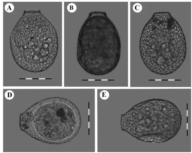
Fig. 1. Light micrographs of clade A cells (Nebela guttata): A. LC-126 from Le Cachot population, Switzerland, B. PE-159 from Pechora
population, Russia, C. LC-118 from Le Cachot population, Switzerland, and D. LC-103 from Le Cachot population, Switzerland. Scale bars represent 50 !m.
an ABI-3130xl or a 3730S 48-capillary DNA sequencer (Applied Biosystems). COI sequences were deposited in GenBank and the accession numbers are given in Table 1.
Phylogenetic analyses
The data set used for phylogenetic analyses (333–665 bp) comprised 32 COI sequences. The sequences were aligned
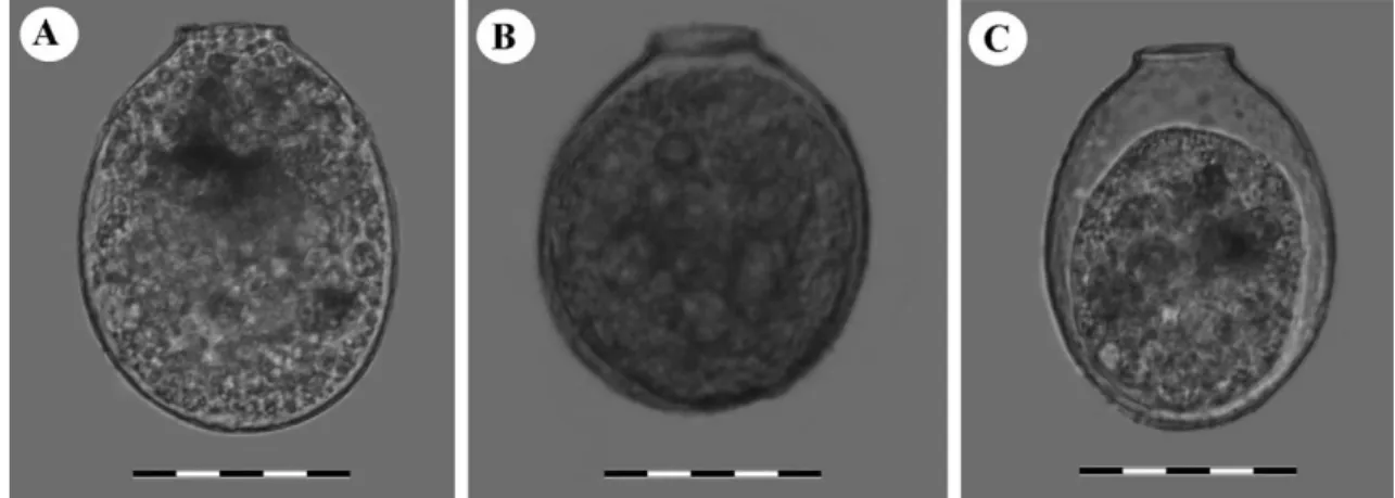
Fig. 2. Light micrographs of clade B cells (Nebela tincta): A. LC-86, B. LC-137, C. LC-117, D. LC-89, E. LC-62 from Le Cachot population,
Fig. 3. Light micrographs of clade C cells (Nebela collaris): A. LC-75, B. LC-69, C. LC-64, D. LC-55 from Le Cachot population, Switzerland.
Scale bars represent 50 !m.
manually using BioEdit software (Hall 1999). The align-ment is available from the authors upon request. Trees were reconstructed using alternatively a maximum likeli-hood and a Bayesian approach. The maximum likelihood tree was built using the RAxML v7.2.8 algorithm (Stamatakis et al. 2008) as proposed on the Black Box portal (http://phylobench.vital-it.ch/raxml-bb/) using the GTR+!+I model. Model parameters were estimated in RAxML over the duration of the tree search. We used sequences from
Certesella martiali(GenBank number JN849064) and from
Nebela tubulosa(JN849020, JN849021, JN849061) to root all tree, based on the fact that these species appear rela-tively closely related to the N. collaris s.l. group in the COI gene-based phylogeny of Hyalospheniidae (Kosakyan et al. 2012). Bayesian Markov Chain Monte Carlo analy-ses were performed using MrBayes v3.1 (Ronquist et al. 2005) with a general time reversible model of sequence evolution with four gamma-distributed rate variation across sites and a proportion of invariable sites. Bayesian MCMC analyses were carried out with two simultaneous chains, and 1,000,000 generations were performed. The generations were added until the standard deviation of split frequen-cies fell below 0.01, according to the manual of MrBayes 3.1 (2005). For every 1000th generation, the tree with
the best likelihood score was saved, resulting in 10,000 trees. The burn in value was set to 25%. Trees were viewed using Fig Tree (program distributed as part of the BEAST package http://tree.bio.ed.ac.uk/software/figtree/). The divergences between sequences were calculated using the package ape in R version 2.10 (R Development Core Team 2010). Missing data were not considered in the calculation (Supplementary Table S1).
Results
A total of 32 COI sequences were obtained from 24 sin-gle cells plus 6 sequences (one Nebela aliciae which was reported as N. tincta var. galeata, one N. flabellum, three
N. tubulosaand one Certesella martiali)) from a previous study (Kosakyan et al. 2012). Three Nebela tubulosa and one
Certesella martialisequences were used as an outgroup. Sin-gle cells investigated in this present study were documented by light microscopy (Figs 1–6). From each population (a population is defined here as “several individuals of a given morphospecies collected from a given moss sample”) a rep-resentative cell was documented by electron microscopy and kept as a voucher species (Fig. 7).
Fig. 4. Light micrographs of clade D cells (Nebela rotunda): A. LC-58, B. LC-71, C. LC-74 from Le Cachot population, Switzerland. Scale
Fig. 5. Light micrographs of clade E cells (Nebela pechorensis): A. PE-149 from Le Pechora population, Russia, with its detailed picture of
lateral pores, B. PE-151 from Pechora population, Russia, with its detailed picture of lateral pores, C. LC-135 from Le Cachot population, Switzerland, with its detailed pictures of lateral pores, D. PE-150 from Pechora population, Russia, E. PE-148 from Pechora population, Russia, F. PE-156 from Pechora population, Russia, G. LC-147 from Pechora population, Russia, H. PE-144 from Pechora population, Russia, I. PE-145 from from Pechora population, Russia. Scale bars represent 50 !m.
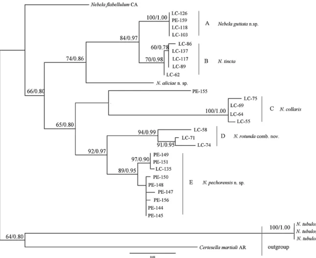
The COI fragment lengths of the newly sequenced cells ranged from 300 bp to 499 bp (Table 1). Our phylogenetic reconstructions showed that taxonomical positions of species within Nebela collaris s.l. must be reconsidered (Fig. 9). Topologies of both the strict consensus ML and Bayesian trees were identical. The tree revealed the existence of
five main clades (A–E) plus a sequence that could not assign to any group, PE-155. Further phylogenetic analy-ses together with detailed morphological observations (see Methods) confirmed the existence of five clear morphotypes within the N. collaris s.l. complex (see Taxonomic actions) that corresponded well with the clades obtained by genetic
Fig. 6. Light micrographs of the remaining forms: A. Nebela aliciae n. sp. from Costa Rica, B. PE-155 from Pechora population, Russia, C. N. flabellulumfrom Canada. Scale bars represent 50 !m for A and B, and 60 !m for C.
means. The cell from which sequence PE-155 derived was clearly distinct from groups A–E. Information on the mor-phology of the cells is summarized in Table 2.
Clade A is supported respectively with 100% bootstrap (B) and 1.00 posterior probabilities (PP) values (Figs 1, 9).
It includes 3 cells (LC-126, LC-118 and LC-103) from Le Cachot (Switzerland) and one cell (PE-159) from Pechora (Russia) populations (Table 1). Cells of this clade are tear-or drop-shaped, with a protruding neck (7.4 ± 0.3 !m high) and with a slightly curved and narrow aperture measuring Table 2. Morphological characteristics of the studied cells.
Clades Cells Aperture (!m) Length/breadth (!m) L/B ratio Test shape
A LC-126 20, curved 83/53 1.5 Tear-shaped LC-159 20, curved 89/59 1.5 Tear-shaped LC-118 21, curved 89/63 1.4 Tear-shaped LC-103 20, curved 80/53 1.5 Tear-shaped B LC-86 25, linear 94/71 1.3 Round-elliptic LC-137 24, linear 90/62 1.4 Round-elliptic LC-117 25, linear 95/70.5 1.3 Round-elliptic LC-89 26, linear 93/71 1.3 Round-elliptic LC-62 25, linear 93/67 1.4 Round-elliptic
C LC-75 32, slightly curved 112/81 1.4 Wide pear-shaped
LC-69 29, linear 112/80 1.4 Wide pear-shaped
LC-64 30, linear 112/77.5 1.4 Wide pear-shaped
LC-55 28, curved 109/74 1.4 Wide pear-shaped
D LC-58 26, linear 94/74 1.2 Round-shaped
LC-71 25, linear 87.5/73.5 1.2 Round-shaped
LC-74 24, linear 88/67 1.3 Round-shaped
E LC-149 20, linear 84.6/53.8 1.4 Tear-shaped
PE-151 19, linear 86/57 1.5 Tear-shaped
PE-135 19, linear 88.5/63.5 1.5 Tear-shaped
PE-150 21, linear 90/69 1.4 Tear-shaped
PE-148 23, linear 92/62 1.3 Tear-shaped
PE-147 23, linear 90/64 1.5 Tear-shaped
PE-156 21, slightly curved 90/64 1.5 Tear-shaped
PE-144 20, linear 92/69 1.3 Tear-shaped
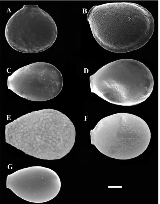
Fig. 7. Scanning electron micrographs of cells from each species-population: A. Nebela flabellulum from Lynn Peak, Canada, B. N. aliciae
n. sp. from Volcán Poás, Costa Rica, C. Nebela guttata n. sp. from Le Cachot, Switzerland, D. N. tincta from Le Cachot, Switzerland, E. N.
collarisfrom Le Cachot, Switzerland, F. N. rotunda comb. nov. from Le Cachot, Switzerland, G. N. pechorensis n. sp., from Pechora, Russia. Scale bars represent 20!m.
24–26 !m wide, covered with a thick organic lip (Figs 2, 9 and Table 2).
Clade C receives maximum support (100% B and 1.00 PP). All four cells from this clade were collected in Le Cachot (Switzerland) (LC-75, LC-69, LC-64 and LC-55) and share exactly the same morphological characters: wide pyriform shape, relatively large L = 109–112 !m, B = 74–81 !m, and
L/B ratio = 1.4 (Table 2). The neck is almost absent or very short (2.7 ± 0.9 !m high), aperture is wide 28–32 !m, linear, slightly curved or curved, and covered with thick organic lip (Figs 3, 9).
Clade D, also strongly supported (94% B and 0.99 PP), is composed of three cells from Le Cachot population (LC-58, LC.71 and LC-74). Cells from this clade have a typical very rounded shape, a short neck (4.2 ± 0.4 !m high) and a wide linear aperture, which gives an impression of a some-what square shape (Figs 4, 9). Cells are intermediate sized:
L= 87.5–94 !m, B = 67–74 !m, L/B = 1.2–1.3, and the aper-ture is 24–26 !m wide (Table 2).
Clade E with 89% B and 0.95 PP support comprises 8 cells from Pechora (PE-149, PE-151, PE-150, PE-148, PE-156, PE-147, PE-144 and PE-145) and one cell (LC-135) from Le Cachot populations. The tests are tear-shaped, small to medium size L = 84.6–92 !m, B = 54–69, L/B = 1.3–1.5, with a slightly protruding neck (6.2 ± 0.8 !m high) and narrow linear aperture 19–23 !m (Fig. 5).
Nebela flabellulum, and the sequence PE-155 from Pechora population have uncertain positions in the tree, and one species N. aliciae sp. nov. branches robustly with clade A and B.
Discussion
DNA-based studies often show that traditional taxonomy underestimates diversity of both macroscopic and micro-scopic organisms (Harper et al. 2009; Hebert et al. 2004a,b). Cytochrome Oxidase Subunit 1 (COI) was shown to be a good barcoding gene and successfully separated all studied mor-phospecies within the family Hyalospheniidae (Arcellinida) (Kosakyan et al. 2012). In this study we used COI together with morphological analyses to assess the phylogenetic rela-tionships within the Nebela collaris s.l. species complex and
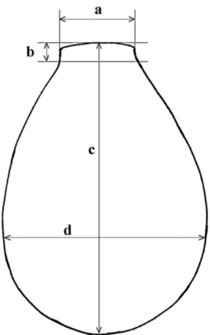
Fig. 8. Schematic sketch and position of the measured axes of the
test: a. diameter of aperture, b. length of the neck, c. length of the test, d. breadth of the test.
related taxa, and to revise the rank of each species and infra-specific taxa within this group.
Taxonomic relevance of the characters used to
discriminate species within the N. collaris s.l.
complex
The taxonomic position of species within N. collaris s.l. has long been a subject of confusion (Heal 1963; Hoogenraad and de Groot 1937; Lüftenegger et al. 1988). The main char-acters that defined the species within this complex were the shape and the size of the test, the shape (curved or linear) and size of the aperture, the length of the neck, and the composi-tion of the test: the size and shape of platelets, the presence or absence of an organic layer on the test, and the presence or absence of the lateral pores. However in most cases these characters were used in a confusing and often contradictory way by different authors. For instance, the length of Nebela
tincta varies between 71–83 !m (Leidy 1879), 85–90 !m and up to 110 !m and more (Cash and Hopkinson 1909), 76–94 !m (Ogden and Hedley 1980), 85–106 !m (Gnekow 1981), 80–110 !m (Lüftenegger et al. 1988). Heal (1963) measured, 1060 individuals and restricted N. tincta to the range of 78–97 !m. Larger forms were referred to as N.
tinctavar. major, N. tincta f. stenostoma, N. bohemica, or
N. collaris.
Another variable character is the shape and size of the aper-ture. Heal (1963) separated large sized taxa (i.e. >100 !m) into two groups: N. collaris sensu stricto, with curved aper-tural lips, and N. collaris sensu lato with straight aperture (such as N. bohemica). Deflandre (1936) observed tests with
Fig. 9. Maximum likelihood bootstrap consensus tree of 31 Nebela collaris s.l. testate amoeba COI sequences based on 300–665 nucleotide
positions. The numbers along the branches represent respectively the bootstraps obtained by maximum likelihood method and the posterior probabilities as calculated with Bayesian analyses. Only values above 50/0.50 are shown. The tree was rooted with outgroup Certesella
martialiand Nebela tubulosa.
the features of N. collaris sensu stricto, which can usually be distinguished from its varieties, but still the mouth width and general shape do vary (Deflandre 1936, plates XIV and XV). Another controversial character is the composition of the test. Given the observation that Nebela species use prey mate-rial to build their tests, Van Oye (1933) tried to separate species that would have different food regimes on the base of the composition of their tests. He discriminated a new species
N. sphagnophila from N. collaris by its angular, irregular plates, which are never round or oval. Ogden and Hedley (1980) illustrated a specimen of N. tincta composed of oval or circular test plates, with a thin layer of organic cement over-lay. Heal (1963) observed on Penard’s slide (20.12.8.501)
N. tinctaindividuals with membranous tests and with tests covered with platelets. Some authors documented that N.
col-larisspecies complex feeds on a wide range of prey and that test composition may change depending on the food source in single clones (Gilbert et al. 2003; MacKinlay 1936). Our molecular data confirm that the type and arrangement of the plates and overall composition of the test cannot be used as a
taxonomic character for discrimination of species within N.
collaris s.l.
The presence or absence of lateral pores is probably the most controversial taxonomic character for N. collaris s.l. Many species were described based on this character. Cash and Hopkinson (1909) described N. parvula as differing from
N. tincta only by the absence of lateral pores. N. minor described by Penard (1902) differing from N. tincta mainly by the absence of lateral pors. Large forms of N. tincta such as N.
tinctavar. major or N. tincta f. stenostoma differ from N.
col-larismainly by the presence of two lateral pores. Deflandre (1936) and Jung (1942) illustrate N. collaris without lateral pores. However, Mazei and Tsyganov (2000) and Ogden and Hedley (1980) illustrate it with pores. Hoogenraad and de Groot (1952) describe N. collaris var. galeata with pores, and Klitzke (1913) describes N. collaris var. bohemica with pores. In addition to this controversy, Heal (1963) notes that pores are impossible to distinguish when the test is com-pletely covered with platelets. We observed pores in all our specimens, with a number that varies from 1 to 4 per cell,
neck, and the shape and size of the aperture. Characters of no proven taxonomic validity were: the composition of the test, the size and shape of platelets, the presence or absence of an organic layer on the test, and the presence or absence of lateral pores.
Phylogenetic analyses of Nebela collaris s.l.
We obtained molecular data for a wide range of morpho-types from the Nebela collaris s.l. group. Our phylogenetic analyses separate the studied morphospecies into 5 groups (A–E), which are robustly supported by molecular analy-ses (B, PP), and also morphologically easy to discriminate. According to Heal’s (1963)definition, all the species within
N. collaris s.l. with test length ranging from 75 to 95 !m belong to the species N. tincta. However, our phylogenetic data revealed four distinct clades (A, B, D, E) within this size range (75–95 !m).
Closely related clades can be morphologically quite dis-tinct. For instance, individuals from clade A differ strongly from clade B by their smaller size (length <90 vs. 90–95 !m), general drop-like versus elliptic and rounded shape, and narrow curved vs. straight aperture. However, they branch together in the tree with a strong support (B = 84%, PP = 0.97) and share about 96% genetic similarity on the gene consid-ered. In our opinion the combination of morphological and molecular differences clearly indicate that the two clades cor-respond to two different species. Whether representatives of these clades can be considered separated specific entities is however open to debate, since there is no commonly accepted threshold to separate amoebozoan species, as we do not know how far the biological speciesconcept (Mayr 1964) applies to microbial eukaryotes. In animals, a divergence of 4% is considered as sufficient to separate species in a barcoding approach (Hebert et al. 2004a; Witt et al. 2006). Here, a 4% threshold separates efficiently the different morphotypes, and can be used by analogy with animals, especially if we con-sider Arcellinida in general (and certainly the Nebela group) as mostly sexual (Lahr et al. 2011), and therefore following the same modalities of speciation as metazoans. A similar gap was observed in vannellid naked amoebae, another group of amoebozoa (Nassonova et al. 2010).
Representatives of clade D correspond perfectly to the descriptions of N. tincta var. rotunda. Genetic distances (up
The validity of certain characters can vary among taxa. Aperture shape (i.e. straight or curved) has been shown to be a valid criterion for species discrimination in this study, and was used notably to distinguish N. collaris from other large forms of the species complex (N. tincta f. stenostoma,
N. tincta var. major, N. bohemica and N. sphagnophila). Although our molecular data clearly separates all larger sized (L = 109–112 !m) specimens from all smaller forms into the well-supported (100% B and 1.00 PP) Clade C, their aperture varies from straight to strongly curved; different morpholo-gies form a continuum of shapes, and individuals share an important degree of genetic identity (seeFig. 4). Our results suggest that all large-sized (>100 !m length) N. collaris s.l. species and infra-specific taxa such as N. tincta f.
stenos-toma, N. tincta var. major, N. bohemica and N. collaris, N.
sphagnophilacorrespond to one single species: N. collaris (see Taxonomic actions).
Some morphospecies such as Nebela flabellulum, N.
ali-ciaen. sp. and PE-155 branched as different entities than the five main described groups. N. flabellulum is a morphologi-cally well-defined species that differs from other N. collaris
s.l.by being wider than long. N. aliciae n. sp. resembles N.
tinctaf. galeata (Hoogenraad and de Groot 1952; Jung 1936). The 7% genetic divergence between N. tincta and N. aliciae n. sp., brings further support to considering it as an independent taxon (see Taxonomic actions). PE-155 probably constitutes another species given its particular morphology(see Fig. 6B); further investigation will be necessary to describe it as a new taxonomic entity.
Our molecular and morphological analyses show that the main characters that define the species within N. collaris s.l. are the size and the shape of the cells, and probably the size and shape of aperture (as in case of clade A and E). This generally agrees with our previous observations (Kosakyan et al. 2012; Lara et al. 2008).
However, we should not ignore intra-species morpho-logical variability, which may or may not be driven by environmental conditions, including food sources, etc. (Wanner 1991). Such phenotypic plasticity which can lead to morphological difference that do not correspond to molecular differences is a source of confusion for morphology-based taxonomy and studies such as ecology and palaeoecology that are typically based only on morphological characters. Detailed studies combining morphological and molecular
tion compressed oval; composed of pale yellow transparent, structureless, chitinoid membrane; mouth transversely oval. Sarcode colorless; pseudopods digitate, usually two, three, or more. Size – Smallest specimen, 0.076 mm long, 0.056 mm broad, 0.028 mm thick, with the mouth 0.02 mm by 0.008 mm; second specimen broader than long, 0.06 mm long, 0.08 mm broad with the mouth as in the former; third specimen, 0.08 mm long and broad, 0.026 mm thick, and mouth same as in the former; largest specimen, 0.092 mm long, 0.064 broad, and mouth as in the others”. Leidy distinguished it from H.
cuneataby its much more pyriform shape, pale tinted test, and habitat (Sphagnum mosses vs. ponds). The specimens he observed were laterally compressed, oval, with a short neck composed of a pale yellow or straw-colored transparent chiti-noid membrane, without trace of definite structure. Leidy also noted the presence of lateral pores “below the middle” and sometimes also “above the middle” (i.e. at about 1/3 and 2/3, of the distance from the pseudostome to the fundus). Later on Awertinzew (1906) noticed that the test of many of the specimens corresponding to this description bore platelets and the test was not simply a homogenous organic mem-brane. He therefore transferred this taxon to genus Nebela and reported a broader range of test length than in the origi-nal description: 70–120 !m.Heal (1963)considered N. tincta as a well-defined species with a length ranging from 75 to 95 !m. Two other species, N. parvula and N. minor with similar length were described byCash and Hopkinson (1909) and byPenard (1902)respectively as distinct species based on the absence of lateral pores. Our molecular data sug-gests that the presence of pores is not a valid taxonomical criterion and therefore that these tree species need to be syn-onymized. The name Nebela tincta Awertinzew 1906 takes precedence according to the principle of priority (article 23 of the international code of zoological nomenclature).
Diagnosis of Nebela tincta (Leidy) sensuKosakyan et Lara Taxonomic summary:
Arcellinida Kent 1880
Hyalospheniidae (Schulze) Kosakyan et Lara Syn.: Nebela tincta (Leidy 1879) Awerintzew 1906
Nebela bursellaVejdovsky 1882
Hyalosphenia tinctaLeidy 1879
Nebela minorPenard 1902
Nebela parvulaCash 1909
on, whenLeidy (1879)separated Nebela from Difflugia based on the structure of the test, he considered all pyriform species with test length around 150 !m and breadth 72 !m, as Nebela
collaris, which then became the type species of genus Nebela. Heal (1963) noted that, within this group, individuals with curved apertural lips can be separated from the rest. He referred to these morphotypes as N. collaris sensu stricto. However it remained problematic to separate among sev-eral large taxa ranging from 95 to 155 !m in length, such as N. tincta f. stenostoma, N. tincta var. major, N. bohemica,
N. sphagnophilaand N. collaris; these taxa were generally referred to N. collaris sensu lato. The main discriminating character between all these taxa is (1) the size, which often overlaps, (2) the presence or absence of lateral pores and (3) the shape of the platelets (as in case of N. sphagnophila).
We observed pyriform species with the length 109–112 !m length and 74–81 !m breadth, with variable aperture: linear, slightly curved or strongly curved, which together form a separate clade with high 99 B and 100 PP values. We sug-gest synonymizing the above-mentioned species with Nebela
collaris. Further extensive sampling focusing especially on this group would be needed to ascertain if Nebela collaris is a homogenous species or not.
Diagnosis of Nebela collaris (Ehrenberg 1848)sensu Kosakyan et Gomaa
Taxonomic summary: Arcellinida Kent 1880
Hyalospheniidae (Schulze) Kosakyan et Lara
Nebela collarissensu Kosakyan et Gomaa Syn.: Nebela collaris (Ehrenberg 1848) Leidy 1879
Difflugia collarisEhrenberg 1848
Diffluga cancellataEhrenberg 1848
Difflugia reticulataEhrenberg 1848
Difflugia carpioEhrenberg 1854
Difflugia laxaEhrenberg 1871
Difflugia celluliferaEhrenberg 1874
Nebela numataLeidy 1874
Nebela bohemicaTaranek 1882
Nebela sphagnophila(Steinecke) Van Oye 1933
Nebela tinctavar. major Deflandre 1936
Nebela tinctaf. stenostoma Jung 1936
The test is large, pyriform, slightly yellowish or brown-ish, laterally compressed with small lateral pores (number of pores can vary), which can be difficult to observe. The test can
According to article 45.6.3, as the name was published before 1961 using the abbreviation var., it is deemed to be subspecific rather than infra-subspecific and therefore falls under rulings for species-group nominal taxa (Chapter 10). (2) Accord-ing to article 46.1, names established at either species ranks (species or subspecies) are simultaneously established at the other rank, with same author and same type. Authority thus is unchanged.
4. Description of new species: Nebela guttata n. sp. Kosakyan et Lara
Taxonomic summary: Arcellinida Kent 1880
Hyalospheniidae (Schulze) Kosakyan et Lara
Nebela guttataKosakyan et Lara
Description: The test is colorless or slightly brown-ish, tear- or drop-shaped, with a protruding narrow neck (7.4 ± 0.3 !m high), laterally slightly compressed, with small lateral pores (number of pores can vary) (Figs 1A–D, 7C). Test composed of small particles (likely obtained from preys, e.g. euglyphid testate amoebae), which often can be cov-ered with thin layer of organic cement. The aperture is oval, curved (Figs A–D, 7C). Dimensions (based on 5 individuals): length: 80–89 !m, breadth: 53–65 !m, width of aperture: 20–22 !m.
Hapantotype: The tests were collected from Sphagnum mosses in a peatland in Le Cachot, Vallée de la Brévine, Switzerland (47.5◦N 6.4◦E), except one PE-159, which was collected from Pechora, Russia (62◦05,449#N 58◦19,050#E). One SEM stub with several specimens is deposited at the Natural History Museum of Neuchâtel (Ref Nr. SEM-A-2, UniNe-EM-2). COI sequences were deposited in GenBank with accession numbers JX682598, JX682585, JX682588, and JX682587.
Etymology: The name of this species is derived from the Latin word “gutta” which means drop or tear.
Note: Nebela guttata resembles N. tincta, from which it differs by its narrow protruding curved aperture and slender drop shape of the test. Our molecular data clearly separates these two species (sequence divergence up to 4%).
5. Description of new species: Nebela pechorensis n. sp. Kosakyan et Mitchell
length: 84–92 !m, breadth: 54–69 !m, width of aperture: 19–23 !m.
Hapantotype: The tests were collected from Sphagnum mosses in a peatland in Pechora, Russia (62◦05,449N; 58◦19,050E), and only one was collected from Le Cachot, Switzerland. Dry moss samples containing this species are deposited in the sample collection of the laboratory of Soil Biology, University of Neuchâtel, Switzerland (codes: EM-1614). One SEM stub with several specimens is deposited at the Natural History Museum of Neuchâtel (Ref. Nr.: SEM-A-3, UniNe-EM-3). COI sequences were deposited in GenBank with accession numbers JX682581, JX682583, JX682593, JX682582, JX682580, JX682579, JX682584, JX682577, JX682578.
Etymology: The name of this species is derived from the name of Pechora River, and the general region where moss samples containing this species were collected.
Note: Nebela pechorensis by shape very much resembles
N. guttata, from which it differs only by the linear aperture. Despite the similar morphology, these two species are clearly genetically different (sequence divergence up to 12%). These two species can therefore be considered as examples of pseu-docryptic species.
6. Description of new species: Nebela aliciae n. sp. Mitchell et Lara
Taxonomic summary: Arcellinida Kent 1880
Hyalospheniidae (Schulze) Kosakyan et Lara
Nebela aliciaeMitchell et Lara
Description: The test is wide pyriform, with a lateral keel about 5 !m wide, laterally compressed, with a small lateral pore on each side (Figs 6A, 7B). The test is composed of small oval particles likely obtained from preys (i.e. euglyphid tes-tate amoebae). The aperture is oval, linear (Figs 6A, 7B). Dimensions (based on 7 individuals): length: 104–115 !m, breadth: 76–93 !m, width of aperture: 24–27 !m. (Note: This description is based on Nebela tincta var. galeata data pub-lished inKosakyan et al. 2012).
Hapantotype: The tests were collected from mosses Vol-cán Poás, Costa Rica (10.11◦N 84.13◦W). Dry moss samples containing this species are deposited in the sample collection of the laboratory of Soil Biology, University of Neuchâtel, Switzerland (code: EM-1451). One SEM stub with several
Nebela tinctaf. galeata). Our molecular and morphological data suggests that N. aliciae is indeed an independent species from other studied taxa. Molecular data on Nebela tincta f.
galeatais however lacking.
Identification key of N. collaris s.l.
1. → Test wider than long. . .. . .. . .. . .. . .. . .. . .. . .. . .. . .. . .. . .. . .. . .. . .. . .. . .. . .. . .. . .. . .. . .. . .. . .. . .2 ← Test longer than wide. . .. . .. . .. . .. . .. . .. . .. . .. . .. . .. . .. . .. . .. . .. . .. . .. . .. . .. . .. . .. . .. . .. . .3 2. → L. 72–111 !m, B. 90–133 !m, strongly flattened, with short neck, with linear or slightly curved aperture
19–34 !m. Mostly in moist Sphagnum mosses in raised bogs and heathlands.Fig. 7A Nebela flabellulum Leidy 1874
← Smaller species, L. 60 !m, B. 70 !m, without neck. In wet Sphagnum
N. acolla Cash 1909
3. → Larger species, test longer than 100 !m . . .. . .. . .. . .. . .. . .. . .. . .. . .. . .. . .. . .. . .. . .. . .. . .. . .. . .. . .. . .. . .. . .. . .. . .. . .4 ← Smaller species, test shorter than 100 !m . . .. . .. . .. . .. . .. . .. . .. . .. . .. . .. . .. . .. . .. . .. . .. . .. . .. . .. . .. . .. . .5 4. → Species with lateral ridge, wide ovoid, L. 104–115 !m, B. 76–93, with wide linear aperture 24–27 !m. In mosses.
Fig. 7B N. aliciae n. sp.
← Species without lateral ridge, L > 100 !m, L/B = 1.4, pyriform, with very short (2.7 ± 0.9 !m high or almost absent) neck, with aperture slightly or strongly curved, or sometimes linear, 28–32 !m. In moist and wet
Sphagnumand other mosses in peatlands, forests and acidic humic ponds. Fig. 3 N. collaris (Ehrenberg 1848) Leidy 1879
5. → Species with protruding neck and narrow aperture 19–23 !m, up
twisted. . .. . .. . .. . .. . .. . .. . .. . .. . .. . .. . .. . .. . .. . .. . .. . .. . .. . .. . .. . .. . .. . .. . .. . .. . .6 ← Species with short neck (4.6 ± 0.2 high), with linear wide aperture 24–26 !m . . .. . .. . .. . .. . .. . .. . .. . .. . .. . .. . .. . .. . .. . .. . .. . .. . .. . .. . .. . .. . .. . .. . .. . .. . .7
6. → Test drop- or tear-shaped, L. 80–89 !m, B. 53–65 !m, L/B = 1.4–1.5, curved aperture. In Sphagnum mosses. Fig. 1 N. guttata n. sp.
← Aperture not curved, L. 84–90(92) !m, B. 54–64(69) !m, L/B = 1.3–1.5. In Sphagnum mosses. Fig. 5 N. pechorensis n. sp.
7. → Test ovoid or elongated elliptic, L. 90–95, B. 62–71, L/B = 1.3–1.4, aperture linear. In Sphagnum mosses. Fig. 2 N. tincta (Leidy 1879) Awerintzew 1906
← Test rounded, L. 87.5–94 !m, B. 67–74 !m, L/B = 1.2–1.3, aperture linear. Lives Sphagnum mosses. Fig. 4 N. rotunda comb. nov.
thank two anonymous reviewers for commenting on the manuscript.
River floodplain. Eur. J. Protistol. 48, 169–177.
Alves, G.M., Velho, L.F.M., Simões, N.R., Lansac-Tôha, F.A., 2010. Biodiversity of testate amoebae (Arcellinida and Euglyphida) in different habitats of a lake in the upper Paraná River floodplain. Eur. J. Protistol. 46, 310–318.
Awerintzew, S., 1906. Freshwater Rhizopoda. Proc. St. Petersburg Nat. Soc. 36, 121–351.
Barth, D., Krenek, S., Fokin, S.I., Berendonk, T.U., 2006. Intraspe-cific genetic variation in Paramecium revealed by mitochondrial cytochrome c oxidase 1 sequences. J. Eukaryot. Microbiol. 53, 20–25.
Booth, R.K., Meyers, B., 2010. Environmental controls on pore number in Hyalosphenia papilio: implications for paleoenviron-mental reconstruction. Acta Protozool. 49, 29–35.
Cash, J., Hopkinson, J., 1909. British Freshwater Rhizopoda and Heliozoa. II. The Ray Society, London.
Chantangsi, C., Lynn, D.H., Brandl, M.T., Cole, C.J., Hetrick, N., Ikonomi, P., 2007. Barcoding ciliates: a comprehensive study of 75 isolates of the genus Tetrahymena. Int. J. Syst. Evol. Micro-biol. 57, 2412–2425.
Charman, D.J., Hendon, D., Woodland, W.A., 2000. The Identi-fication of Testate Amoebae (Protozoa: Rhizopoda) in Peats. Quaternary Research Association, London.
Cotterill, F.P.D., 1995. Systematics, biological knowledge and envi-ronmental conversation. Biodivers. Conserv. 4, 183–205. Deflandre, G., 1936. Etude monographique sur le genre Nebela
Leidy. Ann. Protistol. 5, 201–286.
Ehrenberg, C.G., 1848. Fortgesetzte Beobachtungen über jetzt herrschende atmosphärische mikroskopische Verhaltnisse. Ber. Verh. Akad. Wiss. Berlin 13, 370–381.
Finlay, B.J., Esteban, G.F., Fenchel, T., 2004. Protist diversity is different? Protist 155, 15–22.
Foissner, W., 1997. Global soil ciliate (Protozoa, Ciliophora) diver-sity: a probability-based approach using large sample collections from Africa, Australia and Antarctica. Biodivers. Conserv. 6, 1627–1638.
Foissner, W., 1998. An updated compilation of world soil ciliates (Protozoa Ciliophora), with ecological notes, new records, and descriptions of new species. Eur. J. Protistol. 34, 195–235. Foissner, W., 1999. Protist diversity: estimates of the
near-imponderable. Protist 150, 363–368.
Foissner, W., 2006. Biogeography and dispersal of micro-organisms: a review emphasizing protists. Acta Protozool. 45, 111–136.
Foissner, W., 2008. Protist diversity and distribution: some basic considerations. Biodivers. Conserv. 17, 235–242.
Folmer, O., Black, M., Hoeh, W., Lutz, R., Vrijenhoek, R., 1994. DNA primers for amplification of mitochondrial cytochrome c
Harper, J.T., Gile, G.H., James, E.R., Carpenter, K.J., Keeling, P.J., 2009. The inadequacy of morphology for species and genus delineation in microbial eukaryotes: an example from the parabasalian termite symbiont Coronympha. PLoS ONE 4 (8), e6577.
Heal, O.W., 1961. The distribution of testate amoebae (Rhizopoda: Testacea) in some fens and mires in northern England. Zool. J. Linn. Soc. 44, 369–382.
Heal, O.W., 1963. Morphological variation in certain Testacea (Pro-tozoa: Rhizopoda). Arch. Protistenkd. 106, 351–368.
Hebert, P.D.N., Cywinska, A., Ball, S.L., de Waard, J.R., 2003b. Biological identifications through DNA barcodes. Philos. Trans. Royal Soc. Lond. B 270, 313–321.
Hebert, P.D.N., Penton, E.H., Burns, J.M., Janzen, D.H., Hallwachs, W., 2004b. Ten species in one: DNA barcoding reveals cryptic species in the neotropical skipper butterfly Astraptes fulgerator. Proc. Natl. Acad. Sci. U. S. A. 10, 14812–14817.
Hebert, P.D.N., Ratnasingham, S., de Waard, J.R., 2003a. Barcoding animal life: cytochrome c oxidase subunit 1 divergences among closely related species. Philos. Trans. Royal Soc. Lond. B 270, 96–99.
Hebert, P.D.N., Stoeckle, M.Y., Zemlak, T.S., Francis, C.M., 2004a. Identification of birds through DNA barcodes. PLoS Biol. 2, 1657–1663.
Heger, T.J., Lara, E., Mitchell, E.A.D., 2011b. Arcellinida tes-tate amoebae (Arcellinida: Amoebozoa): model of organisms for assessing microbial biogeography. In: Fontaneto, D. (Ed.), Biogeography of Microscopic Organisms, Is Everything Small Everywhere? Systematics Association & Cambridge University Press, Cambridge, pp. 111–129.
Heger, T.J., Mitchell, E.A.D., Golemansky, V., Todorov, M., Lara, E., Leander, B.S., Pawlowski, J., 2010. Molecular phy-logeny of euglyphid testate amoebae (Cercozoa: Euglyphida) suggests transitions between marine supralittoral and freshwa-ter/terrestrial environments are infrequent. Mol. Phylogenet. Evol. 55, 113–122.
Heger, T.J., Mitchell, E.A.D., Ledeganck, P., Vincke, S., Van De Vijver, B., Beyens, L., 2009. The curse of taxonomic uncertainty in biogeographical studies of free-living terrestrial protists: a case study of testate amoebae from Amsterdam Island. J. Bio-geogr. 36, 1551–1560.
Heger, T.J., Pawlowski, J., Lara, E., Leander, B.S., Todorov, M., Golemansky, V., Mitchell, E.A.D., 2011a. Comparing poten-tial COI and SSU rDNA barcodes for assessing the diversity and phylogenetic relationships of Cyphoderiid testate amoebae (Rhizaria: Euglyphida). Protist 162, 131–141.
Hoogenraad, H.R., de Groot, A.A., 1937. Biometrische Unter-suchungen an Süsswasserrhizopoden (Rhizopoden und
415–434.
Lahr, D.J.G., Parfrey, L.W., Mitchell, E.A.D., Katz, L.A., Lara, E., 2011. The chastity of amoebae: re-evaluating evidence for sex in amoeboid organisms. Proc. Royal Soc. Lond. B 278 (1715), 2081–2090.
Lara, E., Heger, T.J., Ekelund, F., Lamentowicz, M., Mitchell, E.A.D., 2008. Ribosomal RNA genes challenge the monophyly of the Hyalospheniidae (Amoebozoa: Arcellinida). Protist 159, 165–176.
Leidy, J., 1879. Fresh-water Rhizopods of North America. USGS Terr. Rep. 12, 1–32.
Lin, S., Zhang, H., Hou, Y.B., Zhuang, Y.Y., Miranda, L., 2009. High level diversity of dinoflagellates in the natural environment, revealed by assessment of mitochondrial cox1 and cob genes for dinoflagellate DNA barcoding. Appl. Environ. Microbiol. 75, 1279–1290.
Lüftenegger, G., Petz, W., Berger, H., Foissner, W., Adam, H., 1988. Morphologic and biometric characterization of twenty-four soil testate amoebae (Protozoa, Rhizopoda). Arch. Protistenkd. 136, 153–189.
MacKinlay, R.B., 1936. Observations on Nebela collaris Leidy (pro-parte), a testate amoeba of moorland waters: Part I. J. R. Microsc. Soc. 56, 307–325.
Mayr, E., 1964. Systematics and the Origin of Species: From the Viewpoint of a Zoologist (with a new introduction by the author). Dover Publications, Inc.
Mazei, Y.A., Tsyganov, A.N., 2000. Freshwater Testate Amoebae. KMK Publishing, Moscow.
Ogden, C.G., Hedley, R.H., 1980. An atlas of Freshwater Testate Amoebae. Oxford University Press, Oxford.
Penard, E., 1902. Faune Rhizopodique du Bassin du Léman. Henry Kundig, Geneve.
Ronquist, F., Huelsenbeck, J.P., van der Mark, P., 2005. MrBayes 3.1.http://mrbayes.csit.fsu.edu/index.php
R Development Core Team, 2010. R: a Language and environment for statistical computing, Foundation for Statistical Comput-ing, Version 2.8.0. R Development Core Team, Vienna, Austria. http://www.R-project.org
Smith, H., Bobrov, A., Lara, E., 2008. Diversity and biogeography of testate amoebae. Biodivers. Conserv. 17, 1822–1831. Stamatakis, A., Hoover, P., Rougemont, J., 2008. A rapid
boot-strap algorithm for the RAxML web servers. Syst. Biol. 57, 758–771.
Taranek, K.J., 1882. Monographie der Nebeliden Böhmens Ein Beitrag zur Kenntnis der Süsswasser-Monothalamien. Abh. Königl. Böhm. Ges. d. Wiss.
Van Oye, P., 1933. Rhizopodes du district sub-alpin de la Belgique. Arch. Naturgesch. 2, 538–573.
Wanner, M., 1991. Studies on the morphology of testate amoebae (Protozoa, Rhizopoda) in forests of Southern Germany. Arch. Protistenkd. 140, 45–66.
Witt, J.D.S., Threloff, D.L., Hebert, P.D.N., 2006. DNA barcoding reveals extraordinary cryptic diversity in an amphipod genus: implications for desert spring conservation. Mol. Ecol. 15, 3073–3082.