HAL Id: insu-00684987
https://hal-insu.archives-ouvertes.fr/insu-00684987
Submitted on 5 Mar 2021
HAL is a multi-disciplinary open access
archive for the deposit and dissemination of
sci-entific research documents, whether they are
pub-lished or not. The documents may come from
teaching and research institutions in France or
abroad, or from public or private research centers.
L’archive ouverte pluridisciplinaire HAL, est
destinée au dépôt et à la diffusion de documents
scientifiques de niveau recherche, publiés ou non,
émanant des établissements d’enseignement et de
recherche français ou étrangers, des laboratoires
publics ou privés.
Revision and new data on the Early and Middle
Miocene soricids (Soricomorpha, Mammalia) from
Central and South-Eastern France
Marguerite Hugueney, Pierre Mein, Olivier Maridet
To cite this version:
Marguerite Hugueney, Pierre Mein, Olivier Maridet. Revision and new data on the Early and Middle
Miocene soricids (Soricomorpha, Mammalia) from Central and South-Eastern France. Swiss Journal
of Palaeontology, Springer, 2012, 131 (1), pp.23-49. �10.1007/s13358-011-0036-1�. �insu-00684987�
Revision and new data on the Early and Middle Miocene soricids
(Soricomorpha, Mammalia) from Central and South-Eastern
France
Marguerite Hugueney,Pierre Mein, Olivier Maridet
Introduction
The Early and Middle Miocene fossil record of Central and South-Eastern France comprises numerous and often very rich localities, either from stratified sediments or fissure-fillings, all of which yielded Soricids. The initial studies of these taxa date back several decades and most of them have never been restudied since then. Furthermore, newly dis-covered specimens have never been described. We survey and update taxa that belong to the subfamilies: Heteroso-ricinae, AllosoHeteroso-ricinae, Paenelimnoecinae, Soricinae and Crocidosoricinae, the last of which is paraphyletic (as suggested by Furio´ et al. 2007; Hugueney and Maridet
2011 and others) but it is convenient to separate the taxa whose phylogenetic positions are uncertain from those whose assignment is certain.
Biochronologic background of Limagne Basin localities
The Limagne Basin yielded the types of several soricid genera (Crocidosorex Lavocat, 1952, Oligosorex Kretzoi, 1959, Meingensorex Hugueney and Maridet, 2011; see Fig. 1). The basin includes for instance the well-known locality Montaigu-le-Blin (Allier; reference locality for MN2a), but surrounding localities are not of exactly the same age (Hugueney et al.2006) and yielded different taxa of soricids. We present here the soricids of Gannat and Chavroches, localities never studied before and whose age is a little older than Montaigu-le-Blin.
The soricids of Gannat come from three superposed levels at the top of a quarry which was excavated by M. Hugueney and was subsequently destroyed due to mining activities; they constitute three different localities: (1) the lower level (locality called Gannat-81, G-81) is located
Abstract New or poorly known Soricids from the Early Miocene of Central France (Limagne d’Allier) and from the Middle Miocene of South-Eastern France (near Lyon) are studied; one new genus (Viretia) and a new species (Carposorex burkarti) are described. The soricid assemblages of the Early and late Early/Middle Miocene localities are totally different from each other and new information about appearance and evolution of the taxa are introduced. The following are lists of studied taxa for the beginning of the Early Miocene:Clapasorex bonisi, Clapasorex aff. sigei, Carposorex burkarti nov. sp., Soricellacf.discrepans,Oligosorexaff. antiquusand Crocid-osoricinae indet, and for the late Early/Middle Miocene Dinosorex sansaniensis,Dinosorex pachygnathus,Heterosorex delphinensis, Miosorex desnoyersianus, Miosorex grivensis, Lartetium dehmi,Lartetium ziegleri, Turiasorex pierremeini, Viretianov. gen.gracilidens,Paenelimnoecus micromorphus, Paenelimnoecus crouzeliandHemisorex robustus.
Keywords Soricidae, Heterosoricinae,
Crocidosoricinae,Allosoricinae,Paenelimnoecinae, Soricinae,Early Neogene,France,Central Massif, Region of Lyon,New taxa
M. Hugueney (corr. author),P. Mein,O. Maridet
Laboratoire de ge´ologie de Lyon: terre, plane`tes, environnement, UMR CNRS 5276, universite´Lyon 1, 43, boulevard du 11-novembre, 69622 Villeurbanne cedex, Francee-mail: [email protected]. Meine-mail: [email protected]
O. Maridet
Key Laboratory of Evolutionary Systematics of Vertebrates, Institute of Vertebrate Paleontology and Paleoanthropology, Chinese Academy of Sciences, Beijing 100044, Chinae-mail: [email protected]
Postprint of the manuscript published in Swiss Journal of Palaeontology:
Hugueney, M., Mein, P., Maridet, O., 2012. Revision and new data on the Early and Middle
Miocene soricids (Soricomorpha, Mammalia) from Central and South-Eastern France. Swiss
Journal of Palaeontology 131, 23–49.
roughly 6 m beneath the top of the quarry and is irreg-ularly stratified. (2) The intermediate level (locality called Gannat-sommet, G-som) is located about 2 m higher (but intercalated between stromatolitic construc-tions). The eomyids taxa produced by these localities display a morphology which is more advanced than those of Coderet (MP 30), but a little less advanced than those of Paulhiac (MN1) indicating an intermediate age between the two localities. (3) The upper layer (locality called Gannat-sup., G-sup), near the top of the quarry, yielded a relatively poorly diversified fauna. Plesios-minthus myarion Schaub, 1930 is the most abundant species recovered from this layer (Plesiosminthus is absent in the two lower layers). P. myarion is a species characteristic of the Early Miocene as opposed to P. schaubi Viret, 1936 from the Late Oligocene of Coderet (Hugueney
1997). This layer also yielded Cainotherium miocaenicum Crusafont et al., 1955, a minute cainotheriid, hitherto only known from and abundant in Miocene, also reported in lower levels (G-81 and G-som; Berthet 2003). The three localities of Gannat can thus be ascribed to the lowermost Miocene (MN1).
The fossils of Saulcet were collected by J. Hu¨rzeler around 1930 and are mainly housed in the Natural History Museum Basel, but some are in the Lyon Museum. Their age is difficult to estimate, MN1/MN2a, but it is younger than Gannat and older than Chavroches.
The locality Chavroches comprises irregularly stratified sandy/argillaceous layers which are overlain by stromato-litic formations embedded in identical sediments. The small mammals are recovered from the interstitial deposits and are often strongly encrusted and difficult to observe. This locality was also excavated by J. Hu¨rzeler in 1930 but the soricid material was never studied; in addition, D. Berthet recently made new excavations that yielded abundant material. The eomyids of this locality indicate a correlation MN2a but slightly older than Montaigu-le-Blin (Hugueney et al.2006).
The locality of Cluzel 1 yielded eomyids which corre-late MN2a a bit younger than Chavroches but slightly older than the lower levels of Montaigu-le-Blin (Hugueney et al.
2006).
Biochronologic background of Lyon region localities
In the neighbourhood of Lyon, the two famous localities Vieux-Collonges and La Grive-Saint-Alban which corre-late with the corre-late Early and Middle Miocene, respectively, yielded rich micromammal faunas. Vieux-Collonges, the older of the two, is an isolated fissure-filling in a lime-stone quarry near the village of Vieux-Collonges (Mein
1958). Based on the evolutionary stages of the cricetid
rodents its fauna can be ascribed to MN5 (Mein 1975; Maridet 2003).
La Grive Saint Alban was exploited for limestone; several quarries were worked at the beginning of the 20th century. The fissures in the limestone are filled with red clay which contains rich faunas of small and large mam-mals (Mein and Ginsburg 2002). These localities were excavated by P. Mein, but nowadays most of the quarries have disappeared. Each fossiliferous fissure is treated as a separate locality, with noticeably different ages (Mein and Ginsburg2002; Maridet2003), and is distinguished with a letter and a number (La Grive L3, L5, L7, M; the former being the reference locality for the MN7–8). La Grive L3 and L5 are the oldest fissure-fillings in the area of La Grive-Saint-Alban as indicated by the occurrence of Democricetodon aff. brevis (Schaub, 1925) of which the morphology is intermediate between D. gracilis Fahlbusch, 1964 of Sansan (see Maridet and Sen2012) and D. brevis from La Grive M and L7 (Maridet 2003). However, the occurrence of the genus Hispanomys Mein and Freuden-thal, 1971 in La Grive L3 and L5 (Lo´pez-Anton˜anzas and Mein2009) suggests a correlation of the two localities to biozone MN7–8 rather than to MN6 (reference locality Sansan). The very similar faunal composition of La Grive L3 and L5 suggest a close age, possibly ‘early’ MN7–8. Mein and Ginsburg (2002), however, suggested that La Grive L3 might be slightly older than La Grive L5. The two other localities La Grive L7 and M yielded similar faunas indicating a close age. However, as demonstrated by Maridet (2003), the species Spermophilinus bredai von Meyer, 1848, Heteroxerus grivensis (Major, 1893), Eomuscardinus vallesiensis Hartenberger, 1966 and Democricetodon (Falhbuschia) larteti (Schaub, 1925), which increase in size over time, are slightly larger in La Grive M than in La Grive L7 suggesting that La Grive M is the youngest of the two.
Materials and methods
The classification follows Furio´ et al. (2007) and the ter-minology and measurements follow Reumer (1984). For the dental formula we follow the quotation of Dannelid (1998) counting ‘upper/lower’ incisor, antemolars, pre-molars and pre-molars (e.g. genus Sorex 1.5.1.3/1.1.1.3). Upper and lower cheek-teeth are indicated by upper case (I, A, P, M) and lower case letters (i, a, p, m), respectively. Other abbreviations are maxilla (Mx) and mandible (Md). Mea-surements are given in millimetres: L, maximal length; W, maximal width; H, maximal height; TrW, trigonid width; TaW, talonid width. Mandibular measurements are taken in buccal view unless otherwise indicated. In addition, H/m2
designates the height of the mandibles under m2. Mea-surement tables are synthetic ones but details of measure-ments are available on request.
SEM photographs of the material from Vieux-Collonges and La Grive were taken by P. Bert and D. Jammot (Dijon University) a long time ago but have not been previously published.
Institutional abbreviations University Lyon1 (FSL), Museum of Lyon (ML or MLMC), Muse´e national d0Histoire naturelle de Paris (MNHN); Naturhistorisches Museum of Basel (NMB).
Systematic palaeontology
Class Mammalia Linnaeus, 1758 Order Soricomorpha Gregory, 1910 Family Soricidae G. Fischer, 1814
Subfamily Heterosoricinae Viret and Zapfe,1952
Genus Dinosorex Engesser,1972
Type species Dinosorex pachygnathus Engesser,1972
Type locality Anwil (Switzerland), late Middle Miocene (MN7–8)
Dinosorex sansaniensis (Lartet, 1851)
Material Vieux-Collonges: FSL 66665, A2; MLMC 409, left mandible with i, m1, m2 and m3 (i crown length = 3.87, vertical diameter = 1.92, transversal diameter = 1.28, L m1–3 = 5.96; height under m3 = 2.75), FSL 66666, right m1.
Measurements Table1
Description The tips of molar cusps are pigmented and the incisor is darker than the molars; this pigmentation is more visible on the buccal side. According to Engesser (2009), this species possesses four upper antemolars, A2 being the only triangular-shaped antemolar. The A2 from Vieux-Collonges (FSL 66665) displays a triangular shape. On the mandible (MLMC 409), the number of antemolars is difficult to estimate because the upper part of the man-dible is broken. Despite the fact that Vieux-Collonges
(MN5) is older than Sansan (MN6, S-W France), the comparison of the mandible from Vieux-Collonges with the numerous mandibles recovered from Sansan shows no differences, either in length or in morphology. This species was precisely described in Engesser (2009, p. 9–13) and the mandible, the incisor and the condyle are figured (Figs. 1a–c, 3a–c).
Dinosorex pachygnathus Engesser1972
Figure1a–c
Material The bulk of the material comes from the La Grive L7 fissure. But other fissures from La Grive L3 and M produced comparable material. All these fissures cor-relate to MN7–8. Fissure L3: FSL 66657–66660, four isolated teeth (M2, m2, m3). Fissure L7: FSL 66641–FSL 66656, two maxilla and six mandibles, plus several isolated teeth (I, A, P4, M1, M2, M3, i, a, m1, m2 and m3). Fissure M: FSL 66661–66664, one maxilla and two mandibles, plus isolated P4, p4 and m1.
Measurements Table2
Description The upper incisor of Dinosorex pachygna-thus was figured by Engesser (1972). As in the Anwil locality, the incisors are larger than those of Heterosorex delphinensis and the incisor tip is slightly bifid: on the side of the main point, there is a secondary anterolabial tip. The main point is prolonged by a coloured crest joining the basal cingulum. The lingual edge furrow is wide and deep. The labial side is lined by a slight basal cingulum.
The labial side of M3 has no post-metacone ridge and does not show a W (parastyle-paracone-mesostyle-meta-cone). From the metacone, a posterolingual ridge closes the posterior edge; it turns on the lingual edge to the protocone base. The protocone, very open V-shaped, shows a branch reaching the anterior edge at the back of the paracone. This tooth has a great central transverse valley; its lower point is at the centre of the tooth. The labial edge shows a small cingulum between the parastyle and the mesostyle.
The morphology of the other teeth is identical to the descriptions given by Engesser (1972,1975).
Discussion The main differences between D. pachy-gnathus and H. delphinensis are (1) the number of upper antemolars: three in H. delphinensis and four in D. pachy-gnathus, (2) the morphology of the P4: this tooth is trian-gular in H. delphinensis with a very long buccal edge, but rectangular for D. pachygnathus with the buccal and lin-gual edges about the same length, (3) the morphology of the mandible: the upper edge of the ascending ramus is located posteriorly to the middle of the masseteric fossa in H. delphinensis, whereas it is located anteriorly in D. pachygnathus; furthermore, the inferior edge of the masseteric fossa is below the molar row in H. delphinensis but above it in D. pachygnathus.
Table 1 Measurements of Dinosorex sansaniensis from Vieux-Col-longes (in mm) N° Element L W FSL66665 A2 1.80 1.52 FSL66666 m1 2.64 1.68 MLMC409 m1 2.82 1.62 m2 2.40 1.48 m3 1.70 1.30 I Total length 3.9 Vertical diameter 1.9 Thickness 1.3
Genus Heterosorex Gaillard,1915
Type species Heterosorex delphinensis Gaillard,1915
Heterosorex delphinensis Gaillard,1915
Holotype Muzzle and associated mandible ML Lgr.209 (Gaillard1915; Figs. 4, 5, 6) and figured in Viret and Zapfe (1952, Fig. 11a–c); from La Grive-Saint-Alban, exact ori-gin unknown.
Material The holotype is the only material referred to this species.
Description The three upper antemolars of H. delphinensis decrease in size from A1 to A3. The mandible possesses only two alveoli before m1, one for p4 and one for a1.
Discussion Since the study of Gaillard (1915), no new material from La Grive has been ascribed to this species. The precise origin of these specimens is completely unknown and, since 1915, some quarries have been destroyed. The only complementary indication is the presence of a small glirid Miodyromys hamadryas
hamadryas (Major 1899), stored in the collections of the British Museum (M5298a), with the same imprecise origin (likely the same fissure-filling as H. delphinensis). H. del-phinensis is unknown in the Middle Miocene, faunas from La Grive fissures, but Miodyromys hamadryas hamadryas (also known for instance in Germany from Langenmoosen, MN5) proves that older fissure fillings where excavated in the area of La Grive at the beginning of the 20th century. According to a reduction of the number of antemolars with time, H. delphinensis is so far the most derived spe-cies of the genus with only 3/1 antemolars and it is also the last record of this genus.
Remarks Heterosoricinae are rare in the faunas studied here. Although they were present in the Upper Oligocene, they are almost unknown in the Earliest Miocene of Limagne, Heterosoricinae indet. from Saulcet (MN1/2) being the only taxon reported so far. For the Middle Miocene the subfamily comprises only three species: D. sansaniensis, D. pachygnathus and H. delphinensis. Among the localities
Fig. 1 Dinosorex pachygnathusEngesser,1972from La Grive L7 (a, c) and M (b): a left maxillary fragment with I and one antemolar A1, FSL 66641(a1 labial view, a2 occlusal view); b left ascending ramus
with condyle, FSL 66663 (posterior view); c left mandible with incisor and m1–m3, FSL 66652 (occlusal view)
studied here, Vieux-Collonges is the only locality which yielded a few specimens of Dinosorex sansaniensis. Subfamily Crocidosoricinae Reumer,1987
Genus Clapasorex Crochet,1975
Type species Clapasorex sigei Crochet,1975
Type locality Bouzigues 1 (France, He´rault), Early Mio-cene (MN2b)
Clapasorex bonisi Crochet,1975
Figure2a–f, h–k
Material and measurements. Gannat-81: I, P4,M1,M2, FSL 98200–98204; mandibular fragments, FSL 98205–98208; isolated i and m1–2, FSL 98209–98210. Gannat-sommet: isolated incisors, FSL 98211; mandibular fragment with p4–m2, FSL 98212; isolated teeth, FSL 98213–98219 (Table3).
Description Very small-sized soricid (L m1–3 = 2.62–2.76) which possesses apparently three single-rooted antemolars anterior to the double-rooted p4. The antemo-lars are unknown, but the second alveolus behind the incisor is proportionally large and seems to contain the roots of a single-rooted a2 and of a minute single-rooted a3 hidden under p4. Consequently the lower dental formula is 1.3?.1.3. All teeth are pigmented.
The lower part of the mandible (FSL 98212; Fig.2f) is slightly damaged under the molars, but its better preserved
anterior part shows a large alveolus for a1, a hardly smaller one for a2 and 3 under the small p4 which is very pro-cumbent. The mental foramen is located directly anterior to the posterior root of p4; the anterior part of the mandible remains high but is very short so that the posterior border of the alveolus of i is situated rather close to the anterior part of p4. Another mandible with m1–3 shows part of the ascending ramus, which is almost perpendicular to the horizontal branch with an inferior undulating base and a very long angular process, denticulated on its upper part.
Among the lower teeth, six isolated lower incisors were found in G-som: all are bicuspulate and small, faintly fal-ciform and with a marked posterocuspule (FSL 98211). Only one p4 on a mandible is known in G-som; it is rather worn and its apex only shows a flat triangular surface with two divergent symmetrical posterior crests ending before the basal cingulum. The rather high cingulum surrounds the entire tooth but disappears under the anterior emargination; a small median posterocuspule is present.
The morphology of the molars is characterised by acute tubercles lying on a massive base and a thin but marked basal cingulum, only slightly enlarged anteriorly; the internal cingulum is also present. The length of m1 is not very different of that of m2. The oblique crest is ascending on the posterior wall of the trigonid; the entocristid is short but well developed and acute on m1, a little rounded and
Table 2 Measurements of Dinosorex pachygnathus from La Grive localities L3, L7 and M (in mm)
Locality N L TrW TaW
Range Mean Range Mean Range Mean
m1–3 L7 1 – 5.80 – – – – m1 L7 11 2.45–2.74 2.59 1.43–1.75 1.56 1.57–1.85 1.69 M 2 2.76–2.80 2.78 1.53–1.77 1.65 1.66–1.88 1.77 m2 L3 1 – 2.38 – 1.59 – 1.47 L7 8 2.20–2.54 2.35 1.50–1.73 1.59 1.33–1.62 1.50 M 1 – 2.23 – 1.55 – 1.58 m3 L3 1 – 1.85 – 1.46 – 1.02 L7 5 1.58–1.89 1.70 1.03–1.30 1.18 0.92–1.04 0.96 P4–M2 L7 1 – 6.10 – – – – A1 L7 1 – 2.45 – – – – P4 L7 1 – 2.07 – 2.58 – – M 1 – 2.12 – 2.20 – – M1 L7 1 – 2.26 – 2.65 – – M2 L3 2/1 2.23–2.23 2.23 1.86–2.46 2.16 1.77–1.77 1.77 L7 1 – 2.13 – 2.53 – – Locality N L H H/m2
Range Mean Range Mean Range Mean
Md M 2 – – – – 3.23–3.40 3.31
lower on m2; the talonids of m1–2 are well developed; the talonid of m2 is about as large as the trigonid. The talonid of m3 is closed by two low crests ascending to only one cuspide, the hypoconid.
The upper incisor is very small, the buccal side of its wide and flattened root showing only a labial furrow; the apex is well rounded but points anteriorly; the talon is also rounded and the buccal cingulum is well marked.
The antero-labial border of P4 (FSL 98201) is rounded; the parastyle is labially located, next to paracone and
protocone; a short parastylar crest joins the base of the paracone; hypocone and posterior emargination are absent; a faint and regular cingulum surrounds the hypoconal flange.
M1 is short and compressed antero-posteriorly, the posterior emargination is lingual and faint and hypoconal flange is short. Tubercles are acute and the labial border is clearly indented; the parastyle shows a short distal crest. The preprotocrista ends a little before the paracone and the postprotocrista goes abruptly down and ends anteriorly to a
Fig. 2 Clapasorex bonisifrom Gannat (a–f, h–k) and Clapasorex aff. sigei from Cluzel 1 (g): a left m1, FSL 98213 (labial view, G-som.); b left m2, FSL 98214 (labial view, G-som.); c left m3, FSL 98215 (labial view, G-som.); d left i, FSL 98211 (labial view, G-som.); e left mandibular fragment with m1–3, FSL 98205 (labial view, G-81); f left mandibular fragment with p4–m2, FSL 98212
(labial view; G-som.); g left p4, FSL 98222 (g1 labial view, g2 occlusal view, Cluzel1); h left I, FSL 98200 (labial view; G-81); i left P4 on maxillary fragment, FSL 98201 (palatal view, G-81); j left M1, FSL 98202 (occlusal view, G-som.); k left M2, FSL 98203 (occlusal view, G-81)
small but well-rounded hypocone with distal cingulum; a thin but well-marked cingulum starts before hypocone base toward the anterior part of the tooth. M2 (FSL 98203) is a little smaller than M1, also with a more symmetric W-shaped wear facet; the preprotocrista is ascending labially under the paracone; the heel is short with a very faint tubercle and an internal cingulum. M3 is unknown.
Discussion The material from Gannat is homogeneous regarding size and, even if a part of the dentition, the con-dyles and the skull are missing, facilitates the distinction of the main characteristics of the genus Clapasorex: the shape of the anterior part of the mandible and of the antemolars alveoli, the pattern of premolars and molars (and particularly the upper molars with lingual cingulum, the small but double-rooted p4, the moderate width of m1–2 talonids). Furthermore, the tiny size is similar to that of the coeval Clapasorex bonisi from Paulhiac (MN1; L m1–3 = 2.82).
Remarks The lower incisor of Clapasorex from Gannat is morphologically not very different from that of Oligos-orex even if it is a little more falciform and much smaller; it differs from the Clapasorex sigei incisor figured by Crochet (1975) in having only two denticles. It is however possible, based on the illustration published by Crochet (1975) that some of the five mentioned denticles of Cla-pasorex sigei are an artefact due to the preservation.
In the description of the p4 of C. sigei, Crochet (1975) notices that the lingual crest of p4 disappears always rap-idly but, on his Fig. 11, this lingual crest is very long and was distinguished by Reumer (1987) as a special type of Crocidosoricinae. In fact, the pattern of the p4 is variable on the material from Bouzigues: in Lyon collections, in some p4 the lingual crest seems to be longer than the labial one, but the p4 often shows two subequal divergent crests or even a junction between the postero-labial crest and the postero-cingulum (see C. aff. sigei below).
The occurrence of C. bonisi in Gannat, whereas no equivalent morphology is known in the European Upper Oligocene, suggests that Clapasorex is likely an immigrant genus arriving in Europe at the beginning of the Miocene. The teeth of Crocidosorex sp. from Ulm-Westtangente (MN2a; Ziegler 1989) show roughly the same length as C. bonisi from Gannat but the width of the German specimens seems to be smaller. The talonid of m3 is also more rect-angular and the upper molars have no internal cingulum in these German specimens; we consequently agree that they do not belong to the genus Clapasorex. As well as the small soricid from Anatolia (Soricid III; MN1; van den Hoek Ostende2001): its size is identical to that of C. bonisi and its morphology quite similar, but the upper molars show only ‘‘a short and narrow cingulum against the anterior flank of the protocone’’ and the upper incisor shows a strong anterior curve and its apex points backward. Clapasorex aff. sigei Crochet,1975
Figure2g
Material and measurements Chavroches: NMB CHAV 1127, Md (L m1–3 = 3.25; p4 = 0.58 9 0.54; m1 = 1.20 9 0.78; m2 = 1.17 9 0.77; m3 = 0.96 9 0.67). Cluzel 1: FSL 98222, p4 (0.70 9 0.51); FSL 98223, m2 (1.09 9 0.82); FSL 98224, M3 (0.95 9 1.19).
Description The Chavroches sample includes a small mandible (NMB CHAV 1127) with a very short anterior part. The specimen possesses a two-rooted and very short p4 with triangular apex, three one-rooted antemolars and teeth with thin and regular cingula.
The locality Cluzel 1 yielded also a few small teeth. The p4 (Fig.2g) shows a particularity: the postero-labial and postero-lingual arms are sub-equal, each with a fold joining the basal cingulum, but, a crest starting from the median postero-cuspule joins the postero-labial crest in its middle height, creating a postero-lingual basin.
Discussion The morphology of these teeth corresponds to Clapasorex; they are a little larger than the teeth of Gannat and their size fits better that of C. sigei. Moreover, in Bouzigues some p4 show the same pattern as that of Cluzel 1.
Starting from MN2, the local lineage of Clapasorex grows larger and reaches a size even larger than C. sigei (L m1–3 = 3.09 in Bouzigues). van den Hoek Ostende (2003) notices a size increase from MN1 to MN3, as the species C. alvarezae (MN3, Ramblar 3, Spain) is larger than C. sigei. C. bonisi from Gannat (MN1) and the more recent C. aff. sigei from Chavroches and Cluzel 1 (MN2) could confirm this evolutionary trend and could support the hypothesis formulated by van den Hoek Ostende (2003) that these three species of Clapasorex could belong to a same lineage, lineage that would become extinct after the MN3.
Table 3 Measurements of the lower and upper dentitions of Cla-pasorex from Gannat (in mm)
Locality N L W
Range Mean Range Mean p4 G-som. 1 – 0.66 – 0.53 m1 G-som. 2 0.92–1.08 1.00 0.60–0.61 0.60 m2 G-som. 4 0.92–1.09 1.03 0.65–0.77 0.68 m3 G-som. 3 0.86–0.95 0.90 0.54–0.70 0.61 p4–m3 G-som. 1 – 3.25 – – m1–3 G-som. 1 – 2.62 – – P4 G-81 1 – 1.06 – 0.92 M1 G-81 2 0.96–1.09 1.02 1.30–1.40 1.35 M2 G-81 2 0.85–0.95 0.90 1.25–1.26 1.25 G-som. 2 0.94–0.96 0.95 1.21–1.23 1.22 G-som. Gannat sommet
Genus Carposorex Crochet,1975
Type species Carposorex sylviae Crochet,1975
Type locality Laugnac (France, Lot-et-Garonne), Early Miocene (MN2b)
Carposorex burkarti nov. sp. Figure3a–f
Holotype left Md with p4–m3 and fragmentary ascend-ing ramus (ML CHAV 20.270300; Fig.3c)
Type-locality Chavroches (France, Allier), Early Mio-cene (MN2a)
Etymology Dedicated to Burkart Engesser appreciating his great contributions to mammalian palaeontology and especially to our knowledge of soricids.
Diagnosis Medium-sized soricid (range of L m1–3 = 3.12–3.37) with strongly pigmented teeth; lower dental formula = 1.3.1.3 with two-rooted p4. Falciform, bicusp-ulate lower incisor; lower molars with the distolabial crest of the protoconid joining the posterolabial cingulum and m3 with entoconid. P4 with strongly projecting parastyle, M1 and M2 with a rounded hypocone and M3 reduced posteriorly.
Differential diagnosis C. burkarti is ascribed to the genus Carposorex because, during the European Early Miocene, it is the unique genus with the disto-labial crest of the protoconid joining the distal cingulid. It differs from Carposorex sylviae by its smaller size, pigmented teeth and less roughly wrinkled enamel, in the presence of three
Fig. 3 Carposorex burkarti nov. sp. from Chavroches (a–f) et Carposorex aff. burkarti from Gannat sup. (g): a left (reversed) mandibular fragment with internal temporal fossa and condyle, ML CHA 20.270301 (a1 lingual view, a2 sketch of condyle in caudal view); b left i, ML CHAV 20.270302 (labial view); c left mandible with p4–m3 and fragmentary ascending ramus, ML CHAV
20.270300, holotype (c1 occlusal view, c2 labial view); d left mandible with I, a1, a3 and p4–m2, ML CHAV 20.270303 (d1 labial view, d2 occlusal view, d3 lingual view); e left maxilla with P4–M3, ML CHAV 20.270304 (palatal view); f right (reversed) I, CHAV 20.270305 (labial view); g left maxilla with P4–M3, FSL 98227 (palatal view)
lower antemolars instead of two; p4 shows two well-sep-arated roots and two subequal posterior crests instead of only the postero-labial crest developed in C. sylviae and the molar talonids are not reduced.
Paratypes left Md with ascending ramus and condyle (ML CHAV 20.270301); left i (ML CHAV 20.270302); left fragment Mmd with i–m2, except a 2 (ML CHAV 20.270303); left Mx with P4–M3 (ML CHAV 20.270304); right I (ML CHAV 20.270305); fragment Mx, isolated upper teeth (ML CHAV 20.270310-20.270314); fragment Md with teeth (ML CHAV 20.270315-20.270320).
Measurements Table4
Description of the holotype The horizontal branch of the type-mandible (Fig.3c) gets thicker from front to posterior part; the ascending ramus is high (height of the coronoid process = 3.67) and its anterior margin is less oblique than the posterior one, with a rounded coronoid spicule and an external temporal fossa slightly oblique. The mental fora-men is located low under the anterior root of p4.
The p4 has two well-separated roots; its shape is elon-gated with two divergent posterior crests sub-equal in length; it also displays a well-developed cingulum with a minute median postero-tubercle. The labial border of p4 is more rounded and longer than the lingual one.
The molars are characterised by a protoconid-distolabial crest joining the labial cingulum; the cingula are well marked and present also lingually; m2 is generally a little smaller than m1; the entoconids and the curved entocristids are well developed and the hypolophids end before the lingual border. m3 possesses a well-developed and rect-angular talonid with a minute entoconid.
Description of referred material No mandibular con-dyles are well preserved and only their shape can be
observed: they are nearly triangular with rounded angles (Fig.3a2). The mandibular foramen is also low under the internal temporal fossa, the posterior and upper angle of which are rounded (Fig.3a1).
The falciform and bicuspulate lower incisor possesses a strong posterior cingulum; the axes between its crown and its root form an obtuse angle. There are three one-rooted antemolars, the a2 being not preserved. a1 rests on the posterior part of the incisor and possesses two divergent crests, the lingual one being longer; a3 displays a rela-tively large root, aligned with the other roots and a minute crown hidden under the anterior part of p4. On the molars, the distolabial crest of the protoconid is more or less visible—and sometimes disappears in m2 and/or m3 (Fig. 3c, d).
The upper incisor is large with a strong posterolabial cingulum; the heel consists of an acute labial tubercle and a lingual ridge. The number of upper antemolars is unknown. P4 has a strong paracone delimited by a rectilinear labial border and a strongly projecting parastyle with short par-astylar crest. P4 is concave anteriorly and markedly con-cave on posterior side; its lingual border shows an enlarged cingulum without hypocone.
Upper molars—hitherto unknown in Carposorex—are massive, slighty wider than long; their hypocone protrudes strongly lingually and continues in a marked postero-cin-gulum reaching the metastyle; the anterior arm of the protocone continues under the paracone of M1 and M2. M1 is slightly larger than M2. M3 is proportionally wide with a transversally elongated metaloph that constitutes the pos-terior edge; the metacone is not visible.
The maxilla shows a well-developed jugal apophysis; in labial view, the infra-orbital foramen ends above the midpoint of M1 and its posterior margin is perpendicular to the base of the maxilla; the bridge over the infraorbital canal ends posteriorly above the paracone of M2; the lac-rimal foramen is large and high.
Discussion Due particularly to differences not only in size but also to different evolutionary level, C. burkarti seems, at first sight, not very close to C. sylviae. But Crochet (1975) ascribed to this genus some smaller isolated teeth from Paulhiac (MN1) and La Bre`te (MN2); the dental formula of these two earlier Carposorex sp. is unknown. However, the minute a3 present in Chavroches is so tiny that its disappearance seems a likely evolutionary trend of Carposorex and moreover, considering a size increase for the genus—as demonstrated by the locality Stubersheim 3 (MN3, Ziegler1989: m1 = 1.72 9 1.00 vs. 1.56 9 1.01 in the earlier type-locality Laugnac)—all these forms could belong to a same evolutionary lineage. Moreover, the Early Miocene locality G-sup yielded a maxilla and a lower incisor that show all the characteristics of Carposorex but are smaller in size (see below).
Table 4 Measurements of the lower and upper dentitions of Car-posorex from Chavroches (in mm)
N L W
Range Mean Range Mean p4 10 0.78–0.95 0.88 0.48–0.63 0.57 m1 17/16 1.23–1.43 1.30 0.73–0.90 0.80 m2 16 1.14–1.35 1.24 0.74–0.90 0.81 m3 9 0.90–1.10 1.00 0.56–0.87 0.67 p4–m3 3 3.95–4.40 4.13 m1–3 6 3.12–3.37 3.28 P4 2 1.46–1.48 1.47 1.25–1.32 1.29 M1 6 1.10–1.36 1.25 1.32–1.65 1.52 M2 2 1.15 1.50 M3 1 0.67 1.17 P4–M3 1 4.20 M1–3 1 2.91
Teeth with the disto-labial crest of the protoconid join-ing the labial cjoin-ingulum are known in the Oligocene of Europe as soon as the beginning of the Oligocene (MP 21) in Srinitium caeruleum (this characteristic is not seen however in the type-species S. marteli) and in the late Oligocene Ulmensia ehrensteinensis and U. antecedens (Ziegler 1989, 1998a), but these forms are massive with wide cingulids and, at the end of the Oligocene, U. ehrensteinensis displays also some differences in the upper teeth (hypocone absent in M1–2, M3 with visible metacone) and is much larger so that an eventual rela-tionship with Carposorex would imply an abrupt decrease in size.
In the Upper Oligocene of Mongolia, Ziegler et al. (2007) described a new genus and species, Tavoonyia altaica, which displays many similarities with C. burkarti. Common characteristics are m1–2 with the distolabial crest of the protoconid, three antemolars, p4 with two divergent sub-equal crests, and M1 with rounded hypocone. Some differences exist, however: Tavoonyia size seems to be a little smaller and p4 seems to be shorter; the hypoconid flange of M1 is less expanded so that the protocone pro-trudes lingually and, moreover, an incomplete mesoloph is present. But many usual diagnostic features of soricids are unknown for Tavoonyia (such as the lower incisor, pattern of m3, P4, and M2-3) and we consequently prefer to ascribe the Chavroches material to Carposorex as archaic member of the C. sylviae lineage.
Relationships between the Asian group Tavoonyia, Aralosorex Lopatin,2004(this genus with apparently not procumbent p4) and the European groups comprising, on one side, the Oligocene genera Srinitium and Ulmensia and, later, the Miocene Carposorex seems to be evident, but these relationships can not be determined more accu-rately, according to the present available fossil data. Carposorex aff. burkarti
Figure3g
Material and measurements Gannat: G-sup: FSL 98227, left Mx with P4–M3 (P4–M3 = 3.80, M1–3 = 2.60, P4 = 1.16 9 1.49, M1 = 1.06 9 1.39, M2 = 1.06 9 1.39, M3 = 0.62 9 1.15); FSL 98228, left I (1.49 9 0.65); FSL 98229, right m1 (1.24 9 0.75 9 0.75); FSL 98230, right m1 (1.20 9 0.70 9 0.72); FSL 98231, left m2 (1.14 9 0.70 9 0.72). G-81: one damaged lower incisor.
Discussion The maxilla with P4–M3, even if smaller in size, compares morphologically so well with the material from Chavroches that it is ascribed to C. burkarti with some differences, however. P4 displays a faint swelling at the beginning of the hypoconal flange. M1 and M2 are more compressed antero-posteriorly; the posterior arm of the protocone bifurcates in a faint metaloph (present in Tavoonyia) and a posterior crest directed toward the
rounded hypocone; these two crests have disappeared in the Chavroches specimens; the M3 metacone is still visible in the posterior crest. The lower incisor is identical to those of Chavroches.
This material indicates that Carposorex is present in Limagne as soon as the beginning of the Miocene and that it could be also an Early Miocene immigrant.
Genus Soricella Doben-Florin, 1964
Type species Soricella discrepans Doben-Florin, 1964 Type locality Wintershof-West (Germany), Early Miocene (MN3)
Soricella cf. discrepans Doben-Florin, 1964
Material and measurements FSL 4423 coll. Viret, left mandible from Chavroches with m1, m2 and the alveoli of i, a1, a2, two-rooted p4 and m3, ascending ramus without condyle (H coronoid process = 5,1)
Description The molars of the mandible are propor-tionally large (m1 = 1.53 9 0.89, m2 = 1.38 9 0.89?, m1–3 (aveolar length = 3.86) and they display a charac-teristic morphology: the hypolophid joins directly the entoconid and no distolabial crest of the protoconid con-nects to the massive distolabial cingulid. This mandible comes from old collections, and as several quarries were worked in Chavroches, its precise location—and conse-quently its precise level—is unknown.
No other material showing the typical hypolophid-entoconid joint has been found in the recent excavations from Chavroches, but in the lower levels of the Montaigu-le-Blin quarry, one isolated tooth (left m2?, FSL 98235 = 1.36 9 0.95) shows also this characteristic and is ascribed to Soricella.
The figures of Chavroches and Montaigu (lower levels) correspond to the values given for the population from Ulm/Westtangente ascribed by Ziegler (1989) to S. cf. discrepans because of their little smaller size.
Genus Oligosorex Kretzoi, 1959
Type species Sorex antiquus Pomel, 1853
Type locality Saint-Ge´rand-le-Puy (France, Allier), Early Miocene (MN2a)
Oligosorex aff. antiquus (Pomel 1853) Figure4c, e
Material and measurements Table5; Saulcet: NMB Sau 4533, right Mx with P4–M3; NMB Sau 4481, right Mmd with m1–m3 (m1–3 = 3.25); other NMB Mx and Md not studied here; ML SG 841, left Md with m1–3 (m1–3 = 3.26); ML SG 842, left Md with I and m2 (fig-ured in Hugueney 1974). Chavroches: ML CHAV 20. 270308, right Md with p4–m1, ascending ramus and condyle.
Description Oligosorex is a genus often reported from the Early Miocene and even earlier from the Late Oligo-cene; the characteristics of its lower teeth are summarized by Engesser and Storch (2008): lower dental formula with
three single-rooted antemolars, the a3 being minute in front of the double-rooted p4; hypoflexids of m1 and m2 open well above the ectocingulid; no protoconid-disto-labial edge confluent with postero-ectocingulid; ectocingulid
Fig. 4 Crocidosoricinae indet. from Chavroches (a, d), and from Gannat-sommet (b), Oligosorex aff. antiquus from Chavroches (c) and from Saulcet (e): a left mandible with I and p4–m3, ML CHAV 20.270306 [a1 occlusal view, a2 labial view, a3 sketch of condyle in caudal view, b left mandible with p4–m2, FSL 98226
(labial view)]; c right mandible with p4–m1, ML CHAV 20.270308 (c1 lingual view, c2 sketch of condyle, reversed caudal view); d left maxilla with P4–M3, ML CHAV 20.270307 (palatal view); e right maxilla with P4–M3 (reversed), NMB 4533, (palatal view)
irregular, especially thin and often interrupted below the protoconid, precingulid well developed; the cusps of the lower molars are high and sharp, the trigonid basin is deep and very open, especially in m1; slender bicuspulate i.
Maxillary teeth are massive, P4 with moderate project-ing anterocone and without hypocone, M1–M2 with mod-erate posterior emargination, faintly developed hypocone and without lingual cingulum—a short cingulum is, how-ever, often present in front of the hypocone. M3 relatively large, metastyle well marked and more or less independant from the posterior border of the tooth.
Discussion The material from the Early Miocene of Limagne is morphologically similar to O. antiquus but of smaller size than the typical O. antiquus and its size cor-responds better to that of O. thauensis (Crochet1975) from Bouzigues (MN2b). The O. aff. antiquus mandible from Saulcet (Hugueney 1974; Fig. 1; m1–3 = 3.26) is very similar to the type-mandible except for being smaller. In Chavroches, a slightly larger right mandible (Fig.4d) shows a triangular ascending ramus with internal temporal fossa similar to that of the type-mandible (Hugueney and Maridet2011; Fig. 2c); the height of the coronoid process is 3.31. The shape of the condyle is also similar with its horizontally enlarged base and its upper part slightlly excavated lingually
The maxilla from Saulcet has a size similar to that of O. thauensis, but its premolar and molars seem a little more emarginated posteriorly and the emargination is more
lingual; the M3 shows a well-individualised metacone as in O. antiquus (the M3 of O. thauensis is not figured but is described as very reduced).
The lower levels of Montaigu-le-Blin quarry often yield isolated soricid material (particularly more or less frag-mentary mandibles); even if they are a little smaller than those of the upper levels—the locus-typicus of the type— they are generally ascribed to O. antiquus (Hugueney
1974), but some of them could as well pertain to the genus Clapasorex and only very complete and unworn material can be determinated with certainty.
Some German fossils from Upper Oligocene or Early Miocene (Ziegler 1989; Engesser and Storch 2008) have been ascribed to O. thauensis in view of their similar size. But relationships between O. antiquus and O. thauensis are completely unknown and, in the present state of our knowledge, we think it impossible to choose between the two species. In our opinion, however, the Limagne material is more likely a primitive representative of the O. antiquus lineage, in view of its similar geographic location. Crocidosoricinae gen. and sp. indet.
Figure4a, b, d
Material and measurements Chavroches: ML CHAV 20.270306, left Mmd with i, p4, m1–3, ascending ramus and condyle (L p4–m3 = 4.02; L m1–3 = 3.30; i = 2.45 9 0.59; p4 = 0.79 9 0.59; m1 = 1.22 9 0.81; m2 = 1.21 9 0.82; m3 = 1.03 9 0.62; H ascending ramus = 3.49); ML CHAV 20.270309, left Mmd with p4–m3 (p4–m3 = 4.01; m1–3 = 3.34; p4 = 0.79 9 0.57; m1 = 1.24 9 0.81; m2 = 1.23 9 0.80; m3 = 0.97 9 0.63); ML CHAV 20.270307, left Mx with P4–M3 (L P4–M3 = 3.74; L M1–3 = 2.70; P4 = 1.31 9 1.17; M1 = 1.16 9 1.52; M2 = 1.04 9 1.41; M3 = 0.62 9 1.21).
G-som: FSL 98226, left fragmentary mandible with anterior alveoli and p4–m2 (L p4–m2 = 2.82; p4 = 0.70 9 0.63; m1 = 1.18 9 0.82; m2 = 1.17 9 0.78).
Discussion The mandible from Chavroches with i, p4–m3, ascending ramus and condyle is roughly of the same size as Oligosorex of Chavroches, the teeth being not very different except the bicuspulate i that seems a little more curved; the lower dental formula is 1.3.1.3, with the minute root of a3 lingual of the anterior root of the V-crested p4; the mental foramen is under the posterior part of p4. The ascending ramus is nevertheless different, its coronoid process is much more compressed antero-posteriorly and higher (coronoidal H = 3.49) with a double coronoid spicule and the external temporal fossa enclosed between the posterior edge and a long parallel anterior crest; the condyle is placed above the tooth-row, and the condyle base is not horizontal but obliquely directed lingually (Fig.4a1–a3). The internal temporal fossa is large and triangular; its base is parallel to the upper border of the
Table 5 Measurements of the lower and upper dentitions of Oli-gosorex aff. antiquus from Saulcet (Sau) and Chavroches (Chav) (in mm)
Locality N L W
Range Mean Range Mean p4 Sau 2 0.70–0.71 0.70 0.58–0.65 0.61 Chav 1 0.73 0.58 m1 Sau 3 1.14–1.19 1.16 0.70–0.88 0.78 Chav 1 1.23 0.77 m2 Sau 3 1.04–1.16 1.14 0.73–0.88 0.78 m3 Sau 2 0.96–1.01 0.98 0.60–0.63 0.61 p4–m3 Sau 2 3.55–3.87 3.55 Chav 1 3.74 m1–3 Sau 2 3.25–3.26 3.25 Chav 1 3.27 P4 Sau 1 1.37 1.23 M1 Sau 1 1.24 1.55 M2 Sau 1 1.14 1.44 M3 Sau 1 0.66 1.15 P4–M3 Sau 1 3.98 M1–3 Sau 1 2.85
horizontal ramus with the mandibular foramen posterior to the middle of the fossa. Teeth are pigmented.
A left maxilla (Fig.4d) of corresponding size is ascribed to the same form; the number of antemolars is unknown. P4 shows a parastyle not projecting anterolabially, a parastylar crest ascending to the paracone and without hypocone. M1–2 are a little compressed antero-posteriorly, with lin-gual emargination of posterior border and small but marked hypocone but without lingual cingulum. M3 is wide with metacone fused in a crest constituting the posterior border of the tooth.
This form shows a mixture of Oligosorex and Clapas-orex characteristics: parastylar crest of P4, antero-posteri-orly compressed molars, width of M3 proportionally large and metacone disappearing in the posterior crest, lower molars with thin cingula, relatively long m3 remind of Clapasorex (at least those of the Early Miocene levels) but
absence of lingual cingulum in M1–2, larger size remind of Oligosorex. For the mandible, i, coronoid process and condylar process differ from the other species known in the Early Miocene. The material, even if well-preserved, is insufficient to ascribe this form to a known genus or to describe a new taxon.
In Gannat-sommet, the fragmentary mandible shows the same characteristics as in Chavroches but the mental foramen is a little more posterior; p4 seems a little more compressed antero-posteriorly, m1 and m2 display massive and rectangular teeth, but the cingulum of m1 is almost interrupted under protoconid as in Oligosorex; teeth are pigmented.
Genus Miosorex Kretzoi, 1959
Type species Sorex grivensis Deperet1892
Type locality La Grive (France, Ise`re), Middle Miocene
Fig. 5 Miosorex desnoyersianus(Lartet, 1851) from Vieux-Collon-ges: a right maxilla with I and antemolars alveoli, FSL66695 (labial view); b left maxilla with M1 and M2, FSL 66672 (occlusal view); c leftmandibular fragment with i and antemolars alveoli, FSL 66694
(labial view); d left mandible with m1–m3, FSL 66678 (labial view); e rightmandible with p4–m2, MLMC 415, type specimen of Sorex collongensis (Mein1958) (e1 labial view, e2 lingual view, e3 occlusal view plus double-sized close-up of p4)
Miosorex desnoyersianus Lartet, 1851 Figure5a–e
Type locality Sansan (France, Gers), early Middle Miocene (MN6)
Material Vieux-Collonges: FSL 66667–FSL 66697, including two maxillae, a dozen mandibles with preserved ascending ramus and numerous isolated teeth.
Measurements L m1–m3: Mean = 3.13, Min = 2.91, Max = 3.27.
Description In Vieux-Collonges, represented by abun-dant material including many fragments, there is a
medium-sized soricid with two antemolars and a biradi-culate p4; the anterior root of this tooth is smaller than the posterior one. The lower molars m1 and m2 are the same size and they are preceded by four alveoli; the two last belonging to p4, there are only two antemolars. In the upper teeth, M1 and M2, the posterior arm of the protocone descends towards the hypocone without any connection.
Discussion Miosorex desnoyersianus is only present in Vieux-Collonges and represented by an abundant material including hundreds of fragments. This material was first described by Mein (1958), as Sorex collongensis n. sp., the material from Sansan was unknown at the time. Later
Fig. 6 Miosorex grivensisDeperet,1892from La Grive L7 (a) and M (b–g): a left antemolar a1 with vestigial a2 and p4, FSL 66726a (double-sized close-up in labial view); b right mandible with m1–m3, FSL 66748 (labial view); c left maxilla with I–M1, FSL 66732 (c1 labial view, c2 palatal view); d left maxilla with P4–M2, FSL 66736
(occlusal view); e right mandible with ascending ramus, FSL 66750 (labial view); f right mandible with i and p4–m2, FSL 66749 (labial view); g left mandible with m1–m3, FSL 66748 (g1 occlusal view, g2 lingual view)
Baudelot (1972) proposed, in her Ph.D. thesis on Sansan material, attributing S. desnoyersianus to the genus Miosorex Kretzoi, 1959. No specific features can be used to separate S. collongensis from M. desnoyersianus, so we recognize here the synonymy of the Vieux-Collonges S. collongensis with M. desnoyersianus. This species has been described in Engesser (2009) with an emended diagnosis.
Miosorex grivensis Deperet,1892
Figure6a–g
Material La Grive L3: FSL 66698–66702, including one maxilla, five mandibles, and numerous isolated teeth. La Grive L5: FSL 66703–66709 including one I, one P4, one mandible, one i, one p4, one m1 and one m2. La Grive L7: FSL 66710–66731, including maxillae with preserved I, P4, M1, and M2, plus numerous mandibles and isolated molars. La Grive M: from FSL 66732–66756, including maxillae, several isolated upper teeth from I to M3, man-dibles with ascending ramus; several isolated lower teeth from p4 to m3, also post-cranial elements such as humeri and ulna.
Measurements La Grive M: length m1–m3: average 3.61 minimum length: 3.27- maximal length: 3.91. La Grive L7: length m1–m3: average 3.74 minimum length: 3.52-maximal length: 4.05.
Description Miosorex grivensis is the most abundant shrew in all the fissures of La Grive. M. grivensis was initially described from La Grive by Deperet (1892), then studied by several authors such as Major (1899) and Viret and Zapfe (1952) who gave precise descriptions of the mandibular ramus and the position of the mental foramen. In the L7 fissure of La Grive, an orange pigmentation can be seen on the tips of the lower incisor and molars; in the other fissures, this colouration is lost. In some specimens, as in La Grive M (MLMC 257), the antemolar A2 is missing; this feature is also seen in La Grive L3. In the aforementioned specimens, only the alveoli of the anterior teeth are preserved and four sockets can be counted: the two last are for p4, so two antemolars a1and a2 are present; a2 is the smaller and a1 the bigger; the mental foramen is located below the anterior socket of p4.
Discussion Two successive species of Miosorex are found in the aforementioned localities: Miosorex desnoy-ersianus (Lartet, 1851) and Miosorex grivensis (Deperet,
1892). The two species are mainly differentiated by size. Miosorex desnoyersianus is the smaller. The first occur-rence of the genus is known in Germany, MN3 with Miosorex pusilliformis (Doben-Florin, 1964), and the last record is in the Late Miocene of Spain (MN10) with Miosorex grivensis (Van Dam1997; Ziegler2003). Genus Lartetium Ziegler,1989
Type species Sorex prevostianus Lartet, 1851
Type locality Sansan (France, Gers), early Middle Miocene (MN6)
Remarks on the genus Lartetium The genera Lartetium and Miosorex are closely related Crocidosoricinae co-existing in the localities of La Grive and Vieux-Collonges. Despite some common primitive features and a similar size, several morphologic characters distinguish Miosorex from Lartetium. Lartetium possesses three lower antemolars before p4 (often only two for Miosorex as the specimens here studied) and four upper antemolars before P4 (as opposed to five for Miosorex). Further-more, on the maxilla, the three anterior foramina are large and triangular-shaped for Lartetium, whereas only very small foramina can be seen in Miosorex; in the two genera the anterior part of the maxilla, bearing the ant-emolars, makes an angle with the posterior part bearing the molariform teeth (including P4); for Lartetium this angle is near 130°, but is about 170° in Miosorex. In Lartetium the P4 shows a cusp located behind the pro-tocone, which is the hypocone, whereas this cusp is lacking in Miosorex.
Either based on preserved roots or on maxillary sockets, it is possible to compare the number of roots of M1 between the two genera: Miosorex shows three aligned roots on the labial edge, whereas Lartetium possesses only two main labial roots on the same labial edge; however, a third tiny root located behind the first anterolabial root is also present in Lartetium.
Remarks on the different species Viret and Zapfe (1952) described a new species Sorex dehmi from three localities: Vieux-Collonges, Devinska Nova Ves (Neu-dorf) and La Grive. Ziegler (1989) created a new genus Lartetium for Sorex prevostianum Lartet, 1851 with Sansan as the type locality and referred Sorex dehmi from Vieux-Collonges and La Grive as Lartetium dehmi, Vie-ux-Collonges being the type locality. Lartetium dehmi is the most abundant shrew in Vieux-Collonges and seems, from our point of view, to display significant morpho-logical differences with the specimens from Devinska Nova Ves. Further comparisons including more speci-mens from the Slovakian locality will be necessary to confirm whether the two populations can be ascribed to the same species.
In contrast, the genus is very rare in La Grive localities where Mein and Ginsburg (2002) described a new species under the name of Lartetium ziegleri.
Lartetium dehmi Viret and Zapfe,1952
Figure7a–g
Type locality Vieux-Collonges (France, Rhoˆne), late Early Miocene (MN5)
Fig. 7 Lartetium dehmi(Viret and Zapfe1952) from Vieux-Collon-ges: a Partial skull with left P4–M1 and five A alveoli, FSL 65716 (a1 palatal view, a2 right lateral view); b Partial left maxilla with incisor and A alveoli, FSL 66757 (left lateral view); c Partial skull with left P4–M1 and alveoli, FSL 65718 (palatal view); d left mandible with
p4–m3, MLMC 429 (d1 labial view plus double-sized close-up of p4, d2lingual view plus double-sized close-up of p4, d3 occlusal view plus double-sized close-up of m3); e right ascending ramus, FSL 66767b (lingual view); f left i, FSL 66775 (labial view); g left mandible with p4–m3 and ascending ramus, FSL 66763 (labial view)
Lectotype MLMC 230, left mandible with p4 - m2 ? a2 ? a3 (p4 = 1.00 9 0.68; m1 = 1.43 9 0.86 9 0.99; m2 = 1.43 9 0.90 9 0.95), figured in Viret and Zapfe, Fig. 6a–b, p. 416.
Measurements Table6
Referred specimens FSL 65716–65718 and FSL 66757–66779: a partial skull, MLMC 418, with preserved P4–M1, 12 maxillae and 16 mandibles, plus several isolated teeth (I, P4, M1, M2, M3, i, a, p4, m1, m2 and m3).
Remarks The species Lartetium dehmi is well described in Viret and Zapfe (1952) with abundant illustrations of the specimens. Later, more material was found by Mein in Vieux-Collonges and studied in 1958. We can add a lower incisor, never described, which shows on the dorsal edge two small cuspules separated by light hollows. In this paper, this species is illustrated by some new pictures of maxillae from Vieux-Collonges (Fig.7). One strikingly distinctive feature is that in L. dehmi, m2 is longer than m1, contrasting with most of the shrews where m1 is longer than m2.
Lartetium ziegleri Mein and Ginsburg,2002
Type locality: La Grive L3 (France, Ise`re), late Middle Miocene (MN7–8)
Holotype: Right mandible with m1–m3 and alveoli of i, a1, a2, a3, p4 (FSL 66147)
Paratypes: left maxillary fragment with preserved A5, and alveoli of A1–A4 (FSL 66780); left M1 (FSL 66781). Referred specimens: FSL 66782–66788, including one maxilla, five mandibles and more than 600 isolated teeth
Measurements Table7
Diagnosis Lartetium somewhat smaller than L. dehmi from Vieux-Collonges, larger than Lartetium prevostianum (Lartet, 1851) and Lartetium petersbuchense Ziegler,1989
(translated from Mein and Ginsburg2002).
Emended diagnosis: Lartetium somewhat smaller than L. dehmi (Viret and Zapfe, 1952) larger than Lartetium prevostianum (Lartet, 1851).
Differential diagnosis Lartetium ziegleri is smaller than L. petersbuchense, in contradiction with the original diag-nosis (L m1–m3 for L. ziegleri from 3.65 to 3.73 vs. 3.97 for L. petersbuchense). L. ziegleri differs from L. dehmi in having a lower corpus mandibulae tapering forwards, with a very long antemolar part and a smaller and narrower m3. Description The upper dentition is poorly represented. One maxillary fragment (FSL 66780) possesses a preserved A5; this tooth (0.85 9 0.78) is twice as large as the cor-responding tooth in Miosorex grivensis. Two very similar left M1 have been found, they are characterised by a straight labial edge (as in Lartetium petersbuchense from Petersbuch 2) and a low connection between the protocone and the base of the metacone.
The lower dentition shows a mental foramen between the anterior root of p4 and antemolars; the antemolar part of the mandible is very long. The p4 is short but as high as it is long, with two roots; its labial crest is longer than its lingual one; furthermore the p4 covers the a3 socket in occlusal view. m1 and m2 have similar sizes; their entoconids are well developed and are interrupted without connecting the metaconid basis; on the lingual side, the hypoconid crest joins entoconid at the base of this cuspid. m3 is posteriorly reduced, but its talonid always presents a slight basin between the crests. The lingual crest is inter-rupted before joining the metaconid.
Genus Turiasorex van Dam, van der Hoek Ostende, Reu-mer,2011
Type species Turiasorex pierremeini van Dam et al. (2011) Type-locality Casas Altas (Spain), early Late Miocene (MN9)
Turiasorex pierremeini van Dam, van der Hoek Ostende, Reumer, 2011
Figure8f
Referred specimens La Grive L3: FSL 66868–66671, three isolated teeth M1, m1 and m2. La Grive L7: FSL
Table 6 Measurements of Lartetium dehmi mandibles from Vieux-Collonges (in mm)
N L W H/m2
Range Mean Range Mean Range Mean
p4–m3 20 4.52–4.90 4.68 – – – – m1–3 48 3.61–4.00 3.75 – – – m1 47 1.32–1.50 1.41 0.92–1.07 1.01 – – m2 40 1.37–1.54 1.46 0.93–1.04 0.98 – – m3 36/35 1.09–1.26 1.18 0.65–0.76 0.70 – – Md 16 – – – – 1.17–1.53 1.35
Table 7 Measurements of Lartetium ziegleri lower dentition from La Grive L3 (in mm)
N L W
Range Mean Range Mean m1–3 2 3.73–3.85 3.79 – –
p4 1 – 0.96 – 0.77
m1 4 1.50–1.52 1.51 0.99–1.03 1.01 m2 5 1.42–1.51 1.47 0.95–1.03 0.98 m3 4 1.06–1.30 1.20 0.70–0.76 0.72
66860–66867, 13 left mandibles, some isolated teeth (I, M1), and one large lower incisor. La Grive M: FSL 66850–66859, four Mx, I, P4, M1, one mandible, m2, m3
Measurements Table8
Remarks Turiasorex is a new genus of shrew recently described from the Teruel and Calatayud-Daroca basins in the East central Spain; its age is MN7–8 and MN9. In the La Grive collections of the University Lyon 1, this genus is present in fissures L7 and M of La Grive. The remains are rare and co-occur with Miosorex which is hundreds of time more abundant. However, in La Grive, the teeth of Turias-orex being slightly smaller than MiosTurias-orex and with different proportions, they can be primarily differentiated by their size. When mandibles are available the two genera can be easily differentiated by the position of the mental foramen located under the anterior root of m1 in Turiasorex.
Subfamily Allosoricinae Fejfar,1966
Allosoricinae present very narrow lower molars, with a trigonid much longer than the talonid, two-rooted p4 ver-tically implanted; in this tooth, the protoconid is placed in a median position and in the middle of the tooth; the pos-terolabial crest is thick and the posterolingual crest thin; between these two crests is a small depression. The well-developed distal cingulum connects to the posterolabial crest. These characteristic molar features are present in Viretia which is allocated to Allosoricinae.
Viretia nov. gen.
Type species Sorex gracilidens Viret and Zapfe,1952
Type locality Neudorf Spalte (Devinska Nova Ves, Slo-vakei Republik), Middle Miocene (MN6)
Fig. 8 Viretia gracilidensnov. gen. from La Grive L7 (a), L5 (b) et M (c–e) and Turiasorex pierremeini van Dam et al.,2011from La Grive L3 (f): a right maxillary fragment with P4 and alveoli M1, FSL 66791h (palatal view); b left M1, FSL 66791l (b1 occlusal view, b2
view from below); c left M2, FSL 66791g (occlusal view); d right M3, FSL 66791k (occlusal view); e left m1, FSL 66792a (e1 occlusal view; e2 lingual view); f right m1, FSL 66867b (f1 occlusal view; f2 lingual view)
Other localities Oberdorf (Austria), Franzenbad (Tchek Republik), La Grive M (France)
New localities La Grive L5, L7 (France) studied in this paper.
Derivatio nominis Genus dedicated to Jean Viret, former professor at the University Claude Bernard Lyon and Director of the Natural History Museum of Lyon, who was the first palaeontologist to study the soricids of Neudorf in collaboration with Helmuth Zapfe and also the first to state that this species could pertain to a new genus.
Generic diagnosis Small size shrew characterised by very narrow molars, the molar size strongly decreasing from the first to the last. For all the lower teeth, the trigonid is much longer than the talonid; this talonid is more and more reduced from m1 to m3. The p4 is very narrow with an indistinct metaconid; it is vertically implanted as are the molars (Viret and Zapfe 1952; Fig. 9). The mental foramen is located below p4, between its two roots. The ascending ramus is relatively long and high, and has two well-separated articular facets on the condyle.
Differential diagnosis Viretia differs from Allosorex— genus previously proposed for the species gracilidens (see Ziegler1998b)—by its much smaller size. The two-rooted narrow and conical lower p4 is nearly symmetrical antero-posteriorly and without overlapping the anterior tooth; in Allosorex, the lingual edge is longer than the labial one and the protoconid curve is stronger than in Viretia. The p4 in Viretia is nearly as high as the protoconid of m1, but lower in Allosorex. The ascending ramus and condyle show very different patterns. The relative size of lower molars is more contrasted in Allosorex and the teeth are relatively narrower.
Viretia gracilidens (Viret and Zapfe,1952) Figure8a–e
1952—Sorex gracilidens Viret and Zapfe, p. 418 1966—Sorex (s.l.) gracilidens Viret and Zapfe, p. 239 1998—’’Allosorex’’ gracilidens Ziegler, p.68–69 1999—‘‘Sorex’’ gracilidens Mein, p. 30
Lectotype Left mandible with p4–m3 and ascending ramus, NMW C 28, figured in Viret and Zapfe (1952), Fig. 9. (L p4–m3 = 4.6; L m1–m3 = 3.7; H/m2 = 1.4).
Paralectotype from Neudorf FSL 66790, right mandible with m2–m3, ascending ramus and condyle (m2 = 1.22 9 0.60; m3 = 0.98 9 0.44; H/m2 = 1.3).
Studied material La Grive L5, L7 and M: FSL 66790– FSL 66792, four partial maxillae and isolated teeth.
Measurements Table9.
Description of the material from La Grive A maxillary fragment (FSL 66791 e) shows a relatively small infraor-bital foramen, situated above the P4. Backwards the fora-men lacrymal is small and the bridge between the two foramina is narrow.
Upper teeth P4 is dominated by a very long buccal edge which makes a near 90° angle with the anterior edge. The moderately high paracone is extended by a decreasing posterior metastyle crest on the base of the tooth; a basal cingulum is well developed forwards, thinner in the middle part and thicker in the posterior part. The parastyle is completely separated from the paracone; it shows a lingual crest which joins the protocone. The protocone is nearer from the lingual edge; the hypocone is indistinct, incor-porated into a lingual cingulum which continues to the posterior edge of the tooth. The two lingual and buccal edges are not parallel. The posterior emargination is huge. In the M1 from La Grive L5, the buccal edge is much larger than the lingual one, because the metastyle extends far postero-buccally. The very important buccal length of the M1 fits very well the also elongated shape of the lower
Table 8 Measurements of Turiasorex pierremeini from La Grive localities L3 and M (in mm)
Locality N L W
Range Mean Range Mean P4 M 9 1.30–1.48 1.37 1.41–1.58 1.53 M1 L3 2 1.32–1.37 1.34 1.78–1.80 1.79 M 3 1.26–1.38 1.31 1.71–1.93 1.84 M2 M 4 1.12–1.23 1.17 1.60–1.71 1.65 M3 M 6 0.67–0.82 0.78 1.30–1.45 1.35 m1–3 M 5 3.40–3.58 3.48 – – p4 M 3 0.90–0.95 0.92 0.75–0.81 0.77 m1 L3 2 1.32–1.35 1.33 0.87–0.90 0.88 M 8 1.26–1.43 1.33 0.87–0.96 0.91 m2 L3 2 1.40–1.65 1.52 0.81–0.82 0.81 M 9 1.20–1.35 1.29 0.84–0.94 0.88 m3 M 5 1.04–1.09 1.06 0.62–0.75 0.70
Table 9 Measurements of Viretia gracilidens from La Grive L5, L7 and M (in mm)
N L W
Range Mean Range Mean
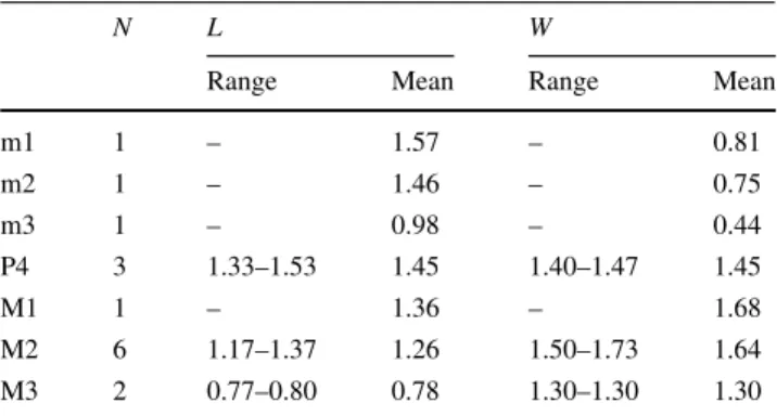
m1 1 – 1.57 – 0.81 m2 1 – 1.46 – 0.75 m3 1 – 0.98 – 0.44 P4 3 1.33–1.53 1.45 1.40–1.47 1.45 M1 1 – 1.36 – 1.68 M2 6 1.17–1.37 1.26 1.50–1.73 1.64 M3 2 0.77–0.80 0.78 1.30–1.30 1.30 Due to the scarcity of specimens the measurements of La Grive localities are given here together
molars, thus supporting the attribution of all specimens to the new genus. The antero-buccal edge is straight. The metacone is twice as big as the paracone. The post-para-cone crista joins the pre-metapost-para-cone crista before the labial edge, so that a little straight mesostyle crista is present; the simple mesostyle is well developed. A protruding parastyle is lacking on the anterior buccal edge. The protocone shows an anterior crest linked with the paracone base and a posterior crest disappears without connection; these two crests make an angle of about 110°. The hypocone is small but well developed, more buccally located than the proto-cone and not connected to it. The lingual edge of the tooth is sinuous between the protocone and the hypocone base. A short postero-lingual cingulum extends backwards. The posterior emargination is relatively short and restricted to the lingual part of the posterior edge. There are four roots, each located under the main cusps, plus the mark of a fifth one under the mesostyle.
M2 is smaller than M1; the buccal edge is sinuous as in the M1 but the distal cingulum, the posterior emargination and the metastyle are weaker than in M1.
M3 is triangular; the metacone is absent and the meta-style crest follows the posterior edge, connecting to a weak protocone and a protruding parastyle on the buccal edge; the mesostyle shows a clear division into two parts. By comparison, the M3 mesostyle is only superficially divided in the Oberdorf sample (Ziegler1998b).
Lower teeth One m1 from La Grive M was briefly mentioned in Mein (1999). It is characterised by a very narrow trigonid; the protoconid is higher than the other cuspids; its tip is curved backwards; the posterior arm of the protoconid ends in the lingual part of the trigonid and is coalescent with the metaconid. This metaconid is higher than the two other lingual cuspids. The buccal cingulum is well marked anteriorly, indistinct in the middle and weak distally. The talonid is short with a flat basin. The anterior arm of the hypoconid ends lingually after the middle of the tooth, and the posterior arm slopes down to the middle of the tooth and then climbs up and joins the entoconid under its tip. On the lingual edge, the entoconid is well marked and joins the metaconid by a curved crest. Because the measurements and the morphology of this m1 are compa-rable to those of the lectotype from Devinska Nova Ves (Neudorf), this tooth is attributed to Viretia gracilidens.
Only one tooth from another fissure (La Grive L7) dis-plays a similar morphology but noticeably smaller (perhaps partly due to the corroded enamel) and has been identified as m2.
No m3 has been recorded in La Grive.
We can compare directly the La Grive material with a right partial mandible from Devinska Nova Ves (Neudorf, FSL 66790). An m3 is preserved on this mandible. The tooth is very small (0.98 9 0.44) and as a very low crown.
The trigonid is about three times longer than the talonid. The talonid basin is completely undistinguishable. This mandible (FSL 66790) also preserves the ascending ramus. The measurements of this mandible are height of the cor-onoid process = 3.79, width of the internal temporal fossa = 1.35, height of the condyle = 1.4, length of the condylar lower facet = 1.3. In posterior view the condyle has two separate articular facets. The superior one is rounded and higher on the buccal side; the inferior articular facet is slightly divided in two parts and twice as long as high. This inferior articular surface is remarkably strong.
Discussion Viret and Zapfe (1952) recognised this new species only in Devinska Nova Ves (Neudorf) and sug-gested that it could be a new sub-genus or even a new genus. They gave as particular characteristics the relatively high mandibular ramus with mental foramen under p4; ascending ramus well developed in length with wide internal temporal fossa; the particularly strong condyle is low, at the level of the teeth. Teeth are particularly narrow, the size of the molars progressively decreasing backwards and characterised by their unusually long trigonid wide open lingually. Weak metaconid close to protoconid. Very reduced m3. The most remarkable tooth, the two-rooted p4, is a high cone-shaped unicuspid, bucco-lingually flattened tooth without an accessory cuspule and with a simple basal cingulum, a pattern unknown so far in soricids; moreover, the crown does not overlap the preceding tooth as is the case in other Miocene or recent soricids.
At the moment, upper molars of this form are unknown. Fejfar (1966) excavated at Devinska Nova Ves; he published pictures of the ‘‘holotype’’ of ‘‘Sorex gracili-dens’’—p4–m1 (Fig. 14d–f) and ascending ramus with condyle (Fig. 13a–d)—together with a newly found m1 (1.40 9 0.76). Based on this new material Fejfar (1966) concluded that S. gracilidens shows noticeable affinities with his new genus Allosorex from Ivanovce (Slovak Republic, Pliocene, MN15), the holotype of which is a toothless mandible with condyle, for which he created the new subfamily Allosoricinae.
Reumer (1992) outlined the problems of the different sub-families of Soricinae as did Ziegler (1998b) who also described more precisely and figured an important sample from Oberdorf 4 (Austria; MN4) as ‘‘Allosorex’’ gracilidens which can undoubtedly be referred to the above described material.
Engesser (1972) also described an m1 called ‘Soricid II’ and compared it with ‘‘Sorex’’ gracilidens. He concluded that there were affinities between the two species (based on the very narrow trigonid, the reduced talonid and the backwardly inclined tip of the protoconid), but because of the size differences, the different location of the mental foramen and the posterior crest connection to the entoconid on m1, he excluded a direct filiation between these two forms.