propagation of prions to CNS
Adriano Aguzzi, Frank L Heppner, Mathias Heikenwalder,
Marco Prinz, Kirsten Mertz, Harald Seeger and Markus Glatzel
Institute of Neuropathology, Universitätsspital Zürich, Switzerland
Prions are not only unique in the way they replicate. Also the sequence of events triggered by peripheral prion infection, generically termed ‘peripheral pathogenesis’, sets prions aside from all other known pathogens. Whereas most bacteria, parasites, and viruses trigger innate and adaptive immune responses, the mammalian immune system appears to be remarkably oblivious to prions. Transmissible spongiform encephalopathies (TSEs) do not go along with inflammatory infiltrates, and antibodies to the prion protein are not typically raised during the course of the disease. On the other hand, there is conspicuous involvement of lymphoid organs, which accumulate sizeable concentrations of the infectious agent early during disease. Moreover, various states of immune deficiency can abolish peripheral pathogenesis and prevent ‘take’ of infection when prions are administered to peripheral sites. Here, we critically re-visit the current evidence for an involvement of the immune system in prion diseases, and will attempt to trace the elaborate mechanisms by which prions, upon entry into the body from peripheral sites, reach the brain.
Biology of prion infection
The fastest and most efficient method for inducing spongiform encephal-opathy in the laboratory is intracerebral inoculation of brain homogenate. Inoculation of 106infectious units (defined as the amount of infectivity that will induce TSE with 50% likelihood in a given host) will yield disease in ~6 months; a remarkably strict inverse relationship can be observed between the logarithm of the inoculated dose and the incubation time1.
However, the above situation does not correspond to what typically happens in the field. There, acquisition of prion infectivity through any of several peripheral routes is the rule. However, prion diseases can also be initiated by feeding2–4, by intravenous and intraperitoneal injection5as well as from the eye by conjunctival instillation6, corneal grafts7and intra-ocular injection8.
Correspondence to: Prof. Adriano Aguzzi, Institute of Neuropathology, Universitätsspital Zürich, Schmelzbergstrasse 12, CH-8091 Zürich, Switzerland
While only less than 1% of all reported cases of Creutzfeldt-Jakob disease (CJD) can be traced to a defined infectious source, the bovine spongiform encephalopathy (BSE) epizootic has highlighted oral exposure as a tremendously efficient vector for bovine prion diseases. BSE is most likely transmissible to humans, and strong circumstantial evidence suggests that BSE is the cause of variant Creutzfeldt-Jakob disease (vCJD).
Several aspects of CJD epidemiology continue to be enigmatic. For example, CJD incidence in Switzerland rose 2-fold in 2001, and increased further in 20029. A screen for recognized or hypothetical risk factors for CJD has, to date, not exposed any causal factors. Several scenarios may account for the increase in incidence, including improved reporting, iatrogenic transmission, and transmission of a prion zoonosis. Prion diseases typically exhibit a very long latency period between the time of infection and the clinical manifestation. From the viewpoint of interventional approaches, this peculiarity may be exploitable, since it opens a possible window of intervention after infection has occurred, but before brain damage is being initiated. Prions spend much of this latency time executing neuro-invasion, which is the process of reaching the central nervous system (CNS) after entering the body from peripheral sites10,11. During this process, little or no damage occurs to brain, and one might hope that its interruption may prevent neuro-degeneration.
Immune cells and the peripheral entry sites of prions
For a long time, two routes of infection have suggested that immune cells might be of importance for peripheral pathogenesis – oral challenge and administration by scarification. Upon oral challenge, an early rise in prion infectivity can be observed in the distal ileum of infected organisms: this applies to several species but was most extensively investigated in sheep12,13. There, Peyer’s patches acquire strong immunopositivity for the prion protein. Immunohistochemical stains with antibodies to the prion protein typically reveal a robust signal in primary B-cell follicles and germinal centres, which roughly co-localizes with the complement receptor, CD35, in a wide variety of secondary
lymphoid organs including appendix and tonsils14. Although
conventional light microscopy does not allow differentiating between PrPC and PrPSc, Western blot analysis has left no doubt that Peyer’s patches do accumulate the disease-associated form of the prion protein. The latter is true also in the mouse model of scrapie, which is being used as a convenient experimental paradigm by many laboratories including ours. Administration of mouse-adapted scrapie prions (Rocky Mountain Laboratory or RML strain, originally derived from the
Chandler sheep scrapie isolate) induces a surge in intestinal prion infectivity as early as a few days after inoculation15.
All of the above evidence conjures the suspicion that Peyer’s patches may represent a portal of entry for orally administered prions, on their journey from the luminal aspect of the gastro-enteric tube to the CNS. However, the question as to whether the same applies for BSE-affected cattle has been answered less unambiguously.
In a monumental study of BSE pathogenesis in cattle, cows of various ages were fed with 100 g, 10 g, 1 g, or 100 mg of brain homogenate derived from BSE-sick cows16. A large variety of tissues was taken at various points in time, homogenized, and transmitted intracerebrally to indicator organisms in order to assess their prion content. This study uncovered a transient surge in infectivity in the distal ileum of cows at approximately 6 months’ post-infection. Infectivity then subsides, but it appears to return to the terminal ileum at the end stages of disease, maybe by a type of retrograde transport17. Although this was not formally confirmed, it appears likely that Peyer’s patches are the sites of prion accumulation in the gastrointestinal tract of cattle challenged orally with prions.
Prions and the gut
Membranous epithelial cells (M-cells) are key sites of antigen sampling for the mucosal-associated lymphoid system (MALT) and have been recognized as major ports of entry for enteric pathogens in the gut via transepithelial transport18. Interestingly, maturation of M-cells is dependent on signals transmitted by intra-epithelial B-cells. The group of Kraehenbuhl (Lausanne) has developed efficient in vitro systems, in which epithelial cells can be instructed to undergo differentiation to cells that resemble M-cells by morphological and functional/physiological criteria. Therefore, we investigated whether M-cells are a plausible site of prion entry in a co-culture model19. Colon carcinoma cells were cultured on the upper face of filters until confluency. Next, B-lymphoblastoid cells were added onto the lower side of the filters. Lymphoid cells migrated through the pores of the filter and settled within the epithelial monolayer (Plate XIII), inducing differentiation of M-cells. Scrapie prions were administered to the apical compartment of co-cultures that combined integrity and active transport of beads. Infectivity was determined within the basolateral compartment by bioassay with tga20 mice, which overexpress a Prnp transgene and develop scrapie rapidly after infection20. Upon challenge with scrapie prions, we consistently recovered prions in the basolateral compartment of co-cultures containing M-cells, suggesting transepithelial prion
transport (Plate XIII). In contrast, there was hardly any prion transport in Caco-2 cultures without M-cells21.
These findings indicate that M-cell differentiation is necessary and sufficient for active transepithelial prion transport in vitro. M-cell-dependent uptake of foreign antigens or particles is known to be followed by rapid transcytosis directly to the intra-epithelial pocket, where key players of the immune system (e.g. macrophages, dendritic cells and lymphocytes18) are located. Therefore, prions may exploit M-cell-dependent transcytosis to gain access to the immune system.
While these findings suggest that M-cells are a plausible candidate for the mucosal portal of prion infection, it still remains to be established whether the pathway delineated above does indeed represent the first portal of entry of orally administered prions into the body.
Prions and the skin
Scarification of the most superficial layers of the skin, and subsequent administration of prions, has been known for a long time to be a highly efficacious method of inducing prion disease22,23. Dendritic cells (professional antigen-presenting cells abundant in the skin) may become loaded with the infectious agent by this method; in fact, recent work has implicated dendritic cells as potential vectors of prions in oral24and haematogenous spread25of the agent. It is equally possible, however, that scarification induces direct neural entry of prions into skin nerve terminals. The latter hypothesis has not yet been studied in much detail, but it would help explain the remarkable speed with which CNS pathogenesis follows inoculation by this route: dermal inoculation of scarified mice yields typical latency periods of the disease that are similar to those obtained by intracerebral inoculation. The possibility of direct neural spread of the agent has also been suggested by a series of elegant experiments in the laboratories of Oldstone and Chesebro: transgenic mice that lack the endogenous prion gene but express a Prnp transgene exclusively in nervous tissue (under transcriptional control of the neuron-specific enolase regulatory elements) can be efficiently infected by the oral route despite lack of prion protein expression in lymphoid organs26. Of course, these experiments do not exclude the possibility that dendritic or other mobile cells may participate to neuro-invasion even if they do not express endogenous PrPC.
Prions and lymphocytes
The normal prion protein is consistently expressed, albeit at moderate levels, in circulating lymphocytes27. Innate or acquired deficiency of
lymphocytes impairs peripheral prion pathogenesis, whereas no aspects of pathogenesis are affected by the presence or absence of lymphocytes upon direct transmission of prions to the CNS28,29. Klein described the requirement for B-cells10,30: this was surprising since there had been no suggestions that humoral immunity would be involved in prion diseases. In the same study, it was shown that T-cell deficiency brought about by ablation of the T-cell receptor α-chain did not affect prion pathogenesis. On the other hand, lymphocytes alone could not account for the entirety of prion pathogenesis, and an additional sessile compartment had to be involved, since adoptive transfer of Prnp+/+ bone marrow to Prnpo/o recipient mice did not suffice to restore infectibility of Prnp-expressing brain grafts, indicating that neuro-invasion was still defective31.
It then emerged that peripheral prion pathogenesis required the physical presence of B-cells, yet intraperitoneal infection occurred efficiently even in cell deficient hosts that had been transferred with B-cells from Prnp knockout mice32. Therefore, the presence of B-cells – but not expression of the cellular prion protein by these cells – is indispensable for pathogenesis upon intraperitoneal infection in the mouse scrapie model33.
These results have been reproduced and confirmed several times over the years by many laboratories in various experimental paradigms: the requirement for B-cells in particular appears to be very stringent in most instances investigated.
Prion strains and lymphotropism
An interesting discrepancy that remains to be addressed concerns the actual nature of the cells that replicate and accumulate prions in lymphoid organs. With prions of the RML strain, four series of rigorously controlled experiments over 5 years34–37 unambiguously reproduced the original observation that transfer of wild-type bone marrow cells (or fetal liver cells) to Prnp-deficient mice restored accumulation and replication of prions in spleen31. By contrast, Brown and colleagues reported a diametrically opposite outcome of similar experiments when mice were inoculated with prions of the ME7 strain38. Since trivial experimental artefacts appear unlikely, this discrepancy may identify yet another significant difference in the cellular tropism of different prion strains.
It is important to note that the Blättler results do not necessarily indicate that lymphocytes are the primary splenic repository of prions. Instead, bone marrow transplantation may: (i) transfer an ill-defined population with the capability to replenish splenic stroma and to replicate prions; or, less probably, (ii) donor-derived PrPC-expressing
haematopoietic cells may confer prion replication capability to recipient stroma by virtue of ‘GPI painting’, i.e. the post-translational cell-to-cell transfer of glycophosphoinositol-linked extracellular membrane proteins39. Stromal splenic follicular dendritic cells (FDCs) have been described by some authors to possibly derive from haematopoietic precursors, particularly when donors and recipients were young40,41. Conversely, instances have been described in which transfer of GPI-linked proteins occurs in vivo with surprisingly high efficiency42. GPI painting has been described specifically for the cellular prion protein43.
While it has been known for a long time that specific strains of prions may preferentially affect specific subsets of neurons, the Blättler/Brown paradox may uncover an analogous phenomenon in peripheral prion pathogenesis. The latter question may be important, since the molecular and cellular basis of peripheral tropism of prion strains is likely to be directly linked to the potential danger of BSE in sheep44–46, as well the potential presence of vCJD prions in human blood47.
Prion reservoirs in the periphery of the body
Prion infectivity rises very rapidly (in a matter of days) in the spleen of intraperitoneally infected mice. Although B lymphocytes are crucial for neuro-invasion, a series of prion titrations in expressing and Prnp-ablated mice, as well as reciprocal bone marrow chimeras thereof, has ruled out that the bulk of infectivity might be contained in lymphocytes. Instead, most splenic prion infectivity may reside in a ‘stromal’ fraction. Therefore, lymphocytes may be important for trafficking prions within lymphoid organs, but they do not appear to represent the major hideout for the infectious agent. Follicular dendritic cells are a prime candidate prion reservoir, since they express large amounts of cellular prion protein and PrP accumulations tend to co-localize with FDCs in light and electron microscopical analyses of prion-infected spleens. Elegant immuno-electron microscopic studies have shown that prion immunoreactivity is situated in the immediate neighbourhood of iccosomes on the surface of FDCs48.
A definitive assessment of the contribution of FDCs to prion patho-genesis continues to be problematic since the histopatho-genesis and the molecular characteristics of these cells are ill-defined. There is a dearth of molecular markers for FDCs. FDCs express S-100 proteins, as well as the complement receptors 2 (CD35) and 4 (identical to the marker FDC-M2). All of these markers, however, are also expressed by additional cell types, even within lymphoid organs49. The FDC-M1 marker recognized by hybridoma clone 4C11 appears to be more specific, but it stains also tingible body macrophages which are most likely of haematopoietic origin.
Gene-deletion experiments in mice have shown that signalling by TNF and lymphotoxins is required for FDC development50–52. Membrane-bound lymphotoxin-α/β (LT-α/β) heterotrimers signal through the LT-β receptor (LT-βR)53 which activates a pathway required for the development and maintenance of secondary lymphoid organs54,55. Membrane LT-α/β heterotrimers are mainly expressed by activated lymphocytes56,57. Maintenance of pre-existing FDCs in a differentiated state requires continuous interaction with B-lymphocytes expressing surface LT-α/β58. Inhibition of the LT-α/β pathway in mice by treatment with LT-βR–immunoglobulin fusion protein (LT-βR–Ig)59 leads to the disappearance of mature, functional FDCs (defined as cells that express markers such as FDC-M1, FDC-M2 or CD35) within 1 day, both in spleen and in lymph nodes55,60. Prolonged administration of the LT-βR–Ig protein leads to disruption of B-cell follicles.
Montrasio and colleagues studied the effect of selective ablation of functional FDCs on the pathogenesis of scrapie in mice. FDC depletion was maintained by weekly administration of the LT-βR–Ig fusion protein Fig. 1 Depletion of follicular dendritic cells by pharmacological inhibition of lymphotoxin
signalling. Time course of FDC depletion in spleen of LT-βR–Ig treated mice. Frozen sections of treated (left) and control mice (right) immunostained with FDC-specific antibody FDC-M1 at different times points after injection of LT-βR–Ig (original magnifications: upper row 8x25; lower row 8x63). Germinal centre FDC networks were already depleted 1 week after treatment60,66. Some FDC-M1 positive cells, which may represent residual FDCs or tingible
for 8 weeks. Histological examination of spleen sections revealed that FDC networks had disappeared 1 week after treatment (Fig. 1), as expected.
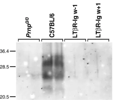
In mice, following peripheral inoculation, infectivity in the spleen rises within days and reaches a plateau after a few weeks61–63. Even after intracerebral inoculation, spleens of C57BL/6 animals already contain infectivity 4 days’ post-infection (p.i.)61. However, Western blot analysis (Plate XIV) revealed that 8 weeks after inoculation spleens of control mice showed strong bands of protease-resistant PrP, whereas mice injected weekly with LT-βR–Ig, starting either 1 week before or 1 week after inoculation, showed no detectable signal (less than 1/50th of the controls).
In spleens of mice treated with LT-βR–Ig 1 week before intraperitoneal inoculation, no infectivity could be detected after 3 or 8 weeks (< 1.0 logID50 units/ml 10% homogenate). In mice treated with LT-βR–Ig 1 week after inoculation the titres were about 2.2 and 4.1 logID50units/ml 10% homogenate at 3 weeks and at borderline detectability at 8 weeks after infection, suggesting that some prion accumulation took place in the first weeks after inoculation but was reversed under treatment with LT-βR–Ig by 8 weeks.
Mice receiving LT-βR–Ig starting 1 week after inoculation developed the disease with about 25 days delay as compared to control mice. If FDC depletion was initiated 1 week before inoculation, the effect on incubation time was more pronounced. This indicates that FDCs are essential for deposition of PrPSc and generation of infectivity in the spleen, and suggest that FDCs participate in the process of neuro-invasion. Therefore, strategies aimed at depleting FDCs might be envisaged for post-exposure prophylaxis64of prion infections initiated at extracerebral sites65.
Some open questions regarding the lymphotropism of prions
Inhibition of the LT-β signalling pathway with a soluble receptor depletes FDCs60 and abolishes prion replication in spleen, thereby prolonging the latency of scrapie after intraperitoneal challenge66. This suggests that B-cell-deficient µMT mice67 may be resistant to intraperitoneal prions30 because of impaired FDC maturation32,66. However, additional experimentation indicates that PrPC-expressing haematopoietic cells are required in addition to FDCs for efficient lymphoreticular prion propagation31,35. This apparent discrepancy called for additional studies of the molecular requirements for prion replication competence in lymphoid stroma. Therefore, we studied peripheral prion pathogenesis in mice lacking TNF-α, LT-α/β, or their receptors.
After intracerebral inoculation, all treated mice developed clinical symptoms of scrapie with incubation times, attack rates, and histopathological characteristics similar to those of wild-type mice, indicating that TNF/LT signalling is not relevant to cerebral prion pathogenesis. Upon intraperitoneal prion challenge, mice defective in LT signalling (LT-α–/–, LT-β–/–, LT-βR–/–, or LT-αTNF-α–/–) proved virtually non-infectible with ≤ 5 logLD50 scrapie infectivity, and establishment of subclinical disease68 was prevented. In contrast, TNFR1–/– mice were almost fully susceptible to all inoculum sizes, and TNF-α-/-mice showed dose-dependent susceptibility. TNFR2–/– mice had intact FDCs and germinal centres, and were fully susceptible to scrapie.
Unexpectedly, all examined lymph nodes (Plate XV) of TNFR1–/–and TNF-α–/– mice had consistently high infectivity titres. Even inguinal lymph nodes, which are distant from the injection site and do not drain the peritoneum, contained infectivity titres equal to all other lymph nodes. Therefore, TNF deficiency prevents lymphoreticular prion accumulation in spleen but not in lymph nodes.
Why is susceptibility to peripheral prion challenge preserved in the absence of TNFR1 or TNF-α, while deletion of LT-signalling components confers high resistance to peripheral prion infection – although each of these defects (except TNFR2–/–) abolishes FDCs? For one thing, prion pathogenesis in the lymphoreticular system appears to be compart-mentalized, with lymph nodes (rather than spleen) being important reservoirs of prion infectivity during disease. Second, prion replication appears to take place in lymph nodes even in the absence of mature FDCs. We generated chimeric mice in order to determine whether haemato-poietic components are involved in prion propagation in TNFR1–/–lymph nodes. In Prnpo/o mice grafted with TNFR1–/– haematopoietic cells, high infectivity loads were detectable in lymph nodes but not spleen, indicating that TNFR1-deficient haematopoietic cells may support prion propagation within lymph nodes. These findings are in line with previous studies, which showed that chimeras of PrP-deficient hosts with PrP-expressing haematopoietic cells can accumulate prions chronically31,35. The PrP signal co-localized with a subset of macrophages in TNFR1–/–lymph nodes. Since marginal zone macrophages are in close contact to FDCs and also interact with marginal zone B-cells, this cell type is a candidate supporter in prion uptake and replication.
Sympathetic nerves: a neuro-immune synapse for prions?
A widely discussed model predicts that prion neuro-invasion consists of two distinct phases – lympho-invasion and neuro-invasion proper69. The second phase has long been suspected to involve peripheral nerves, and
may depend on expression of PrPCby nerves70. Studies focusing on the temporal and spatial dynamics of neuro-invasion have suggested that the autonomic nervous system might be responsible for transport from lymphoid organs to the CNS71–74. We have been attempting to test the requirement for expression of PrPC in peripheral nerves and have developed a gene transfer protocol to spinal ganglia aimed at resolving this question75; however, we were never able to recover infectivity in spinal cords of Prnpo/omice whose spinal nerves had been transduced by
Prnp-expressing adenoviruses (M Glatzel & A Aguzzi, unpublished
results). Also, fast axonal transport does not appear to be involved in prion neuro-invasion as mice that are severely impaired in this transport mechanism experience prion pathogenesis with kinetics similar to that of wild-type mice76.
There is substantial evidence suggesting that prion transfer from the lymphoid system to the CNS occurs along peripheral nerves in a PrPC -dependent fashion31,70,77. The innervation pattern of lymphoid organs is mainly sympathetic78. Sympathectomy delays the transport of prions from lymphatic organs to the thoracic spinal cord, which is the entry site of sympathetic nerves to the CNS. Transgenic mice overexpressing NGF under control of the K14 promoter, whose spleens are hyperinnervated, developed scrapie significantly earlier than non-transgenic control mice. However, many details remain to be elucidated. It is not known whether prions can be transferred directly from FDCs to sympathetic endings, or whether additional cell types are involved. The latter possibility is particularly enticing, as FDCs have not been shown to entertain physical contact with sympathetic nervous system terminals.
Moreover, it is unclear how prions are actually transported within peripheral nerves. Axonal and non-axonal transport mechanisms may be involved. Within the framework of the protein-only hypothesis, one may hypothesize a ‘domino’ mechanism, by which incoming PrPSc converts resident PrPC on the axolemmal surface, thereby propagating spatially the infection. This model may accommodate the finding that the velocity of neural prion spread is extremely slow79 and may not follow the canonical mechanisms of fast axonal transport. Recent studies70,80may favour a non-axonal transport mechanism that results in peri-axonal deposition of PrPSc.
The fact that even denervated mice eventually develop scrapie may be due to: (i) an alternative, low-efficiency route of entry that may become uncovered by the absence of sympathetic fibres; or (ii) because of incomplete sympathectomy. Entry through the vagal nerve has been proposed in studies of the dynamics of vacuolation following oral and intraperitoneal challenge with prions71,81.
The surprising finding that infectious titres in hyperinnervated spleens are at least two logs higher and show enhanced PrPSc accumulations
compared to control mice suggests that sympathetic nerves, besides being involved in the transport of prions, may also accumulate and replicate prions in lymphatic organs72. This has implications related to the perman-ence and possibly eradication of prions in subclinically infected hosts.
Spread of prions within the CNS
Ocular administration of prions has proved particularly useful to study neural spread of the agent, since the retina is a part of the CNS, and intra-ocular injection does not produce direct physical trauma to the brain – which may disrupt the blood-brain barrier and impair other aspects of brain physiology. The assumption that spread of prions occurs axonally rests mainly on the demonstration of diachronic spongiform changes along the retinal pathway following intra-ocular infection8.
To investigate whether spread of prions within the CNS is dependent on PrPC expression in the visual pathway, PrP-producing neural grafts were used as sensitive indicators of the presence of prion infectivity in the brain of an otherwise PrP-less host. Following inoculation with prions into the eye of grafted Prnpo/o mice, none of the grafts showed signs of scrapie. Therefore, infectivity administered to the eye of PrP-deficient hosts cannot induce scrapie in a PrP-expressing neurons located > 1 synapse away82.
Because engraftment of Prnpo/omice with PrPC-producing tissue might lead to an immune response to PrP and possibly to neutralization of infectivity83. In order to rule out the possibility that prion transport was disabled by a neutralizing immune response, Prnpo/omice were rendered tolerant by expressing PrPCunder the control of the lck promoter. These mice overexpress PrP on T-lymphocytes, but are resistant to scrapie and do not replicate prions in brain, spleen or thymus after intraperitoneal inoculation with scrapie prions84. Engraftment of these mice with PrP-overexpressing neuro-ectoderm did not lead to the development of antibodies to PrP after intracerebral or intra-ocular inoculation, presumably due to clonal deletion of PrP-immunoreactive T-lymphocytes. As before, intra-ocular inoculation with prions did not provoke scrapie in the graft, supporting the conclusion that lack of PrPC, rather than immune response to PrP, prevented prion spread82. Therefore, PrPC appears to be necessary for the spread of prions along the retinal projections and within the intact CNS.
Therefore, it appears that intracerebral spread of prions is based on a PrPC-paved chain of cells, perhaps because they are capable of supporting prion replication. When such a chain is interrupted by interposed cells that lack PrPC, no propagation of prions to the target tissue can occur. Perhaps prions require PrPC for propagation across
synapses: PrPC is present in the synaptic region85 and certain synaptic properties are altered in Prnpo/o mice86,87. Perhaps transport of prions within (or on the surface of) neuronal processes is PrPC-dependent. These findings may be accommodated by a ‘domino-stone’ model10 in which spreading of scrapie prions in the CNS occurs per continuitatem through conversion of PrPC by adjacent PrPSc.
Innate immunity and antiprion defence
Cells of the monocyte/macrophage lineage typically represent the first line of defence against a broad variety of pathogens. In the case of prions, it might be conceivable that macrophages protect against prions. However, it would be equally conceivable that macrophages, by virtue of their phagocytic properties and of their intrinsic mobility, may function as Trojan horses that transport prion infectivity between sites of replication within the body. This interesting question has not yet been fully resolved.
In a short-term prion infection paradigm, Beringue and colleagues administered dichloromethylene disphosphonate encapsulated into liposomes to mice: this eliminates for a short period of time all spleen macrophages. Accumulation of newly synthesized PrPScwas accelerated, suggesting that macrophages participate in the clearance of prions, rather than being involved in PrPScsynthesis.
Beringue and colleagues suggested that activation or targeting of macrophages may represent a therapeutic pathway to explore in TSE infection. This was taken up by Sethi and Kretzschmar, who recently reported that activation of Toll-like receptors (TLRs), which function as general stimulators of innate immunity by driving expression of various sets of the immune regulatory molecules, can effect postexposure prophylaxis in an experimental model of intraperitoneal scrapie infection88. Administration of prions intraperitoneally elicited disease after approximately 180 days, whereas administration of CpG oligodeoxynucleotides 7 h after prion inoculation and daily for 20 days led to disease-free intervals of ‘more than 330 days’ – although it appears that all inoculated mice died of scrapie shortly thereafter (communicated by H. Kretzschmar at the TSE conference in Edinburgh, September 2002).
This finding is very surprising, since most available evidence indicates that general activation of the immune system would typically sensitize mice to prions, rather than protect them. The mechanism by which activation of TLR can result in post-exposure prophylaxis is wholly unclear at present, particularly in view of the fact that mice lacking Myd8889, which is an essential mediator of TLR signalling, develop prion disease with exactly the same sensitivity and kinetics as wild-type mice15.
Another prominent component at the crossroad between innate and adaptive immunity is represented by the complement system. Opsonization by complement system components also appears to be relevant to prion pathogenesis: mice genetically engineered to lack
complement factors36, or mice depleted of the C3 complement
component by administration of cobra venom90, exhibit a remarkable resistance to peripheral prion inoculation. This phenomenon may, once again, be related to the pathophysiology of FDCs, which typically function as antigen traps. Trapping mechanisms essentially consist of capture of immune complexes by Fcγ receptors, and binding of opsonized antigens (linked covalently to C3d and C4b complement adducts) to the CD21/CD35 complement receptors.
Capture mediated by Fcγ receptors does not appear to be important in prion disease: knockout mice lacking Fcγ receptors91–94 are just as susceptible to intraperitoneally administered scrapie as wild-type mice. Further, introduction into µMT mice of a generic immunoglobulin µ-chain fully restored prion neuro-invasion irrespective of whether this heavy chain allowed for secretion of immunoglobulins, or only for production of membrane-bound immunoglobulins. Therefore, we conclude that circulating immunoglobulins are certainly not crucial to prion replication in lymphoid organs and to neuro-invasion.
A second mechanism exploited by FDCs for antigen trapping involves covalent linking of proteolytic fragments of the complement components C3 and C495,96. Ablation of C3, or of its receptor CD21/CD35, as well as C1q (alone or combined with BF/C2–/–), delayed neuro-invasion significantly after intraperitoneal inoculation when a limiting dose of prions was administered. These effects suggest that opsonization of the infectious agent may enhance its accessibility to germinal centres by facilitating docking to FDCs.
Large prion inocula (> 106 infectious units) appear to over-ride the requirement for a functional complement receptor in prion pathogenesis. This is similar to systemic viral infections and co-receptor-dependent retention within the follicular compartment, whose necessity can be over-ridden by very high affinity antigens97 or adjuvants98. Additional retention mechanisms for prions may, therefore, exist in FDCs, which are not complement-dependent, or depend on hitherto unidentified complement receptors.
Humoral immunity against prions
For many viruses, vaccination is a most effective method of infection control, but is it possible to induce protective immunity in vivo against prions? Pre-incubation with anti-PrP antisera was reported to reduce the
prion titre of infectious hamster brain homogenates by up to 2 log units99 and anti-PrP antibodies inhibit formation of PrPSc in a cell-free system100. Also, antibodies101and F(ab) fragments raised against certain domains of PrP102 can suppress prion replication in cultured cells. However, it is difficult to induce humoral immune responses against PrPC and PrPSc. This is most likely due to tolerance of the mammalian immune system to PrPCwhich is expressed ubiquitously. Ablation of the
Prnp gene103, which encodes PrPC, renders mice highly susceptible to
immunization with prions82, and many monoclonal antibodies to the prion protein have been generated in Prnpo/o mice104–106. However,
Prnpo/o mice are unsuitable for testing vaccination regimens since they do not support prion pathogenesis61.
Therefore, we have asked whether genes encoding high-affinity anti-PrP antibodies (originally generated in Prnpo/o mice) may be utilized to reprogram B-cell responses of prion-susceptible mice that express PrPC. Indeed, introduction of the epitope-interacting region the heavy chain of 6H4, a high-affinity anti-PrP monoclonal antibody105into the germ line of mice sufficed to produce high-titre anti-PrPC immunity. The build-up of anti-PrPC titres, however, was slower in the presence of endogenous PrPCsuggesting that some clonal deletion is actually occurring.
How can these observations be interpreted? The total anti-PrPC titre results from pairing of one transgenic µ-heavy chain with a large repertoire of endogenous κ- and λ-chains: some pairings may be reactive, while others may be anergic (Plate XVI). Maybe the B-cell clones with the highest affinity to PrPC are eliminated by immune tolerization, and only clones with medium affinity are retained (Plate XVI).
Expression of the 6H4 µ-heavy chain sufficed to confer protection from scrapie upon intraperitoneal inoculation of the prion agent21. This delivers proof-of-principle that a protective humoral response against prions can be mounted by the mammalian immune system, and suggests that B-cells are not intrinsically tolerant to PrPC. If the latter is generally true, lack of immunity to prions may be due to T-helper tolerance. To overcome the latter is not trivial, but there may be ways, some of which are currently being explored in our laboratory. These findings, therefore, encourage a re-assessment of the possible value of active and passive immunization, and perhaps of reprogramming B-cell repertoires by µ-chain transfer, in prophylaxis or in therapy of prion diseases.
PrP immunization: a tool against prions?
Approximately one year later, a first example of a reduction to practice of the approach proposed by Heppner was demonstrated in the laboratory of Wisnieswki. Immunization was simply achieved by
injecting 50 µg of recombinant prion protein emulsified in Freund’s adjuvant. This procedure had been utilized extensively in the Zürich laboratory, but had never produced any reasonable titres in wild-type mice. Upon inoculation with a high dose or with a lower dose of prion inoculum, vaccinated mice exhibited a modest delay in development of prion disease. Although the success of the study, from the viewpoint of survival of the mice, might be regarded as limited, it does indicate a way to translate vaccination into models that are closer to the real-life situation than immunoglobulin-transgenic mice.
Conclusions
Therapy of manifest CJD continues to be unattainable. Therefore, it might be sensible to concentrate efforts on post-exposure and pre-exposure prophylaxis. Many promising approaches are being developed towards these goals: they involve small therapeutic molecules and cytokine antagonists (post-exposure) as well as specific anti-PrPC immunity (pre-exposure). Therapy, however, is unlikely to be profitable for any time to come, since the number of CJD patients is exceedingly small and will hopefully not increase significantly. It is to be hoped that national governments and the European Union will agree to consider this area of funding with some priority, so that the most interesting of the approaches outlined above will be developed further, and their usefulness will be proved in clinical settings.
Acknowledgements
Special thanks are due to Petra Schwarz for maintaining our prion-infected mouse colony in an impeccable shape. This work was supported by grants of the Federal Office (FO) of Education and Science, the FO of Health, the FO of Animal Health, the Swiss National Foundation, the NCCR on neural plasticity and repair, and the Migros foundation.
References
1 Prusiner SB, Cochran SP, Groth DF, Downey DE, Bowman KA, Martinez HM. Measurement of the scrapie agent using an incubation time interval assay. Ann Neurol 1982; 11: 353–8
2 Kimberlin RH, Wilesmith JW. Bovine spongiform encephalopathy. Epidemiology, low dose exposure and risks. Ann NY Acad Sci 1994; 724: 210–20
3 Wells GA, Scott AC, Johnson CT et al. A novel progressive spongiform encephalopathy in cattle. Vet
Rec 1987; 121: 419–420
4 Anderson RM, Donnelly CA, Ferguson NM et al. Transmission dynamics and epidemiology of BSE in British cattle. Nature 1996; 382: 779–88
5 Kimberlin RH, Walker CA. Pathogenesis of mouse scrapie: effect of route of inoculation on infectivity titres and dose-response curves. J Comp Pathol 1978; 88: 39–47
6 Scott JR, Foster JD, Fraser H. Conjunctival instillation of scrapie in mice can produce disease. Vet
Microbiol 1993; 34: 305–9
7 Duffy P, Wolf J, Collins G, DeVoe AG, Streeten B, Cowen D. Possible person-to-person transmission of Creutzfeldt-Jakob disease. N Engl J Med 1974; 290: 692–3
8 Fraser H. Neuronal spread of scrapie agent and targeting of lesions within the retino-tectal pathway.
Nature 1982; 295: 149–50
9 Glatzel M, Rogivue C, Ghani A, Streffer J, Amsler L, Aguzzi A. Incidence of Creutzfeldt-Jakob disease in Switzerland. Lancet 2002; 360: 139–41
10 Aguzzi A. Neuro-immune connection in spread of prions in the body? Lancet 1997; 349: 742–3 11 Nicotera P. A route for prion neuroinvasion. Neuron 2001; 31: 345–8
12 Wells GA, Dawson M, Hawkins SA et al. Infectivity in the ileum of cattle challenged orally with bovine spongiform encephalopathy. Vet Rec 1994; 135: 40–1
13 Vankeulen LJM, Schreuder BEC, Meloen RH, Mooijharkes G, Vromans MEW, Langeveld JPM. Immunohistochemical detection of prion protein in lymphoid tissues of sheep with natural scrapie. J
Clin Microbiol 1996; 34: 1228–31
14 Hill AF, Zeidler M, Ironside J, Collinge J. Diagnosis of new variant Creutzfeldt-Jakob disease by tonsil biopsy. Lancet 1997; 349: 99
15 Prinz M, Huber G, Macpherson AJS, Heppner FL, Glatzel M, Eugster HP et al. Oral prion infection requires normal numbers of Peyer’s patches but not of enteric lymphocytes. Am J Pathol 2003; 162: 1103–11
16 Bradley R. Veterinary research at the Central Veterinary Laboratory, Weybridge, with special reference to scrapie and bovine spongiform encephalopathy. Rev Sci Tech 2000; 19: 819–30
17 Wells GA, Hawkins SA, Green RB et al. Preliminary observations on the pathogenesis of experimental bovine spongiform encephalopathy (BSE): an update. Vet Rec 1998; 142: 103–6
18 Neutra MR, Frey A, Kraehenbuhl JP. Epithelial M-cells: gateways for mucosal infection and immunization. Cell 1996; 86: 345–8
19 Kerneis S, Bogdanova A, Kraehenbuhl JP, Pringault E. Conversion by Peyer’s patch lymphocytes of human enterocytes into M-cells that transport bacteria. Science 1997; 277: 949–52
20 Fischer M, Rülicke T, Raeber A et al. Prion protein (PrP) with amino-proximal deletions restoring susceptibility of PrP knockout mice to scrapie. EMBO J 1996; 15: 1255–64
21 Heppner FL, Christ AD, Klein MA et al. Transepithelial prion transport by M-cells. Nat Med 2001;
7: 976–7
22 Carp RI. Transmission of scrapie by oral route: effect of gingival scarification [letter]. Lancet 1982; 1: 170–1
23 Taylor DM, McConnell I, Fraser H. Scrapie infection can be established readily through skin scarification in immunocompetent but not immunodeficient mice. J Gen Virol 1996; 77: 1595-9 24 Huang FP, Farquhar CF, Mabbott NA, Bruce ME, MacPherson GG. Migrating intestinal dendritic
cells transport PrP(Sc) from the gut. J Gen Virol 2002; 83: 267–71
25 Aucouturier PGF, Damotte D, Saborio GP et al. Infected splenic dendritic cells are sufficient for prion transmission to the CNS in mouse scrapie. J Clin Invest 2001; 108: 703–8
26 Race RE, Priola SA, Bessen RA et al. Neuron-specific expression of a hamster prion protein minigene in transgenic mice induces susceptibility to hamster scrapie agent. Neuron 1995; 15: 1183–91 27 Cashman NR, Loertscher R, Nalbantoglu J et al. Cellular isoform of the scrapie agent protein
participates in lymphocyte activation. Cell 1990; 61: 185–92
28 Kitamoto T, Muramoto T, Mohri S, Dohura K, Tateishi J. Abnormal isoform of prion protein accumulates in follicular dendritic cells in mice with Creutzfeldt-Jakob disease. J Virol 1991; 65: 6292–5 29 Lasmezas CI, Cesbron JY, Deslys JP et al. Immune system-dependent and -independent replication of
the scrapie agent. J Virol 1996; 70: 1292–5
30 Klein MA, Frigg R, Flechsig E et al. A crucial role for B cells in neuroinvasive scrapie. Nature 1997;
390: 687–90
31 Blättler T, Brandner S, Raeber AJ et al. PrP-expressing tissue required for transfer of scrapie infectivity from spleen to brain. Nature 1997; 389: 69–73
32 Klein MA, Frigg R, Raeber AJ et al. PrP expression in B lymphocytes is not required for prion neuroinvasion. Nat Med 1998; 4: 1429–33
33 Aguzzi A, Brandner S, Fischer MB et al. Spongiform encephalopathies: insights from transgenic models. Adv Virus Res 2001; 56: 313–52
34 Aguzzi A, Klein MA, Montrasio F et al. Prions: pathogenesis and reverse genetics. Ann NY Acad Sci 2000; 920: 140–57
35 Kaeser PS, Klein MA, Schwarz P, Aguzzi A. Efficient lymphoreticular prion propagation requires PrP(c) in stromal and hematopoietic cells. J Virol 2001; 75: 7097–106
36 Klein MA, Kaeser PS, Schwarz P et al. Complement facilitates early prion pathogenesis. Nat Med 2001; 7: 488–92
37 Prinz M, Montrasio F, Klein MA et al. Lymph nodal prion replication and neuroinvasion in mice devoid of follicular dendritic cells. Proc Natl Acad Sci USA 2002; 99: 919–24
38 Brown KL, Stewart K, Ritchie DL et al. Scrapie replication in lymphoid tissues depends on prion protein- expressing follicular dendritic cells. Nat Med 1999; 5: 1308–12
39 Kooyman DL, Byrne GW, Logan JS. Glycosyl phosphatidylinositol anchor. Exp Nephrol 1998; 6: 148–51
40 Kapasi ZF, Qin D, Kerr WG et al. Follicular dendritic cell (FDC) precursors in primary lymphoid tissues. J Immunol 1998; 160: 1078–84
41 Szakal AK, Kapasi ZF, Haley ST, Tew JG. Multiple lines of evidence favoring a bone marrow derivation of follicular dendritic cells (FDCs). Adv Exp Med Biol 1995; 378: 267–72
42 Kooyman DL, Byrne GW, McClellan S et al. In vivo transfer of GPI-linked complement restriction factors from erythrocytes to the endothelium. Science 1995; 269: 89–92
43 Liu T, Li R, Pan T et al. Intercellular transfer of the cellular prion protein. J Biol Chem 2002; 277: 47671–8
44 Kao RR, Gravenor MB, Baylis M et al. The potential size and duration of an epidemic of bovine spongiform encephalopathy in British sheep. Science 2002; 295: 332–5
45 Bruce ME, Boyle A, Cousens S et al. Strain characterization of natural sheep scrapie and comparison with BSE. J Gen Virol 2002; 83: 695–704
46 Glatzel M, Aguzzi A. The shifting biology of prions. Brain Res Brain Res Rev 2001; 36: 241–8 47 Aguzzi A. Prion diseases, blood and the immune system: concerns and reality. Haematologica 2000;
85: 3–10
48 Jeffrey M, McGovern G, Goodsir CM, Bruce ME. Sites of prion protein accumulation in scrapie-infected mouse spleen revealed by immuno-electron microscopy. J Pathol 2000; 191: 323–32 49 Bofill M, Akbar AN, Amlot PL. Follicular dendritic cells share a membrane-bound protein with
fibroblasts. J Pathol 2000; 191: 217–26
50 Endres R, Alimzhanov MB, Plitz T et al. Mature follicular dendritic cell networks depend on expression of lymphotoxin beta receptor by radioresistant stromal cells and of lymphotoxin beta and tumor necrosis factor by B cells. J Exp Med 1999; 189: 159–68
51 Koni PA, Sacca R, Lawton P, Browning JL, Ruddle NH, Flavell RA. Distinct roles in lymphoid organogenesis for lymphotoxins alpha and beta revealed in lymphotoxin beta-deficient mice.
Immunity 1997; 6: 491–500
52 Fu YX, Huang G, Matsumoto M, Molina H, Chaplin DD. Independent signals regulate development of primary and secondary follicle structure in spleen and mesenteric lymph node. Proc Natl Acad Sci
USA 1997; 94: 5739–43
53 Ware CF, VanArsdale TL, Crowe PD, Browning JL. The ligands and receptors of the lymphotoxin system. Curr Top Microbiol Immunol 1995; 198: 175–218
54 Matsumoto M, Fu YX, Molina H et al. Distinct roles of lymphotoxin alpha and the type I tumor necrosis factor (TNF) receptor in the establishment of follicular dendritic cells from non-bone marrow-derived cells. J Exp Med 1997; 186: 1997–2004
55 Mackay F, Majeau GR, Lawton P, Hochman PS, Browning JL. Lymphotoxin but not tumor necrosis factor functions to maintain splenic architecture and humoral responsiveness in adult mice. Eur J
Immunol 1997; 27: 2033–42
56 Browning JL, Ngam-ek A, Lawton P et al. Lymphotoxin beta, a novel member of the TNF family that forms a heteromeric complex with lymphotoxin on the cell surface. Cell 1993; 72: 847–56 57 Browning JL, Dougas I, Ngam-ek A et al. Characterization of surface lymphotoxin forms. Use of
specific monoclonal antibodies and soluble receptors. J Immunol 1995; 154: 33–46
58 Gonzalez M, Mackay F, Browning JL, Kosco-Vilbois MH, Noelle RJ. The sequential role of lymphotoxin and B-cells in the development of splenic follicles. J Exp Med 1998; 187: 997–1007
59 Crowe PD, VanArsdale TL, Walter BN et al. A lymphotoxin-beta-specific receptor. Science 1994; 264: 707–10
60 Mackay F, Browning JL. Turning off follicular dendritic cells. Nature 1998; 395: 26–7
61 Büeler HR, Aguzzi A, Sailer A et al. Mice devoid of PrP are resistant to scrapie. Cell 1993; 73: 1339–47 62 Bruce ME. Agent replication dynamics in a long incubation period model of mouse scrapie. J Gen
Virol 1985; 66: 2517–22
63 Rubenstein R, Merz PA, Kascsak RJ et al. Scrapie-infected spleens: analysis of infectivity, scrapie-associated fibrils, and protease-resistant proteins. J Infect Dis 1991; 164: 29–35
64 Aguzzi A, Collinge J. Post-exposure prophylaxis after accidental prion inoculation. Lancet 1997; 350: 1519–20
65 Aguzzi A, Glatzel M, Montrasio F, Prinz M, Heppner FL. Interventional strategies against prion diseases. Nat Rev Neurosci 2001; 2: 745–9
66 Montrasio F, Frigg R, Glatzel M et al. Impaired prion replication in spleens of mice lacking functional follicular dendritic cells. Science 2000; 288: 1257–9
67 Kitamura D, Roes J, Kuhn R, Rajewsky K. A B-cell-deficient mouse by targeted disruption of the membrane exon of the immunoglobulin mu chain gene. Nature 1991; 350: 423–6
68 Frigg R, Klein MA, Hegyi I, Zinkernagel RM, Aguzzi A. Scrapie pathogenesis in subclinically infected B-cell-deficient mice. J Virol 1999; 73: 9584–8
69 Aguzzi A, Montrasio F, Kaeser PS. Prions: health scare and biological challenge. Nat Rev Mol Cell Biol 2001; 2: 118–26
70 Glatzel M, Aguzzi A. PrP(C) expression in the peripheral nervous system is a determinant of prion neuroinvasion. J Gen Virol 2000; 81: 2813–21
71 Beekes M, McBride PA, Baldauf E. Cerebral targeting indicates vagal spread of infection in hamsters fed with scrapie. J Gen Virol 1998; 79: 601–7
72 Clarke MC, Kimberlin RH. Pathogenesis of mouse scrapie: distribution of agent in the pulp and stroma of infected spleens. Vet Microbiol 1984; 9: 215–25
73 Cole S, Kimberlin RH. Pathogenesis of mouse scrapie: dynamics of vacuolation in brain and spinal cord after intraperitoneal infection. Neuropathol Appl Neurobiol 1985; 11: 213–27
74 McBride PA, Beekes M. Pathological PrP is abundant in sympathetic and sensory ganglia of hamsters fed with scrapie. Neurosci Lett 1999; 265: 135–8
75 Glatzel M, Flechsig E, Navarro B et al. Adenoviral and adeno-associated viral transfer of genes to the peripheral nervous system. Proc Natl Acad Sci USA 2000; 97: 442–7
76 Künzi V, Glatzel M, Nakano MY, Greber UF, Van Leuven F, Aguzzi A. Unhampered prion neuroinvasion despite impaired fast axonal transport in transgenic mice overexpressing four-repeat tau. J Neurosci 2002; 22: 7471–7
77 Race R, Oldstone M, Chesebro B. Entry versus blockade of brain infection following oral or intraperitoneal scrapie administration: role of prion protein expression in peripheral nerves and spleen.
J Virol 2000; 74: 828–33
78 Felten DL, Felten SY. Sympathetic noradrenergic innervation of immune organs. Brain Behav Immun 1988; 2: 293–300
79 Kimberlin RH, Hall SM, Walker CA. Pathogenesis of mouse scrapie. Evidence for direct neural spread of infection to the CNS after injection of sciatic nerve. J Neurol Sci 1983; 61: 315–25
80 Hainfellner JA, Budka H. Disease associated prion protein may deposit in the peripheral nervous system in human transmissible spongiform encephalopathies. Acta Neuropathol (Berl) 1999; 98: 458–60
81 Baldauf E, Beekes M, Diringer H. Evidence for an alternative direct route of access for the scrapie agent to the brain bypassing the spinal cord. J Gen Virol 1997; 78: 1187–97
82 Brandner S, Raeber A, Sailer A et al. Normal host prion protein (PrPC) is required for scrapie spread
within the central nervous system. Proc Natl Acad Sci USA 1996; 93: 13148–51
83 Heppner FL, Musahl C, Arrighi I et al. Prevention of scrapie pathogenesis by transgenic expression of anti-prion protein antibodies. Science 2001; 294: 178–82
84 Raeber AJ, Sailer A, Hegyi I et al. Ectopic expression of prion protein (PrP) in T lymphocytes or hepatocytes of PrP knockout mice is insufficient to sustain prion replication. Proc Natl Acad Sci USA 1999; 96: 3987–92
85 Fournier JG, Escaig Haye F, Billette de Villemeur T, Robain O. Ultrastructural localization of cellular prion protein (PrPc) in synaptic boutons of normal hamster hippocampus. C R Acad Sci III 1995; 318:
339–44
86 Collinge J, Whittington MA, Sidle KC et al. Prion protein is necessary for normal synaptic function.
Nature 1994; 370: 295–7
87 Whittington MA, Sidle KC, Gowland I et al. Rescue of neurophysiological phenotype seen in PrP null mice by transgene encoding human prion protein. Nat Genet 1995; 9: 197–201
88 Sethi S, Lipford G, Wagner H, Kretzschmar H. Postexposure prophylaxis against prion disease with a stimulator of innate immunity. Lancet 2002; 360: 229–30
89 Adachi O, Kawai T, Takeda K et al. Targeted disruption of the MyD88 gene results in loss of IL-1-and IL-18-mediated function. Immunity 1998; 9: 143–50
90 Mabbott NA, Bruce ME, Botto M, Walport MJ, Pepys MB. Temporary depletion of complement component C3 or genetic deficiency of C1q significantly delays onset of scrapie. Nat Med 2001; 7: 485–7
91 Park SY, Ueda S, Ohno H et al. Resistance of Fc receptor-deficient mice to fatal glomerulonephritis. J
Clin Invest 1998; 102: 1229–38
92 Takai T, Ono M, Hikida M, Ohmori H, Ravetch JV. Augmented humoral and anaphylactic responses in Fc gamma RII-deficient mice. Nature 1996; 379: 346–9
93 Takai T, Li M, Sylvestre D, Clynes R, Ravetch JV. FcR gamma chain deletion results in pleiotrophic effector cell defects. Cell 1994; 76: 519–29
94 Hazenbos WL, Gessner JE, Hofhuis FM et al. Impaired IgG-dependent anaphylaxis and Arthus reaction in Fc gamma RIII (CD16) deficient mice. Immunity 1996; 5: 181–8
95 Szakal AK, Hanna Jr MG. The ultrastructure of antigen localization and virus-like particles in mouse spleen germinal centers. Exp Mol Pathol 1968; 8: 75–89
96 Carroll MC. CD21/CD35 in B-cell activation. Semin Immunol 1998; 10: 279–86
97 Fischer MB, Goerg S, Shen L et al. Dependence of germinal center B-cells on expression of CD21/CD35 for survival. Science 1998; 280: 582–5
98 Wu X, Jiang N, Fang YF et al. Impaired affinity maturation in Cr2–/–mice is rescued by adjuvants
without improvement in germinal center development. J Immunol 2000; 165: 3119–27
99 Gabizon R, McKinley MP, Groth D, Prusiner SB. Immunoaffinity purification and neutralization of scrapie prion infectivity. Proc Natl Acad Sci USA 1988; 85: 6617–21
100 Horiuchi M, Caughey B. Specific binding of normal prion protein to the scrapie form via a localized domain initiates its conversion to the protease-resistant state. EMBO J 1999; 18: 3193–203 101 Enari M, Flechsig E, Weissmann C. Scrapie prion protein accumulation by scrapie-infected
neuroblastoma cells abrogated by exposure to a prion protein antibody. Proc Natl Acad Sci USA 2001; 98: 9295–9
102 Peretz D, Williamson RA, Kaneko K et al. Antibodies inhibit prion propagation and clear cell cultures of prion infectivity. Nature 2001; 412: 739–43
103 Büeler HR, Fischer M, Lang Y et al. Normal development and behaviour of mice lacking the neuronal cell-surface PrP protein. Nature 1992; 356: 577–82
104 Prusiner SB, Groth D, Serban A et al. Ablation of the prion protein (PrP) gene in mice prevents scrapie and facilitates production of anti-PrP antibodies. Proc Natl Acad Sci USA 1993; 90: 10608–12 105 Korth C, Stierli B, Streit P et al. Prion (PrPSc)-specific epitope defined by a monoclonal antibody. Nature
1997; 390: 74–7
106 Korth C, Streit P, Oesch B. Monoclonal antibodies specific for the native, disease-associated isoform of the prion protein. Methods Enzymol 1999; 309: 106–22
107 Farquhar CF, Dornan J, Somerville RA, Tunstall AM, Hope J. Effect of Sinc genotype, agent isolate and route of infection on the accumulation of protease-resistant PrP in non-central nervous system tissues during the development of murine scrapie. J Gen Virol 1994; 75: 495–504