0 1 Med 1996; 89:571-577
Original papers
QJM
The common 'thermolabile' variant of methylene
tetrahydrofolate reductase is a major determinant
of mild hyperhomocysteinaemia
D.L. H A R M O N
1, J.V. W O O D S I D E
2, J.W.G. YARNELL
2, D. McMASTER
3,
I.S. Y O U N G
4, E.E. M c C R U M
2, K.F. GEY
5, A.S. W H I T E H E A D
1and A.E. EVANS
2From the
1Department of Genetics and Biotechnology Institute, Trinity College, Dublin 2,
Ireland, Departments of
2Epidemiology and Public Health,
3Medicine, and
4CI'mical
Biochemistry, The Queen's University of Belfast, Northern Ireland, and
5Vitamin Unit,
Department of Biochemistry and Molecular Biology, University of Berne, Switzerland
Received 11 March 1996 and in revised form 2 May 1996
Summary
Mild hyperhomocysteinaemia is a major risk factor for vascular disease and neural tube defects (NTDs), conferring an approximately three-fold relative risk for each condition. It has several possible causes: heterozygosity for rare loss of function mutations in the genes for 5,10-methylene tetrahydrofolate reductase (MTHFR) or cystathionine-/?-synthase (CBS); dietary insufficiency of vitamin co-factors B6, B12 or folates; or homozygosity for a common 'ther-molabile' mutation in the MTHFR gene which has also been associated with vascular disease and NTDs. We quantified the contribution of the ther-molabile mutation to the hyperhomocysteinaemic phenotype in a working male population (625 indi-viduals). Serum folate and vitamin B12 concentra-tions were also measured and their relaconcentra-tionship
with homocysteine status and MTHFR genotype assessed. The homozygous thermolabile genotype occurred in 48.4, 35.5, and 23.4% of the top 5 , 1 0 , and 20% of individuals (respectively) ranked by plasma homocysteine levels, compared with a fre-quency of 11.5% in the study population as a whole, establishing that the mutation is a major determinant of homocysteine levels at the upper end of the range. Serum folate concentrations also varied with genotype, being lowest in thermolabile homozy-gotes. The MTHFR thermolabile genotype should be considered when population studies are designed to determine the effective homocysteine-lowering dose of dietary folate supplements, and when prophylactic doses of folate are recommended for individuals.
Introduction
Mild hyperhomocysteinaemia is now recognized as an important risk factor for coronary artery disease (CAD),1'2 peripheral vascular disease,3'4 cerebrovas-cular disease4"6 and recurrent venous thrombosis.7 Elevated maternal levels of homocysteine have recently been associated with the occurrence of NTDs.8
Homocysteine is an unstable thiol amino acid generated solely as a product of transmethylation reactions which consume S-adenosyl methionine (SAM). It is either used to regenerate SAM, in which case it is initially remethylated to methionine by the vitamin-B12-dependent enzyme methionine syn-thase, using 5-methyl tetrahydrofolate as the methyl
Address correspondence to Professor A.S. Whitehead, Department of Genetics, Trinity College, Dublin 2, Ireland © Oxford University Press 1996
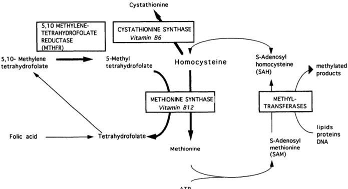
C y s t a t h i o n i n e 5.10METHYLENE-TETRAHYDROFOLATE REDUCTASE (MTHFR) CYSTATHIONINE SYNTHASE Vitamin B6 5,10- Methylene tetrahydrofolate 5-Methyl tetrahydrofolate \
Homocysteine
Folic acid\J
S-Adenosyl homocysteine (SAH) methylated products METHIONINE SYNTHASE Vitamin B12 TetrahydrofolateJ
Methionine METHYL-TRANSFERASES S-Adenosyl methionine (SAM) lipids proteins DNA ATPFigure 1. The production of homocysteine in methionine metabolism and the involvement of MTHFR. Arrows in bold
represent points at which genetic defects in the enzyme or in co-factor biosynthesis are known to cause elevated homocysteine.
donor, or disposed of by the transsulphuration path-way in which the initial step is its condensation with serine to form the thioether cystathionine through the action of the vitamin-B6-dependent enzyme cystathionine-/?-synthase (CBS) (Figure 1).
Of the enzymes involved in recycling or removing homocysteine, severe genetic defects resulting in loss of function are known to affect two: 5,10-methylene tetrahydrofolate reductase (MTHFR) and cystathion-ine- jS-synthase (CBS). The genes encoding each of these two enzymes have been cloned and sequenced, and several mutations which cause such highly deficient phenotypes have been identified. Homozygotes for severe mutations of either the CBS or MTHFR genes have homocystinuria with associ-ated premature vascular disease and thromboembol-ism affecting both large and small arteries and veins.9'10 Heterozygotes for such severe mutations have mild hyperhomocysteinaemia; however, these genotypes are too rare to account for the frequency of mild hyperhomocysteinaemia observed in the general population.
MTHFR is a flavoprotein which catalyses the NADPH-linked reduction of 5,10-methylenetetra-hydrofolate to 5-methyltetra5,10-methylenetetra-hydrofolate, the major circulating form of folate and the methyl donor for the methioninesynthase-catalysed remethylation of homocysteine to methionine. A phenotypic variant of MTHFR with characteristic thermolability after partial denaturation at 46°C for 5 min11 has been identified in 5 - 8 % of the healthy population and
shown to give rise to mild hyperhomocysteinaemia without homocystinuria in healthy controls as well as CAD patients, in whom it is approximately three times more common than in healthy controls.12"14 This biochemically-defined thermolabile variant is the most probable cause of mild hyperhomocysteina-emia in 28% of hyperhomocysteinaemic vascular disease patients.15
The cDNA for MTHFR was recently cloned and sequenced by Goyette ef a/., who identified nine mutations in classically deficient patients.16'17 In addition, a C to T transition at nucleotide 677, resulting in an amino acid change from alanine to valine, correlated with thermolability; homozygotes for the mutation had thermolabile MTHFR (defined by a specific activity of 50% of the normal mean, and residual activity after heat inactivation of < 3 6 % of the initial activity), while heterozygotes had inter-mediate thermolability.18 The mutation resulted in elevated homocysteine levels irrespective of whether measurements were made after fasting or methionine loading. This mutation has subsequently been shown to confer a 2.9-fold risk of cardiovascular disease in Ireland (Gallagher ef a/., submitted) and a 3.1-fold risk of cardiovascular disease in Holland19—figures which are similar to the relative risk associated with elevated homocysteine levels.
The frequency of homozygosity for thermolabile MTHFR in individuals with NTDs is approximately three times the average in the population,20'21 and in Ireland 13% of NTDs may be directly attributed to this factor.20
MTHR genotype and hyperhomocysteinaemia
573
Elevated homocysteine is clearly an important risk indicator for a range of clinical conditions. The magnitude of risk conferred by the thermolabile MTHFR genotype is similar to that associated with elevated homocysteine. To permit a rational assess-ment of the potential for screening the population to identify individuals genetically at risk of homocyst-eine-associated disease, and to devise appropriate treatments with homocysteine-lowering supplements such as folic acid, the proportion of individuals who are hyperhomocysteinaemic due to the thermolabile MTHFR genotype needs to be determined.
Our study examines the extent to which mild hyperhomocysteinaemia in a working male popula-tion is directly attributable to the thermolabile MTHFR mutation.
by Ubbink et al23 Plasma samples were derivatized with ammonium 7-fluoro 2-oxa-1,3 diazole-4-sulph-onate (SBD-F).
Cenotyping for the MTHFR thermolabile mutation was performed by PCR and H/nfl digestion as in Frosst et a/.18
Levels of serum folate and vitamin B12 were measured using an ICN Pharmaceuticals kit.
Statistics were analysed using SPSS for Windows. Distributions of homocysteine, folate and B12 were skewed and were logarithmically transformed when appropriate. Analysis of variance was used to com-pare mean values between groups (genotypes or tenths). Relative risk estimates were calculated using X2 tests with Yates' correction or Fisher's exact probability test as appropriate.
Methods
Males aged 30-49 from an industrial workforce in Belfast, comprising both manual and non-manual workers, were invited to a screening clinic, and after informed consent for all biochemical and genetic analyses had been given, a venous blood sample was taken with minimum haemostasis and anti-coagulated with EDTA. Individuals who were dia-betic, had had a general anaesthetic within the previous 3 months, or were using any form of dietary supplementation (16.6%) were excluded. A total of 625 men were eligible for inclusion in the present analysis.
Total homocysteine (free plus protein-bound) was assayed by high performance liquid chromatography according to the method of Araki & Sako,22 modified
Results
In our study population, the overall frequencies of thermolabile homozygotes, heterozygotes, and non-thermolabile homozygotes were 11.5, 43.7, and 44.8% respectively (in close agreement with the Hardy-Weinberg prediction of 11.1, 44.5, and 44.4%: £ test p > 0.5).
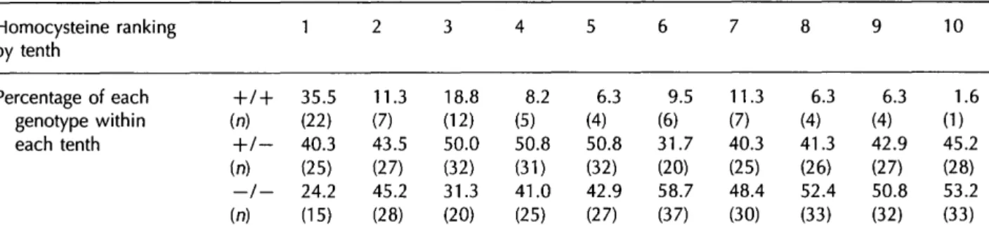
Thermolabile homozygous individuals had mean homocysteine levels significantly higher than those of heterozygotes, whose homocysteine levels were slightly, but not significantly, higher than those of non-thermolabile homozygotes (Table 1). To assess the genotype/phenotype relationship we divided the population into tenths according to homocysteine ranking (Table 2). The thermolabile homozygote fre-quency exhibited a gradual increase from 1.6% in
Table 1 Plasma homocysteine, serum folate and vitamin B12 levels by MTHFR genotype
Genotype
(n = 72) = 273) = 280)
Significance
Mean homocysteine (u.mol/1) Mean homocysteine in
men with folate below median (u.mol/1) Mean homocysteine in
men with folate above median (u.mol/1) Mean serum folate
(nmol/l) Mean vitamin B12 (pmol/l) 9.46(8.40,10.65) 11.22(9.57,13.16) (n = 46) 6.82(6.21,7.49) (n = 24) 9.29(8.33,10.35) 221(198,248) 7.12(6.85,7.40) 7.88(7.47,8.31) (n=134) 6.54(6.23,6.87) (n = 133) 10.76(10.26,11.29) 259(247,272) 6.77(6.55,7.01) 7.43(7.02,7.86) (n=118)
6.32(6.08,6.57)
(n=156)
12.31(11.71,12.93) 266(253,279)Geometric mean values of homocysteine, folate, and vitamin B12 are shown with 95% CIs (in brackets) for thermolabile homozygotes ( + / + ) , heterozygotes (+/—), and non-thermolabile homozygotes (—/—). Mean homocysteine levels for men whose serum folate levels are above and below the median value (11.04 nmol/l) show that the association of high homocysteine with the + / + genotype is highly dependent on folate status. * Significant difference (p<0.003) between the + / + genotype and the other two genotypes; **AII three genotypes are significantly different from each other (p<0.001). Note: folate levels measured for 611 subjects.
Table 2 MTHFR genotypes Homocysteine ranking by tenth Percentage of each genotype within each tenth in -h (n) -h (n) —t (n) relation to 1 / + 35.5 (22) 1- 40.3 (25) / - 24.2 (15) homocysteine concentration 2 11.3 (7) 43.5 (27) 45.2 (28) 3 18.8 (12) 50.0 (32) 31.3 (20) 4 8.2 (5) 50.8 (3D 41.0 (25) 5 6.3 (4) 50.8 (32) 42.9 (27) 6 9.5 (6) 31.7 (20) 58.7 (37) 7 11.3 (7) 40.3 (25) 48.4 (30) 8 6.3 (4) 41.3 (26) 52.4 (33) 9 6.3 (4) 42.9 (27) 50.8 (32) 10 1.6 (1) 45.2 (28) 53.2 (33)
Individuals were assigned to tenths according to their homocysteine ranking: tenth 1 corresponds to highest homocysteine levels. The percentage of individuals within each tenth who are thermolabile homozygotes ( + / + ), heterozygotes ( + / — ) , and non-thermolabile homozygotes {—/—) is shown.
the lowest tenth, to between 1 0 % and 2 0 % in the second and t h i r d highest tenths. In the upper tenth, the percentage of homozygotes rose appreciably to 3 5 . 5 % , b u t there was a striking further increase in the frequency of thermolabile homozygotes a m o n g the individuals w i t h the highest homocysteine rank-ings w i t h i n this group. W e therefore sequentially subdivided the upper tenth, and observed progressive increases in t h e r m o l a b i l e homozygote frequency as the numbers of individuals were progressively restricted a c c o r d i n g t o their ranking, i.e. 11 of the t o p 2 0 (55%), and 7 of the top 10 (70%). W e calculated the relative risk of being in the top 5, 10, 2 0 and 5 0 % of the p o p u l a t i o n w i t h respect to homocysteine for individuals w i t h the thermolabile homozygous genotype relative to non-thermolabile homozygotes and to non-thermolabile homozygotes and heterozygotes c o m b i n e d (Table 3). There was a highly significant 9.7-fold risk of being in the top 5 % for t h e r m o l a b i l e homozygotes relative to n o n -t h e r m o l a b i l e homozygo-tes. This risk was sligh-tly less (7.2-fold) w h e n thermolabile homozygotes were c o m p a r e d w i t h non-thermolabile homozygotes and heterozygotes together. A small risk was observed for heterozygotes relative to nonthermolabile h o m o -zygotes, but this was o n l y statistically significant in the upper 5 0 % of the homocysteine distribution considered as a w h o l e . O v e r a l l the data p o i n t to a
small but significant thermolabile heterozygote effect in elevating homocysteine, w h i c h has not been reported previously.
W h e n serum folate status was taken into account, the relationship between the homozygous thermola-bile genotype and homocysteine levels was restricted to those individuals whose serum folate levels were b e l o w the median value ( p < 0 . 0 0 1 ) . In individuals w i t h folate levels above the median value, homocyst-eine levels d i d not vary significantly between geno-types ( p = 0.3) (Table 1).
Serum folate and v i t a m i n B12 levels were both inversely related to homocysteine levels (Figure 2), and also correlated w i t h each other (not shown). W e observed a relationship between serum folate c o n -centration and MTHFR genotype w i t h homozygous t h e r m o l a b i l e individuals having the lowest serum folate levels and non-thermolabile homozygotes the highest (Table 1). Heterozygotes had intermediate serum folate levels. The differences in folate levels between all three genotypes w e r e significant ( p < 0 . 0 0 1 ) . V i t a m i n B12 levels were also signific-antly lower in thermolabile homozygotes than in individuals w i t h other genotypes (Table 1). The differ-ences in folate and B12 levels between genotypes for each adjacent tenth were not statistically significant due to the small numbers of thermolabile h o m o -zygotes i n v o l v e d , b u t the trend is apparent,
Table 3 The relative risk of mild hyperhomocysteinaemia conferred by MTHFR thermolabile genotypes Homocysteine rank Top 5% Top 10% Top 20% Top 50%
+/ +
Risk 9.72 5.70 2.62 1.69 relative to —/— 95% Cl (3.91,24.17) (3.12,10.42) (1.77,3.89) (1.37,2.08) P < 0.001 < 0.001 < 0.001 < 0.001+/ +
Risk 7.20 4.22 2.34 1.47 relative to ( + / — 95% Cl (3.72,13.93) (2.67,6.88) (1.68,3.28) (1.23,1.75) and - / - ) P < 0.001 < 0.001 < 0.001 < 0.001+
/-Risk 1.71 1.71 1.24 1.31 relative to —/— 95% Cl (0.63,4.64) (0.92,3.17) (0.86,1.79) (1.10,1.57) P 0.29 0.08 0.25 0.003Relative risks of being in the top 5, 10, 20 and 50% of the homocysteine distribution for individuals with the thermolabile homozygous genotype ( + / + ) relative to non-thermolabile homozygotes (—/—), and to non-thermolabile homozygotes and heterozygotes combined, and for thermolabile heterozygotes ( + / — ) relative to non-thermolabile homozygotes.
MTHR genotype and hyperhomocysteinaemia
575
2 . 0 1 . 8 1 . 6 1 . 2 -1.0 1 2 3 4 5 6 7 8 9 10homocysteine tenth
to o c o u 5.6=1 5.4 5.2 homocysteine tenthFigure 2. The relationship between a serum folate and
b vitamin B12 levels and homocysteine. Mean log levels are plotted separately for each genotype within the homo-cysteine distribution (homohomo-cysteine decreases from tenth 1 to 10). Thermolabile homozygotes, filled squares; hetero-zygotes, open triangles; non-thermolabile homohetero-zygotes, open circles. The low B12 value for the homozygous thermolabile genotype in tenth 10 is due to the presence of only one such individual in this tenth.
particularly for serum folate in individuals in the upper half of the homocysteine distribution (Figure 2a).
We further examined the serum folate and
vitamin B12 levels in the 20 individuals with the
highest homocysteine levels. Most of these
indi-viduals had folate and B12 levels at the low end of
the overall range. Consistent with the genotype/folate
interaction already discussed, on average the
thermo-labile homozygotes had lower folate levels (mean
5.45 nmol/l, n = 11), than individuals with the other
two genotypes (combined mean 7.71 nmol/l, n = 9).
Vitamin B12 levels showed a similar trend. Some
individuals in the top 20 are not thermolabile
homo-zygotes and do not have marked deficiency of folate
or vitamin B12, suggesting the existence of other,
possibly genetic, factors influencing homocysteine
levels.
Discussion
We have shown that in a working male population
homozygosity for the thermolabile MTHFR genotype
is a major contributing factor to mild
hyperhomocys-teinaemia, and in particular to homocysteine levels
above the 95th percentile. The approximately
five-fold increase in frequency of the thermolabile
geno-type in the top 5% of the population (to 48%)
relative to its frequency in the bottom 80% is of
particular note because one of the largest studies of
the association of homocysteine with myocardial
infarction (Ml)—a prospective study of 14 916
physi-cians of whom 271 developed Ml—suggested that
the 95th percentile of homocysteine distribution was
the point at which the risk of Ml increased
approxi-mately three-fold.
1Dietary deficiency of the vitamin cofactors B6
(for CBS) and B12 (for methionine synthase), and of
folic acid, the precursor of the methyl donor
5-methyltetrahydrofolate, can all cause elevated
homocysteine levels, although evidence that this is
a major contributing factor to
hyperhomocysteinae-mia only exists for an elderly population studied by
Selhub et al.
24(who estimated that up to 2/3 of
hyperhomocysteinaemic 67-96 year olds from the
Framingham Heart Study had elevated homocysteine
attributable to deficiency of one of these three dietary
components).
Our study identifies two separate aspects of the
interaction between the homozygous thermolabile
MTHFR genotype and folate metabolism.
Firstly, in individuals with serum folate levels
below the median, those with the homozygous
thermolabile genotype have higher homocysteine
levels (50% higher than non-thermolabile
homozy-gotes). Heterozygotes show a smaller increase (6%)
in homocysteine relative to individuals with no
thermolabile allele. Among individuals whose folate
levels are above the median, the homozygous
ther-molabile genotype does not appear to affect homo-cysteine levels. Jacques ef al. recently observed a similar effect in thermolabile homozygotes (but not heterozygotes) with folate levels below the median.25 The folate level below which the thermolabile homozygous genotype becomes a homocysteine-determining factor has yet to be established. However it is apparent that low folate has a particularly detrimental effect on the capacity of thermolabile homozygotes to remethylate homocysteine.
Secondly, the thermolabile homozygotes as a group have significantly lower serum folate levels than the other genotypes, indicating that the thermo-labile homozygous genotype itself causes reduced serum folate levels. It seems likely that the above two interactions between genotype and folate can act in concert to produce the mild hyperhomocystei-naemic phenotype.
The apparently causal relationship between geno-type and serum folate levels indicates that in a proportion of individuals in the higher homocysteine range the lower folate levels are not necessarily attributable to dietary insufficiency alone but are, at least in part, a direct result of the reduced activity of the thermolabile enzyme. In a recent study of women with multiple NTD events, Lucock et al.26 have proposed that the low levels of plasma 5-methyltetrahydrofolate (measured specifically by HPLC) relative to dietary folate intake observed in such cases, may reflect an underlying control or structural defect in the MTHFR gene or in another gene involved in an earlier step in the conversion of dietary folate to 5-methyltetrahydrofolate. They speculated that such cases may need a higher intake of dietary folate (or supplements) to achieve the same plasma 5-methyltetrahydrofolate concentrations as controls. In a subsequent study decreased serum folate levels and elevated red-cell folate levels were observed in thermolabile homozygotes by van der Put ef a/.21 who point out that the MTHFR enzyme product 5-methyltetrahydrofolate is the predominant form of folate in serum, while other folate species including the substrate 5,10-methylenetetrahydro-folate are mainly present in cells.
In conclusion, it appears that homozygosity for the MTHFR thermolabile mutation is a major cause of mild hyperhomocysteinaemia in the study popula-tion. The frequency of the mutation may be expected to vary between populations, and this may account for some of the variation in homocysteine levels observed in different countries and regions (Malinow et al., submitted). The extent to which this is due to variations in MTHFR genotype frequencies will not become clear until significant numbers of individuals from defined populations have been studied.
Dietary supplementation with folic acid given periconceptionally has already been shown to be
effective in the reduction of NTDs,27'28 some of which are caused by the thermolabile MTHFR allele.20'21 Studies of dietary folic acid supplementation of healthy individuals and vascular disease patients indicate that this is a safe and effective method of reducing homocysteine levels (provided B12 is not deficient).29"31 It is likely that extra dietary folic acid can compensate for the suboptimal function of the thermolabile form of MTHFR and can therefore be used to reduce the high homocysteine levels observed in many thermo-labile homozygotes, thus eliminating this major risk factor for vascular and thrombotic disease. Our results establish that for a given homocysteine con-centration thermolabile MTHFR homozygotes have lower serum folate levels, indicating that there is a direct genotype—folate interaction. We suggest that the thermolabile MTHFR genotype should be taken into account in the design of studies aiming to identify the optimum dose of folic acid required to lower homocysteine concentration, as the effect-iveness of folate supplementation is likely to vary with genotype.
Acknowledgements
We acknowledge the technical assistance of Caroline Mercer and Janet Lightbody. This work was funded by a Project Grant from The Irish Heart Foundation and a Unit Grant from The Irish Heart Foundation/ Health Research Board. We acknowledge the support of Sandoz pic.
References
1. Stampfer MJ, Malinow MR, Willett WC, Newcomer LM, Upson B, Ullmann D, Tishler PV, Hennekens CH. A prospective study of plasma homocyst(e)ine and risk of myocardial infarction in US physicians. JAMA 1992; 268:877-81.
2. Genest JJ, Jr, McNamara JR, Salem DN, Wilson PWF, Schaefer EJ, Malinow MR. Plasma homocyst(e)ine levels in men with premature coronary artery disease. J Am Coll
Cardiol 1990; 16:1114-19.
3. Molgaard J, Malinow MR, Lassvik C, Holm A-C, Upson B, Olsson A C Hyperhomocyst(e)inaemia: an independent risk factor for intermittent claudication. J Intern Med 1992; 231:273-9.
4. Taylor LM, Jr, DeFrang RD, Harris E, Jr, Porter JM. The association of elevated plasma homocyst(e)ine with progression of symptomatic peripheral arterial disease.
J VascSurg 1991; 13:128-36.
5. Brattstrom L, Lindgren A, Israelsson B, Malinow MR, Norrving B, Upson B, Hamfelt A. Hyperhomocysteinaemia in stroke: prevalence, cause, and relationships to type of stroke and stroke risk factors. EurJ Clin Invest 1992; 22:214-21.
MTHR genotype and hyperhomocysteinaemia 577 Inahara T, Mukerjee D, Sexton C, Upson B. Prevalence of
hyperhomocyst(e)inemia in patients with peripheral arterial occlusive disease. Circulation 1989; 79:1180-8.
7. den Heijer M, Blom HJ, Gerrits WBJ, Rosendaal FR, Haak HL, Wijermans PW, Bos GMJ. Is hyperhomocysteinaemia a risk factor for recurrent venous thrombosis? Lancet 1995; 345:882-5.
8. Mills JL, McPartlin JM, Kirke PN, Lee YJ, Conley MR, Weir DG, Scott JM. Homocysteine metabolism in pregnancies complicated by neural-tube defects. Lancet 1995; 345:149-51.
9. Mudd SH, Levy HL, Skovby F. Disorders of transsuIfuration. In: Scriver CR, Beaudet AL, Sly WS, Valle D, eds. The
Metabolic Basis of Inherited Disease. New York,
McGraw-Hill, 1989:693-734.
10. Rosenblatt DS. Inherited disorders of folate transport and metabolism. In: Scriver CR, Beaudet AL, Sly WS, Valle D, eds. The Metabolic Basis of Inherited Disease. New York, McGraw-Hill, 1989:2049-64.
11. Kang S-S, Zhou JZ, Wong PWK, Kowalisyn J, Strokosch G. Intermediate homocysteinemia: a thermolabile variant of methylenetetrahydrofolate reductase. Am J Hum Genet 1988; 43:414-21.
12. Kang S-S, Wong PWK, Zhou J, Sora J, Lessick M, Ruggie N, Grcevich G. Thermolabile methylenetetrahydrofolate reductase in patients with coronary artery disease.
Metabolism 1988; 37:611-13.
13. Kang S-S, Wong PWK, Susmano A, Sora J, Norusis M, Ruggie N. Thermolabile methylenetetrahydrofolate reductase: an inherited risk factor for coronary artery disease. Am) Hum Genet 1991; 48:536-45. 14. Kang S-S, Passen EL, Ruggie N, Wong PWK, Sora H.
Thermolabile defect of methylenetetrahydrofolate reductase in coronary artery disease. Circulation 1993; 88:1463-9. 15. Engbersen AMT, Franken DG, Boers GHJ, Stevens EMB,
Trijbels FJM, Blom HK. Thermolabile
5,10-methylenetetrahydrofolate reductase as a cause of mild hyperhomocysteinemia. Amj Hum GeneH 995;
56:142-50.
16. Goyette P, Sumner JS, Milos R, Duncan AMV, Rosenblatt DS, Matthews RG, Rozen R. Human
methylenetetrahydrofolate reductase: isolation of cDNA, mapping and mutation identification. Nature Genet 1994; 7:195-200.
17. Goyette P, Frosst P, Rosenblatt DS, Rozen R. Seven novel mutations in the methylenetetrahydrofolate reductase gene and geneotype/phenotype correlations in severe
methylenetetrahydrofolate reductase deficiency. Am J Hum
Genet 1995; 56:1052-9.
18. Frosst P, Blom HJ, Milos R, Goyette P, Sheppard CA, Matthews RG, Boers GJH, den Heijer M, Kluijtmans LAJ, van den Heuvel LP, Rozen R. A candidate genetic risk factor for vascular disease: a common mutation in
methylenetetrahydrofolate reductase. Nature Genef 1995; 10:111-13.
19. Kluitjmans LAJ, van den Heuvel LPWJ, Boers GHJ, Frosst P, Stevens EMB, van Oost BA, den Heijer M, Trijbels FJM, Rozen R, Blom HJ. Molecular genetic analysis in mild hyperhomocysteinaemia: a common mutation in the methylenetetrahydrofolate reductase gene is a genetic risk factor for cardiovascular disease. Am J Hum Genet 1996; 58:35-41.
20. Whitehead AS, Gallagher P, Mills JL, Kirke PN, Burke H, Molloy AM, Weir DG, Shields DC, Scott JM. A genetic defect in 5,10-methylenetetrahydrofolate reductase in neural tube defects. QJ Med 1995; 88:763-6. 21. van der Put NMJ, Steegers-Theunissen RPM, Frosst P,
Trijbels FJM, Eskes TKAB, van den Heuvel LP, Mariman ECM, den Heyer M, Rozen R, Blom HJ. Mutated methylenetetrahydrofolate reductase as a risk factor for spina bifida. Lancet 1995; 346:1070-1.
22. Araki A, Sako Y. Determination of free and total
homocysteine in human plasma by HPLC with fluorescence detection. J Chromatogr 1987; 422:43-52.
23. Ubbink JB, Vermaak WJH, Bissbort S. Rapid high-performance liquid chromatographic assay for total homocysteine levels in human serum. J Chromatogr 1991; 565:441-6.
24. Selhub J, Jacques PF, Wilson PWF, Rush D, Rosenberg IH. Vitamin status and intake as primary determinants of homocysteinemia in an elderly population. JAMA 1993; 270:2693-8.
25. Jacques PF, Bostom AG, Williams RR, Ellison RC, Eckfeldt JH, Rosenberg IH, Selhub J, Rozen R. Relation between folate status, a common mutation in
methylenetetrahydrofolate reductase, and plasma homocysteine concentrations. Circulation 1996; 93:7-9. 26. Lucock MD, Wild J, Schorah CJ, Levene Ml, Hartley R. The
methylfolate axis in neural tube defects: In Vitro characterisation and clinical investigation. Biochem Med
MetabBiol 1994; 52:101-14.
27. Czeizel AE, Dudas I. Prevention of the first occurrence of neural-tube defects by periconceptual vitamin
supplementation. N Engl J Med 1992; 327:1832-5. 28. MRC Vitamin Study Research Group. Prevention of neural
tube defects: results of the Medical Research Council vitamin study. Lancet WW; 338:131-7.
29. Brattstrom LE, Israelsson B, Jeppsson J-O, Hultberg BL. Folic acid—an innocuous means to reduce plasma homocysteine.
ScandJ Lab Clin Invest 1988; 48:215-21.
30. Landgren F, Israelsson B, Lindgren A, Hultberg B, Andersson A, Brattstrom L. Plasma homocysteine in acute myocardial infarction: homocysteine-lowering effect of folic acid. J Int Med 1995; 237:381 - 8 .
31. Brattstrom L, Israelsson B, Norrvig B, Bergqvist D, Thorne J, Hultberg B, Hamfelt A. Impaired homocysteine metabolism in early-onset cerebral and peripheral occlusive arterial disease. Atherosclerosis 1990; 81:51-60.