European Heart Journal (1983) 4 (Supplement A), 29-34
>
Physiologic or pathologic
hypertrophy
^ H . P. K.RAYENBUEHL, O . M . HESS, J. SCHNEIDER AND M . TURINA
v Medical Policlinic, Cardiology, Institute of Pathology and Surgical Clinic A. University Hospital, Zurich, * Switzerland
KEY WORDS: Myocardial hypertrophy, left ventricular function and structure.
Physiologic hypertrophy occurs as the result of exercise conditioning and is characterized bv normal or " supranormal left ventricular (LV) contractile function and reversibility of structural alterations. Whether hypertrophy produced by chronic abnormal loading can be termed 'physiologic' is a matter of debate because in experimental pressure overload hypertrophy normal in vivo ventricular function may be associated with abnormal I in vitro function of the papillary muscles. In patients with moderate LV hypertrophy from aortic valve disease (angiographic mass < 180 g/m2) ejection fraction (EF) is preserved, but at similar levels ofafterload, when mass -* exceeds 180 g/m2, EF is depressed. Comparison of LV function with myocardial structure (endomyocardial
biopsies) has shown that in patients with compensated LV function and those with left heart failure (EF <57°/o.
LVEDP > 20mm Hg and/or cardiac index < 2-5 l/min/m2) interstitialfibrosis (IF) was increased to a similar
f extent (16 and 18%: normal < 5%), whereas muscle fiber diameter (MFD; normal < 20 /<) was larger (P < 005) i in the patients with failure (30 /J.) than in those with preserved function (27 n). Moreover patients with depressed postoperative function had a larger (P < 001) preoperative MFD (35 /() than those with normal postoperative a function (30 ft). Seventeen months after successful aortic valve replacement IF increased (P < 002) and MFD decreased (P < 0001) but did not become normal regardless whether postoperative function was normal or " \ depressed. Thus in secondary hypertrophy myocardial structure is pathologic even in the presence of normal LV
function and depressed function appears likely to be related to excessive fiber hypertrophy rather than to IF. *" Massive fiber hypertrophy heralds an unfavorable postoperative L V function and fibrosis is irreversible after L surgical correction of the abnormal load.
The predominant compensatory mechanism in alterations which may have occurred during the conditions with chronic abnormal loading of the formation of hypertrophy following the elimination heart is myocardial hypertrophy. When an increase in of the stimulus to hypertrophy. Conversely in muscle mass is associated with normal or enhanced pathological hypertrophy there is no or only myocardial function, hypertrophy is termed physio- incomplete reversibility of morphological alterations. ** logical"1. Biochemically, physiological hypertrophy
„ , is characterized by a normal or increased myosin ™ • • • • . L I . I L L , L • . _ „ . . ,,, , , , . . . Physiological and pathological hypertrophy in the
ATPase activity . In contrast, pathological hyper- . . . . , . , • . . • • j J I experimental animal
i trophy is determined by impaired myocardial r
function and a decreased myosin ATPase activity1". The prototype of physiological hypertrophy is the These definitions do not take into account the hypertrophy consequent to exercise conditioning. I potentials of reversibility of hypertrophy. As will be Myocardial function has been demonstrated to be shown later in this paper, it seems appropriate to add increased in isolated hearts from rats with increased > •* to the characteristics of physiological hypertrophy heart weight/body weight ratio after training by the capacity of full reversibility of any myocardial swimming121 and in intact dogs trained by treadmill
i running131 as compared to function in
cor-sequesis for rtprims w H. P• Krayenbuehl. M c'•• Ch.ef or r e s p o n d i n g sedentary animals. However, in vitro ™ Cardiology, Medical Policlinic of Ihe University. CH-8091 Zurich. F 6 J '
Switzerland studies carried out in papillary muscles of trained cats 0195-668X/83/04A029+ 06 $02.00/0 ( 1983 The European Society of Cardiology'
did not elicit a higher than normal contractile state'41. The diastolic properties of the left ventricle evaluated by the diastolic pressure-length relation-ship have been found to be normal in trained dogs<3). Cardiac myosin ATPase activity is typically increased in exercise-conditioned animals as shown by several studies summarized in the overview by Scheuer and Bhan|6). There is excellent reversibility of exercise-induced hypertrophy because in the myocardium of rats two weeks following cessation of training, the structural alterations typical of hyper-trophy regressed substantially and the light micro-scopic picture was almost indistinguishable from that of normal rats'7'.
Of importance is the question of whether a chronic abnormal mechanical burden by pressure or volume overload leads to physiological or pathological hypertrophy. M o s t '8" "1 but not all"2 1 animal models with chronic volume overload did show essentially normal in vivo and in vitro function of the hypertrophied myocardium. In contrast myocardial hypertrophy consequent to the production of severe pressure overload with sudden onset was associated with depressed contractility"3"1" which sometimes improved with time although the pressure loading persisted"6'. These pressure overload models have however little analogy to the more progressive development of pressure loading in man. Hence only models in which a gradually increasing pressure load was applied are considered in the present discussion. Progressive pressure loading was achieved by a slight banding of either the aorta or the pulmonary artery in young animals"7 '"'. With normal growth of the animal a considerable stenosis developed and myocardial hypertrophy ensued. In dogs"7' and in cats"8' the ejection fraction of the overloaded ventricle remained within normal limits 37 and 60 weeks respectively after banding. No consistent findings were obtained in vitro. In the cat papillary muscle Cooper et a/."81 found a decrease in maximal velocity of shortening and maximal active isometric tension with increasing duration of pressure loading (25-60 weeks), whereas with a similar duration (24-52 weeks) of progressive pressure loading Williams el a/."9' could demonstrate no change in contractility. This difference can probably be explained by the fact that in the study of Cooper et a/."81 the hydroxyproline concentration was signifi-cantly increased in the hypertrophied muscle, whereas it was similar in banded and non-banded animals in the study of Williams et a/."9'. Thus the connective tissue content is likely to have been more marked in
the former"81 animals and may have interfered with normal contractile behavior. Furthermore, an increased hydroxyproline content has been shown not to decrease after debanding in rats with left ventricular hypertrophy from constriction of the ascending aorta'20'.
In summary these studies in animals with progressive pressure load indicate that despite preserved pump function in vitro muscle function can be depressed and probably irreversible increase in connective fibrous tissue may occur. Hence the apparent physiological hypertrophy as judged from the normal pump function is in fact already an early form of pathological hypertrophy.
Nature of secondary hypertrophy in man. Systolic function and relaxation
In contrast to the studies in the papillary muscles, where an unequivocal assessment of contractility is possible, the evaluation of contractility in man is complicated by the fact that there is no contractile index which is not influenced by either afterload, preload or heart rate. Recently we have studied by left ventricular (LV) micromanometry and cineangio-graphy 47 patients with aortic valve disease before and 18 months after successful valve replacement. LV contractile state was assessed by the relationship between ejection fraction (EF) and peak systolic circumferential wall stress (PSWS)'2" (Fig. 1) and by a function diagram where LV isovolumic maximal rate of rise of circumferential wall stress (max d<r/dt) was plotted v. end-diastolic wall stress (EDS)'221 (Figs 2 and 3). Patients with mild to moderate LV hypertrophy having an angiographic mass < 180 g/m2 (GR 1) and patients with severe hypertrophy in whom mass was 5= 180 g/m2 (GR 2) were analyzed separately. Moreover, the patients were divided in those with pure or predominant aortic stenosis (AS, n = 27) and those with pure or predominant aortic insufficiency (AI, n = 20). Fourteen control patients (C) were studied by the same techniques. At the preoperative evaluation the GR 1 (AS and AI) as well as the GR 2 (AS and AI) patients had a significantly increased PSWS as compared to C (Fig. I). No difference existed among the four groups with aortic valve disease. The GR 2 patients with severe hypertrophy showed however a significantly lower EF than the GR 1 patients which was indicative of depressed contractility. In turn the EF in the GR 1 patients was only slightly and insignificantly smaller than in C. This mildly
1
5
4 V
Physiologic or pathologic hypertrophy 31 !
> 1
I
0 8 0 7 0 6 0 5 0 4 -4 0 0 6 0 0Peak systolic circumf«rentiol wall stress ( I 03 dynes/cm2 )
Figure I Relation between left ventricular (LV) ejection
fraction (EF) and peak systolic circumferential wall stress (PSWS) in 14 controls (C), 17 patients with aortic stenosis (AS) and an LV angiographic mass (LMMI) < 180 g/m2
(AS GR 1) ( • ) , 10 patients with AS and an LMMI £ 1 8 0 g/m2 (AS GR 2) (O), 10 patients with aortic
insufficiency (Al) and an LMMI < 180 g/m2 (AI GR 1) (A)
and 10 patients with AI and an LMMI > 180 g/m2 (AI GR
2) (A)- Mean values + 1 standard error are presented. The arrows are directed from the preoperative to the postoperative values. At the preoperative evaluation PSWS was increased significantly in all patients groups as compared to the C (asterisks to the right of the symbols). In both patient groups having an LMMI < 180 g/m2 EF was
slightly and insignificantly decreased probably consequent to the increased PSWS (afterload). In contrast, both patient groups with severe hypertrophy (LMMI > 180 g/m2)
elicited a significantly decreased EF at similar PSWS to the GR I patients indicative of depressed LV contractile function (asterisks above the symbols for comparison with C). *, P< 0 05, •*, P - 0 02, *•*, P - 0 0 1 ; * * " ,
P < 0-001) Following successful aortic valve replacement
the relation between EF and PSWS became normal (not different from C) except in the patients with AI and severe preoperative hypertrophy whose EF remained depressed despite normalization of PSWS.
decreased EF was probably due to the increased afterload at an apparently unchanged contractile state. From Fig. 2 it is however evident that a beginning depression of contractile state was also present in the GR 1 patients, because the AS group and especially the AI group were located rightward
7000
5000
3000
* * *
0 25 50 75 IOO
Circumferential end - diastobc stress (10 3 dynes/cm2 )
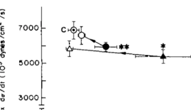
Figure 2 Left ventricular function diagram opposing
iso-volumic maximal rate of rise of circumferential wall stress (max d<x/dt, ordinate) to circumferential end-diastolic wall stress (EDS, abscissa) in the control group (C) and in the patients with aortic stenosis ( • . preoperatively; O-postoperatively) and aortic insufficiency ( A , preoperatively; A . postoperatively) having an LMMI < 180 g/m2.
Pre-operatively the relationship between maximum dff/dt and EDS was located rightward and downward to the control group indicating an impaired contractile state. After surgery the relationship between max du/dt and EDS became normal (not different from C). Mean values ± 1 standard error are presented. Asterisks to the right of the symbols are for comparison of EDS and above the symbols for comparison of max du/dt. *, P < 0 0 5 ; **. P < 0 0 2 ; *•*,
P < 001 (unpaired t-test).
and downward to the control group eliciting less isovolumic performance at an increased preload. An even more marked rightward and downward shift was observed in the GR 2 patients (Fig. 3).
Following surgery the relationship between EF and PSWS and maximum dcr/dt and EDS respectively became normal in the GR 1 patients and in the GR 2 patients with preoperative AS (Figs 1-3). In contrast the GR 2 patients with preoperative AI elicited no normalization of the EF despite a postoperative PSWS close to that of the controls (Fig. 1). Preload also became normal in AI GR 2 but max dff/dt remained depressed (Fig. 3).
From these observations one can conclude that in AI with severe increase of angiographic mass there is pathological hypertrophy with at best partial reversibility of depressed function after surgery-Although all the AS patients and the AI GR 1 patients elicited a postoperative contractile function not different from that in C they probably do not represent groups with true physiological hypertrophy because there were clear-cut (AS GR 2) or beginning functional deficits (AS GR 1. AI GR 1) prior to surgery.
X 7000
s
• & " 5000 b 3000 -O ^ 0 25 75 100 125Circumferential end-diattolic stress ( I 03 dynes / c m2 )
Figure 3 Left ventricular function diagram opposing
isovolumic maximal rate of rise of circumferential wall stress (max dff/dt) to circumferential end-diastolic wall stress (EDS) in the control group ( Q and in the patients with aortic stenosis and insufficiency having an LMMI >180g/m2. Preoperatively the relationship between max
dtr/dt and EDS was shifted rightward and downward to the control group indicating an impaired contractile state. The shift was clearly more marked in the patients with AI than in those with AS. Moreover the shift was greater than in the patients with an LMMI < 180 g/m2 (vide Fig. 2). After
surgery the relationship between max du/dt and EDS became practically normal in the patients with AS whereas in the putients with AI max d<r/dt remained significantly depressed. Symbols as in Fig. 2.
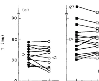
A further argument that even patients with well compensated pump function having a normal LV ejection fraction may not have true physiological, but rather beginning pathological hypertrophy stems from studies on the LV pressure decay (time constant, T) during isometric exercise1231. In 14 patients with aortic valve disease having a LV EF not different from control subjects T was significantly (P < 0-05) greater (60 ms) than in 12 controls (38 ms). Because the prolonged T in the patients with aortic valve disease may solely be the consequence of the increased LV peak systolic pressure'241 and the hypertrophy of the left heart chamber per se12^ and may therefore not be indicative of an intrinsic relaxation disturbance the reaction of T to an additional load produced by handgrip was evaluated. It was found (Fig. 4) that, unlike in the controls who showed a decrease of T during handgrip, T did not change in the patients with aortic valve disease. The systolic response to handgrip evaluated by the change of LV end-diastolic pressure and peak measured velocity of the contractile elements did not differ between the patients with aortic valve disease and the controls.
From these observations it was concluded that in patients with aortic valve disease and normal ejection
Figure 4 Time constant of LV pressure decay (T) in (a) 12
control subjects and (b) 14 patients with aortic valve disease (AVD) at rest (C) and during handgrip (HG, 3 min at 30% of maximal voluntary contraction). The reaction to HG was different in the two groups because T decreased in the controls but remained unchanged in AVD. P = probability (paired t-test); NS = not significant
performance the reaction of LV relaxation to handgrip is abnormal despite a normal contractile response and is likely to represent an early sign of impaired myocardial function.
Correlations between function and structure in secondary hypertrophy
In recent years the morphometric analysis of endomyocardial .biopsies taken at cathetenzation or of biopsies obtained at surgery has added con-siderably to our concept of the nature of hypertrophy in man. In patients with aortic valve disease it has been shown, that already in the stage of compensated LV function there is marked increase in interstitial fibrosis'26 ~29'. In 51 patients (24 aortic stenosis, nine aortic insufficiency, 18 combined lesion) with preserved LV function (biplane EF ^ 57%, LV end-diastolic pressure ^ 20 mm Hg, cardiac index > 2-5 1/min/m2) we have found interstitial fibrosis to be increased to 16% (normal < 5%)l 2 9 '. The mean muscle fiber diameter (MFD) amounted to 27// (normal ^ 2 0 / ; ) - 'n 20 patients with aortic valve disease (11 aortic stenosis, six aortic insufficiency, three combined lesion) presenting with left heart failure (EF < 57%, LV end-diastolic pressure > 2 0 m m H g and/or cardiac index < 2-5 1/min/m2) the interstitial fibrosis was only insignificantly higher
1
Physiologic or pathologic hypertrophy 33
>
s
i
(18%) than in the group with compensated LV function, but MFD was increased (P < 005) to 30 /*. Massive cellular hypertrophy has also been shown to be indicative of persistent postoperative LV dysfunction12830'. In 20 patients with aortic valve disease LV endomyocardial biopsies were obtained preoperatively as well as 17 months after successful aortic valve replacement. Eight of these patients had a depressed LV ejection fraction after surgery. Preoperative MFD was 35 n in this group whereas in the 12 patients with normal postoperative ejection fraction preoperative MFD was 30 ft (P < 001) (Fig. 5). Hence it does appear that advanced cellular hypertrophy rather than excessive interstitial fibrosis is at the origin of depressed contractile function. In this context it is also noteworthy that Schwarz et al.ai) found a reduction of myofibrils with respect to the cell volume in patients with decompensated pressure overload from aortic stenosis. They speculated that this deficit of myofibrils might be the cause for the depressed contractile quality of the LV
£
40 30 20 I 0 -(a)-•in
f
30 20 I 0 (b) 1 <OO5-<0'0l GRI <OO2 GR2 1-<005J 1<OO9J ' MS 1 GRI GR2Figure 5 (a) Muscle fiber diameter (MFD) and (b)
interstitial fibrosis (IF) in 12 patients with aortic valve disease and normal ( ^ 57%) postoperative left ventricular biplane ejection fraction (EFpo) (GR 1) and 8 patients with aortic valve disease and depressed ( < 57%) E Fr o (GR 2).
The bars to the left indicate the preoperative and the bars to the right the postoperative values. In both groups MFD decreased after surgery. However, GR 2 patients with depressed postoperative ventricular function had a signifi-cantly larger preoperative MFD than did GRI patients with normal postoperative function. In both groups IF increased significantly after surgery. There were, however, no significant differences between the two groups neither before nor after surgery. The horizontal dashed lines indicate the upper limit of normality for both MFD and IF. P values were obtained by the paired or the unpaired t-test as appropriate; NS = not significant.
myocardium. In turn, massive interstitial fibrosis appears to play a major role in altering the passive diastolic properties of the myocardium1311.
Reversibility of cellular hypertrophy after aortic valve replacement was incomplete in both patients with normal and depressed postoperative LV function (Fig. 5). The upper limit of normality of the MFD was reached in no instance despite the fact that LV angiographic mass decreased to normal values in almost 70% of the patients'281. Interstitial fibrosis increased significantly (P < 002) from 17 to 26°O in the 20 patients as a group as well as in the two subgroups with normal and depressed postoperative LV function (Fig. 5). LV fibrous content (°0 interstitial fibrosis x angiographic mass) remained unchanged (preoperative 29 g/m2, postoperative 28 g/m2).
In summary the bulk of functional and morpho-logical data suggests that secondary hypertrophy in patients with aortic valve disease is not a physio-logical adaptation but a pathophysio-logical process because:
(1) even in moderate hypertrophy when ejection performance is preserved abnormalities of isovolumic contraction as well as of relaxation are detectable; (2) interstitial fibrosis is augmented not only in patients with left heart failure, but also in those with normal LV ejection fraction;
(3) reversibility of function, i.e. a normal ejection fraction after surgery is not associated with normalization of myocardial structure in that interstitial fibrosis increases, fibrous content remains the same and cellular hypertrophy regresses incompletely.
From the experiments with trained animals it is apparent that the occurrence of true physiological hypertrophy is probably limited to exercise con-ditioning. In secondary hypertrophy from chronic mechanical loading various stages of severity of pathological hypertrophy are possible.
This work was supported by a grant from the Swiss National Science Foundation.
References
(1) Wikman-Coffelt J. Parmley WW, Mason DT. The cardiac hypertrophy process. Analyses of factors determining pathological vs. physiological develop-ment. Circ Res 1979; 45: 697-707.
(2) Penpargkul S, Scheuer J The effect of physical training upon the mechanical and metabolic performance of the rat heart. J Clin Invest 1970; 49: 1859-68.
(3) Riedhammer HH. Rafflenbeul W, Weihe WH, Krayenbuehl HP. Left ventricular contractile function in trained dogs with cardial hypertrophy. Basic Res Cardiol 1976; 71: 297-308.
(4) Williams JF Jr, Potter RD. Effect of exercise conditioning on the intrinsic contractile state of cat myocardium. Circ Res 1976; 39: 425-8.
(5) Carew TE, Covell JW. Left ventricular function in exercise-induced hypertrophy in dogs. Am J Cardiol
1978; 42: 82-8.
(6) Scheuer J. Bhan AK. Cardiac contractile proteins: Adenosine triphosphatase activity and physiological function. Circ Res 1979; 45: 1 12.
(7) Frenzel H, Holtermann W, Schnurch HG, Novi A, Hort W. Experimentelle morphologische und bio-chemische Untersuchungen am Herzen wahrend der Ruckbildung einer Hypertrophie. Verh Dtsch Ges Pathol 1981; 65: 481.
(8) Taylor R, Covell JW, Ross J Jr. Left ventricular function in experimental aorto-caval fistula with circulatory congestion and fluid retention. J Clin Invest
1968; 47: 1333^42.
(9) Turina M. Bussmann WD, Krayenbuehl HP. Contrac-tility of the hypertrophied canine heart in chronic volume overload. Cardiovasc Res 1969; 3: 486-95. (10) Cooper G, Puga FJ, Zujko KJ, Harrison CE. Coleman
HN. Normal myocardial function and energetics in volume-overload hypertrophy in the cat. Circ Res
1973; 32: 140-8.
(11) Carey RA. Natarajan G. Bove AA. Coulson RL, Spann JF. Myosin adenosine triphosphatase activity in the volume-overloaded hypertrophied feline right ventricle. Circ Res 1979; 45: 81-7.
(12) Pinsky WW, Lewis RM, Hartley CJ, Entman ML. Permanent changes of ventricular contractility and compliance in chronic volume overload. Am J Physio!
1979; 237- H575-83.
(13) Spann JF Jr, Buccino RA. Sonncnblick EH. Braunwald E. Contractile state of cardiac muscle obtained from cats with experimentally produced ventricular hypertrophy and heart failure. Circ Res
1967; 21. 341 54.
(14) Bing OHL. Matsushita S. Fanburg BL. Levine HJ. Mechanical properties of rat cardiac muscle during experimental hypertrophy. Circ Res 1971; 28: 234-45 (15) Cooper G. Satava RM Jr, Harrison CE. Coleman HN.
Mechanism for the abnormal energetics of pressure induced hypertrophy of cat myocardium. Circ Res 1973; 33: 213-23.
(16) Williams JF Jr. Potter RD. Normal contractile state of hypertrophied myocardium after pulmonary artery constriction in the cat. J Clin Invest 1974; 54: 1266-72. (17) Carabello BA. Mee R. Collins JJ Jr. Kloner RA. Levin D, Grossman W. Contractile function in chronic gradually developing subcoronary aortic stenosis. Am J Physiol 1981; 240: H80-6.
(18) Cooper G IV, Tomanek RJ, Ehrhardt JC. Marcus ML. Chronic progressive pressure overload of the cat right ventricle. Circ Res 1981; 48: 488-97.
(19) Williams J F J r , Potter RD, Hern DL, Mathew B, Deiss WP Jr. Hydroxyproline and passive stiffness of pressure-induced hypertrophied kitten myocardium. J Clin Invest 1982; 69: 309-14.
(20) Cutilletta AF, Dowell RT, Rudnik M, Arcilla RA, Zak R. Regression of myocardial hypertrophy. I. Experi-mental model, changes in heart weight, nucleic acids and collagen. J Mol Cell Cardiol 1975; 7: 767-81. (21) Huber D, Gnmm J, Koch R, Krayenbuehl HP.
Determinants of ejection performance in aortic stenosis. Circulation 1981; 64. 126-34.
(22) Krayenbuehl HP, Huber D, Koch R, Turina M, Senning A. Contractilite du ventricule gauche hyper-trophie par suite d'une lesion de la valvule aortique. Coeur 1980; 11:429-39.
(23) Deuel W, Hess OM, Krayenbuhl HP. Isometrische Belastung mittels Handgrip zur Beurteilung der linksventrikularen Relaxation bei Patienten mil Aortenvitien. Z Kardiol 1982; 71: 236 (Abstract). (24) Gaasch WH, Blaustein AS, Andrias CW, Donahue
RP, Avitall B. Myocardial relaxation. II. Hemo-dynamic determinants of the rate of left ventricular isovolumic pressure decline. Am J Physiol 1980, 239: H I - 6
(25) Eichhorn P, Gnmm J. Koch R, Hess OM, Carroll J, Krayenbuehl HP. Left ventricular relaxation in patients with left ventricular hypertrophy secondary to aortic valve disease. Circulation 1982; 65: 1395-404.
(26) Schwarz F, Flameng W, Schaper J, Langebartels F. Sesto M, Hehrlein F, Schlepper M. Myocardial structure and function in patients with aortic valve disease and their relation to postoperative results. Am J Cardiol 1978; 41: 661-9.
(27) Schwarz F, Schaper J, Kittstein D, Flameng W. Walter P, Schaper W. Reduced volume fraction of myofibrils in myocardium of patients with decompensated pressure overload. Circulation 1981, 63. 1299-304. (28) Krayenbuehl HP, Schneider J, Turina M, Senning A.
Myocardial function and structure in aortic valve disease before and after surgery. Eur Heart J 1982; 3 (suppl A). 149-53.
(29) Hess OM, Schneider J. Krayenbuhl HP. Slruklur des Myokards bci Patienten mit Aortenvitien und Links-lnsuffizienz. Z Kardiol 1982; 71: 216 (Abstract). (30) Donaldson RM. Left ventricular hypertrophy in aortic
valve disease Eur Heart J 1982; 3 (suppl A): 179-86. (31) Hess OM, Schneider J. Koch R. Bamert C, Grimm J, Krayenbuehl HP. Diastolic function and myocardial structure in patients with myocardial hypertrophy. Circulation 1981: 63: 360-71.