772
Growth-Deficient Mycobacteria in Patients with AIDS: Diagnosis by Analysis
of DNA Amplified from Blood or Tissue
Stefan Emler, Eric C. Btittger, Barbara Broers, Ignazio Cassis, Luc Perrin, and Bernard Hirschel
From the Division des Maladies Infectieuses, HOpital Cantonal Universitaire de Genêve, Geneva, Switzerland; the Institut fur medizinische Mikrobiologie, Medizinische Hochschule, Hannover, Germany; and the Ufficio del Medico Cantonale, Bellinzona, Switzerland Amplification and sequencing of mycobacterial ribosomal RNA genes (16S rDNA) may
per-mit the detection of growth-deficient species (i.e., those exhibiting no growth or those whose growth is delayed for more than 12 weeks). Of blood samples from 26 patients with AIDS and a liver sample from one additional AIDS patient, three samples (two of blood and the one of liver) were positive by polymerase chain reaction only; cultures of these three samples remained nega-tive for more than 12 weeks. Analysis of amplified 16S rDNA from blood revealed a sequence characteristic of Mycobacterium genavense in the first case, in which one of many previous blood cultures had also been positive for M. genavense. The sequences found in the second and third cases were characteristic of Mycobacterium avium. The sample from the second patient was a liver biopsy specimen in which acid-fast bacilli were visualized; the culture of this specimen yielded M. avium after 7 months. The third sample was a blood sample from a patient in whom a relapse of treated M. avium infection was suspected. These results indicate that amplification and sequencing of mycobacterial 16S rDNA may permit early diagnosis and provide a rationale for treatment of infections due to growth-deficient mycobacteria.
Conventional methods for the detection and identification of mycobacteria depend on the organisms' growth in culture and subsequent analysis by hybridization with nucleic acid probes [1] or by biochemical testing on solid media [2]. New techniques involve analysis of mycobacterial DNA and may be very useful in the identification of positive cultures [3]: with use of oligonucleotides complementary to sequences common to all mycobacteria, a part of the 16S rRNA gene (16S rDNA) is amplified by PCR, and species-specific vari-able regions are analyzed by sequencing [4]. By this ap-proach, several new mycobacterial species have been de-tected and identified [5-7]. Such techniques are usually applied to material derived from cultures; however, growth-deficient species or isolates from antibiotic-treated patients may fail to grow in cultures.
We amplified mycobacterial DNA directly from the blood of 26 patients with AIDS and from a livei biopsy sample of one additional AIDS patient. Herein we report in detail our findings for the three patients whose PCR result was positive but whose culture either was negative or exhibited only de-layed growth (i.e., growth after >12 weeks).
Methods
Patients with HIV infection and suspected mycobactere-mia underwent venous blood sampling. The blood sample
Received 8 April 1994: revised 8 August 1994. Grant support: The Swiss National Foundation.
Reprints or correspondence: Dr. Bernard Hirschel, Division of Infectious Diseases, Geneva University Hospital, CH-121 I Geneva 14, Switzerland.
Clinical Infectious Diseases 1995;20:772-5
© 1995 by The University of Chicago. All rights reserved. 1058-4838/95/2004-0006$02.00
was divided into two 5-mL portions. One portion was di-rectly cultured on liquid Middlebrook 7H11 medium (Bac-tec 13A; Becton-Dickinson, Towson, MD). The other was collected in EDTA tubes, and WBCs were prepared with a Ficoll gradient (Ficoll-Paque; Pharmacia P-L Biochemicals, Milwaukee).
For PCR, a 100-4 volume of WBCs was lysed twice with 300 AL of NP40 (0.4%) in water, and DNA was extracted as described previously [4]. In one case (patient 2; see Results), a portion of a frozen liver biopsy sample (-3 mm3) was minced and extracted; this sample was then subjected to PCR. In order to control contamination, each step was per-formed under a specially equipped hood in a separate labora-tory, and samples from patients were interspersed with nega-tive control samples.
A 10-AL portion of extracted DNA was used for amplifica-tion of a 1,039-bp fragment of the mycobacterial 16S rRNA gene by PCR; one biotinylated primer (285) specific for bac-terial 16S rRNA and one ,mycobacbac-terial genus-specific primer (264) were employed [4]. PCR was conducted as de-scribed previously [4]. Amplification of a fragment of the correct size was monitored by agarose gel electrophoresis and ethidium bromide staining. A single-strand sequencing tem-plate was then purified by binding of the amplified DNA strand containing the biotinylated primer to streptavidin-coated magnetic beads (Dynal; Oslo) according to the manu-facturer's instructions [8]. Manual sequencing of a species-specific region of the 16S rRNA gene was undertaken by the Sanger method [9]. Sequenase 2.0 (USB, Cleveland) was used as recommended by the manufacturer; a bacterium-spe-cific internal primer (244) was also employed [4].
speci-CID 1995;20 (April) PCR-Based Mycobacterial Diagnosis 773
Table 1. Comparison of mycobacterial culture and PCR results for samples from 27 patients with AIDS.
Culture result
No. of patients with indicated PCR result
Positive Negative Total
Positive Negative Total 8 3 3 13 11 16 11 16 27 NOTE. Twenty-six blood samples and one liver sample were tested.
men were cultured on liquid Middlebrook 7H 1 1 medium (Bactec 13A). These cultures were checked twice weekly during the first 2 weeks and then weekly; after 2 months of incubation, cultures were monitored at 2-week intervals for at least 4 months. Mycobacterium avium isolates from liquid
cultures were identified with nucleic acid probes (Accu-Probe; Gen-Probe, San Diego) in accordance with the manu-facturer's instructions.
Results
Each of 27 HIV-positive patients with CD4+ lymphocyte
counts of <100/AL provided one sample for examination by PCR (table 1). All patients were febrile and had lost weight. Mycobacterial infection was suspected and mycobacterial culture was attempted in all cases. Cultures yielded M. avium
in nine cases. In two additional instances, cultures showed limited mycobacterial growth after 6 weeks; the organism was later identified as Mycobacterium genavense by
molecu-lar techniques [5]. Eight (73%) of the 11 culture-positive pa-tients also had positive results of PCR, while three were posi-tive only by culture. Conversely, three patients with posiposi-tive PCR results had cultures that remained negative for more than 12 weeks.
The first of these patients (patient 1), a 28-year-old woman, was hospitalized in January 1992 with fever, ane-mia, and weight loss. Her CD4+ lymphocyte count was 20/
AL. Repeated blood cultures were negative with one excep-tion: in September 1992, mycobacterial DNA with the sequence of M. genavense was amplified from a single blood
culture bottle showing limited growth (growth index, <400). Treatment was started but had to be discontinued because of severe intolerance. In January 1993, blood was collected in EDTA tubes and subjected to PCR, which revealed a se-quence characteristic of M. genavense (figure 1). Cultures of
blood collected at that time have remained sterile for 6 months. The patient died in January 1993; no autopsy was done.
Patient 2 was a 29-year-old HIV-positive injection drug user who had had Pneumocystis carinii pneumonia in 1990
and who presented in the autumn of 1991 with intermittent
fever, weakness, and weight loss. Her CD4+ lymphocyte
count was 8/4. Bacterial pneumonia and oral candidiasis were diagnosed and treated, but weight loss and intermittent fever persisted. In October 1991 and March 1992, several blood cultures were negative. In May 1992, a liver biopsy revealed granulomatous hepatitis, with rare acid-fast rods within macrophages. A portion of this biopsy sample was processed for mycobacteria by PCR, which revealed a se-quence characteristic of M. avium (figure 1). The patient
re-ceived clarithromycin, ethambutol, rifampin, and amikacin. She became afebrile and gained 5 kg. After 7 months, a cul-ture of her liver biopsy specimen yielded an acid-fast bacte-rium, which was identified as M. avium by both the 16S
rDNA sequence and nucleic acid probes. Subcultured on solid medium (Middlebrook 7H11), this isolate took 7 weeks to attain visible growth.
Patient 3 was a 48-year-old homosexual man who had had Kaposi's sarcoma since 1988 and who developed P. carinii
pneumonia and cytomegalovirus retinitis in the summer of 1992. His CD4+ lymphocyte count was 4/AL. Blood cultures
were positive for M. avium in September 1992, when the
patient presented with fever, weight loss, and hepatomegaly. Treatment with amikacin, clofazimine, clarithromycin, and ethambutol was accompanied by the disappearance of fever and hepatomegaly. In November 1992, therapy with amika-cin and clofazimine was discontinued. In January 1993, while still being treated with clarithromycin and ethambutol, the patient was hospitalized because of a relapse of fever and hepatomegaly. Venous blood was collected in EDTA tubes for PCR, which revealed sequences characteristic of M. avium (figure 1). Treatment was adjusted, but the patient
died 4 weeks later; no autopsy was conducted. Blood cul-tures remained negative for more than 6 months.
Discussion
Confirming suspected infection with atypical mycobac-teria is often an exercise in frustration. Clinical symptoms and signs are nonspecific, and weeks pass while the results of culture are awaited. A negative culture result does not settle the issue because some mycobacteria, such as M. genavense
[5] and Mycobacterium ulcerans [10], do not grow at all on
most media. One study has shown that supplementation with mycobactin J may promote the growth of M. genavense [11].
However, in our experience, this approach is not always suc-cessful.
DNA-based diagnosis is undergoing extensive trials, most often for the detection of Mycobacterium tuberculosis in
spu-tum [12]. Diagnosis based on analyses of blood and tissue, particularly when atypical mycobacteria are involved, has been hindered by difficulties with DNA extraction and by the inhibition of PCR by blood constituents [13]. Therefore, no standard protocols exist for the routine use of these tech-niques in laboratories. Even with perfect DNA extraction
774 Emler et al. CID 1995;20 (April)
5' ATA GGA CCA CGG GAT GCA TGT CT- TGT GGT GGA M. tuberculosis
5' ... T.. .. • ... A.0 ... ... T.- . . . ... . . . M. genavense
5' ... T.. . . . ... A.0 ... T.- .. .. ... ... Patient 1
5' . . . . . ..- .C. . .. .. . Patient 2
... . - .C. .. .. ... Patient 3 5' ... ... ..T .AA ..0 ... . . .. -. .C. ... . . . AL avium
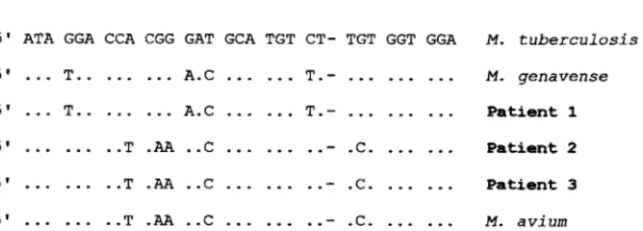
Figure 1. Part of the 16S rRNA gene of various mycobacterial species and clinical strains: positions 179-215 (E. coli alignment). Only nucleotides different from those of M. tuberculosis are shown. Dashes indicate deletions.
. • ... ..T .AA ..0 5' ... ..T .AA ..0
and amplification, PCR is probably not as sensitive as cul-ture for the detection of growth-proficient mycobacteria: my-cobacteremia is of variable intensity, with <1 to >5 X 10" microorganisms/mL [ 1 4]. In our assay, however, the maxi-mal amount of DNA input into the PCR reaction did not exceed 2 lig, and thus the inhibition of amplification was avoided (data not shown). This amount of DNA is extracted from <250 ps of blood or tissue. There may be no mycobac-terial DNA in such a sample, while a conventional culture using 5 mL of blood is still positive [15]. This circumstance could explain the three PCR-negative but culture-positive samples (table 1).
In most studies comparing DNA-based techniques with conventional methods, some samples were DNA-positive but culture-negative. In cases of suspected tuberculosis, the proportion was 54 of 271, or 20% [ 13]. These samples were usually from antibiotic-treated patients and from patients who had M. tuberculosis cultured simultaneously from other sites. This fact suggests that the PCR results reflected true infection and not laboratory contamination. In cases of sus-pected M. avium bacteremia in patients with AIDS, 32 (14%) of 228 culture-negative specimens tested positive by PCR [16]. However, since no details were given regarding the ori-gin of these specimens, it is difficult to judge the clinical significance of these results. The sensitivity of PCR raises difficult issues of interpretation that are particularly trouble-some when DNA from facultative pathogens (such as atypi-cal mycobacteria) is found in normally unsterile specimens, such as sputum, urine, feces, and gastric contents. Con-versely, amplification of mycobacterial genes from normally sterile specimens such as blood, CSF, or liver biopsy samples is probably significant.
In the three cases presented in detail herein, the circum-stances suggest that contamination was unlikely and that the laboratory findings reflect true infection. In case 1, M.
gena-vense, a mycobacterium that is notoriously difficult to culture [5, 17], had grown once before in a blood culture. In case 2, the culture confirmed the PCR result after an extremely long delay. In case 3, cultures had previously been positive for M.
avium but became negative during treatment; the results of PCR, but not of culture, became positive when the patient had a clinical relapse. Thus, as in many other infections,
positive cultures may be more difficult to obtain in mycobac-teremia if the patient has been treated. The diagnosis of re-lapse will then depend on amplification and sequencing of bacterial DNA.
Our report demonstrates that in selected cases the direct amplification of DNA from blood or biopsy samples may be the only means of diagnosing infections with growth-defi-cient mycobacteria. The diagnosis is valuable mainly be-cause it justifies appropriate treatment of patients with AIDS, in whom the administration of any additional drug requires critical evaluation.
References
1. Musial CE, Tice LS, Stockman L, Roberts GD. Identification of myco-bacteria from culture by using the Gen-Probe Rapid Diagnostic Sys-tern for Mycobacterium avium complex and Mycobacterium tubercu-losis complex. J Clin Microbiol 1988;26:2120-3.
2. Roberts GD, Koneman EW, Kim YK. Mycobacterium. In: Balows A, Hausler WJ Jr, Herrmann KL, Isenberg HD, Shadomy HJ, eds. Man-ual ofclinical microbiology. 5th ed. Washington, DC: American Soci-ety for Microbiology, 1991:304-39.
3. Kirschner P, Springer B, Vogel U, et al. Genotypic identification of mycobacteria by nucleic acid sequence determination: report of a 2-year experience in a clinical laboratory. J Clin Microbiol 1993; 31:2882-9.
4. Kirschner P, Meier A, Winger EC. Genotypic identification and detec-tion of mycobacteria—facing novel and uncultured pathogens. In: Persing DH, Smith TF, Tenover f-C, White TJ, eds. Diagnostic mo-lecular microbiology: principles and applications. Washington, DC: American Society for Microbiology, 1993:173-90.
5. }Ringer EC, Teske A, Kirschner 13, et al. Disseminated "Mycobacterium genavense" infection in patients with AIDS. Lancet 1992;340: 76-80.
6. Meier A, Kirschner P, SchrOder KH, Wolters J, Kroppenstedt RM, Wager EC. Mycobacterium intermedium sp. nov. Int J Syst Bacteriol 1993; 43:204-9.
7. Emler S, Rochat T, Rohner P, et al. Chronic destructive lung disease associated with a novel mycobacterium. Am J Respir Crit Care Med 1994;150:261-5.
8. Tong X, Smith LM. Solid-phase method for the purification of DNA sequencing reactions. Anal Chem 1992;64:2672-7.
9. Sanger F, Nicklen S, Coulson AR. DNA sequencing with chain-termin-ating inhibitors. Proc Natl Acad Sci USA 1977; 74:5463-7. 10. Hofer M, Hirschel B, Kirschner P, et al. Brief report: disseminated
os-CID 1995;20 (April) PCR-Based Mycobacterial Diagnosis 7
teomyelitis from Mycobacterium ulcerans after a snakebite. N Engl J Med 1993; 328:1007-9.
11. Coyle MB, Carlson LC, Wallis CK, et al. Laboratory aspects of "Myco-bacterium genavense," a proposed species isolated from AIDS pa-tients. J Clin Microbiol 1992; 30:3206-12.
12. Jonas V, Alden MJ, Curry JI, et al. Detection and identification of Mycobacterium tuberculosis directly from sputum sediments by am-plification of rRNA. J Clin Microbiol 1993; 31:2410-6.
13. Brisson-Noel A, Aznar C, Chureau C, et al. Diagnosis of tuberculosis by DNA amplification in clinical practice evaluation. Lancet 1991; 338:364-6.
14. Havlir D, Kemper CA, Deresinski SC. Reproducibility of
lysis-centrifu-gation cultures for quantification of Mycobacterium avium compl bacteremia. J Clin Microbiol 1993; 31:1794-8.
15. Persing DH. Target selection and optimization of amplification res tions. In: Persing DH, Smith TF, Tenover F-C, White Ti, eds. Dis nostic molecular microbiology: principles and applications. Washir ton, DC: American Society for Microbiology, 1993:88-104. 16. Iralu JV, Sritharan VK, Pieciak WS, Wirth DF, Maguire JH, Barker B
Jr. Diagnosis of Mycobacterium avium bacteremia by polymers chain reaction. J Clin Microbiol 1993; 31:181 1-4.
17. Hirschel B, Chang HR, Mach N, et al. Fatal infection with a nol unidentified mycobacterium in a man with the acquired immunode ciency syndrome. N Engl J Med 1990;323:109-13.