Dietary factors and low-grade inflammation
in relation to overweight and obesity
Philip C. Calder
1, Namanjeet Ahluwalia
2, Fred Brouns
3,21, Timo Buetler
4,22, Karine Clement
5,
Karen Cunningham
6, Katherine Esposito
7, Lena S. Jo¨nsson
8, Hubert Kolb
9, Mirian Lansink
10,
Ascension Marcos
11, Andrew Margioris
12, Nathan Matusheski
13, Herve Nordmann
14, John O’Brien
4,
Giuseppe Pugliese
15, Salwa Rizkalla
5, Casper Schalkwijk
16, Jaakko Tuomilehto
17,
Julia Wa¨rnberg
11,18, Bernhard Watzl
19and Brigitte M. Winklhofer-Roob
201. School of Medicine, University of Southampton, Southampton SO16 6YD, UK 2. INSERM U557, University of Paris, 93017 Bobigny Cedex, France
3. Cargill R&D Centre Europe, 1800 Vilvoorde, Belgium
4. Nestle´ Research Centre, Vers-chez-les-Blanc, 1000 Lausanne, Switzerland
5. Department of Nutrition, INSERM U872, Research Center on Human Nutrition, Pitie Salpetriere Hospital, 75013 Paris, France
6. Coca-Cola Europe, Hammersmith, London W6 9HQ, UK
7. Division of Metabolic Diseases, University of Naples, 80138 Naples, Italy 8. ILSI Europe a.i.s.b.l., Avenue E. Mounier 83, Box 6, 1200 Brussels, Belgium
9. Research Group Immunobiology, Medical Faculty, University of Dusseldorf, 40225 Dusseldorf, Germany 10. Danone Research, Centre for Specialised Nutrition, 6700, CA, Wageningen, The Netherlands
11. Department of Metabolism and Nutrition, CSIC, Spanish National Research Council, 28040 Madrid, Spain 12. School of Medicine, University of Crete, 71409 Heraklion, Greece
13. Nutrition Research, Kraft Foods, Inc., Glenview IL 60025, USA 14. Ajinomoto Europe, 75817 Paris, France
15. Department of Clinical and Molecular Medicine, “La Sapienza” University of Rome, 00161 Rome, Italy 16. Internal Medicine, University of Maastricht, 6202 AZ Maastricht, The Netherlands
17. University of Helsinki, 00014 Helsinki, Finland
18. Department of Preventive Medicine and Public Health, University of Navarra, 31080- Pamplona, Spain 19. Max-Rubner Institut, Federal Research Centre for Nutrition and Food, 76131 Karlsruhe, Germany 20. Institute of Molecular Biosciences, Human Nutrition and Metabolism Research and Training Center,
Karl-Franzens University of Graz, 8010 Graz, Austria
21. Department of Nutrition, Maastricht University Medical Centre, 6200 MD Maastricht, The Netherlands 22. XeRR, Institute of Pharmacology and Toxicology, University of Zu¨rich, 8057, 8057 Zu¨rich, Switzerland
Commissioned by the
ILSI Europe Metabolic Syndrome and Diabetes Task Force
Correspondence: ILSI Europe a.i.s.b.l. - Avenue E. Mounier 83, Box 6 – 1200 Brussels, Belgium,
fax +32 2 762 00 44, email publications@ilsieurope.be
British Journal of Nutrition
Volume 106, 2011 ISSN: 0007-1145
Publishing, Production, Marketing, and Subscrip-tion Sales Office:
Cambridge University Press The Edinburgh Building Shaftesbury Road Cambridge CB2 8RU, UK For Customers in North America: Cambridge University Press Journals Fullfillment Department 100 Brook Hill Drive
West Nyack
New York 10994-2133 USA
Publisher:Katy Christomanou Special sales and supplements:
This Journal accepts relevant advertisements and in-serts. We also provide bulk reprints of suitable papers to meet teaching or promotional requirements. The journal also publishes supplements on behalf of academic and corporate collaborators. Please contact Katy Christoma-nou at the Cambridge address for further details. E-mail: kchristomanou@cambridge.org
Subscription information:
British Journal of Nutrition is an international journal published by Cambridge University Press on behalf of The Nutrition Society. The twelve issues starting January 2011 comprise Volume 105, the twelve issues starting July 2011 comprise Volume 106.
Annual subscription rates: Volumes 105/106 (24 issues):
Internet/print package £1021/$1990/e1635 Internet only: £835/$1630/e1334 Print only: £973/$1895/e1580
Any supplements to this journal published in the course of the annual volume are normally supplied to subscribers at no extra charge.
Back volumesare available. Please contact Cambridge University Press for further information.
Claims for non-receipt of journal issues will be considered on their merit and only if the claim is received within six months of publication. Replacement copies supplied after this date will be chargeable.
US POSTMASTERS:please send address corrections to British Journal of Nutrition, Cambridge University Press, 100 Brook Hill Drive, West Nyack, New York 10994-2133.
Directions to Contributors are available from the Society at the address below or can be found on the Society’s website at http://www.nutritionsociety.org (an abbreviated Notes for Authors can be found inside the back cover).
Offprints:The author (or main author) of an accepted paper will receive a copy of the PDF file and a voucher copy of the issue in which their paper has been published. There will be an option to purchase paper offprints, these should be ordered at proof stage. No page charges are levied by this journal.
Copyright: As of 1 July 2000 the copyright of all articles submitted to British Journal of Nutrition are retained by the authors or their institutions. For articles prior to this date permission for reproduction of any part of the journal (text, figures, tables or other matter) in any form (on paper, microfiche or electronically) should be sought directly from the Society, at: The Publications Office, The Nutrition Society, 10 Cambridge Court, 210 Shepherds Bush Road, Hammersmith, London W6 7NJ, UK.
Disclaimer: The information contained herein, including any expression of opinion and any projection or forecast, has been obtained from or is based upon sources believed by us to be reliable, but is not guaranteed as to accuracy or completeness. The information is supplied without obligation and on the understanding that any person who acts upon it or otherwise changes his/her position in reliance thereon does so entirely at his/her own risk. Neither the Society nor Cambridge University Press accepts respon-sibility for any trade advertisement included in this publication.
This journal is printed on acid-free paper from renewable sources. Printed in the UK by Bell & Bain Ltd., Glasgow.
This journal issue has been printed on FSC-certified paper and cover board. FSC is an independent, non-governmental, not-for-profit organization established to promote the responsible management of the world’s forests. Please see www.fsc.org for information.
British Journal of Nutritionis covered in Current Contentsw/Agriculture, Biology & Environmental Sciences,
SciSearchw, Research Alertw, Current Contentsw/Life Sciences, Index Medicusw (MEDLINEw),
AGRICOLAw, CAB AbstractsTM, Global Health, BIOSISw Database, EMBASE/Excerpta Medica and
Vol. 106
Supplement No. S3
December 2011
British Journal of Nutrition
Table of Contents
Preamble S5
Concept and markers of low-grade inflammation S5 – S11
Chronic low-grade inflammation and insulin resistance S11 – S13
Postprandial inflammatory response S13 – S14
Ageing and low-grade inflammation S14 – S15
Exercise and low-grade inflammation S15 – S18
A consideration of different approaches to identify relationships between diet and its
components and markers of chronic low-grade inflammation S18
Dietary patterns and low-grade inflammation S18 – S28
Process-related compounds: advanced glycation end products and advanced lipoperoxidation end
products S28 – S37
Macronutrients and low-grade inflammation S37 – S46
Micronutrients and phytochemicals S46 – S52
Other factors S52 – S53
Summary, conclusions and research gaps S53 – S54
Acknowledgements S54 – S55
(Received 20 January 2011 – Revised 8 August 2011 – Accepted 7 September 2011)
Key words:Inflammation: Cytokines: Adipose: Obesity: Diet
Correspondence: ILSI Europe a.i.s.b.l., Avenue E. Mounier 83, Box 6 – 1200 Brussels, Belgium, fax þ 32 2 762 00 44, email publications@ilsieurope.be
Abbreviations:AGE, advanced glycation end products; AGE-R, advanced glycation end product recep-tor; AHEI, alternate healthy eating index; ALE, advanced lipoxidation end products; CCL, chemokine (C – C motif) ligand; CCR, CC chemokine receptor; CFU, colony forming units; CLA, conjugated lino-leic acids; CRP, C-reactive protein; DQI, Diet Quality Index; DQI-R, revised Diet Quality Index; GI, glycaemic index; GL, glycaemic load; HEI, healthy eating index; IFN, interferon; IL-1ra, IL-1 receptor antagonist; IRS, insulin receptor substrate; LPS, lipopolysaccharide; MAPK, mitogen-activated protein kinase; MCP, monocyte chemoattractant protein; MIF, macrophage migration inhibitory factor; MIP, macrophage inflammatory protein; MRP, Maillard reaction products; NHANES, National Health and Nutrition Examination Survey; PAI-1, plasminogen activator inhibitor 1; Q, quintile; RAGE, receptor for advanced glycation end products; RANTES, regulated on activation, normal T expressed and secreted; ROS, reactive oxygen species; SAA, serum amyloid A; sE-selectin, soluble E-selectin; sICAM-1, soluble intercellular adhesion molecule-1; sP-selectin, soluble P-selectin; STAMP, six-trans-membrane protein of prostate; sTNFR, soluble receptors of TNF; sVCAM-1, soluble vascular cell adhesion molecule-1; TGF, transforming growth factor; TLR, Toll-like receptors.
Low-grade inflammation is a characteristic of the obese state, and adipose tissue releases many inflammatory mediators. The source of these mediators within adipose tissue is not clear, but infiltrating macrophages seem to be especially important, although adipocytes them-selves play a role. Obese people have higher circulating concentrations of many inflammatory markers than lean people do, and these are believed to play a role in causing insulin resistance and other metabolic disturbances. Blood concentrations of inflammatory markers are lowered following weight loss. In the hours following the consumption of a meal, there is an elevation in the concentrations of inflam-matory mediators in the bloodstream, which is exaggerated in obese subjects and in type 2 diabetics. Both high-glucose and high-fat meals may induce postprandial inflammation, and this is exaggerated by a high meal content of advanced glycation end products (AGE) and partly ablated by inclusion of certain antioxidants or antioxidant-containing foods within the meal. Healthy eating patterns are associated with lower circulating concentrations of inflammatory markers. Among the components of a healthy diet, whole grains, veg-etables and fruits, and fish are all associated with lower inflammation. AGE are associated with enhanced oxidative stress and inflammation. SFA and trans-MUFA are pro-inflammatory, while PUFA, especially long-chain n-3 PUFA, are anti-inflammatory. Hyperglycaemia induces both postprandial and chronic low-grade inflammation. Vitamin C, vitamin E and carotenoids decrease the circulating concentrations of inflammatory markers. Potential mechanisms are described and research gaps, which limit our understanding of the interaction between diet and postprandial and chronic low-grade inflammation, are identified.
Preamble
Inflammation is a normal defence mechanism that protects the host from infection and other insults; it initiates pathogen killing as well as tissue repair processes and helps to restore homeostasis at infected or damaged sites. It is typified by redness, swelling, heat, pain and loss of function, and involves interactions among many cell types and the pro-duction of, and responses to, a number of chemical mediators. Self-regulation of the inflammatory response involves the activation of negative feedback mechanisms such as the secretion of anti-inflammatory cytokines, inhibition of pro-inflammatory signalling cascades, shedding of receptors for inflammatory mediators and activation of regulatory cells. As such, and controlled properly, regulated inflammatory responses are essential to remain healthy and maintain homeostasis. Pathological inflammation involves a loss of tolerance and/or of regulatory processes. Where this becomes excessive, irreparable damage to host tissues and disease can occur(1). Such diseases are characterised by markedly elevated concentrations of inflammatory markers and of activated inflammatory cells at the site of tissue damage and in the systemic circulation. While the existence of inflammatory diseases has been long recognised, it is only more recently that the condition of chronic low-grade inflammation has received attention, particularly in relation to obesity, the metabolic syndrome and CVD. Chronic low-grade inflam-mation is characterised by raised concentrations of inflamma-tory markers in the systemic circulation. This article sets out to explain the nature of chronic low-grade inflammation in the context of overweight and obesity, and to describe the factors that might influence it, in particular those related to diet. The literature in the areas of adipose tissue, obesity and inflammation, and dietary components and inflammation is vast, and it is not possible to mention all studies here. In particular, the review of diet and its components and inflammation is not exhaustive, although the main studies of relevance are included.
Concept and markers of low-grade inflammation Obesity and low-grade inflammation
The concept of systemic, chronic, but low-grade inflammation as a risk factor for the metabolic syndrome and for type 2 diabetes is based on the observation of elevated blood levels of inflammation-associated markers in people with incident type 2 diabetes or with the metabolic syndrome(2,3). The up-regulation of systemic indicators of inflammation such as leucocyte count, and serum and plasma concen-trations of acute-phase proteins, pro-inflammatory cytokines, chemokines, soluble adhesion molecules and prothrombotic mediators is modest, usually less than 2-fold above what is observed in controls. Diagnostic criteria for low-grade inflammation have not been precisely defined, but the phenotype per se is not disputed.
Systemic concentrations of pro-inflammatory mediators are higher in obese (BMI . 30 kg/m2) than in normal-weight per-sons(4,5). Serum or plasma concentrations of TNF-a or IL-6 in healthy adults are typically 0·01 – 2 pmol/l(6). Other inflamma-tory mediators, such as monocyte chemoattractant protein (MCP)-1, interferon (IFN)-g-induced protein-10 and IL-18, may reach mean concentrations of 10 pmol/l; macrophage migration inhibitory factor (MIF) and regulated on activation, normal T expressed and secreted (RANTES) concentrations may get close to the nanomolar range; and C-reactive protein (CRP) concentration is often above 10 nmol/l. The variation in concentrations of most mediators among non-obese or obese individuals is at least 10-fold. Hence, there is a substantial overlap between non-obese and obese persons. However, there is a positive relationship between BMI and other measures of obesity such as waist circumference and circulat-ing concentrations of CRP and other inflammatory markers(7). A mechanistic link between obesity and low-grade inflam-mation was first proposed by Hotamisligil et al.(8)who showed that white adipose tissue synthesises and releases the pro-inflam-matory cytokine TNF-a. The expression of TNF-a is elevated in adipocytes of obese and insulin-resistant mice, while insulin sensitivity is improved following administration of anti-TNF-a antibodies. Based on these data, it was suggested that adipose tissue plays an important immune role and may be a major
British
Journal
of
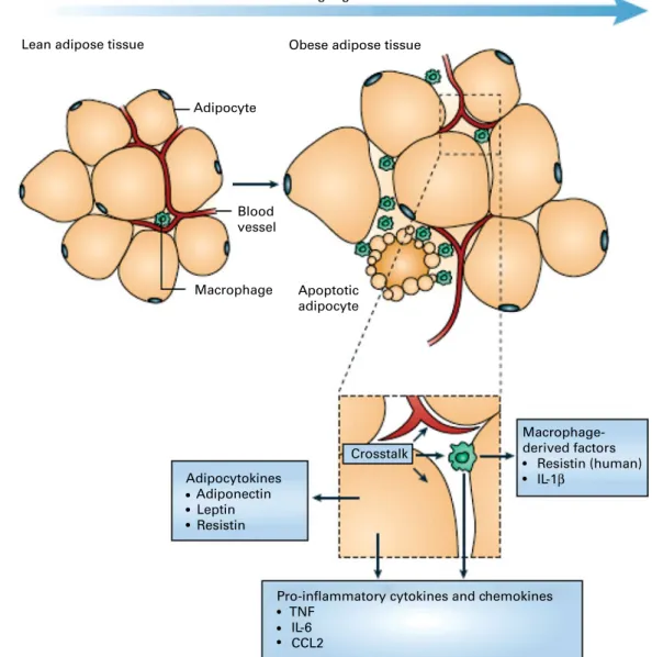
source of pro-inflammatory mediators which initiate the development of chronic inflammation, insulin resistance and atherosclerosis, all of which are characteristics of the metabolic dysregulation associated with obesity. The discovery of leptin modified the view of adipose tissue as being an ‘inert’ energy store to being the largest endocrine gland in the body. Leptin is produced and secreted by white adipose tissue. The discovery of leptin introduced the concept of ‘adipocytokines’ or ‘adipokines’, substances produced by adipose tissue and which circulate in the bloodstream, so exerting systemic effects as hormones(9). Some adipokines are produced within adipose tissue exclusively by adipocytes (e.g. leptin, adiponectin, serum amyloid A (SAA)), while others are produced by both adipo-cytes and other cell types of the non-adipocyte fraction of adipose tissue. It is now recognised that macrophages accu-mulate in the adipose tissue in obesity(10,11) (Fig. 1) and that these may represent major contributors to the production of adipokines(12,13).
Adipose tissue as a source of inflammatory mediators
Adipose tissue expresses and secretes into the systemic circu-lation a growing list of hormones, inflammatory mediators and immune system effectors. The products of adipose tissue can be categorised into several groups (Table 1):
(1) Hormones: many of the hormones produced by adipose tissue affect the immune system and insulin sensi-tivity. Leptin appears to be pro-inflammatory(14), while adiponectin appears to be anti-inflammatory and insulin-sensitising(15). Similarly, visfatin(16)and resistin(17) contribute to the development of insulin resistance, while omentin(18,19) appears to be an insulin sensitiser. It should be noted that most studies identifying the roles of these hormones have been performed in rodents and the immune and insulin-sensitising effects of these hormones in humans remain unclear(20,21).
(2) Acute-phase proteins: these proteins are secreted in the acute phase of inflammation and include plasminogen activator inhibitor 1 (PAI-1)(22), pentraxine-3, lipocalin 24p3, haptoglobin, SAA(23)and a1-glycoprotein. (3) Cytokines: these are the classic peptide mediators of
inflammation and include IL-1, IL-1 receptor antagonist (IL-1ra)(24 – 26), IL-6, IL-7, IL-18(27 – 30), IL-10(31,32), MIF(33) and TNF-a.
(4) Chemokines: these include IL-8(34,35), MCP-1, -3, -4, RANTES (now known as chemokine (C–C motif) ligand (CCL) 5), angiopoietin, metallothioneins, macrophage inflammatory protein (MIP)-1a and -1b (now known as CCL3 and CCL4, respectively)(36), and induced protein-10(37).
(5) Growth factors: transforming growth factor (TGF)-b(38). (6) Components of the alternative complement system:
adipsin and factors C2, C3, C4, C7, B and D (these are expressed more highly in omental compared with subcutaneous adipose tissue(39,40)).
(7) Retinol-binding protein 4 which is linked with insulin resis-tance(41,42), although its precise role has been debated(43).
Adipose tissue distribution and its impact on inflammation
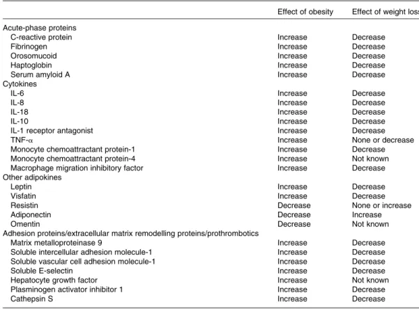
Obesity is characterised by an expansion of the mass of adipose tissue and dramatic changes in its distribution in the body. Simplistically speaking, accumulation of adipose tissue in the thorax and abdomen (variously termed abdominal, central, visceral, splanchnic or android obesity) results in an increased risk for diabetes and atherosclerosis, while excess adipose tissue in the lower part of the body (termed gynoid obesity) does not appear to be associated with major metabolic consequences(44,45). The increase in abdominal fat mass is associated with a chronic elevation of the circulating concentrations of inflammatory mediators including several acute-phase inflammatory proteins such as CRP(46,47), pro-and anti-inflammatory cytokines, adhesion molecules pro-and pro-thrombotic molecules(22,47 – 49). It should be noted that the liver and the lymphoid organs are usually the major pro-duction sites of these inflammatory mediators but in obesity, adipose tissue becomes a major producer resulting in a chronic and constant local and systemic inflammatory milieu (Table 2). Abdominal obesity is a risk factor for type 2 diabetes, hyperten-sion, dyslipidaemia and CVD(50), and also probably obesity-associated hepatic diseases (non-alcoholic fatty liver disease and non-alcoholic steatohepatitis). Glucose intolerance is sig-nificantly more common in subjects with abdominal obesity compared with those with fat mass accumulation in their lower part of the body. Plasma TAG concentrations are also sig-nificantly more elevated in individuals with abdominal obesity. It appears that the anatomical localisation of adipose tissue is of paramount importance in relation to its physiological func-tion, i.e. handling of lipids (lipogenesis, lipolytic activity), expression of multiple genes, and response to insulin, catechol-amines, sex hormones and cortisol(51). In addition, the profile of adipokines produced is dissimilar between the subcutaneous and abdominal adipose tissues. Thus, leptin is preferentially expressed and secreted by subcutaneous adipose tissue(52), while the expression of adiponectin, visfatin, omentin, resistin, PAI-1, IL-8, IL-7, IL-1a, MCP-1, TGF-b, growth-related oncogen-a, CCL5 and MIP-1b is higher in abdominal fat. In contrast to such distributions, there are reports that IL-6 and TNF-a seem to be equally synthesised by the different sites(28,36,53 – 59). It is important to mention that in severe obesity, the part played by the abdominal or the very abundant subcutaneous adipose tissue in the systemic delivery of inflammatory mediators is still not well understood. Nevertheless, the distinct profile of adipokine secretion between the abdominal and subcu-taneous adipose tissues probably contributes to the increased risk of metabolic and cardiovascular complications and to the development of other complications such as hepatic steatosis and non-alcoholic steatohepatitis in obese individuals. Finally, other adipose tissue depots in so-called ‘ectopic sites’, such as within the liver, heart or skeletal muscle, may contribute to the production of inflammatory mediators in the absence of obesity. In this regard, the local production of the inflamma-tory molecules by adipose tissue within the heart may be important; the amount of this tissue and its proximity to the coronary vessels could contribute to the development of coronary pathologies(60,61).
British
Journal
of
Cell populations of adipose tissue
Adipose tissue is a heterogeneous tissue composed of several cell types: mature adipocytes, pre-adipocytes, fibroblasts, endothelial cells, mast cells, granulocytes, lymphocytes and macrophages. Cells within adipose tissue, apart from mature adipocytes, are collectively termed the stroma-vascular fraction. The various cell types have not been precisely characterised, nor has the relative change of their quantitative contributions to the tissue in obesity. Because of the hetero-geneity of cells in the adipose tissue, the cellular source of the inflammatory factors secreted by the tissue into the sys-temic circulation remains unknown. In vitro studies have demonstrated that mature adipocytes express inflammatory factors such as TNF-a(56). SAA is overexpressed and secreted in abundance by isolated adipocytes from obese subjects, as is leptin, while secretion of adiponectin is suppressed. SAA
and leptin production by adipose tissue depends on adipocyte size(18,19,23,62). Adipocyte size also influences the expression of other inflammatory mediators as demonstrated by fraction-ation studies of adipocytes coupled with studies of gene expression profiles(62). Adipocyte size, for example, deter-mines the production of IL-6, IL-8, MCP-1 and granulocyte colony-stimulating factor(29). Although adipocyte hypertrophy precedes the development of type 2 diabetes(63), a growing number of studies indicate that the principal site of production of inflammatory mediators appears to be the stroma-vascular fraction(36,64 – 67). More recent studies in mice have suggested that the infiltration of obese adipose tissue by macrophages is accompanied or even preceded by an influx of T-lympho-cytes(68 – 71) and that T-cells have a key early role(69). Early work indicated the presence of CD3-positive T-lymphocytes in human adipose tissue(72), and more recent studies have shown high numbers of T-cells in the adipose tissue of
Lean adipose tissue
Weight gain
Obese adipose tissue
Adipocyte Blood vessel Macrophage Apoptotic adipocyte Macrophage-derived factors Resistin (human) IL-1β Crosstalk
Pro-inflammatory cytokines and chemokines TNF IL-6 CCL2 Adipocytokines Adiponectin Leptin Resistin
Fig. 1. Schematic representation of the interaction between adipocytes and macrophages showing some of the molecules released. Expansion of adipose tissue during weight gain leads to the recruitment of macrophages through various signals (e.g. chemokines such as chemokine (C – C motif) ligand 2 (CCL2)) released by adipocytes. Macrophages accumulate around apoptotic adipocytes. Mediators synthesised by adipocytes and resident macrophages contribute to local and systemic inflammation. Reproduced with permission from Tilg & Moschen(10).
British
Journal
of
diet-induced obese insulin-resistant mice(71). Furthermore, Wu et al.(71) demonstrated the presence of CD3-positive T-lymphocytes in human adipose tissue and described the expression of RANTES, a T-cell-specific chemokine, and its respective receptor CCR5 in the visceral adipose tissue of mor-bidly obese patients. A recent study in mice reported mainly CD8-positive lymphocyte infiltration in hypoxic areas within the adipose tissue(70). Most recently, it has been shown that pro-inflammatory T-lymphocytes are present in visceral adipose tissue and may contribute to local inflammatory cell activation before the appearance of macrophages, suggesting that these cells could play an important role in the initiation and perpetuation of adipose tissue inflammation as well as the development of insulin resistance(69).
It has been proposed that macrophages and mature adi-pocytes are derived from the same precursor cells and show close gene expression profiles including the Toll-like recep-tors (TLR). Pre-adipocytes exert ‘macrophage-like’ effects when exposed to strong pro-inflammatory environments(73,74). It should be noted, however, that the vast majority of the macrophage infiltration in adipose tissue in obesity originates most probably from the circulation. In obese subjects, these macrophages typically aggregate in crowns around apoptotic adipocytes(31) (Figs. 1 and 2). Although these macrophages express activation markers, they could be pro- or anti-inflammatory depending on the degree of obesity and its evol-ution as suggested by studies in mice showing that weight gain is accompanied by transformation from a macrophagic M2 (anti-inflammatory) phenotype towards an M1 (pro-inflammatory) profile(75) (Fig. 3)(76). Consequently, secretion profiles of the adipose tissue can change depending on the phenotype of the cell population infiltrating it during the different stages of obesity (initiation, aggravation, mainten-ance and weight loss; Fig. 4)(77).
Adipose tissue macrophages may contribute to the mainten-ance of the low-grade inflammatory state linked to obesity(36). Factors that induce the infiltration and activation of macro-phages in the adipose tissue are probably multifactorial. Para-crine, autocrine and endocrine signals, as well as mechanical modifications (hypertrophy and adipocyte hyperplasia), could play a role in this phenomenon. Many adipokines synthesised by the adipose tissue are candidates to attract
inflammatory cells. In vitro studies have suggested that leptin itself (at supra-physiological levels) induces adhesion proteins, hence facilitating the migration of monocytes(78). Conversely, adiponectin may inhibit this process(79). Very little is known about the role of selectins, integrins and elements of adhesion to the extracellular matrix, in the process of macrophage attraction to the adipose tissue. Gene expression studies with human adipose tissue have demon-strated that the levels of expression of MCP-1, colony-stimulat-ing factor-3 and the urokinase plasminogen activator CD87 increase significantly in the adipose tissue of subjects with morbid obesity(31). MCP-1 is a strong chemoattractant and it acts via its receptor CCR2. MCP-1 is synthesised predominantly by macrophages and endothelial cells and, to a lesser extent, by adipocytes. In one study, CCR2 gene knockout mice showed a reduction of macrophage infiltration in the adipose tissue and improvement of insulin sensitivity(80). This led to the suggestion that MCP-1 and its receptor CCR2 are major players in the macrophage accumulation within the adipose tissue(80). However, contradictory data suggest that MCP-1 might not be such a crucial candidate(81). The role of MCP-1 in the macrophage accumulation in human obesity needs to be established. Other candidate molecules and other mechan-isms continue to be explored. Local hypoxia could also play an important role in the attraction and retention of macro-phages within the adipose tissue(82). Hypoxia-inducible factor-1a, a transcription factor normally induced by hypoxia, is overexpressed in the subcutaneous adipose tissue of obese subjects and its expression is decreased during weight reduction(31). Tissue hypoxia induces macrophage attraction into solid tumours as well as into atherosclerotic plaques. Adi-pose tissue of obese subjects could be hypoxic in some areas and a local expression of chemokines could be induced. It should be noted that leptin, which possesses indirect chemoattractant properties, is induced by hypoxia(83).
It is generally considered that macrophage accumulation in adipose tissue is detrimental. However, macrophage accumu-lation could be related to an adaptation process associated with the augmentation of fat mass, and macrophage accumu-lation could be necessary for the upkeep of the tissue and perhaps to limit its expansion. It appears that macrophage aggregates within the adipose tissue are localised around apoptotic adipocytes, suggesting that one of their functions is to clean up the debris of dying and dead cells(84). In addition to their role in cleaning up the old cells, the accumu-lation of macrophages may also be useful for the formation of new vessels, particularly at the site of inflammation and in ischaemic zones when adipose tissue grows(85). Macrophages also control fat mass growth and modify the biology of adipo-cytes and pre-adipoadipo-cytes in a paracrine manner. The specific effects of TNF-a and IL-6 on different adipocyte functions (increased lipolysis, modification of adipocyte secretion patterns and induction of adipocyte insulin resistance) have been shown(86). In the presence of a medium derived from human macrophages, human pre-adipocytes showed a drastic change in their phenotype, acquiring a pro-inflammatory phenotype and secreting significant amounts of IL-6 and IL-8, and they grew well(87), but differentiated poorly(87,88).
Table 1. Cytokines expressed or secreted by human adipose tissue Family Example(s)
Chemokines MCP-1 (known as CCL2), MCP-3, MCP-4, RANTES (known as CCL5), MIP-1a (known as CCL3)
IL IL-6, IL-8 (acts as a chemokine), IL-1ra, IL-10, IL-18 Interferons IP-10
TNF TNF-a
Growth factors Vascular endothelial growth factor, TGF-b, hepatocyte growth factor
Others Leptin
MCP, monocyte chemoattractant protein; CCL, chemokine (C – C motif) ligand; RANTES, regulated on activation, normal T expressed and secreted; MIP, macrophage inflammatory protein; IL-1ra, IL-1 receptor antagonist; IP, interferon-g-induced protein; TGF, transforming growth factor.
British
Journal
of
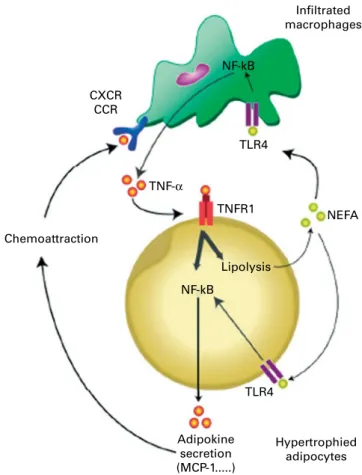
These cellular alterations are induced in co-cultures without direct cellular contact, suggesting the key role of soluble factors, although it cannot be excluded that a direct cell – cell interaction also plays a role in modifying pre-adipocyte or adipocyte biology. The mature adipocyte also endures pro-found modifications of its biology in culture systems with a medium from activated macrophages. Other than the pro-inflammatory state, increased lipolysis and resistance to insulin have been observed(89,90). TNF-a has been proposed to mediate these effects. From the molecular point of view, the NF-kB pathway, implicated in the primary regulation of inflammatory responses(91 – 93) (Fig. 5)(94), is induced in the pre-adipocyte(87) and in the adipocyte in the presence of a medium from macrophages(95). The NF-kB pathway is also brought into play in macrophages in contact with a medium from adipocytes. TLR appear to be important players which lead to the induction or suppression of genes orchestrating the inflammatory response. TLR-4 is the bacterial lipopolysac-charide (LPS) receptor, but data have shown that the NEFA produced by adipocytes after adrenergic stimulation are also strong inducers of the TLR-4/NF-kB system(95)(Fig. 5). TLR-4 is expressed by adipocytes and is overexpressed during obesity(96). TLR-4 knockout mice are protected from insulin resistance induced by lipid infusions(97).
Based on these different studies, a dual effect of macro-phages of the adipose tissue could be expected: first, a local ‘beneficial’ effect in clearing out old adipocytes, and in the control and of the development of fat mass and second, a deleterious local and systemic effect via the increase in the
production and secretion of adipokines, promoting the pro-gression of complications of obesity and the induction of insu-lin resistance.
Adipokines and chronic low-grade inflammation
Adipokines and the complications of obesity. Inflammatory molecules are likely candidates exerting a molecular link between the adipose tissue and the metabolic, cardiovascular, hepatic and thrombotic complications, and certain cancer types occurring in conjunction with or as a consequence of human obesity. A myriad of candidate adipokines are proposed to play this role(98 – 100). In the cardiovascular field, they can be considered as risk factors, and even directly play a pathophysiological role favouring the initiation and progression of atherosclerosis. Relationships between abnormalities of cardiac function in obese subjects, the accumulation of abdominal fat and low-grade inflammation have been suggested(101,102). Among the candidates secreted by the adipose tissue, the increase in IL-6, IL-8 and MCP-1 and the decrease in adiponectin are considered to be particu-larly important(101,102). The studies of the pathophysiological links between adipokines and cardiovascular health can be illustrated by the analysis of the effects of adiponectin in rodents. Overexpression of adiponectin results in diminished size of the lesions observed following acute ischaemic myocardial infarction, increased angiogenic properties and reduced size of atheromatous plaques in the genetically pre-disposed apoE2/2 mouse(103).
Table 2. Modification of circulating inflammatory marker concentrations in relation to obesity and weight loss
Effect of obesity Effect of weight loss Acute-phase proteins
C-reactive protein Increase Decrease
Fibrinogen Increase Decrease
Orosomucoid Increase Decrease
Haptoglobin Increase Decrease
Serum amyloid A Increase Decrease
Cytokines
IL-6 Increase Decrease
IL-8 Increase Decrease
IL-18 Increase Decrease
IL-10 Increase Decrease
IL-1 receptor antagonist Increase Decrease
TNF-a Increase None or decrease
Monocyte chemoattractant protein-1 Increase Decrease
Monocyte chemoattractant protein-4 Increase Not known
Macrophage migration inhibitory factor Increase Decrease Other adipokines
Leptin Increase Decrease
Visfatin Increase Decrease
Resistin Decrease None or increase
Adiponectin Decrease Increase
Omentin Decrease Not known
Adhesion proteins/extracellular matrix remodelling proteins/prothrombotics
Matrix metalloproteinase 9 Increase Decrease
Soluble intercellular adhesion molecule-1 Increase Decrease Soluble vascular cell adhesion molecule-1 Increase Decrease
Soluble E-selectin Increase Decrease
Hepatocyte growth factor Increase Not known
Plasminogen activator inhibitor 1 Increase Decrease
Cathepsin S Increase Decrease
British
Journal
of
Several inflammatory mediators produced by adipose tissue, such as CCL5, IL-1b and IL-8, as well as markers of oxidative stress, are increased in diabetic or glucose-intolerant patients, and the amelioration of hyperglycaemia by insulin therapy reduces circulating concentrations of these molecules(104,105). The increase in the concentrations of TNF-a, IL-6, IL-1b, IL-8, resistin and many other factors produced by macrophage activation could contribute to the deterioration of insulin sensitivity (Fig. 6)(106). The precise relationship between the importance of macrophage and T-cell accumulation in adi-pose tissue depots, adipokine secretion and the modifications of insulin sensitivity needs to be further addressed in humans. Macrophage accumulation is more abundant in the abdomi-nal adipose tissue(65)than in the subcutaneous tissue, and this could explain some of the risks associated with the accumu-lation of intra-abdominal fat. For example, a reaccumu-lationship between the increase in macrophages in the abdominal adi-pose tissue and hepatic inflammation and fibrosis has been reported(65). In another study, the expression of the MCP-1 and colony-stimulating factor-1 genes and proteins was associ-ated with macrophage accumulation in obese subjects(107). Since the abdominal adipose tissue is partly drained by the portal system, it cannot be excluded that some adipokines, together with high NEFA fluxes and hormones delivered by the adipose tissue, could contribute to the alteration of hepatic function observed in obese subjects, the mechanism of which needs to be better dissected.
Adipokines and weight loss. Even modest weight reduction improves the metabolic and cardiovascular risks associated with human obesity. Measures of endothelial acti-vation also improve after weight reduction(108 – 110). Many studies have shown that weight loss induced by a decrease in energy intake, and sometimes an increase in exercise, reduces systemic inflammation. A reduction in concentrations of numerous inflammatory molecules and endothelial risk
factors, and an increase in adiponectin concentration have been observed during weight-loss programmes, and these are sometimes associated with improvement of insulin sensi-tivity(111). Such changes have been described for CRP(112), IL-6(113), IL-18(114), IL-1ra(26), PAI-1(115), SAA(23,116), cathepsin S(117), matrix metalloproteinase-9(118), soluble adhesion mol-ecules (soluble intercellular adhesion molecule-1 (sICAM-1), soluble vascular cell adhesion molecule-1 (sVCAM-1))(110), tissue factor(119), MIF(120), MCP-1(121), soluble receptors of TNF (sTNFR) and for eotaxin, an inflammatory factor implicated in asthma, another complication of obesity(122). Weight loss induced by gastric bypass reduced the circulating concentrations of visfatin(123) and TNF-a(124,125). One study followed sixty obese patients during the course of weight loss induced by bariatric surgery and reported a reduction of 30 % of initial weight, a decrease in CRP, SAA, orosomucoid protein, IL-6, TNF-a and fibrinogen concentrations, and an increase in adiponectin concentration(126). After the surgery, the concentration of IL-6 dropped slowly while the concen-trations of SAA and CRP dropped more quickly(126).
There was a significant modification in the expression of inflammatory genes in the subcutaneous adipose tissue of obese women following a hypoenergetic diet(127). The expression of 100 genes linked to inflammatory processes was modified after 4 weeks (41 % increased and 59 % decreased). These genes belonged to at least twelve functional families including cytokines, the complement factor cascade, acute-phase proteins of inflammation, and molecules involved in cellular adhesion and in the remodelling of the extracellular matrix. The improvement of the inflammatory profile (at the level of gene expression) involved both the decreased expression of pro-inflammatory factors and the increased expression of anti-inflammatory factors such as IL-10 and IL-1ra. The modification of the inflammatory gene expression profile was very similar in subjects following bariatric
Non-obese subject
Macrophages
Obese subject
Obese subject
Fig. 2. Adipose tissue from non-obese and obese human subjects showing macrophage infiltration. Macrophages are stained with HAM56 antibody. Reproduced with permission from Cancello et al.(65).
British
Journal
of
surgery and was associated with a reduction of macrophage infiltration. In this study, the protein expression of IL-10 increased, suggesting a possible M1 to M2 (pro-inflammatory to anti-inflammatory) switch of macrophage phenotypes(31). Overall, the mitigation of the systemic inflammatory profile observed during weight loss is associated with modifications of adipose tissue inflammatory gene expression, and this may be linked with altered profiles of inflammatory protein secretion. The eventual consequence of this phenomenon on the local biology of the adipose tissue remains to be identified.
Chronic low-grade inflammation and insulin resistance Experimental model systems in vitro
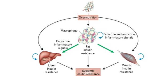
In obesity and the metabolic syndrome, key organs displaying increased insulin resistance are the liver, skeletal muscle, adipose tissue and the endothelium (Fig. 6). Experimental model systems in vitro have shown that hepatocytes as well as muscle cells, adipocytes and endothelial cells respond to exposure to the pro-inflammatory cytokines TNF-a, IL-6 and/or IL-1b with impaired insulin signalling(128). Since these cytokines mediate their effects by binding to their cognate receptors, downstream components of the signalling cascade must interfere with insulin receptor function. Several such pathways have been identified (Fig. 7)(128). Pro-inflammatory cytokine signalling usually activates the kinases
IkB kinase b and C-jun N-terminal kinase 1, both of which in turn phosphorylate the Ser 307 residue of insulin receptor substrate (IRS)-1. This prevents the phosphorylation of tyrosine residues of IRS-1 by the insulin receptor and the downstream signalling cascade. TNF-a mediates insulin resist-ance also via the activation of p38 mitogen-activated protein kinase (MAPK) which interferes with the IRS – phosphatidyl-inositol 3-kinase – Akt pathway(129). Another mechanism involves cytokine-induced suppressor of cytokine signalling 1 and 3, which also prevent tyrosine phosphorylation of IRS proteins, by direct interference, or ubiquitylation and sub-sequent degradation(130,131). Induction of suppressor of cyto-kine signalling 3 by IL-6 occurs via the signal transducer and activator of transcription 3 (STAT3) mammalian target of rapamycin (mTOR) pathway(132).
In vivo models
The impairment of insulin signalling by TNF-a has also been observed in vivo after infusion of the cytokine into rodents(133,134). A critical mediator downstream of TNF-a appears to be MIF, since mice with a disrupted MIF gene pre-serve normal insulin signalling(135). In this context, it is of interest that adipocytes are able to secrete MIF(33). The latter finding demonstrates that there are still substantial gaps in our understanding of the pro-inflammatory cytokine signalling
Classically activated macrophage LPS, IFN-γ TNF-γ , IL-Iβ, IL-6, resistin, NO, etc. Alternatively activated macrophage Lean insulin sensitive Obese insulin resistant Anti-inflammatory NEFA IL-13 IL-4 Proinflammatory NEFA Chemokines TNF-α Adipocyte IL-10 IL-4, IL-3 IL-10
Fig. 3. Schematic representation of factors regulating macrophage polarity and insulin resistance in adipose tissue. Under lean conditions, adipocytes secrete factors, such as IL-13, that promote alternative activation of macrophages. Alternatively activated (M2) macrophages secrete anti-inflammatory mediators, such as IL-10, and may secrete insulin-sensitising factors. Obesity induces changes in adipocyte metabolism and gene expression, resulting in increased lipolysis and the release of pro-inflammatory NEFA and factors that recruit and activate macrophages, such as chemokines and TNF-a. Activated M1 macrophages produce large amounts of pro-inflammatory mediators, such as TNF-a, IL-1b and resistin, that act on adipocytes to induce an insulin-resistant state. This establishes a positive feedback loop that further amplifies inflammation and insulin resistance. IFN, interferon; LPS, lipopolysaccharide. Reproduced with permission from Olefsky & Glass(76).
British
Journal
of
cascade leading to insulin resistance. Most importantly, it remains unclear where other known regulators of insulin sensitivity fit into the chain of events. There is convincing evidence that reactive oxygen species (ROS) are critical to the effects of TNF-a on insulin signalling(136), and also that mitochondrial dysfunction is involved(137). The impact of insulin on cellular metabolic activity, proliferation and differ-entiation can also be impaired by inflammatory mediators via an indirect pathway, i.e. by enhancing or suppressing the production of hormones that modulate cellular responses to insulin. These effects include the up- or down-regulation of the synthesis of resistin(138), leptin, adiponectin(139), lipocalin 2(140), osteopontin(141), and of insulin itself. When considering the subnanomolar systemic concentration of many pro-inflammatory mediators (see section ‘Obesity and low-grade inflammation’), it is possible that only a few of them contri-bute to the metabolic derangements seen in obesity and the metabolic syndrome.
A different situation emerges when paracrine effects of inflammatory mediators are considered. As described above, there is substantial local production of inflammatory mediators in organs affected by insulin resistance. Hepatocytes, adipo-cytes, muscle cells and the endothelium are sites of inflamma-tory mediator synthesis, but local activated macrophages appear to be the dominant site of synthesis and secretion,
which leads to spillover into the general circulation(66,67,142). Paracrine concentrations of inflammatory mediators are suffi-cient to induce insulin resistance(128). Indeed, co-culture of adipocytes with macrophages caused impairment of insulin signalling(75). In addition to paracrine effects, it is conceivable that the functions of liver cells are affected by inflammatory mediators released from the abdominal adipose tissue because of their blood link.
Evidence supporting the link between inflammatory mediators and insulin resistance
In human subjects, the most direct approach to assess the contribution of low-grade inflammation to the development of insulin resistance and the metabolic syndrome is to analyse the consequences of anti-inflammatory pharmacotherapy. The longest experience is with the use of salicylates which are weak inhibitors of IkB kinase b and of serine phosphoryl-ation of IRS proteins(143,144). Early clinical trials with high doses of salicylates, notably aspirin, yielded both positive and negative effects on glycaemia and insulin resistance. Later studies have revealed that only very high doses are effective in improving glucose metabolism(145). A randomised placebo-controlled pilot trial of salsalate treatment for 1 month in twenty non-diabetic obese individuals found decreased
Over-nutrition Adipose tissue expansion/inflammation Obesity-associated pathologies Lean/healthy state Insulin sensitivity
Normal endothelial function
Insulin sensitivity Endothelial dysfuction
Anti-inflammatory adipokines Adiponectin, omentin
TNF-α, IL-6, IL-8, MIF, MCP-1, RANTES Leptin, adipsin, chemerin, visfatin, apelin, vaspin SAA, haptoglobin, PAI-1 HGF, NGF, TGFβ, VEGF Activation of COX, NOS, RAS and MMP Insulin sensitivity Pro-inflammatory adipokines Anti-inflammatory adipokines Pro-inflammatory adipokines Hypoxia/inflammation
Fig. 4. Schematic representation of the alterations in adipose tissue that accompany body-weight gain. In the lean state, the tissue secretes elevated levels of adi-ponectin and other anti-inflammatory adipokines and is insulin responsive. Energy intake in excess of expenditure is followed by adipocyte hypertrophy and death and chemotactic adipokine release (see Fig. 1). This facilitates macrophage infiltration into the tissue and exacerbates the inflammatory response. These secretory changes are accompanied by local insulin resistance and hypoxia. Many of the adipokines released by inflamed adipose tissue cause insulin resistance and endothelial dysfunction. COX, cyclo-oxygenase; HGF, hepatocyte growth factor; MIF, macrophage migration inhibitory factor; MMP, matrix metalloproteinase; NGF, nerve growth factor; NOS, NO synthase; PAI-1, plasminogen activator inhibitor-1; RANTES, regulated on activation, normal T expressed and secreted; SAA, serum amyloid A; TGF, transforming growth factor; VEGF, vascular endothelial growth factor; MCP, monocyte chemoattractant protein; RAS, renin-angiotensin system. Reproduced with permission from Karastergiou & Mohamed-Ali(77).
British
Journal
of
blood glucose and insulin responses to an oral glucose challenge consistent with improved insulin sensitivity(146). A secondary analysis of a prospective multicentre observa-tional study of 4905 adults with rheumatoid arthritis, of whom 1808 had taken hydroxychloroquine, indicated a reduced risk of diabetes in patients using this drug(147). More specific anti-inflammatory intervention is possible through the use of recombinant proteins antagonising pro-inflammatory mediators. A first controlled double-blind trial was performed with daily injections of recombinant human IL-1ra for 13 weeks. This resulted in decreased glycated Hb levels and enhanced endogenous insulin production(148). However, although there was a significant decrease in sys-temic CRP and IL-6 concentrations in response to anti-inflam-matory treatment, there was no change in insulin resistance (homeostasis model assessment index and euglycaemic – hyperinsulinaemic clamp studies). It is difficult to judge the extent of inflammation persisting during therapy because absolute serum concentrations of CRP and IL-6 were not reported. There was no significant decrease in circulating
TNF-a, MCP-1 or IL-8 concentrations, which indicates that there was no general down-regulation of pro-inflammatory cytokines. Another target-specific approach is the neutralis-ation of TNF-a by injections of recombinant antibodies or sTNFR. In animal models of insulin resistance, infusion of TNF-a antibodies has been reported to ameliorate insulin sig-nalling(8,149). In obese non-diabetic or diabetic individuals, several studies have observed improvement of insulin sensi-tivity after prolonged treatment with neutralising TNF-a anti-bodies(150,151), whereas other trials did not report such effects of TNF-a antibody injections, despite dampening of systemic inflammation(152). Possible explanations are that the recombinant antibodies do not reach sufficiently high concen-trations in target tissues, or that TNF-a neutralisation is effective only in skeletal muscle tissue but not in adipose tissue as has been observed in rats(153). The overall conclusion is that results of studies of anti-inflammatory therapy generally support the concept of inflammatory mediators as contributors to the pathogenesis of insulin resistance, but have as yet not provided clear evidence of a critical pathogenic role of TNF-a or IL-1.
Postprandial inflammatory response
The foregoing discussion has dealt with chronic changes in concentrations of inflammatory mediators but a rise in inflammation also appears to take place acutely following meals. The postprandial inflammatory response lasts for only few (4 – 8) h but it recurs several times a day following eating. Although the postprandial inflammatory response has been known for several years(154), it is only recently that its probable importance in the generation of insulin resistance and atherosclerosis has been appreciated(155 – 157). Several cells in the body associated with the innate immune system, including abdominal adipocytes and monocytes/ macrophages, respond to acute postprandial elevation of several components of a meal by mounting a transient inflammatory response. The most efficient triggers of the postprandial inflammatory response appear to be TAG, SFA, oxysterols and glucose(158 – 161). The pathophysiological significance of a postprandial inflammatory response in causing insulin resistance, the metabolic syndrome and atherosclerosis is currently under investigation, and this response appears to play a much more crucial role than pre-viously thought(162).
Non-dietary factors influencing the magnitude of the postprandial inflammatory response
Body weight. Obesity is considered an important determi-nant of the magnitude of the postprandial inflammatory response(163), perhaps being more important than any specific component of a meal inducing the response. The exaggerated postprandial inflammatory response of the obese is reversible upon reduction of body weight(155,164).
Hyperglycaemia and type 2 diabetes. Patients with type 2 diabetes exhibit a higher postprandial inflammatory response than non-diabetics, irrespective of their body weight(165,166).
Infiltrated macrophages NF-kB NF-kB TLR4 TNF-α Chemoattraction Lipolysis Adipokine secretion (MCP-1...) Hypertrophied adipocytes TNFR1 TLR4 NEFA CXCR CCR
Fig. 5. Schematic representation of the cross-talk between adipocytes and macrophages of adipose tissue in obesity. TNF-a produced by macrophages activates adipocytes via TNF-a-receptor-1 (TNFR1) and the NF-kB pathway. TNF-a also induces lipolysis leading to the release of NEFA. Saturated NEFA in turn activate the Toll-like receptor 4 (TLR4)/NF-kB pathway in both macrophages and adipocytes, thereby further amplifying the inflammatory process. Some of the adipokines produced (e.g. monocyte chemoattractant protein-1 (MCP-1)) exert chemoattractant activity through binding to specific receptors (CXC chemokine receptor (CXCR) and CC chemokine receptor (CCR)) of macrophages, leading to their infiltration in obese adipose tissue. Reproduced with permission from Maury & Brichard(94).
British
Journal
of
The magnitude of the postprandial inflammatory response appears to correlate with the degree of insulin resistance(165). Drugs. Certain medications including statins and angioten-sin II receptor antagonists ameliorate the postprandial inflam-matory response in obese patients(167).
Pathophysiology of the postprandial inflammatory response
The daily influx of TAG, SFA, glucose and other food com-ponents initiates an acute innate immune (i.e. inflammatory) response that lasts for a few hours. Meals or food components may contain LPS which directly triggers systemic inflam-mation. Related to this effect, the absorptive process may allow translocation of LPS from gut bacteria into the systemic circulation(168). Meals may contain oxidised components which initiate oxidative stress and/or inflammatory responses upon absorption. Postprandial hyperglycaemia can suppress antioxidant capacity(169) and thus its ability to curb an inflammatory reaction. Hyperglycaemia induces the pro-duction of free radicals which themselves initiate an inflam-matory reaction. A six-transmembrane protein of prostate 2 (STAMP2) has been proposed as a major determinant of the postprandial inflammatory response(170), acting to block activated inflammatory signalling pathways in adipocytes and possibly in macrophages. In vivo, feeding induces STAMP2 expression in visceral white adipose tissue(170). Fur-thermore, the visceral tissue of STAMP gene knockout mice is resistant to insulin action(170,171).
Ageing and low-grade inflammation
Ageing is associated with complex changes in, and a dysregul-ation of, the immune system, including its inflammatory com-ponent. The ageing of the immune system, immunosenescence,
has been suggested to be a consequence of continuous attrition caused by chronic antigenic overload(172). Ageing is accompanied by a low-grade, chronic inflammatory state clearly shown by 2- to 4-fold increases in serum levels of several inflammatory mediators in older persons(173). Studies have reported increased plasma/serum levels of the pro-inflammatory cytokine IL-6 in healthy subjects with advanced age (55 – 75 v. 26 – 54 years)(174), an increase of 0·016 pg/ml per year of life(175) and a significant increase with overall age (from 20 to 102 years)(176), and in elderly diabetic subjects (65 – 80 years)(177). Ageing is also associated with increased concentrations of TNF-a(178,179), CRP(180) and IL-1ra(176,181). It is hypothesised that failure of anti-inflammatory mechanisms to neutralise inflammatory processes that are continuously triggered lifelong plays a role in chronic low-grade inflam-mation in the elderly(182). In line with this, it has recently been shown that ageing (two groups with a mean age of 77·9 and 102·5 years, respectively v. a group with a mean age of 43·5 years) is characterised by a profound reduction in anti-inflammatory lipoxin A4levels(183).
The effect of ageing on the immune system, however, cannot be completely separated from the contribution of co-morbidity, medication use or malnutrition(184,185). Since several inflammatory markers act as disease markers, it is possible that some of the chronic low-grade inflammation patterns found in the elderly may be related to the presence of co-morbidities(180,186). Interestingly, however, successful ageing (ageing without co-morbidities) has also been associated with chronic low-grade inflammation(173). Other factors that may affect and modulate circulating levels of inflammatory mediators, including obesity, infections, physical activity, age-related decline in sex hormones and altered host – microbiota interaction at the gut level, may also be involved in the age-associated increase in low-grade inflammation(172,187 – 189). Furthermore, high plasma
Over-nutrition Macrophage Endocrine inflammatory signals Fat insulin resistance
Paracrine and autocrine inflammatory signals Muscle insulin resistance Liver insulin resistance Systemic insulin resistance
Fig. 6. Schematic representation of the role of adipose tissue inflammation in the initiation and maintenance of systemic insulin resistance. Reproduced with permission from de Luca & Olefsky(106).
British
Journal
of
levels of IL-6 (and TNF-a) in the elderly were associated with truncal fat mass(177), suggesting that some of this effect might be mediated with age-associated increase in fat mass.
There is strong evidence that low-grade elevations of circulating inflammatory mediators are associated with the development of age-related conditions such as atherosclero-sis, cognitive decline and frailty. This may in part reflect the inflammatory nature of these conditions which involve local or generalised inflammation (e.g. neuroinflammation in cognitive decline), with the increase in circulating con-centrations of inflammatory mediators reflecting overspill from the inflammatory lesion(s). Additionally, the increased inflammatory burden could make a contribution to the ongoing pathology and to a worsening clinical situation. Increases in the levels of circulating TNF-a, IL-6, IL-2R and CRP are also strong predictors of all-cause mortality risk in longitudinal studies of several elderly cohorts(190 – 193). However, whether increased inflammatory activity causes age-associated pathology or reflects the sum of ongoing pathological processes(173,194) remains uncertain. Survival analyses in studies from the USA and Europe with several
populations (healthy, non-disabled, $65-year-old sub-jects(191), high-functioning subjects aged 70 – 79 years(192), disabled women aged $ 65 years(193) and relatively healthy 80-year-old people(190)), however, show that effects of inflammatory mediators were independent of pre-existing morbidity and other traditional risk factors for death. This indicates that these inflammatory mediators influence pathological processes or act as very sensitive markers of subclinical disorders in the elderly(173).
Exercise and low-grade inflammation
Influence of acute and regular physical activity and fitness on low-grade inflammation
The health benefits of a physically active lifestyle are well recognised. Physical inactivity and obesity are also increas-ingly recognised as modifiable behavioural risk factors for a wide range of chronic diseases, and in particular for CVD(195). Physical fitness, physical exercise and physical activity are often used as interchangeable concepts, but it is important to point out the differences among these. Physical activity is any body movement that increases energy expenditure(196).
Insulin receptor Insulin CAP/CbI Gqα/11 IRS Shc SOCS Ikkb JNK P13K NO Akt Glucose transport Glucogen synthesis Protein synthesis Proliferation differentiation ER stress Inflammatory gene expression Impaired expression/function insulin signaling components NF-κB AP-1 Fos/Jun Gluconeogenesis Lipolysis Ras/MAPK pathway TLR and Cytokine receptor NEFA
Fig. 7. Schematic representation of the direct interaction between inflammatory and insulin signalling pathways. The insulin signalling cascade branches into two main pathways. The PI3K-Akt pathway mediates insulin action on nutrient metabolism including glucose uptake. The Ras-mitogen-activated protein kinase (MAPK) pathway mediates the insulin’s effect on gene expression, but also interacts with the PI3K-Akt pathway to control cell growth and differentiation. Acti-vation of the insulin receptor leads to tyrosine phosphorylation of insulin receptor substrate (IRS)1, thereby initiating signal transduction. Stimulation of the NF-kB and activator protein-1 (AP-1) Fos/Jun inflammatory pathways results in the activation of serine kinases, Ikkb and C-jun N-terminal kinase 1, which reduce the signalling ability of IRS1. Related negative regulators of IRS proteins include the suppressor of cytokine signalling proteins and NO, which are induced in inflammation, and promote IRS degradation. NO also reduces PI3K-Akt activity by nitrosylation of Akt. Reproduced with permission from de Luca & Olefsky(128). TLR, Toll-like receptors.
British
Journal
of
Self-reported data of physical activity are easy and feasible to ask in a questionnaire or interview in large populations but are a measurement subject to recall and reporting biases. Exer-cise is planned, structured and repetitive physical activity, while physical fitness is the capacity to perform physical activity, and makes reference to a full range of physiological and psychological qualities. To eliminate reporting bias that could be present in self-reported physical activity measure-ment, several studies have examined the relationship between cardiorespiratory fitness and inflammatory markers. Maximal oxygen consumption (VO2max) attained during a graded maxi-mal exercise to voluntary exhaustion is considered as the single best indicator of cardiorespiratory fitness(197). There are excellent reviews of the evidence addressing the influence of physical activity and fitness on low-grade inflammation from epidemiological studies as well as clinical trials on the general adult population(198 – 202), athletes(203,204), and in children and adolescents(205).
Acute v. regular exercise. IL-6 and other cytokines that are produced and released by skeletal muscles have been suggested to be involved in mediating the health-beneficial effects of exercise and to play important roles in the protection against diseases associated with low-grade inflammation. The following chain of events is based on observations by Pedersen and colleagues and has been excellently reviewed elsewhere(206 – 208):
(1) Contracting skeletal muscle is a major source of circulat-ing IL-6 in response to acute exercise. Plasma IL-6 increases in an exponential fashion with exercise and is related to exercise intensity, duration, the mass of muscle recruited and endurance capacity. During heavy exercise, such as a marathon, there is up to a 60-fold increase in plasma IL-6 concentration(209), with the dur-ation of the event explaining more than 50 % of the vari-ation in concentrvari-ation(210). Interestingly, IL-6 shows a markedly lower response to acute exercise in trained subjects.
(2) Physiological concentrations of IL-6 stimulate the appear-ance in the circulation of the anti-inflammatory cytokines IL-1ra and IL-10 and inhibit the production of the pro-inflammatory cytokine TNF-a. The health benefits of long-term regular exercise are ascribed to the anti-inflam-matory response elicited by an acute bout of exercise, which is partly mediated by muscle-derived IL-6. (3) The anti-inflammatory effects of exercise may therefore
offer protection against TNF-induced insulin resistance. Moreover, IL-6 stimulates lipolysis as well as fat oxidation. The increase in IL-6 at the end of exercise is responsible for the increased CRP levels during late recovery. In response to regular physical activity, basal as well as post-exercise plasma concentrations of IL-6 will decrease by mechanisms that might include increased glycogen content, improved antioxidant capacity and improved insulin sensi-tivity. The lower concentrations of IL-6 in the circulation will subsequently result in lower CRP levels.
Few studies have prospectively examined the effect of exer-cise training on low-grade inflammatory status, and the data
obtained from intervention studies are less consistent when compared with cross-sectional population studies. A lower number of subjects or a good physical condition in the start of some intervention studies may explain a part of this incon-sistency. Nevertheless, two longitudinal studies in athletes show that regular training induces a reduction in CRP concen-tration(211,212). Conflicting findings exist in clinical trials that have involved exercise only. Several training interventions have not produced changes in basal IL-6 or CRP concen-trations(213 – 218), while significant reductions in inflammatory markers have been observed following training without changes in BMI or body fat in elderly participants(219,220). The largest trial was performed in 652 sedentary healthy, young and middle-aged, white and black women and men in the HEalth, RIsk factors, exercise Training And Genetics (HERITAGE) Family Study(221). They were subjected to a 20-week standardised exercise training programme; there was no control group, and each subject served as its own con-trol. A non-significant reduction in CRP concentration was consistent across all groups and varied between 1·2 and 2·2 mg/l. Considering that the over-time variation in CRP in healthy individuals with stable lifestyle is small(222), the reduction, although not significant, could nevertheless reflect the true effect of exercise training. Further stratification according to basal CRP levels showed a reduction by about 1·3 mg/l in subjects with initial CRP levels above 3·0 mg/l.
Effects of exercise in elderly people. Elderly people have higher basal levels of inflammation independently of disease status, and a considerable number of studies have been carried out in this population to assess associations bet-ween physical activity and inflammatory markers(190,223 – 230). Rather consistent inverse, BMI-independent, associations are found and the associations are suggested to be dose-dependent; the more physically active the person, the lower the inflammatory markers(208,224). Also subjects over 80 years of age show consistent inverse associations between inflam-mation and physical activity(190). Functional fitness was inversely associated with IL-6 and IL-1ra concentrations (but not with CRP, TNF-a, IL-10 or IL-1b) in a prospective popu-lation-based study of 1020 participants aged 65 years and older(223,231). Muscle strength was also evaluated in this study and low hand-grip strength was associated with high levels of CRP and IL-6(231). Other studies have also shown a negative association of CRP, IL-6 and TNF-a with muscle strength(228,232). Exercise intervention in elderly people or in patients with CVD shows consistent anti-inflammatory effects. After a 6-month individualised, supervised exercise programme for forty-three subjects at high risk of IHD, a 35 %, albeit non-significant, reduction in CRP concentration was observed. The subjects exercised for a mean of 2·5 (range 0·3 – 7·4) h/week(233). One reason for the lack of a significant effect despite the fairly large reduction in CRP concentration is the small size of the study. Another study reported a decrease in basal plasma IL-6 concentration after aerobic train-ing in patients with coronary artery disease(234). A randomised trial of thirty-nine patients with intermittent claudication demonstrated that both serum CRP and SAA concentrations were significantly reduced after 3 and 6 months of supervised