Mini-review
QJM
D-Dimer testing in suspected venous thromboembolism: an
update
H. BO UNA ME AUX, P.
DEM OERL OOS E, A . PERRI ER a nd M. -J . MI RON
From the Division of Angiology and Haemostasis, Department of Internal Medicine, University
Hospital of Geneva, Geneva, Switzerland
Introduction
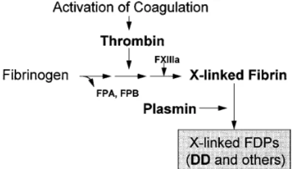
Fibrin is the main thrombus component. Its production being definitely lower in in-patients than in out-patients.
is followed by activation of the fibrinolytic system, resulting in plasmin generation and subsequent fibrin lysis (Figure 1). Dissolution of crosslinked fibrin leads
to formation of specific degradation products, includ-
Methods for measuring DD in blood
ing D-dimer (DD),1 which can be easily detected andThe advantages and disadvantages of the various measured in both whole blood and plasma using
methods that measure DD in blood are displayed in monoclonal antibodies directed against epitopes
Table 1. For clinical use in emergency situations, located in the DD fragment. In the past decade, DD
rapid tests are mandatory. Among them, a whole testing has been established as a useful aid in the
blood latex test (Simplired, Agen) that gives a diagnosis of venous thromboembolism (VTE).2 The
present review updates experience on the use of DD testing for diagnosis of deep-vein thrombosis (DVT) of the lower limbs and pulmonary embolism (PE). Briefly, DD was found in the late 1980s and early 1990s to be highly sensitive (but non-specific) to the presence of VTE where clinically suspected in out-patients. In this population, the negative predictive value of a plasma DD concentration below a certain cutoff (usually about 500 mg/l) was more than 95%, thereby allowing one to rule out the disease in a substantial proportion (about one third) of patients.
However, the diagnostic performances were strongly Figure 1. Generation of D-dimer (DD) fragments. Activation of blood coagulation leads to thrombin
genera-dependent upon the assay used, latex tests being
tion. Thrombin binds to the fibrinogen central domain
definitely less sensitive than the more cumbersome
and liberates fibrinopeptides A and B, resulting in fibrin
enzyme-linked immunosorbent assays (ELISA), which
monomers and polymer formation. The fibrin network is
in turn were not suited for emergency use.2 Moreover,
subsequently stabilized (‘cross-linked’) under the effect of
this heterogeneity of tests raised uncertainty among
activated coagulation factor XIII. Plasmin induces lysis of
clinicians, who called for rigorous evaluation and cross-linked (X-linked) fibrin, resulting in formation of standardization of the various assays.3 Lastly, the various X-linked fibrin degradation products (FDPs) includ-utility of the measurement was dependent on the ing D-dimer (DD) and fragments containing the DD
epitope.
population to which it was applied, the specificity
© Oxford University Press 1997
Address correspondence to Dr H. Bounameaux, Division of Angiology and Haemostasis, Hopital Cantonal, CH-1211 Geneva 14, Switzerland
Table 1 Comparison of the different assay methods for DD
Single-test Rapid Quantitative Sensitive Specific
Latex assays
Classical latex tests on plasma + + − − −
Latex test on whole blood (SimpliredA) + + − +/− −
ELISA assays
Classical ELISA tests − − + + −
Rapid ELISA test (Vidas DD) + + + + −
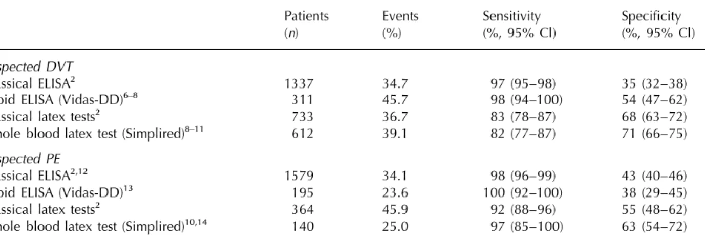
Table 2 Diagnostic performances of DD measurement in patients suspected of DVT or PE
Patients Events Sensitivity Specificity
(n) (%) (%, 95% Cl) (%, 95% Cl)
Suspected DVT
Classical ELISA2 1337 34.7 97 (95–98) 35 (32–38)
Rapid ELISA (Vidas-DD)6–8 311 45.7 98 (94–100) 54 (47–62)
Classical latex tests2 733 36.7 83 (78–87) 68 (63–72)
Whole blood latex test (Simplired)8–11 612 39.1 82 (77–87) 71 (66–75)
Suspected PE
Classical ELISA2,12 1579 34.1 98 (96–99) 43 (40–46)
Rapid ELISA (Vidas-DD)13 195 23.6 100 (92–100) 38 (29–45)
Classical latex tests2 364 45.9 92 (88–96) 55 (48–62)
Whole blood latex test (Simplired)10,14 140 25.0 97 (85–100) 63 (54–72)
Sensitivity and specificity are given as weighted means with 95% Cls; n represents the number of patients, and Events the percentage of patients with confirmed deep-vein thrombosis (DVT) or pulmonary embolism (PE).
qualitative result in 2 min, and a rapid ELISA assay are increasingly referred to diagnostic centres, in spite of a low or very low clinical suspicion. This on plasma (Vidas DD, bioMerieux) that requires
change in practice resulted in a decrease in the 35 min to produce a quantitative result, were
submit-prevalence of DVT among clinically-suspected ted to clinical testing over the past few years.
patients, which has dropped in our centre from 50% Unfortunately, latex tests, including the whole blood
to 25% over the past 15 years. Even though non-Simplired test, have the disadvantages of the visual
invasive diagnostic tests are less harmful and much reading (with potential interobserver disagreement)
cheaper than venography, they are still relatively and a limited sensitivity to the presence of VTE
expensive and require technical skills. Thus, a highly (Table 2). On the other hand, the rapid ELISA test
sensitive and simple test used as initial screening Vidas DD is highly sensitive to the presence of VTE,
and ruling out DVT in a substantial proportion of but needs to be run on a commercial automated
subjects, might save time and money, as demon-device. Other new quick tests, based on the ELISA or
strated in the setting of PE diagnosis.5 The more latex agglutination principle, should undergo
system-specific ultrasonographic test would be applied only atic testing in clinical settings before recommending
in patients presenting with a DD concentration above their use in suspected venous thromboembolism.
the cutoff.
Table 2 summarizes the diagnostic performances of the various methods to assay DD in suspected
DD for ruling out deep-vein
DVT. ELISA tests have a superior sensitivity to the presence of DVT compared with latex tests. Atthrombosis (DVT)
present time, the superiority of the Simplired test In patients with clinical signs and symptoms, B-mode over other latex tests is far from evident in patients venous compression ultrasonography or duplex scan- with suspected DVT. Published reports of sensitivity ning are presently used in clinical practice,4 contrast vary widely from paper to paper,8–11 ranging from venography still being the diagnostic gold standard. an extremely poor 61%8 up to 100%.10 However, On the other hand, with increasing fear for DVT and data are still limited, and studies with larger numbers its immediate and late consequences (pulmonary of patients and adequate methodology are urgently
needed. embolism and post-thrombotic syndrome), patients
tic trials but still needs to be evaluated properly in
DD for ruling out pulmonary
specially designed, prospective studies.
embolism (PE)
Diagnosis of recurrent venous
Clinical diagnosis of PE is unreliable, and the
dia-gnostic gold standard, pulmonary angiography, is
thromboembolism
cumbersome, invasive and not without risk.
It may be very difficult to distinguish recurrent Perfusion/ventilation lung scintigraphy allows ruling
venous thromboembolism from the sequelae of a out or ruling in PE in less than half of cases, and
previous event. Sie´ et al. showed that DD levels patients with an abnormal, non-high-probability lung
returned to normal values within 3 months after an scan pattern should theoretically undergo pulmonary acute DVT20 in most subjects, thereby implying that angiography. Recently, strategies were developed a low DD concentration measured in a patient that include lower-limbs venous compression ultra- suspected of a recurrent event could be used to rule sonography, which is highly specific for venous out recurrence.
thromboembolism. Such policies reduce substantially the need for pulmonary angiography.15,16 On the
Potential use of DD testing for confirming
other hand, because sensitivity of DD to the presence
PE
of PE is extremely high (Table 2), a concentration
below the cutoff allows ruling out the disease with The poor specificity of elevated DD plasma concen-a negconcen-ative predictive vconcen-alue of more thconcen-an 95% when tration is well known.2,21 Indeed, several conditions established methods are used. are associated with fibrin formation and degradation, Table 2 summarizes the diagnostic performances such as infectious or inflammatory diseases, and of the various methods to assay DD in suspected PE. cancer. Only 20% or even less of patients who are Sensitivity of all assays appears to be better for PE admitted with these conditions will present with a DD level below the critical cutoff of 500 mg/l, than for DVT, which may reflect a larger amount of
which decreases the usefulness of testing in case of fibrin formed and degraded in the former. In addition,
concomitant suspected venous thrombosis.21 ELISA assays and the whole-blood latex test seem to
Nonetheless, in out-patients clinically suspected exhibit comparable performances (sensitivity to
pres-of PE, the specificity pres-of the classical ELISA was 41.4% ence of PE of >95%) but confidence intervals for
(95% CI 37.0%–45.9%) in a large series of 671 sensitivity of the new tests are still wide, because of
patients.12 This figure was greatly influenced by age, the relatively small patient populations studied so far.
ranging from 72% (30–39 year-old group) to 9% (>80 years of age). As a consequence, DD measure-ment would allow excluding PE in 2/3 out-patients without the disease under 60 years of age, but only
Other applications of DD
in 1/5 patients older than 60.
measurement in suspected venous
The overall specificity of 41.4% of DD for thethromboembolism
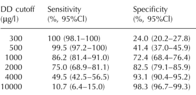
presence of PE was obtained for a cutoff of 500 mg/l (for the ELISA assay Asserachrom DD from Stago)Screening of DVT in asymptomatic high-risk
which was associated with a sensitivity of 99.5%patients
(95% CI 97.2%–100%). Setting the cutoff at 4000 mg/lwould result in a dramatic increase of specificity In a prospective trial of 185 consecutive patients
(93.1%) while sensitivity would be lowered undergoing elective abdominal surgery who were
to 49.5%.12 Detailed operating characteristics of DD submitted to bilateral ascending venography on the
in suspected PE are displayed in Table 3. These data 8th postoperative day, a plasma DD cutoff of
imply that an out-patient clinically suspected of PE 3000 mg/l was able to discriminate between patients
who presents with a DD level above 4000 mg/l has a with and without postoperative DVT with a sensitivity
551 likelihood of having PE, which may be sufficient and a specificity of 89% and 48%, respectively.17
to initiate anticoagulant treatment, provided the prior We, therefore suggested that plasma measurement of
clinical probability is sufficiently high. DD might be useful in thromboprophylactic studies
for initial screening of patients, a level below 3000 mg/l excluding DVT (negative predictive value
Standardization of DD assays:
of 93%) whereas a concentration above the cutoffnecessity or wishful thinking?
would require phlebographic confirmation. Similarresults have been reported by others in general
Different methods, different results
surgery patients18 and by our group in patients
undergoing total hip arthroplasty.19 Such an approach Several types of assays for DD are available (Table 1) and within each type, numerous commercial kits would be particularly valuable in
thromboprophylac-Table 3 Characteristics of DD measurement in out- (iv) cost-effectiveness analyses comparing the ‘new’
patients suspected of PE strategy with other management policies.
DD cutoff Sensitivity Specificity
(mg/l) (%, 95%Cl) (%, 95%Cl)
The place of DD testing in an
300 100 (98.1–100) 24.0 (20.2–27.8)
integrated diagnostic approach of
500 99.5 (97.2–100) 41.4 (37.0–45.9)
venous thromboembolism
1000 86.2 (81.4–91.0) 72.4 (68.4–76.4)
2000 75.0 (68.9–81.1) 82.5 (79.1–85.9) No single diagnostic test has a sufficiently high 4000 49.5 (42.5–56.5) 93.1 (90.4–95.2) sensitivity and specificity to be used alone in the 10000 10.7 (6.4–15.0) 98.3 (96.7–99.3) diagnostic approach of patients suspected of venous
thromboembolism, and this is particularly true for DD
Adapted from reference 12.
testing, which is non-specific. Thus, test combina-tions will be used depending upon their performances but also upon their availability, and the costs. These exist which differ in many aspects, including the tests include, besides DD measurement, venous capture and the tagging antibody, the required compression ultrasonography,4 ventilation/perfusion sample dilution, the detection limit, and the incuba- lung scintigraphy,24 to some extent also echocardio-tion time. In a systematic comparison of four ELISA graphy and spiral CT, and lastly, contrast venography assays in 151 plasma samples from patients clinically of lower limbs and pulmonary angiography. Test suspected of pulmonary embolism, van Beek et al. combinations will also be based on the fact that DVT found intra-assay coefficients of variation in the and PE represent two clinical pictures of a common median values of 3.5% to 17%22 while the inter- condition, venous thromboembolic disease, and that assay variation coefficients were very similar among diagnosing DVT in a patient clinically suspected the four assays (15–20%). This rather poor reprodu- of PE allows to diagnose PE. In addition, the treatment cibility might account, at least in part, for the only of DVT and PE is basically the same.
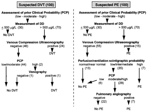
fair correlation (regression coefficients of about 0.60) Finally, a diagnostic strategy should be viewed in among the various ELISA assays. Finally, the normal a Bayesian perspective, every test result being integ-values in a reference healthy population differed rated with the clinical probability of having the considerably between the tests. disease determined prior to any test.2,25,26 Figure 2 On the other hand, because the assays recognize displays two examples of simple, sequential dia-more or less different components in plasma or in gnostic algorithms for suspected DVT or PE which blood and because various techniques are used, are presently being tested in a large-scale Swiss-heterogeneity of the results might have been anticip- Canadian management trial. The sequences were ated. Thus, rather than calling for an unlikely stand- derived from a clinical decision-making model,25 ardization, efforts should focus on determining a a subsequent management trial16 and a cost-critical cutoff for each individual assay, based on effectiveness analysis.5 In the strategy chosen for the test performances established in clinical studies suspected PE, DD testing as an initial screening, that were conducted according to the four steps followed by venous compression ultrasonography, described hereafter. yielded a 10% incremental cost reduction and a 40% reduction of necessary angiograms compared to the reference strategy.5 This sequence would in
Four-step evaluation of a diagnostic test
addition permit district hospitals without lung-scan Systematic evaluation of a new diagnostic test in facilities to manage approximately 50% of outpatients suspected venous thromboembolism23 includes with suspected PE without referral.
(i) technical description of the method; (ii) systematic comparison with a diagnostic standard in order to
establish the critical cutoff of DD concentration
Conclusions and perspectives
(using Receiver Operating Characteristics curveana-lysis), and values of sensitivity and specificity of the The place of DD measurement in the diagnostic workup of suspected venous thromboembolism is test to the presence of venous thromboembolism, for
the cutoff chosen; (iii) use of the test in so-called now well established. When using assays assessed in well-conducted clinical trials, the test can safely management trials, in which anticoagulation is
with-held in patients with a DD concentration below the rule out DVT and PE if the concentration falls below a certain, assay-dependent cutoff. When used as the cutoff. A systematic three-month follow-up would
allow detection of delayed events and establishment initial diagnostic step in an out-patient population with a prevalence of the disease of about 25%, this of the true diagnostic performance of the test;
Figure 2. D-Dimer (DD) testing in diagnostic algorithms of suspected DVT or PE. Due to its high sensitivity to venous thromboembolism, DD measurement can be used early in the diagnostic approach of deep-vein thrombosis (DVT) or pulmonary embolism (PE), a value below the critical cutoff allowing to rule out these diseases. If the DD concentration is above the cutoff, more specific tests are to be used along with prior clinical probability (PCP) assessment. The figures between brackets represent the numbers of patients with the corresponding outcome in a hypothetical cohort of 100 outpatients with clinically suspected DVT or PE, given a prevalence of the disease of about 25% in the population studied (estimations from data obtained in our institution, published in part11,15).
simple test will exclude VTE in about 30% of patients the Ernst and Lucie Schmiedheiny Foundation, the Socie´te´ Acade´mique de Gene`ve, and the Safra at a low cost. A widespread clinical application was
made possible thanks the development of rapid, Foundation. single-test, and quantitative DD assays (and, possibly,
more reliable latex tests) that can be used in the
References
Emergency Department.Further potential uses of the test include diagnosis 1. Kroneman R, Nieuwenhuizen W, Knot EAR. Monoclonal of VTE in symptomatic in-patients, screening of antibody-based plasma assays for fibrin(ogen) and
derivatives, and their clinical relevance. Blood Coag
asymptomatic high-risk patients in
thromboprophyl-Fibrinolysis 1990; 1:91–111.
actic trials, ruling out suspected recurrent VTE, and
2. Bounameaux H, de Moerloose P, Perrier A, Reber G.
ruling in VTE in suspected outpatients when DD
Plasma measurement of D-dimer as diagnostic aid in
concentration is above a certain cutoff. These
suspected venous thromboembolism: an overview. Thromb
applications look promising but deserve further Haemostas 1994; 71:1–6.
investigation. Lastly, attempts to standardize the 3. Moser KM. Diagnosing pulmonary embolism: D-dimer various DD assays should be abandoned, and needs rigorous evaluation. Br Med J 1994; 309:1525–6.
replaced by systematic studies of the diagnostic 4. White RH, McGahan JP, Daschbach MM, Hartling RP. performances of each individual assay in various Diagnosis of deep-vein thrombosis using duplex ultrasound.
Ann Intern Med 1989; 111:297–304.
clinical settings.
5. Perrier A, Buswell L, Bounameaux H, de Moerloose P, Slosman D, Unger PF, Junod A. Non-invasive diagnostic aids are cost-effective in suspected pulmonary embolism.
Acknowledgments
Arch Intern Med 1997; in press.6. D’Angelo A, D’Alessandro G, Tomassini L, Pittet JL,
The work performed at the University Hospital of
Dupuy G, Crippa L. Evaluation of a new rapid quantitative
Geneva and reviewed in the present paper, was D-dimer assay in patients with clinically suspected deep supported in part by grants from the Swiss National vein thrombosis. Thromb Haemostas 1996; 75:412–16.
Science Foundation to HB and AP (#32-027515.89, 7. Borg JY, Le´vesque H, Cailleux N, Franc C, Hellot MF, Courtois H. Rapid quantitative D-dimer assay and clinical
evaluation for the diagnosis of clinically suspected deep ultrasonography: A management study. Arch Intern Med 1996; 156:531–6.
vein thrombosis. Thromb Haemostas 1997; 77:602–3.
17. Bounameaux H, Khabiri E, Huber O, Schneider PA, 8. Janssen MCH, Heebels AE, de Metz M, Verbruggen H,
Didier D, de Moerloose P, Reber G. Value of liquid crystal Wollersheim H, Janssen S, Schuurmans MMJ,
contact thermography and plasma level of D-dimer for Novakova IRO. Reliability of five rapid D-dimer assays
screening of deep venous thrombosis following general compared to ELISA in the exclusion of deep venous
abdominal surgery. Thromb Haemostas 1992; 67:603–6. thrombosis. Thromb Haemostas 1997; 77:262–6.
18. Rowbotham BJ, Whitaker AN, Harrison J, Murtaugh P, 9. Wells PS, Brill-Edwards P, Stevens P, Panju A, Patel A,
Reasbeck P, Bowie EJW. Measurement of crosslinked fibrin Douketis J, Massicotte P, Hirsh J, Weitz JI, Kearon C,
derivatives in patients undergoing abdominal surgery: use in Ginsberg JS. A novel and rapid whole-blood assay for
the diagnosis of postoperative venous thrombosis. Blood D-dimer in patients with clinically suspected deep vein
Coag Fibrinolysis 1992; 3:25–31.
thrombosis. Circulation 1995; 91:2184–7.
19. Bongard O, Wicky J, Peter R, Simonovska S, Vogel JJ, de 10. Turkstra F, van Beek EJR, ten Cate JW, Bu¨ller HR. Reliable Moerloose P, Reber G, Bounameaux H. D-Dimer plasma
rapid blood test for the exclusion of venous measurement in patients undergoing major hip surgery: Use thromboembolism in symptomatic outpatients. Thromb in prediction and diagnosis of postoperative proximal vein
Haemostas 1996; 76:9–11. thrombosis. Thromb Res 1994; 74:487–93.
11. Brenner B, Pery M, Lanir N, Jabareen A, Markel A, Kaftori 20. Sie´ P, Cadroy Y, Elias A, Boccalon H, Boneu B. D-dimer JK, Gaitini D, Rylatt D. Application of a bedside whole levels in patients with long-term antecedents of deep venous blood D-dimer assay in the diagnosis of deep vein thrombosis. Thromb Haemostas 1994; 72:161–2.
thrombosis. Blood Coag Fibrinolysis 1995; 6:219–22. 21. Raimondi P, Bongard O, de Moerloose P, Reber G, Waldvogel F, Bounameaux H. D-Dimer plasma 12. Perrier A, Desmarais S, Goehring C, de Moerloose P,
concentration in various clinical conditions: Implication for Morabia A, Unger PF, Slosman D, Junod A, Bounameaux H.
the use of this test in the diagnostic approach of venous D-Dimer testing for suspected pulmonary embolism in
thromboembolism. Thromb Res 1993; 69:125–30. outpatients. Am J Resp Crit Care Med 1997; in press.
22. van Beek EJR, van den Ende B, Berckmans RJ, van der 13. de Moerloose P, Desmarais S, Bounameaux H, Reber G,
Heide Y, Brandjes DPM, Sturk A, ten Cate JW. A Perrier A, Dupuy G, Pittet JL. Contribution of a new, rapid,
comparative analysis of D-Dimer assays in patients with individual and quantitative automated D-dimer ELISA to
clinically suspected pulmonary embolism. Thromb exclude pulmonary embolism. Thromb Haemostas 1996;
Haemostas 1993; 70:408–13.
75:11–13.
23. Bu¨ller HR, Lensing AWA, Hirsh J, ten Cate JW. Deep vein 14. Ginsberg JS, Wells PS, Brill-Edwards P, Donovan D, thrombosis: new non-invasive diagnostic tests. Thromb
Panju A, van Beek EJR, Patel A. Application of a novel and Haemostas 1991; 66:133–7. rapid whole blood assay for D-dimer in patients with
24. The PIOPED Investigators. Value of the ventilation-perfusion clinically suspected pulmonary embolism. Thromb
scan in acute pulmonary embolism. JAMA 1990;
Haemostas 1995; 73:35–8.
263:2753–9.
15. Stein PD, Hull RH, Saltzmann HA, Pineo G. Strategy for 25. Perrier A, Bounameaux H, Morabia A, de Moerloose P, diagnosis of patients with suspected acute pulmonary Unger PF, Slosman D, Junod AF. Contribution of plasma embolism. Chest 1993; 103:1553–9. D-dimer and lower limb venous ultrasound to the diagnosis
of pulmonary embolism: a decision analysis. Am Heart J 16. Perrier A, Bounameaux H, Morabia A, de Moerloose P,
1994; 127:624–35. Slosman D, Didier D, Unger PF, Junod A. Diagnosis of
pulmonary embolism by a decision analysis-based strategy 26. Sox HC, Blatt MA, Higgins MC, Marton KI. Medical
decision making. Boston, Butterworths, 1988:67–101.