Annals of Oncology 26: 126–132, 2015 doi:10.1093/annonc/mdu499 Published online 30 October 2014
Prognosis of stage II and III colon cancer treated with
adjuvant 5-
fluorouracil or FOLFIRI in relation to
microsatellite status: results of the PETACC-3 trial
†D. Klingbiel
1,2,‡, Z. Saridaki
3,4,‡, A. D. Roth
5, F. T. Bosman
6, M. Delorenzi
2,7,8& S. Tejpar
4,9*
1
SAKK Swiss Group for Clinical Cancer Research, Coordinating Center, Bern;2
SIB Swiss Institute of Bioinformatics, Lausanne, Switzerland;3
Laboratory of Tumor Cell Biology School of Medicine, University of Crete, Heraklion, Greece;4
Center for Human Genetics O&N1, Katholieke Universiteit Leuven, Leuven, Belgium;5
Oncosurgery Unit, Geneva University Hospital, Geneva;6
Department of Pathology, Lausanne University, Lausanne;7
Ludwig Center for Cancer Research;8
Department of Oncology, University of Lausanne, Lausanne, Switzerland;9
Laboratory of Molecular Digestive Oncology, Department of Oncology, KU Leuven, Leuven, Belgium
Received 28 June 2014; revised 18 September 2014 and 15 October 2014; accepted 21 October 2014
Background:Although colon cancer (CC) with microsatellite instability (MSI) has a more favorable prognosis than micro-satellite stable (MSS) CC, the impact varies according to clinicopathological parameters. We studied how MSI status affects prognosis in a trial-based cohort of stage II and III CC patients treated with 5-fluorouracil (5-FU)/leucovorin or FOLFIRI.
Materials and methods:Tissue specimens of 1254 patients were tested for 10 different loci and were classified as MSI-high (MSI-H) when three or more loci were unstable and MSS otherwise. Study end points were overall survival (OS) and relapse-free survival (RFS).
Results:In stage II, RFS and OS were better for patients with MSI-H than with MSS CC [hazard ratio (HR) 0.26, 95% CI 0.10–0.65, P = 0.004 and 0.16, 95% CI 0.04–0.64, P = 0.01). In stage III, RFS was slightly better for patients with MSI-H CC (HR 0.67, 95% CI 0.46–0.99, P = 0.04), but the difference was not statistically significant for OS (HR 0.70, 95% CI 0.44–1.09, P = 0.11). Outcomes for patients with MSI-H CC were not different between the two treatment arms. RFS was better for patients with MSI-H than with MSS CC in the right and left colon, whereas for OS this was significant only in the right colon. For patients with KRAS- and BRAF-mutated CC, but not for double wild-type patients, RFS and OS were significantly better when the tumors were also MSI-H. An interaction test was statistically significant for KRAS and MSI status (P = 0.005), but not for BRAF status (P = 0.14).
Conclusions:Our results confirm that for patients with stage II CC but less so for those with stage III MSI-H is strongly prognostic for RFS and OS. In the presence of 5-FU treatment, stage II patients with MSI-H tumors maintain their survival advantage in comparison with MSS patients and adding irinotecan has no added benefit.
ClinicalTrials.gov Identifier:NCT00026273.
Key words:colon cancer, microsatellite instability, survival, adjuvant treatment, translational research
introduction
Approximately 15% of colon cancer (CCs) are characterized by incompetence of the DNA mismatch repair (MMR) system, leading to abnormal shortening or lengthening of repeating base pair units of DNA, a phenomenon known as microsatellite in-stability (MSI) [1]. In sporadic CC, MSI is largely due to MLH1
inactivation through hypermethylation of the promoter [2]. In familial CC, MSI is mostly due to inherited germline mutation of a MMR gene (notably MLH1 and MSH2) [3]. In sporadic CC, MSI is more frequent in stage II (almost 20%) and III (12%) tumors than in stage IV tumors (4%) [4].
Patients with MSI-Η tumors evolve more favorably than those with MSS. Several retrospective studies [1,5,6], a meta-analysis [7], and recent large trials [8–12] support the notion that stage-adjusted prognosis is more favorable for MSI-H than for MSS CC patients, but the difference in prognosis is larger for stage II than for stage III patients.
According to the current guidelines, adjuvant chemotherapy is the treatment of choice for stage III and a minority of high-risk stage II patients [13]. Disease stage remains the key determinant of prognosis and treatment, but more accurate prognostic and
†Previous presentations: Presented in part as oral presentation at the 45th Annual
Meeting of the American Society of Clinical Oncology, 29 May–2 June 2009, Orlando, FL, USA.
‡Both authors contributed equally to this work.
*Correspondence to: Prof. Sabine Tejpar, Molecular Digestive Oncology Unit, Leuven University Hospital, Herestraat 49, Leuven B-3000, Belgium. Tel: +32-16-34-42-25; Fax: +32-16-34-44-19; E-mail: [email protected]
© The Author 2014. Published by Oxford University Press on behalf of the European Society for Medical Oncology. All rights reserved. For permissions, please email: [email protected].
predictive markers are urgently needed. MSI, 18q loss of hetero-zygosity, KRAS, BRAF, and TP53 mutations have been intensive-ly investigated in this context [4, 14–16], but most are not incorporated into the treatment guidelines nor have they been confronted in large series to traditional stage II high-risk fea-tures [13] or to more recent gene expression-based prognostic signatures [17].
A putative predictive role of MSI for response to 5-fluoroura-cil (5-FU)-based adjuvant chemotherapy has been a more con-tentious issue. Some reports have suggested that disease outcome after chemotherapy does not differ between patients with MSI-H and MSS CC [18], whereas others showed increased sensitivity to 5-FU for the patients with MSI-H CC [19]. Data from randomized clinical trials of 5-FU-based therapy versus surgery only, however, suggested that patients with MSI-H CC do not benefit from 5-FU-based adjuvant chemotherapy com-pared with surgery-alone [6]. This was confirmed in a pooled
analysis [10], which added 457 cases to the previously published 570 [6]. In a recent study, however, the survival benefit of stage III MSI-H CC patients was maintained under 5-FU [11].
An issue remains the impact of MSI on the choice of adjuvant therapy. One trial initially suggested a differential effect of irino-tecan-based adjuvant chemotherapy (CALGB 89803) in favor of MSI-H patients [20], but this became marginal in an updated report [8].
To clarify these controversies, we studied stage-specific prog-nostic effects of MSI in the homogeneous PETACC-3 trial colon cancer population treated with 5-FU or FOLFIRI. The key ques-tion was whether patients with MSI-H CC maintain their sur-vival benefit under 5-FU treatment, when stratified for stage and treatment. In addition, we investigated how tumor site, BRAF and KRAS status, and high-risk stage II factors modulate the prognostic effect of MSI.
patients and methods
patient characteristics
All eligible patients were randomly assigned to receive 6 months of either 5-FU/leucovorin (LV) alone or with irinotecan [21]. MSI status could be determined for 1254 of the 1564 patients of whom tissue was available for analysis (89%), out of the total trial population of 3278 patients [14]. Earlier reports describe how further molecular parameters ( p53 expression, SMAD4 expression, 18q LOH, BRAF, and KRAS mutation status) were obtained [4,14,15]. End points were overall survival (OS), defined as the time from randomization until death, and relapse-free survival (RFS), defined as the time from randomization to local, regional, or distant relapse, the occurrence of a second primary colon cancer or death.
MSI determination
MSI was evaluated at 10 different microsatellite loci containing mono- or di-nucleotide repeated sequences. The panel consisted of thefive markers from the Bethesda reference panel, with the addition offive markers which were also suggested during the International Workshop on HNPCC in 1997 (BAT-25, BAT-26, D2S123, D5S346, TGFBR2, BAT-40, D17S787, D18S69, D17S250, and D18S58) [22]. The amplified PCR products were analyzed
using the automated ABI Prism Sequencer Model 3100 Genetic Analyzer (Applied Biosystems, Foster City, USA). A locus was called unstable if un-equivocal instabilities were seen in the tumor sample in comparison with the paired normal DNA of the same patient. MSI was graded as high (MSI-H)
when three or more markers were unstable, low (MSI-L) when one or two markers were unstable, and stable (MSS) when all markers were stable. For analysis, MSI-L and MSS populations were pooled to MSI-L/S.
The determination of the other markers has been described before [4,14,15].
statistical analyses
Survival curves were determined using Kaplan–Meier methods and com-pared using the log-rank test. Frequencies were comcom-pared using Fisher’s exact and Pearson’s χ2tests. Continuous variables were compared by MSI
status using Wilcoxon’s rank sum test. Hazard ratios (HRs) with 95% confi-dence intervals (CIs) were computed with uni- and multivariable propor-tional hazard models. Interactions were assessed by likelihood ratio tests. P-values are two-sided, not adjusted for multiple testing, and considered sig-nificant if <0.05. Analyses were carried out using the free R software package
(www.r-project.org) version 2.13.0 or later.
results
patient and tumor characteristics in relation to MSI status
Patients and tumor characteristics by MSI status are summar-ized in Table1. The MSI-H frequency was almost twice as high in node-negative, compared with node-positive, patients. The proportion of MSI-H tumors was higher with higher T-stage, in the right colon, when poorly differentiated, mutated for BRAF or with high thymidylate synthase (TYMS) expression, but lower for mucinous tumors and those with SMAD4 loss, high TP53 expression and (weakly) a KRAS mutation.
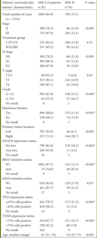
prognostic value of MSI varies according to stage After a median follow-up of 69.1 months, RFS (HR 0.48, 95% CI 0.34–0.69, P < 0.001) as well as OS (HR 0.47, 95% CI 0.31– 0.72, P < 0.001) were better for patients with MSI-H than with MSI-L/S CC. This was most striking in patients with stage II CC with a strong effect of MSI status on RFS and OS, still significant but weaker for RFS in stage III patients but not significant for OS in stage III (Figure1A and B). A statistically significant
inter-action between stage and MSI status was found for OS (P = 0.047), still borderline significant for RFS (P = 0.06). the prognostic value of MSI is not affected by 5-FU/ LV versus FOLFIRI treatment
For stage II 5-FU/LV- as well as FOLFIRI-treated patients, RFS and OS were better for MSI-H than for MSI-L/S CC (Figures1C and D, and2).
For stage III 5-FU/LV-treated patients, the MSI-H effect was weaker compared with stage II 5-FU/LV-treated patients. For stage III FOLFIRI-treated almost no difference was found by MSI status, neither on RFS nor on OS (Figures1E and F, and2). An interaction test between treatment and MSI status within stage III patients, however, was not significant (P = 0.31 for RFS and P = 0.18 for OS).
When patients were stratified according to MSI status, RFS and OS were similar in both treatment arms. We could not confirm the benefit suggested by Bertagnolli et al. [8] for irinote-can addition in MSI-H tumors (supplementary Table S1, avail-able at Annals of Oncology online).
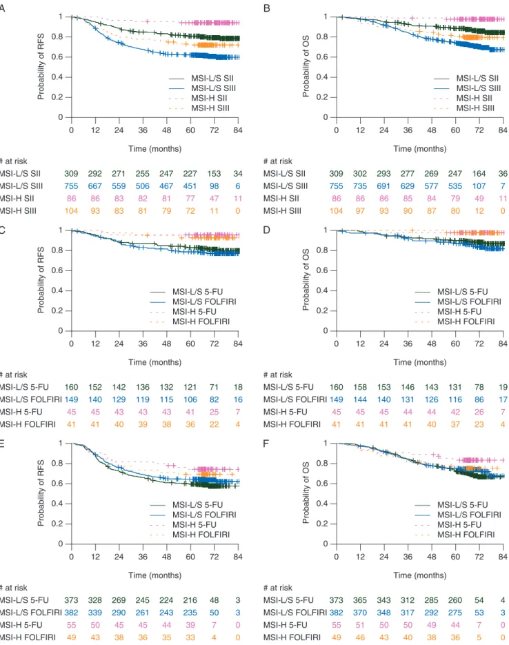
MSI is prognostic in both the right and left colon Right-sided carcinomas were almostfive times more often MSI-H than left-sided carcinomas. More precisely, we found a gradual pattern of MSI-H incidence as reported elsewhere [23]. RFS was better for patients with MSI-H than for MSI-L/S CC, regardless of side. In the right colon, OS was statistically signi fi-cantly different between MSI-H and MSI-L/S CC, but not in the left. Of note is the similarity of the two HRs (Figure2), with a non-significant interaction. As left CCs are less frequently MSI-H than right CCs (n = 46versus 144), the power of tests in this subgroup is lower.
For patients with a stage II carcinoma in the left colon, RFS was similar for MSI-H and MSI-L/S CC, whereas in the right colon, RFS was significantly better for patients with MSI-H CC. For patients with stage II carcinomas in the left colon, MSI status had no effect on OS. There was no event for the 64 MSI-H CC in the right colon. For stage III patients, RFS and OS tended to be better for MSI-H carcinomas, irrespective of site, but this trend was not significant. These observations were confirmed in multivariable models including BRAF mutation status and gender.
MSI status and BRAF/KRAS status
BRAF-mutated CCs were almost four times more often MSI-H than BRAF wild-type CC. In contrast, KRAS-mutated CCs were 1.5 times less often MSI-H than KRAS wild-type CC (Table1). In patients with a double wild-type CC, MSI status had no effect on RFS or OS (Figure2). For patients with a KRAS-mutated CC, however, RFS and OS were clearly better. An interaction test for KRAS and MSI status was significant (P = 0.005). For BRAF-mutated carcinomas, the CIs were larger, but the effect was still significant for both RFS and OS. A test for interaction between MSI and BRAF status, however, was not significant (P = 0.14). Conversely, BRAF status was not prognostic in patients with a MSI-H CC (RFS: HR = 1.26, 95% CI 0.59–2.70, P = 0.55; OS: HR = 1.53, 95% CI 0.63–3.70, P = 0.35). Similar results were obtained in multivariable analyses when stratified by stage (sup-plementary Table S2, available at Annals of Oncology online). MSI status and stage II risk factors
As shown in supplementary Table S3, available at Annals of Oncology online, the distribution of MSI-H and MSI-L/S according to stage II risk factors was similar in both groups, except for poorly differentiated CC, which are rare (5.3% of all stage II tumors) but more often MSI-H. Small patient numbers allowed only univariate analyses of these risk factors in the MSI-H group. T stage (MSI-HR = 5.28, 95% CI 0.88–31.64, P = 0.07), posi-tive margins (HR = 11.53, 95% CI 1.28–104.15, P = 0.03), and perforation (HR = 8.40, 95% CI 1.40–50.38, P = 0.02) were prog-nostic but with high uncertainty due to small patient numbers.
When combining risk factors into a high-risk group with at least one risk factor and a low-risk group without any risk factor, the HRs of patients with MSI-L/S CC were higher than those of high-risk patients both in RFS (HR = 3.63, 95% CI 1.46–9.04, P = 0.006 versus HR = 2.40, 95% CI 1.28–4.47, P = 0.006) and OS (HR = 6.03, 95% CI 1.46–24.91, P = 0.01 versus HR = 2.80, 95% CI 1.25–6.28, P = 0.01). This suggests that the prognostic value of MSI status is stronger than that of the combined classical risk factors for stage II patients. In a Table 1. Association between MSI status, patients’ characteristics,
and tumours’ molecular characteristics
Patients’ and molecular tumors’ characteristics MSI-L/S patients n (%) MSI-H n (%) P-value Total number of cases
(n = 1254) 1064 (84.9) 190 (15.1) Stage II 309 (78.2) 86 (21.8) <0.001 III 755 (87.9) 104 (12.1) Treatment group 5-FU/FA 533 (84.2) 100 (15.8) 0.53 FOLFIRI 531 (85.5) 90 (14.5) N-stage N0 309 (78.2) 86 (21.8) <0.001 N1 495 (88.4) 65 (11.6) N2 260 (87.0) 39 (13.0) T-stage T1/2 69 (93.2) 5 (6.8) 0.03 T3 815 (85.1) 143 (14.9) T4 180 (81.1) 42 (18.9) Grade G-1/2 993 (87.8) 138 (12.2) <0.001* G-3/4 63 (55.3) 51 (44.7) No result 8 1 Mucinous features Yes 898 (88.6) 115 (11.4) <0.001* No 158 (68.1) 74 (31.9) No result 8 1 Primary tumor location
Left 707 (93.9) 46 (6.1) <0.001 Right 357 (71.3) 144 (28.7)
SMAD4 expression status
No loss 799 (81.8) 178 (18.2) <0.001* Any loss 249 (95.8) 11 (4.2)
No result 16 1 BRAF mutation status
Wt 1002 (87.5) 143 (12.5) <0.001* mut 53 (54.6) 44 (45.4)
No result 9 3 KRAS mutation status
Wt 626 (83.0) 128 (17.0) 0.03* mut 421 (87.7) 59 (12.3) No result 17 3 TP53 expression status ≤45% cells positive 641 (78.7) 173 (21.3) <0.001* >45% cells positive 410 (96.5) 15 (3.5) No result 13 2 TYMS expression status
>75% cells positive 254 (67.7) 121 (32.3) <0.001* <75% cells positive 708 (92.2) 60 (7.8)
No result 102 9
Age, median (range) 61 (21–76) 54 (25–75) <0.001 MSI, microsatellite instability; MSI-L/S, MSI-low/stable, MSI-H, MSI-high; TYMS, thymidylate synthase.
*Missing values have not been considered for the calculation of P-values. Except for TYMS expression, there was no significant difference in terms of missingness between MSI-H and MSI-L/S tumors.
Time (months) Probability of RFS 0 0.2 0.4 0.6 0.8 1 # at risk MSI-L/S SII MSI-L/S SIII MSI-H SII MSI-H SIII 309 292 271 255 247 227 153 34 755 667 559 506 467 451 98 6 86 86 83 82 81 77 47 11 104 93 83 81 79 72 11 0 MSI-L/S SII MSI-L/S SIII MSI-H SII MSI-H SIII A Time (months) Probability of OS 0 0.2 0.4 0.6 0.8 1 # at risk MSI-L/S SII MSI-L/S SIII MSI-H SII MSI-H SIII 309 302 293 277 269 247 164 36 755 735 691 629 577 535 107 7 86 86 86 85 84 79 49 11 104 97 93 90 87 80 12 0 MSI-L/S SII MSI-L/S SIII MSI-H SII MSI-H SIII B Time (months) Probability of RFS 0 0.2 0.4 0.6 0.8 1 # at risk MSI-L/S 5-FU MSI-L/S FOLFIRI MSI-H 5-FU MSI-H FOLFIRI 160 152 142 136 132 121 71 18 149 140 129 119 115 106 82 16 45 45 43 43 43 41 25 7 41 41 40 39 38 36 22 4 MSI-L/S 5-FU MSI-L/S FOLFIRI MSI-H 5-FU MSI-H FOLFIRI C Time (months) Probability of OS 0 0.2 0.4 0.6 0.8 1 # at risk MSI-L/S 5-FU MSI-L/S FOLFIRI MSI-H 5-FU MSI-H FOLFIRI 160 158 153 146 143 131 78 19 149 144 140 131 126 116 86 17 45 45 45 44 44 42 26 7 41 41 41 41 40 37 23 4 MSI-L/S 5-FU MSI-L/S FOLFIRI MSI-H 5-FU MSI-H FOLFIRI D Time (months) Probability of RFS 0 0.2 0.4 0.6 0.8 1 # at risk MSI-L/S 5-FU MSI-L/S FOLFIRI MSI-H 5-FU MSI-H FOLFIRI 373 328 269 245 224 216 48 3 382 339 290 261 243 235 50 3 55 50 45 45 44 39 49 43 38 36 35 33 MSI-L/S 5-FU MSI-L/S FOLFIRI MSI-H 5-FU MSI-H FOLFIRI E Time (months) Probability of OS 0 12 24 36 48 60 72 84 0 12 24 36 48 60 72 84 0 12 24 36 48 60 72 84 0 12 24 36 48 60 72 84 0 12 24 36 48 60 72 84 0 12 24 36 48 60 72 84 0 0.2 0.4 0.6 0.8 1 # at risk MSI-L/S 5-FU MSI-L/S FOLFIRI MSI-H 5-FU MSI-H FOLFIRI 373 365 343 312 285 260 54 4 382 370 348 317 292 275 53 3 55 51 50 50 49 44 49 46 43 40 38 36 7 0 4 0 7 0 5 0 MSI-L/S 5-FU MSI-L/S FOLFIRI MSI-H 5-FU MSI-H FOLFIRI F
Figure 1. Kaplan–Meier plots showing outcome according to tumor stage, treatment, and microsatellite status. (A) Relapse-free survival (RFS), (B) overall survival (OS), (C) RFS in stage II, (D) OS in stage II, (E) RFS in stage III, (F) OS in stage III. SII, stage II; SIII, stage III; MSI, microsatellite instable; MSI-H, MSI-high; MSI-L/S, MSI-low/stable.
multivariable model, T stage alone was stronger than all other risk factors combined and equaled the effect of MSI status (sup-plementary Figure S1 and Table S4, available at Annals of Oncology online).
discussion
Our results confirm earlier reports [1,5–8,10,18] that MSI-H colon cancer patients have a better survival than those with MSI-L/S tumors (supplementary Table S5, available at Annals of Oncology online). Furthermore, we confirm MSI-H tumors to be more often stage II, located in the proximal colon, and of poor or undifferentiated histology, in line with previous reports [8,
10,11,24].
We found the MSI-H effect on RFS and OS to be stronger in stage II than in stage III patients [9]. The striking effect in stage II, even though in both arms patients were treated with 5-FU, suggests that patients with MSI-H tumors have a good progno-sis, even when treated. Earlier work of Sargent et al. [10] has suggested a lack of benefit from 5-FU-based chemotherapy, but in the absence of an untreated control arm, however, our dataset cannot assess directly the effect of 5-FU on MSI-H patients.
Furthermore, we have shown that MSI-H colon cancer patients treated with FOLFIRI do not fare better than those treated with only 5-FU/LV [12], in contrast to an earlier report
of Bertagnolli et al. [20]. More in line with our data, Bertagnolli et al. [8] recently reported, in a larger patient cohort, an only marginally significant increase in RFS for 5-FU-/irinotecan-treated MSI-H patients, when all other risk factors were taken into account. We found no evidence for stage-specific or overall interactions between treatment and MSI status. Nevertheless, in the above-mentioned two trials, different irinotecan-based regi-mens were used. These differences must be taken into consider-ation in cross-trial comparisons.
We confirm the high frequency of BRAF mutations in the MSI-H population [25]. As a novel observation, wefind MSI to be prognostic in KRAS- and BRAF-mutated, but not in double wild-type, patients. BRAF, however, had no prognostic impact in MSI-H patients, possibly limited by sample size [26].
A still unanswered question involves the potential impact of the site of the primary tumor, for which we report novel data on the PETACC3 cohort. We found no difference in RFS, between right- and left-sided MSI-H carcinomas. OS of patients with a right-sided MSI-H CC was significantly better compared with those with a right-sided MSI-S/L CC, as previously reported [27]. Our data, however, do not provide convincing evidence in favor of or against a benefit for patients with a left-sided MSI-H CC. In a recent publication, Sinicrope et al. [28] reported that, although patients with a MSI-H right-sided CC have a statistic-ally significant DFS advantage, the outcome of those with a left-RFS HR (95% Cl) OS HR (95% Cl) Overall Stage ll Stage lll Left Right Stage ll, left Stage ll, right 0.48 (0.34–0.69) 0.26 (0.10–0.65) 0.67 (0.46–0.99) 0.46 (0.23–0.94) 0.50 (0.33–0.77) 0.39 (0.09–1.62) 0.23 (0.07–0.77) 0.47 (0.31–0.72) 0.16 (0.04–0.64) 0.70 (0.44–1.09) 0.53 (0.23–1.20) 0.41 (0.24–0.68) 0.61 (0.15–2.57) 0.00 (0.00–lnf) Stage lll, left Stage lll, right 5-FU/FA 5-FU/FA, Sll 5-FU/FA, Slll FOLFlRl FOLFlRl, Sll FOLFlRl, Slll Double wt KRAS mut BRAF mut 0.61 (0.27–1.36) 0.71 (0.45–1.11) 0.41 (0.25–0.69) 0.22 (0.05–0.91) 0.56 (0.32–0.96) 0.57 (0.35–0.93) 0.30 (0.09–0.96) 0.82 (0.48–1.40) 0.72 (0.45–1.16) 0.19 (0.08–0.47) 0.40 (0.19–0.86) 0.60 (0.22–1.61) 0.63 (0.38–1.07) 0.37 (0.19–0.70) 0.18 (0.02–1.32) 0.51 (0.26–1.00) 0.58 (0.33–1.04) 0.14 (0.02–1.03) 0.94 (0.52–1.72) 0.58 (0.30–1.10) 0.27 (0.11–0.67) 0.31 (0.13–0.72) RFS OS 0 1 Hazard ratio 2
Figure 2. Forest plots of the prognostic value of MSI status in selected patient groups. RFS, relapse-free survival; OS, overall survival; HR, hazard ratio; 95% CI, 95 percent confidence interval; SII, stage II; SIII, stage III; wt, wild type; mut, mutated.
sided MSI-H CC was worse, which we did notfind. A larger val-idation series is needed to settle this question, especially given the evidence for stage-specific effects. The improvement in RFS and OS for stage II patients with a MSI-H CC seemed stronger in the right than in the left colon, with the notable observation that out of 64 patients not a single patient with a stage II right-sided MSI-H CC had died and only three had relapsed. Such differences were not found in stage III patients. Another hypoth-esis emerging from our data, but in need of validation, is the potential interaction between KRAS mutation and MSI status.
An open issue is how useful MSI status is as a marker for stage II patients considered for 5-FU chemotherapy. We compared MSI status with conventionally applied high-risk factors [29]. In terms of RFS and OS, MSI-H status was slightly stronger than the com-bination of high-risk factors. Among high-risk factors, T-stage was the strongest. Comparison between T-stage and MSI status resulted in a similar effect on outcome, as we previously reported [15]. Previously published data from the Sargent group [10,30] advocated that MSI-H patients might be spared adjuvant treat-ment. The lack of untreated patients in our study prevents a direct comparison, but we found that stage II patients with T3 and MSI-H CC fare very well. Given the modest treatment effect of 5-FU in this population, they seem to represent the best candidate group for omitting adjuvant treatment. Conversely, patients with MSI-S/ L T4 CC fared much worse, even though they received chemother-apy. For the intermediate-risk patient with a H T4 or a MSI-S/L T3 CC, other factors need to be considered before a conclusion can be reached.
In conclusion, our results confirm that MSI-H is strongly prognostic for RFS and OS for stage II patients, and less so for stage III patients. In the presence of 5-FU treatment, stage II patients with MSI-H tumors maintain their survival advantage in comparison with MSI-L/S patients and adding irinotecan has no added benefit. Additional parameters (including gene expres-sion profiling, ploidy, methylation, and microRNA expression) have to be explored in order to more accurately define stage II patients who require adjuvant treatment and to predict which patients will respond. Based on new emerging information, further exploratory analyses in large patients’ cohorts looking also at the impact of site, mutation profile, and genomic signa-tures will be necessary to further appreciate the molecular and prognostic impact of MSI status in colon cancer.
acknowledgements
We thank all the clinicians who enrolled patients and participated in the PETACC-3 trial (see Appendix at:http://jco.ascopubs.org/ content/27/19/3117.long), in particular the coordinators D. Cunningham, R. Labianca, and E. Van Cutsem. Finally, we also thank Vasso Athanasaki for help with the references.
funding
ZS is a recipient of a research fellowship from the Hellenic Society of Medical Oncology (Hesmo). AR and MD gratefully acknowledge financial support of the Swiss National Science Foundation (grant SNF 320030_135421), the Krebsliga Schweiz (KFS 0269708-2010), and the Fondation Medic. ST is a senior clinical investigator of the fund for Scientific Research Flanders
(FWO-Vlaanderen) and holder of a research grant by the Belgian National Cancer Plan (Nationaal Kankerplan), and the King Baudouin Foundation and the Fondation Majoie.
disclosure
AR: advisor Pfizer; ST: speaker fee Merck Serono, advisor Merck Serono and Sanofi, past research grant Pfizer. ZS: speaker fee Janssen-Cilag, advisor Amgen. All remaining authors have declared no conflicts of interest.
references
1. Thibodeau SN, Bren G, Schaid D. Microsatellite instability in cancer of the proximal colon. Science 1993; 260: 816–819.
2. Thibodeau SN, French AJ, Cunningham JM et al. Microsatellite instability in colorectal cancer: different mutator phenotypes and the principal involvement of hMLH1. Cancer Res 1998; 58: 1713–1718.
3. Peltomaki PT. Genetic basis of hereditary nonpolyposis colorectal carcinoma (HNPCC). Ann Med 1994; 26: 215–219.
4. Roth AD, Tejpar S, Delorenzi M et al. Prognostic role of KRAS and BRAF in stage II and III resected colon cancer: results of the translational study on the PETACC-3, EORTC 40993, SAKK 60–00 trial. J Clin Oncol 2010; 28: 466–474.
5. Gryfe R, Kim H, Hsieh ET et al. Tumor microsatellite instability and clinical outcome in young patients with colorectal cancer. N Engl J Med 2000; 342: 69–77. 6. Ribic CM, Sargent DJ, Moore MJ et al. Tumor microsatellite-instability status as a
predictor of benefit from fluorouracil-based adjuvant chemotherapy for colon cancer. N Engl J Med 2003; 349: 247–257.
7. Popat S, Hubner R, Houlston RS. Systematic review of microsatellite instability and colorectal cancer prognosis. J Clin Oncol 2005; 23: 609–618.
8. Bertagnolli MM, Redston M, Compton CC et al. Microsatellite instability and loss of heterozygosity at chromosomal location 18q: prospective evaluation of biomarkers for stages II and III colon cancer—a study of CALGB 9581 and 89803. J Clin Oncol 2011; 29: 3153–3162.
9. Roth AD, Tejpar S, Yan P. Correlation of molecular markers in colon cancer with stage-specific prognosis: results of the translational study on the PETACC3— EORTC 40993-SAKK 60-00 trial. In ASCO Gastrointestinal Cancers Symposium (abstr 288), 2009.
10. Sargent DJ, Marsoni S, Monges G et al. Defective mismatch repair as a predictive marker for lack of efficacy of fluorouracil-based adjuvant therapy in colon cancer. J Clin Oncol 2010; 28: 3219–3226.
11. Sinicrope FA, Foster NR, Thibodeau SN et al. DNA mismatch repair status and colon cancer recurrence and survival in clinical trials of 5-fluorouracil-based adjuvant therapy. J Natl Cancer Inst 2011; 103: 863–875.
12. Tejpar S, Bosman F, Delorenzi M et al. Microsatellite instability (MSI) in stage II and III colon cancer treated with 5FU-LV or 5FU-LV and irinotecan (PETACC 3-EORTC 40993-SAKK 60/00 trial). J Clin Oncol 2009; 27: 15s.
13. Morris EJ, Maughan NJ, Forman D, Quirke P. Who to treat with adjuvant therapy in Dukes B/stage II colorectal cancer? The need for high quality pathology. Gut 2007; 56: 1419–1425.
14. Bosman FT, Yan P, Tejpar S et al. Tissue biomarker development in a multicentre trial context: a feasibility study on the PETACC3 stage II and III colon cancer adjuvant treatment trial. Clin Cancer Res 2009; 15: 5528–5533.
15. Roth AD, Delorenzi M, Tejpar S et al. Integrated analysis of molecular and clinical prognostic factors in stage II/III colon cancer. J Natl Cancer Inst 2012; 104: 1635–1646.
16. Tejpar S, Bertagnolli M, Bosman F et al. Prognostic and predictive biomarkers in resected colon cancer: current status and future perspectives for integrating genomics into biomarker discovery. Oncologist 2010; 15: 390–404.
17. Gray RG, Quirke P, Handley K et al. Validation study of a quantitative multigene reverse transcriptase-polymerase chain reaction assay for assessment of recurrence risk in patients with stage II colon cancer. J Clin Oncol 2011; 29: 4611–4619. 18. Kim GP, Colangelo LH, Wieand HS et al. Prognostic and predictive roles of
high-degree microsatellite instability in colon cancer: a National Cancer
National Surgical Adjuvant Breast and Bowel Project Collaborative Study. J Clin Oncol 2007; 25: 767–772.
19. Jover R, Zapater P, Castells A et al. The efficacy of adjuvant chemotherapy with 5-fluorouracil in colorectal cancer depends on the mismatch repair status. Eur J Cancer 2009; 45: 365–373.
20. Bertagnolli MM, Niedzwiecki D, Compton CC et al. Microsatellite instability predicts improved response to adjuvant therapy with irinotecan,fluorouracil, and leucovorin in stage III colon cancer: Cancer and Leukemia Group B Protocol 89803. J Clin Oncol 2009; 27: 1814–1821.
21. Van Cutsem E, Labianca R, Bodoky G et al. Randomized phase III trial comparing biweekly infusionalfluorouracil/leucovorin alone or with irinotecan in the adjuvant treatment of stage III colon cancer: PETACC-3. J Clin Oncol 2009; 27: 3117–3125. 22. Boland CR, Thibodeau SN, Hamilton SR et al. A National Cancer Institute
Workshop on Microsatellite Instability for cancer detection and familial predisposition: development of international criteria for the determination of microsatellite instability in colorectal cancer. Cancer Res 1998; 58: 5248–5257. 23. Missiaglia E, Jacobs B, D’Ario G et al. Distal and proximal colon cancers differ in
terms of molecular, pathological and clinical features. Ann Oncol 2014; 25: 1995–2001.
24. French AJ, Sargent DJ, Burgart LJ et al. Prognostic significance of defective mismatch repair and BRAF V600E in patients with colon cancer. Clin Cancer Res 2008; 14: 3408–3415.
25. Rajagopalan H, Bardelli A, Lengauer C et al. Tumorigenesis: RAF/RAS oncogenes and mismatch-repair status. Nature 2002; 418: 934.
26. Popovici V, Budinska E, Bosman FT et al. Context-dependent interpretation of the prognostic value of BRAF and KRAS mutations in colorectal cancer. BMC Cancer 2013; 13: 439.
27. Jernvall P, Makinen MJ, Karttunen TJ et al. Microsatellite instability: impact on cancer progression in proximal and distal colorectal cancers. Eur J Cancer 1999; 35: 197–201.
28. Sinicrope FA, Mahoney MR, Smyrk TC et al. Prognostic impact of deficient DNA mismatch repair in patients with stage III colon cancer from a randomized trial of FOLFOX-based adjuvant chemotherapy. J Clin Oncol 2013; 31: 3664–3672. 29. Benson AB, III, Schrag D, Somerfield MR et al. American Society of Clinical
Oncology recommendations on adjuvant chemotherapy for stage II colon cancer. J Clin Oncol 2004; 22: 3408–3419.
30. Sinicrope FA. DNA mismatch repair and adjuvant chemotherapy in sporadic colon cancer. Nat Rev Clin Oncol 2010; 7: 174–177.
Annals of Oncology 26: 132–140, 2015 doi:10.1093/annonc/mdu474 Published online 15 October 2014
Abituzumab combined with cetuximab plus irinotecan
versus cetuximab plus irinotecan alone for patients
with
KRAS wild-type metastatic colorectal cancer:
the randomised phase I/II POSEIDON trial
E. Élez
1, I. Kocáková
2, T. Höhler
3, U. M. Martens
4, C. Bokemeyer
5, E. Van Cutsem
6, B. Melichar
7,
M. Smakal
8, T. Cso
˝ szi
9, E. Topuzov
10, R. Orlova
11, S. Tjulandin
12, F. Rivera
13, J. Straub
14,
R. Bruns
14, S. Quaratino
14& J. Tabernero
1*
1
Vall d’Hebron University Hospital and Institute of Oncology (VHIO), Universitat Autònoma de Barcelona, Barcelona, Spain;2
Department of Comprehensive Cancer Care, Masarykuv Onkologicky Ustav, Brno, Czech Republic;3
Medical Clinic I, Prosper-Hospital, Recklinghausen;4
Department of Hematology/Oncology, Cancer Center Heilbronn-Franken, Heilbronn;5
Department of Oncology/Hematology, University Hospital Hamburg, Hamburg, Germany;6
Department of Digestive Oncology, University Hospital Gasthuisberg Leuven and KULeuven, Leuven, Belgium;7
Department of Oncology, Palacký University Medical School and Teaching Hospital, Olomouc; 8
Department of Oncology, Horovice, Czech Republic;9
Department of Oncology, Jasz-Nagykun-Szolnok Megyei Hetenyi Geza Korhaz-Rendelointezet, Szolnok, Hungary; 10
GOU VPO St-Petersburg SMA, n/a Mechnikov Federal Agency of Healthcare, St Petersburg;11
City Clinical Oncology Dispensary, St Petersburg;12
S.I. Russian Cancer Research Center, Moscow, Russia;13
University Hospital Marques de Valdecilla, Santander, Spain;14
Merck KGaA, Darmstadt, Germany
Received 5 September 2014; revised 1 October 2014; accepted 2 October 2014
Background:Integrins are involved in tumour progression and metastasis, and differentially expressed on colorectal cancer (CRC) cells. Abituzumab (EMD 525797), a humanised monoclonal antibody targeting integrinαν heterodimers, has demonstrated preclinical activity. This trial was designed to assess the tolerability of different doses of abituzumab in combination with cetuximab and irinotecan ( phase I) and explore the efficacy and tolerability of the combination versus that of cetuximab and irinotecan in patients with metastatic CRC (mCRC) ( phase II part).
Methods:Eligible patients had KRAS (exon 2) wild-type mCRC and had received prior oxaliplatin-containing therapy. The trial comprised an initial safety run-in using abituzumab doses up to 1000 mg combined with a standard of care (SoC: cetuximab plus irinotecan) and a phase II part in which patients were randomised 1 : 1 : 1 to receive abituzumab *Correspondence to: Dr Josep Tabernero, Department of Medical Oncology, Vall
d’Hebron University Hospital, Vall d’Hebron Institute of Oncology (VHIO), P. Vall d’Hebron 119–129, 08035 Barcelona, Spain. Tel: +34-93-489-4301; Fax: +34-93-274-6059; E-mail: [email protected]
© The Author 2014. Published by Oxford University Press on behalf of the European Society for Medical Oncology. All rights reserved. For permissions, please email: [email protected].