Cite this article as: Zientara A, Genoni M, Schwegler I. Type B dissection reveals a rare anomaly of left cervical arch of Haughton D type. Eur J Cardiothorac Surg 2015;47:1109.
Type B dissection reveals a rare anomaly of left cervical
arch of Haughton D type
Alicja Zientara*, Michele Genoni and Igor Schwegler
Department of Cardiac and Vascular Surgery, Triemli Hospital, Zürich, Switzerland
* Corresponding author. Department of Cardiac Surgery, City Hospital Triemli, Birmensdorferstrasse 497, 8063 Zürich, Switzerland. Tel: +41-44-4661650; fax: +41-44-4662745; e-mail: [email protected] (A. Zientara).
Received 1 May 2014; received in revised form 26 June 2014; accepted 27 June 2014
Keywords:
Left cervical arch aneurysm
• Vascular malformations • Thoracic aorta • Mediastinum
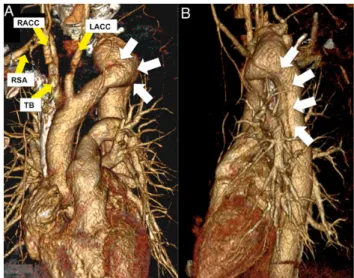
A 32-year-old woman with a history of lumbar-vertebral syndrome
presented with acute back pain. The imaging showed a rare
anomaly of a left cervical arch of Haughton D type complicated by
a dissection arising from the arch aneurysm (Fig.
1
) to both iliac
ar-teries (Fig.
2
A and B).
Figure 1:Computed tomography; arrow points to the left subclavian artery arising from the arch aneurysm (*greatest extent of 41 mm).
Figure 2:(A and B) Computed tomography reconstruction of the aortic arch; arrows show the course of the dissecting membrane, which starts from the an-eurysm. LACC: left common carotid artery; RACC: right common carotid artery; TB: truncus brachiocephalicus; RSA: right subclavian artery.
© The Author 2014. Published by Oxford University Press on behalf of the European Association for Cardio-Thoracic Surgery. All rights reserved.
IM AG ES IN C A RD IO -THO R A C IC SU RGE R Y
European Journal of Cardio-Thoracic Surgery 47 (2015) 1109