Published by Churchill Livingstone, London. © European Orthodontic Society, 1979
Maxillary dentoalveolar problems conflicting
with a skeletal Class II correction: a case
report
U. TeuscherZahrarztliches Institut der Universitat Zurich, CH Zurich 8028, Switzerland
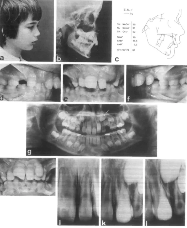
Summary. In a nine year old boy, maxillary dentoalveolar problems and a deviating eruption path of an upper canine delayed the correction of the intermaxillary Class II relation for an extended period of time. It eventually became necessary to conclude treatment rapidly and this was achieved by simultaneous use of heavy extraoral forces, fixed applicance and functional orthopedic therapy.
A boy, 9 years of age presented a skeletal and dental Class II, div. 1 malocclusion (pre-treatment records Fig. 1). In the maxillary arch the right central incisor was a megalo-dont and instead of the left central incisor a small supernumerary tooth had erupted. In the position of the left lateral incisor two laterals erupted: one of them was of normal size but the other however was intermediate in size between a lateral and central incisor. X-rays revealed the left upper canine develop-ing in a pronounced mesiobuccal direction (Fig. lg) with its cusp located between the roots of the two laterals (Fig. li-1). During the observation period, the incisal edge of the megalodont was fractured by accident (Fig. lh).
Treatment
Apart from the correction of the antero-posterior skeletal discrepancy an aligned upper incisor segment had to be established. Since the labially erupted lateral incisor was of acceptable size and marginal circumference to serve as a central incisor, it was decided to
extract the small supernumerary tooth (Fig. 2a).
Nine months after the start of therapy an incisor segment was established. Anterior high pull traction of low force level had been applied for 4 months but because the canine position had further worsened (Fig. 2c) during these treatment procedures, extraoral traction had to be discarded and for 2 years 4 months no attempt was made to correct the Class II malocclusion. At the end of this period the upper canines and first premolar had erupted and anterior high pull traction could be reinstated.
Nine months later, however, i.e. 3 years 10 months after the start of therapy, inter-digitation was still Class II (Fig. 2d-2f). Superimposition of the cephalogram at this stage of treatment with the pretreatment cephalogram indicated above average man-dibular growth (Fig. 2i). The sagittal effect of this growth, however, was cancelled out by vertical growth components. Forward and downward displacement of the maxillary complex (Fig. 2h) was not sufficiently inhibited because of the inadequate time and force (300 g) of headgear application. Vertical
132 CLASS II TREATMENT PROBLEM
Figure 1 Pretreatment records, a-f: class II, div. 1, deep overbite, tooth size discrepancies, g: the left upper
central is a supernumerary tooth, two left laterals are erupting and the left upper canine is developing in a pro-nounced mesial direction, h: shape of the upper incisors, i-lc: the cusp of the canine is located between the roots of the two left laterals.
s
Figure 2 Course of treatment, a, b: incisor alignment, c: worsening of the canine position, d—i: 3 years 10 months
after start of therapy; class IT interdigitation; cephalograms and superimposed tracings of this period of treatment, k: root resorption of the upper incisors. 1, m: simultaneous use of an activator-headgear device with the facebow attached directly to the activator, n - p : interdigitation 4 months after this combined therapy, q-s: cephalograms before and after simultaneous use of the activator-headgear appliance (4 months); superimposition of tracings.
134
CLASS II TREATMENT PROBLEMdentoalveolar development of the buccal segments (Fig. 2i) can also be seen. In the upper arch, this vertical development was accelerated by intramaxillary vertical control of the upper incisors.
The situation was complicated by the radiographic diagnosis of apical root resorp-tion of the upper incisors, especially of the megalodont which had been traumatised (Fig. 2k). This resorption was considered to
be the result of prolonged orthodontic stresses, and it was therefore decided that treatment must be concluded as quickly as possible. All palatal and lingual attachments were accordingly removed from the bands. In addition to the existing fixed appliance, a Class II activator-headgear device was applied during the night. The facebow was mounted directly to the activator (Fig. 21 and 2m). Bodily tooth control by the edgewise appliance
SH M*Go-NL MaGo* SN Occ* SNA' SNB-AN8* PFH/AFHt 9y 39 31 22 79 71.5 7.5 61 e.A.rf 9 , —14,2 II: 4>2 14,2™ 35 25 17 75 733 1.3 64 • 3
Figure 3 Post-treatment records, a-c: grinding of the right upper central (megalodont) and addition of compo-site material to the left central, d: orthopantomogram. e-g: profile of the boy at the end of treatment; overall treatment, superimposed tracings.
allowed an extra-oral force of 800 gr on each side. Within only 4 months, Class I arch relationship was achieved. The initial labially erupted upper left lateral incisor, now in the position of the left central incisor (Fig. 26), had considerably improved the tooth size discrepancies of the original upper incisor segment. Superimposition of cephalograms before and after simultaneous use of the activator-headgear appliance (Figs. 2r and 2s) demonstrated combined dentoalveolar and skeletal changes. The upper dentition was moved distally, the lower dentition was moved mesially. The inhibition of overall vertical growth resulted in mandibular growth mani-festing itself primarily in the horizontal direction. After removal of the bands treat-ment was continued for an additional three months using the activator-headgear ap-pliance with torque control auxiliaries for the upper incisors. Extraoral force level, now substantially reduced, was 200 g on each side.
To restore normal appearance the right upper central incisor was reshaped and composite material was added to the left central incisor (Figs. 3a to 3c). Overall treat-ment resulted in an anterior rotation of the mandible. The maxillo-mandibular plane
angle decreased by 6°, the occlusal plane angle by 5°. The ANB reduction was 6°. Retrospective comment
Considering the canine problem at the outset, overall treatment could have been postponed if the decision to extract the labially erupted left upper lateral had been made but, because of future incisor aesthetics, the supernumerary incisor was extracted. The root of the buccally erupted left lateral incisor now had to be moved away from the unerupted left canine. Orthodontic treatment was started but the worsening canine position conflicted with simultaneous correction of the Class II malocclusion for an extended period. Intra-maxillary dental alignment was finally achieved and, in view of the root resorption of the upper incisors, Class II correction and con-clusion of overall treatment became very urgent. This was realised by simultaneous use of heavy extraoral forces, fixed appliance and functional orthopaedic therapy.
In Class II cases, given that the upper and lower dental arches are already co-ordinated and the correction of the intermaxillary relation presents difficulties, this combined approach may be of interest.