Single photon emission computed tomography (SPECT) perfusion imaging for detection of subendocardial extent of myocardial infarction compared with contrast-enhanced magnetic resonance.
O. Osvaldo Masoli1, M. Redruello2, F. Ogresta3, C. Collaud3, P. Koslowski2, N. Perez Balin˜o2, M. Eleta3, P. Arce2, L. Vidal.2 1Buenos Aires, Argentina,2Hospital General de Agudos Dr Cosme Ar, Nuclear Cardiology, Buenos Aires, Argentina,3TCba, MRI, Buenos Aires, Argentina
Background: Contrast-enhanced magnetic resonance has significantly improved image
quality and delayed hyperenhancement identify subendocardial infarction accurately when⬎50% transmural extent of the left ventricular wall is compromised. We compared the delayed hyperenhancement cardiac resonance (DE-CR) with the visual quantification of the SPECT perfusion myocardial imaging (PMI) for the assessment of subendocardial myocardial infarction (MI).
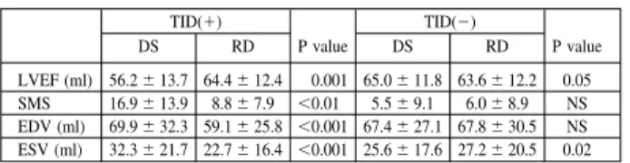
Methods and Results: 22 patients (55⫾9 y/o), 18 males, with first MI and significant obstruction only in the infarct responsibly artery (IRA) were studied with DE-CR with Gd-DTPA and 99mTc-sestamibi SPECT PMI at rest (40⫾5 days post-MI). Both images modalities were analyzed in a 17 segment model, considering for DE-CR subendocar-dial infarction when ⬎50% transmural extent of the left ventricular wall was compromised and a score⫽1 for 99mTc-sestamibi SPECT PMI at rest, in a 5-points, semi-quantitative nominal scoring system representing perfusion in each segment (0⫽normal, 1⫽mildly, 2⫽moderate, 3⫽severe reduction in counts and 4⫽absent uptake). The localization of the MI related with the IRA, 13: inferior (59%) and 9: anterior (41%). 99mTc-sestamibi SPECT PMI at rest found 121 hypoperfused segments and 18(15%) met the score⫽1. DE-CR found 112 delayed hyperenhancement segments and 19 (17%) had⬎50% transmural extent of the left ventricular wall. The sensitivity and specificity of 99mTc-sestamibi SPECT PMI to detect subendocardial infarction was 97% and 64% respectively.
Interpretation: SPECT PMI at rest using a semi-quantitative nominal scoring system may be useful to recognize segments with subendocardial infarction.
10.2
Diagnostic value of transthoracic ultrasound for assessment of distal graft anastamosis patency.
O. Kardash1, N. Natalia Maroz-Vadalajskaya.2 1Scientific-Practical Centre Cardiology, Cardiovascular Surgery, Minsk, Belarus,2Scientific-Practical Center Cardiology, Heart Surgery Laboratory, Minsk, Belarus
The finding of simple tool for visualisation of bypass graft and coronary artery blood flow is in patients after coronary surgery. However there have been problems of assessment of internal thoracic artery (ITA) graft with systolic/diastolic ratio (s/dR)⬎ 1 and of examination of vein graft to coronary artery another than the left anterior descending artery (LAD).
The aim of this study was evaluation of LAD blood flow if ITA graft s/dR was⬎1 and possibility of visualization of vein graft to major diagonal branch (DB) of the left main artery with combination of vascular and cardiac echo.
Methods. We compared ultrasound and angiographic images of 14 ITA grafts and 20
saphenous vein grafts to DB. Ultrasound examination of blood flow in bypass grafts and recipient coronary arteries was performed with combined two-dimensional and Doppler echocardiography (Sonos 5500, probe 2-4MHz).
Results. The ITA graft patterns before distal anasthamosis and in site of distal
anasthamosis were biphasic with higher systolic velocity and were characterized with high systolic/diastolic ratio 1.4⫾0.2. The flow characteristics within the LAD distal to anasthamosis were diastolic with either normal (45.3⫾ 6.5cm/s) or diminished (22.2⫾5.2cm/s) peak velocity. By comparing ultrasound images with angiographic data we concluded that ITA graft flow with s/dR⬎1 and diminished velocity in the distal to anasthamosis portion of LAD was relative to poor coronary bed run-off.
On the other hand the blood flow of the saphenous vein grafts to DB was detected in 8 patients, whereas the blood flow of the recipients DB was obtained in 18 persons. If saphenous vein grafts to DB was patent we registrated diastolic blood flow in the DB with diastolic peak velocity 60.5⫾10.1 cm/s.
Conclusion. Assessment of distal anasthamosis and coronary artery distal to
anast-hamosis may clarify the ITA graft function with systolic/diastolic ratio⬎ 1. Modern ultrasound technology let to evaluate vein graft to major diagonal branch of the left main artery.
Improving the image quality of pinhole gated SPECT by modeling the finite dimensions of the pinhole opening during reconstruction.
C. Christian Vanhove, T. Lahoutte, M. Defrise, A. Andreyev, A. Bossuyt, PR. Franken. AZVUB, Nuclear Medicine, Jette, Belgium
Background: Pinhole collimation is typically used to improve spatial resolution
required for small animal SPECT imaging. During the reconstruction process it is usually assumed that the pinhole opening is infinitesimally small. This assumption is acceptable for very small pinhole openings but may introduce artifacts when using larger pinhole openings (3mm) required to increase the sensitivity necessary for cardiac gated SPECT.
Purpose: To investigate the potential of resolution recovery (RR) in pinhole gated myocardial perfusion SPECT by modeling the finite dimensions of the pinhole opening during reconstruction.
Methods. A dynamic mathematical cardiac-torso phantom was designed to simulate
circular orbit pinhole gated myocardial perfusion SPECT projections of a rat heart. The phantom included the finite dimension of the pinhole aperture (3 mm), camera blurring and Poisson noise. The projection data were reconstructed using OSEM (5 iterations; 16 subsets) incorporating the pinhole geometry and taking into account the finite dimension of the pinhole opening, for which we have implemented a forward projector, where each projection pixel is calculated as a weighted average of seven rays. The seven rays intersect the circular opening of the pinhole in a hexagonal pattern with one of the points at the center. The systematic error (SysErr) and statistical error (StatErr) were calculated on the reconstructed data. The StatErr was determined using five noise generations with a global count density of 6.75 million counts.
Results: Results are summarized in Table 1. The SysErr decreased and the StatErr
increased when RR was applied during reconstruction. The StatErr could be decreased by reducing noise amplification during the reconstruction process using a median root prior (MRP) and/or using a temporal Fourier filtering (TFF) during reconstruction.
Conclusion: Modeling the pinhole opening during reconstruction decreases the SysErr
but increases the StatErr. Consequently, additional spatial and/or temporal filtering is required to improve image quality.
Table 1 No RR-No MRP-No TFF RR-No MRP-No TFF RR⫹ MRP-No TFF RR⫹ TFF-No MRP RR⫹ MRP ⫹ TFF SysErr (%) 23.6 10.6 13.8 10.6 13.9 StatErr (%) 25.3 65.1 6.7 33.9 4.1
Systematic and statistical errors
T
U
E
S
D
A
Y
M
A
Y
10
T
U
E
S
D
A
Y
M
A
Y
10
Journal of Nuclear Cardiology
10.4
Left ventricular remodeling after coronary artery ligature demonstrated by serial pinhole gated SPECT in rats.
P.R. Philippe Franken1, T. Lahoutte1, F. Maskali2, C. Vanhove1, J. Nloga3, N. Tran3, P.-Y. Marie.2 1Academic Hospital VUB, Nuclear Medicine, Brussels, Belgium, 2CHU-Nancy, Nuclear Medicine, Vandoeuvre, France,3CHU-Nancy, Laboratory of Experimental Surgery, Vandoeuvre, France
Background. We have previously shown that accurate and reproducible measurements
of myocardial perfusion and function can be obtained in rats using pinhole gated SPECT.
Aim. To develop a rat model of left ventricular remodeling following the ligature of the
left anterior descending coronary artery (LAD).
Methods. Male Wistar rats (n⫽ 18, body weight 360-540g) were studied 7 days, 1 month and 2 months after the ligature of the mid portion of the LAD. Imaging was performed under pentobarbital anesthesia (60 mg/kg intraperitoneal) 1 hour after intravenous administration of Tc99m sestamibi (500 MBq). ECG gated SPECT (64 projections, 20 sec per step, 360-degree rotation and 16 time bins) was acquired on a single head gamma camera equipped with a pinhole collimator (3mm opening, focal length 171 mm, radius of rotation 44mm). Data were reconstructed using PH-OSEM 4D, an iterative reconstruction algorithm adapted for pinhole geometry and incorporat-ing a temporal filter based on Fourier series (Univ. of Brussels). Myocardial perfusion polar maps were generated from the reconstructed short axis slices and used to quantify the extent of perfusion defects defined as the area below 2 SD of a normal database obtained in 12 age matched rats. Left ventricular (LV) volumes and ejection fraction (EF) were calculated using the 4DMSPECT program (Univ. of Michigan).
Results. Perfusion defects were detected in all animals at 7 days after the ligature.
Defect size ranged from 3% to 55% of the LV surface. Abnormal LVEF and volumes were only observed in animals with perfusion defects⬎ 35% of LV surface. Two animals with the largest defects died during follow-up. Perfusion defect sizes decreased slightly between 7 days and 1 month but were unchanged thereafter. Volumes increased and EF decreased progressively at 1 month and 2 months after the ligature in rats with perfusion defects⬎ 35% of LV area. No significant changes in LV function were observed in animals with perfusion defects⬎35%.
Conclusion. The ligature of the LAD results in variable perfusion defects size in rats.
Changes in LV volumes and EF indicating progressive remodeling were observed only when perfusion defects exceed 35% of the LV surface. Pinhole gated SPECT can be useful to select animals with similar infarct size and to monitor both myocardial perfusion and LV function over time in the same animal.
10.5
Ischemia in women with angina and normal coronary angiograms.
A. Amalia Peix, D. Garcia-Barreto, F. Ponce, L.O. Cabrera, J. Valiente, F. Tornes, I. Guerrero, F. Heres, E.J. Garcia, B. Cabale. Institute of Cardiology, Nuclear Medicine, La Habana, Cuba
Women without angiographic coronary heart disease but with persistent angina constitute a challenge for diagnosis and treatment. Noninvasive tests that suggest myocardial ischemia have limited utility due to anatomical differences and technical artifacts specific to women.
Aim: To assess the presence of coronary microvascular disease in postmenopausal
women. Methods: Twenty-five patients (mean age: 55⫾6 years) with typical angina and normal coronary angiograms were included. All underwent technetium-99m methoxy-isobutyl-isonitrile myocardial scintigraphy (protocol stress-rest); endothelial function measured by ultrasonography at brachial artery; 24 hours ambulatory electrocardio-graphic recording (Holter) and lipidogram.
Results: The mean body mass index was 28⫾4 (overweight) and the mean waist/hip
index was 0.83⫾0.05. Regarding coronary risk factors, there was only one smoker (4%), but 12% were ex-smokers, 28% had diabetes, 40% dyslipidemia and 76% hypertension. Sixty percent of patients experienced stress and rest angina, 20% predominantly at stress and the other 20% at rest. Anemia was constated in only one patient. In eight cases, perfusion defects were detected in the scintigraphy, all in anteroapical and septal territories, which in half of cases coincided with the presence of endothelial dysfunction, but not with ischemia during the Holter recording. Endothelial dysfunction was founded in 40% of patients and ambulatory ischemia by Holter in 12%. Thirty-six percent had supraventricular arrythmias and 8% intermitent left bundle branch block.
Conclusion: In women with typical angina and normal coronary angiograms, the
presence of ischemia due to coronary microvascular disease, with alterations of myocardial perfusion and endothelial dysfunction, should be taken into account as cause of their symptoms.
10.6
Cell proliferation and differentiation of 111In-labelled CD34- human endothelial progenitor cells.
L. Lene Bindslev1, KB. Kirsten Bisgaard2, MHS. Mandana Haack-Soerensen1, SM. Steen Mortensen1, LK. Linda Kragh2, AK. Andreas Kjaer2, JK. Jens Kastrup3, BH. Birger Hesse.2 1Herlev University Hospital, Hematology Laboratory 54P4, Herlev, Denmark,2Rigshospitalet, Univ. of Copenhagen, Clinical Physiology & Nuclear Medicine, Copenhagen, Denmark,3The Heart Centre, Rigshospitalet, Cardiac Cath-eterisation Laboratory, Copenhagen, Denmark
Aim: Endothelial progenitor cells (EPCs) from bone marrow is a promising therapy for
neovascularization in patients with severe ischaemic heart disease, but data concerning the fate of the transplanted cells remain poor. Non-invasive monitoring of In-111 labelled EPCs would be a valuable tool for evaluation of the success of monitoring homing and myocardial retention of the cells within the first few days after intramyo-cardial transplantation.
Methods: EPCs in culture (CD45-/CD34- adherent cells) were labeled directly in the
culture flask with 10 MBq In-111 tropolone for 15 min in 5 ml medium (3x10⁵ cells/ flask). The cells were then washed with PBS and new medium added to the flask. 10-20% labeling efficiency was measured. Membrane markers (FACS) and gene expression (Taqman real-time PCR) were studied following 10 days of growth in cell culture. For measuring proliferation the cells were labeled with the membrane marker PKH26-GL and grown in 24 well trays. The cells were labeled with In-111 tropolone 24 hours after the PKH labeling. PKH was measured by FACS every 24 hours on cells from 4 wells and a doubling time was calculated. Cell survival was also measured in cells grown in 24 well trays. Every 24 hours after indium labeling live cells from 3 wells were counted in a microscope using trypan-blue as an indicator of vitality.
Results: No change in common progenitor and endothelial markers (the phenotype) by
FACS (CD10, CD13, CD31, CD34, CD45, CD73, CD90, CD105, CD106, CD166, VEGFR-2) was observed. However, cell survival was reduced by 30% after 3 days, compared to control cells. This difference increased during the next days: After 7 days 85% more control cells had survived compared to labeled cells. Preliminary results also indicate that the labeled cells do not proliferate as fast as unlabeled cells. The doubling time for unlabeled cells is app. 2-3days compared to 5-6 days for labeled cells.
Conclusions: Cell viability was reduced after In-111 labeling with the present
technique. This harmful effect may be less with lower activity amount used for In-labeling. The unchanged phenotype indicates that In-labeling does not affect cell function. The differentiation potential of these cells will be measured after VEGF stimulation and with angiogenesis assays, which may give more information about their ability to differentiate into endothelial cells after labeling.
10.7
Myocardial perfusion SPECT in hypertensive patients with microvascular angina; predictors of false positive perfusion defect and the role of coronary flow reserve.
F. Fatma Aboul-Enein1, A. Battah2, A. Abdel Fattah2, A. Abdel Atty1, A. Allam.3 1Alexandria University, Cardiology, Alexandria, Egypt,2Cairo University, Critical Care Medicine, Cairo, Egypt,3Alazhar University, Cardiology, Cairo, Egypt
Background: We sought to evaluate patients (pts) with hypertension (HT) presenting
with angina (AG) and a positive stress/rest Tl-201 myocardial perfusion SPECT (MPS) in the setting of normal coronary angiogram (NCA). These patients presenting with false positive defects are a source of clinical dilemma.
Patients: our study encompassed 43 pts (age 51⫾8.8, 64% females) with mild to moderate HT, AG and NCA who underwent stress/rest MPS within 1 month of NCA, and a control group of 20 subjects.
Methods: Images were scored on a 5 point 20-segment scale (0⫽normal, 4⫽no
uptake), MPS was considered positive when SSS was⬎3. Echo-Doppler study was done within 1 week of MPS for LV mass index (LVMI), diastolic function: E/A and isovolumic relaxation time (IVR). In a sub group of 11 pts and 10 controls Dipyrid-amole (D) trans-esophageal echo (TEE) was done within a week of MPS to evaluate coronary flow reserve (CFR) at rest (R) and after D injection (0.56mg/kg). Peak systolic and diastolic velocities and systolic, diastolic and total velocity integrals were estimated at rest (R) and after (D) injection; these were expressed as ratios of D/R (CFR).
Results: MPS was positive in 25 (58%). Discriminate function coefficient analysis
showed the predictors of abnormal MPS were as follows in descending order; E/A, duration of HT, DBP, duration of AG, LVMI, IVR, SBP. SSS correlated with LVMI R⫽0.79, P⬍0.001. In the subgroup that underwent D-TEE, 7 pts had limited CFR ⬎1.85 vs 2.8 in the controls, p⫽.0.001. However, none had regional wall motion abnormalities.
Conclusion: False positive perfusion defects may frequently be encountered in HT pats
with AG. Diastolic dysfunction and decreased CFR are usually present in these pats and may explain the underlying pathophysiology of these defects.
T
U
E
S
D
A
Y
M
A
Y
10
T
U
E
S
D
A
Y
M
A
Y
10
10.8
Assessment of 99mTcN-NOET and thallium-201 myocardial uptake in experimen-tal models of chronic non-reperfused and reperfused myocardial infarction.
L. Laurent Riou1, A. Broisat1, C. Ghezzi1, C. Lartizien2, C. Berthonne`che3, MC. Toufektsian3, S. Maitrejean4, M. Janier2, G. Vanzetto1, D. Fagret.1 1Radiopharmaceutiques Biocliniques,
INSERM 340, La Tronche, France,2CERMEP, Plateforme ANIMAGE, Lyon, France,3Laboratoire
NVMC, Universite´ de Grenoble, Grenoble, France,4Socie´te´ Biospace, Paris, France
Background: TcN-NOET (NOET) is a pure myocardial blood flow tracer. As flow and
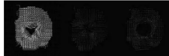
viability are correlated in chronic non-reperfused and reperfused myocardial infarction, we hypothesized that the uptake of NOET would also reflect myocardial viability and would be similar to the uptake of the flow/viability tracer thallium-201 (Tl201) in this setting. Methods and Results: Rats were subjected to permanent coronary occlusion (OCC, n⫽8) or to a 45 min occlusion and reperfusion (REP, n⫽11). Seven days later, the tracers were co-injected and the animals were euthanized 15 min afterwards. Infarct size determination and NOET and Tl201 ex vivo imaging were performed. Regional flow and tissue oedema were quantified using radioactive microspheres and Tc-DTPA, respectively. In both groups, mean NOET and Tl201 defect extents were not significantly different from infarct size. NOET and Tl201 defect magnitudes were similar in OCC animals (0.11⫾0.01 vs. 0.13⫾0.01). In REP animals, Tl201 defect magnitude (0.25⫾0.02) was significantly lower than the magnitude of NOET and flow defects (0.14⫾0.03 and 0.17⫾0.01, respectively, P⬍0.05) (see Figure for representative examples). Tc-DTPA indicated the presence of oedema in the reperfused area. Blood distribution studies showed that, unlike NOET, Tl201 plasma activity was mostly unbound to plasma proteins. Conclusions: NOET delineated the viable area in chronic non-reperfused and reperfused myocardial infarction. The signifi-cantly decreased Tl201 defect in reperfused infarction was likely due to partial diffusion of the tracer from the plasma into the oedema present in the infarcted area. NOET might represent a valuable diagnostic tool for the clinical assessment of microvascular obstruction following reperfusion of acute myocardial infarction.
Figure (defect magnitude in parenthesis)
10.9
Arrhythmogenic right ventricular cardiomyopathy in patients with right ventric-ular outflow tract premature contractions.
F. Francois Rouzet1, D. Daou2, R. Lebtahi3, R. Frank4, A. Leenhardt5, M. Slama6, D. Le Guludec.3 1Paris, France,2Hopital Lariboisie`re, Me´decine Nucle´aire, Paris, France, 3Hopital Bichat, Medecine Nucleaire, Paris, France,4Hopital Pitie´-Salpeˆtrie`re, Institut de Cardiologie, Paris, France,5Hopital Lariboisie`re, Cardiologie, Paris, France,6 Ho-pital Antoine Be´cle`re, Cardiologie, Clamart, France
Background. Vertical axis premature ventricular contractions (PVCs) originating from
the right ventriclar outflow tract (RVOT), in the absence of overt heart disease, are generally considered as benign. Nevertheless, they may also represent the first manifestation of arrhythmogenic right ventricular cardiomyopathy (ARVC) which is a cause of sudden death. The high diagnostic and prognostic value of equilibrium radionuclide angiocardiography (ERNA) with multiharmonic Fourier analysis has been previously validated in patients with right ventricular tachycardia. The aim of this study was to investigate the prevalence of ARVC in patients referred for frequent and complex RVOT-PVCs, without ventricular tachycardia and without known heart disease, using ERNA with Fourier analysis.
Methods. The study population included 276 consecutive patients (age: 38⫾15 years,
males: 52%) referred as part of an investigation on frequent (more than 1000/24 hours) vertical axis PVCs (electrical axis between⫹60° and ⫹ 120°) without suspected or known heart disease and without documented ventricular tachycardia. Prior to ERNA, the diagnosis criteria of the Task Force on ARVC were quoted (1 minor criteria: 110 patients (40%), 2 minor: 129 (47%), 3 minor: 31 (11%), 4 minor and more: 6 (2%)). ERNA was used to detect global and/or regional dysfunction of both ventricles.
Results. The criteria of ARVC were fulfilled in 46 patients (17%), among whom the RV
involvement was segmental in 44 (1 segment: 22 patients, 2 segments: 17, 3 segments: 5), and diffuse in 2. In 3 additional patients, the diagnosis of ARVC was not achieved despite demonstrable segmental dyskinesia of the RV (1 segment in 2 and 2 segments in 1). Left ventricular segmental dyskinesia occured in association with typical RV involvement in 5 patients (18%).
Conclusions. In a specific population with frequent and complex RVOT-VPCs, the
diagnosis of ARVC was achieved in 17% of patients (according to Task Force criteria), including diffuse abnormalities in some of them. Consequently, ERNA with multihar-monic analysis is useful to screen noninvasively patients with RVOT-PVCs in order to identify which may require a specific management.
10.10
Biodistribution and myocardial uptake after ischemia-reperfusion of 99mTc-AnxD1, a new marker of cell apoptosis: a preliminary study.
L. Laure Sarda-Mantel1, JB. Michel2, F. Rouzet1, G. Martet3, A. Meulemans1, JM. Vrigneaud1, D. Le Guludec.1 1Bichat Hospital, Nuclear Medicine, Paris, France,2UFR Bichat, INSERM U460, Paris, France,3UFR Bichat, EA 3512, Paris, France
Objectives: 99mTc-annexinV binds selectively to phosphatidylserins (PS), and is used
for non invasive measurement of cell apoptosis, especially myocardial apoptosis induced by ischemia -reperfusion (IR). 99mTc-AnxD1 is a 4 to 5 fold smaller peptide derived from the annexinV’s domain 1, with high affinity and specificity for PS. We evaluated the tissue biodistribution of this new tracer in rats, and its myocardial uptake after experimental IR.
Methods: Sixteen normal male Wistar rats were injected with AnxD1 (patented by the
CEA, 7.4 MBq /100g). The labelling efficiency was always superior to 97%. Blood kinetic studies (n⫽10), and biodistribution studies in organs (n⫽6) 4h post injection were performed: after killing, aliquots of tissues were taken, weighed and placed in tubes for scintillation well counting. Myocardial IR lesions were induced in 6 anaesthetized Wistar rats by tying during 20 min then opening the left coronary artery. Two hours after reperfusion, AnxD1 was injected, then planar thoracic images were obtained using a pinhole collimator on a gamma-camera (SOPHA medical), 2h post injection. After killing, quantitative autoradiography of myocardial slices (Instant Imager, Packard) was performed. The activity ratios between the lesion area and the remote myocardium (AR) were calculated on the autoradiograms.
Results: The blood kinetic of AnxD1 fitted a two component curve with a fast
decreasing phase then a plateau reached at 80 min. The t1/2 was calculated at 5.8 min, the residual blood activity at 80 min was 5.7%. Biodistribution studies showed high AnxD1 uptake in the kidneys (23.6⫾5.5%ID) and the liver (21.5⫾5.5%ID), 0.87⫾0.06%ID in the spleen, 0.25⫾0.07%ID in the lungs, and only 0.03⫾0.02%ID in the heart myocardium. The scintiscans showed faint AnxD1 myocardial uptake in 1 case, were doubtful in 1 case and negative 4 cases. On autoradiograms, significant AnxD1 uptake was observed in all myocardial lesions, with mean AR at 3.85⫾0.83.
Conclusions: In rats, AnxD1 is rapidly cleared from blood, and non specific AnxD1
uptakes in the heart and the lungs are low. However it has poor sensitivity to detect IR induced myocardial lesions on scintigraphic images, because of low (despite significant) uptake in lesions.
10.11
Validation of an automatic SPECT radionuclide angiography processing software program for the measurement of right ventricular function against echocardiography.
D. Doumit Daou1, C. Coaguila1, A. Benada1, M. Razzouk1, I. Idy-Peretti1, D. Le Guludec.2 1Lariboisiere Hospital, AP-HP, Nuclear Medicine Dept, Paris, France, 2Bichat Hospital, AP-HP, Nuclear Medicine Dept, Paris, France
Aim: Recently, completely automatic software (Quantitative blood pool SPECT
software developed by Cedars Sinai, QBS) was developed to process SPECT radionu-clide angiography (RNA) acquisitions. It allows the automatic calculation of both left ventricular (LV) and right ventricular (RV) ejection fraction (EF) and end diastolic and end systolic volumes (EDV, ESV). However, its value for RV function evaluation is still debated. We aimed to evaluate the performance of QBS for the calculation of RVEF (%) and RV volumes (ml) as compared to the RV fractional area shortening (FAS, %) and RV end diastolic and end systolic area (cm2, EDA and ESA) echocardiography (gold-standard). In parallel, we compared its performance to the semi-automatic manual segmentation software used in our laboratory (based on maximal activity threshold method MAT-35%).
Methods: Forty-two patients with post-embolic pulmonary hypertension were studied:
18 patients before and 24 patients after pulmonary thrombo-endarterectomy. All patients had SPECT RNA and echocardiography within a 2-days period. SPECT RNA acquisitions were processed with QBS and MAT-35%: RV EDV, ESV, and EF were noted. For echocardiography, RV EDA, ESA, and FAS were noted. For the analysis, EDV and ESV measurements were combined.
Results: RV-FSA, RVEF-QBS, and RVEF-35% were respectively 28⫾7 %, 53⫾14 %, and 48⫾17 % (p⬍0.0001 between the 3 groups). Both RVEF-QBS and RVEF-35% were well correlated to RV-FSA: RV-FAS⫽11.28⫹0.31*RVEF-QBS with an r⫽0.64, ser⫽5.2, and p⬍0.0001 and RV-FAS⫽15.18⫹0.25*RVEF-35% with an r⫽0.65, ser⫽5.1, and p⬍0.0001.
RV-A, RVV-QBS, and RVV-35% were respectively 24⫾7 cm2, 104⫾49 ml, and 162⫾73 ml (p⬍0.0001 between the 3 groups). Both RVV-QBS and RVV-35% were well correlated to RV-A: RV-A⫽12.13⫹0.12*RVV-QBS with an r⫽0.78, ser⫽4.7, and p⬍0.0001 and RV-A⫽11.17⫹0.08*RVV-35% with an r⫽0.80, ser⫽4.5, and p⬍0.0001.
Conclusion: As compared to the manual segmentation semi-automatic MAT-35%
method, the completely automatic QBS software provides RVEF and RV volumes that are similarly well correlated to echocardiographic RV functional parameters.
T
U
E
S
D
A
Y
M
A
Y
10
T
U
E
S
D
A
Y
M
A
Y
10
10.12
Relative impact of left ventricular (LV) dyssynchonism (D), right ventricular (RV) D, and interventricular D for the determination of LV function.
D. Doumit Daou1, C. Coaguila1, A. Benada1, A. Leenhardt.2 1Lariboisiere Hospital, AP-HP, Nuclear Medicine Dept, Paris, France,2Lariboisiere Hospital, AP-HP, Cardi-ology Dept, Paris, France
Aim: Recently, a lot of debate has been reported as to the interaction between intra and
inter ventricular (V) LV and RV mechanical D and LV function. Actually, these latter are being used as the basis for biV pacing in the treatment armamentarium of heart failure, particularly the D between LV lateral and septal walls. However, the relative importance of each of these parameters is still unclear. Planar radionuclide angiography (RNA) using Fourier phase analysis (FPA) has been used for the characterization of intra-V and inter-V LV and RV mechanical D. We aimed to evaluate in patients with ischemic hear disease the contribution of the different quantitative parameters reflecting LV and RV intra-V and inter-V D using planar-RNA FPA.
Methods: Our study included 103 consecutive patients with ischemic heart disease
addressed for LV function evaluation with planar-RNA. Mean age was 61⫾15 years, 83% were men, 60% had previous myocardial infarction (MI) with 29% having a previous anterior MI, 35% an inferior MI, and 5% a lateral MI. Planar-RNA (best septal) were processed for FPA with XT-RNA software program (Vision, GEMS, France). Global LV and RV FPA were calculated as the first harmonic mean⫾SD (normalized to heart rate and expressed in degrees) of all corresponding LV and RV pixels: LV-SD was considered to reflect the heterogeneity in global LV contraction, (intra-V global LV mechanical D). Inter-V D was calculated as the difference between mean LV and mean RV FPA. Similarly, two different ROIs were manually drawn on the antero-lateral (AL) and antero-septal (AS) regions of the LV. And corresponding mean⫾SD FPA were calculated. AL-AS D was calculated as the difference between mean AL and AS FPA.
Results: Mean LVEF, LV-SD, and AL-AS D were respectively 40⫾18%, 7⫾17 degrees,
and⫺4⫾27 degrees. LVEF was highly correlated to LV-SD: LVEF⫽71*exp(⫺0.025*LV-SD) , r⫽0.84, ser⫽7.7, P ⬍0.0001. LVAF was mildly and linearly correlated to inter-V D (r⫽0.34, P ⬍0.0001). And there was a trend for a linear correlation between LVEF and AL-AS D (r⫽0.18 , P⫽0.074). On stepwise regression analysis, the only correlated factor was LV-SD (F⫽228, r⫽0.832, ser⫽10, P ⬍0.0001) while the inter-V D (F⫽0.001) and AL-AS D (F⫽1.553) were no longer significant factors.
Conclusion: In patients with ischemic heart disease, there is a high correlation between
LV function and global LV D (reflected by LV-SD) which appears to be the major influencing factor. The influence of interV D and AL-AS D seem much less important. This should be considered in the work-up of patients considered for BiV pacing.
10.13
Impact of patient movement on gated myocardial perfusion SPECT (GSPECT) and radionuclide angiography SPECT (RNA SPECT) for the quantification of left ventricular (LV) function.
D. Doumit Daou1, C. Coaguila1, D. Vilain2, A. Benada1, R. Lebtahi3, D. Le Guludec.3 1Lariboisiere Hospital, AP-HP, Nuclear Medicine Dept, Paris, France,2Foch Hospital, Nuclear Medicine Dept, Suresnes, France,3Bichat Hospital, AP-HP, Nuclear Medicine Dept, Paris, France
Aim: Both GSPECT and SPECT RNA provide reliable estimation of LV Ejection
Fraction (EF). A previous study has demonstrated the better robustness to patient movement of planar RNA as compared to GSPECT for the measurement of LVEF. What remains unclear is whether this better robustness is due to the difference in acquisition methods (planar versus SPECT) or whether it is due to different types of exams (RNA versus myocardial perfusion). We aimed to compare the robustness to patient movement of GSPECT and SPECT RNA for the measurement of LVEF.
Methods: The population included 20 patients with CAD having both rest Tl-201 GSPECT
and SPECT RNA. All acquisitions were done on a DST-XL two-head gamma camera (GEMS, France) with 90 seconds/projection, 32 projections/180°. Patient movement was simulated by applying 1-pixel, 2-pixels, 3-pixels, 4-pixels, and 5-pixels deviation in the z axis (caudal to cranial axis) on each acquired study from the 9th projection to the 16th projection and from the 25th projection to the 32 projection. Eighty GSPECT studies and 80 SPECT RNA studies were generated: 0-pixel, 1-pixel, 2-pixels, 3-pixels, 4-pixels, and 5-pixels acquisitions. GSPECT was processed with the QGS software and SPECT RNA with the QBS software and corresponding LVEF were noted.
Results: For the 4-pixels and 5-pixels GSPECT studies, the LV myocardial wall presented
double contour walls and it was impossible to generate acceptable LV contours and therefore calculate LV volumes with QGS. At 0-pixel, 1-pixel, 2-pixels, and 3-pixels QGS LVEF were respectively 36⫾12, 35⫾11, 32⫾10 °, and 27⫾9 * % (° : p⬍0.02 versus the other groups; * : p⬍0.0002 versus the other groups). For SPECT RNA, the aspect of the LV cavity was still acceptable for the 4-pixels and 5-pixels reconstructed studies. For QBS, the 0-pixel, 1-pixel, 2-pixels, 3-pixels, 4-pixels, and 5-pixels were respectively 47⫾17, 46⫾16, 44⫾14, 44⫾17, 43⫾16, and 45⫾19 for EF (NS).
Conclusion: Despite similar gated SPECT acquisition techniques, SPECT RNA seems
to be less sensitive to patient movement than GSPECT for the quantification of LVEF.
10.14
Repeatability of two different ECG gated blood pool SPECT processing software for the quantification of left ventricular function.
D. Doumit Daou1, C. Coaguila1, A. Benada1, M. Razzouk1, I. Idy-Peretti1, D. Le Guludec.2 1Lariboisiere Hospital, AP-HP, Nuclear Medicine Dept, Paris, France, 2Bichat Hospital, AP-HP, Nuclear Medicine Dept, Paris, France
Aim: Despite some limitations inherent to its 2-dimensional nature, planar equilibrium
radionuclide angiography (RNA) remains the clinical gold-standard for the measure-ment of left ventricular (LV) ejection fraction (EF). The 3-dimensional ECG-gated blood pool SPECT (GBPS) has been proposed to surpass the planar RNA limitations. However, its use has been limited by the unavailability of an automatic processing software. We previously validated the use of a manual maximal activity threshold (35%) method for the processing of GBPS (GBPS-35%) for LV function including its limits of repeatability. Recently, an interesting automatic processing software has been developed (QBS, Cedars-Sinai). We previously defined its value for LV function. We aimed to define its limits of repeatability for LVEF and volume (V) measurement as compared to planar equilibrium radionuclide angiography (RNA) and GBPS-35%.
Methods: Ten patients with CAD had RNA studies acquired as follows. First, planar
left anterior oblique (LAO) then GBPS RNA acquisitions were realized by one technician. Then, the patients were allowed to rest in the waiting room for at least 15 min and a second set of acquisitions (LAO and GBPS) were realized by another technician. GBPS acquisitions were processed with QBS and GBPS-35%. All 4 acquired studies were processed by 2 different observers.
Results: For LVEF the intraobs, interobs, and interstudy reproducibility (absolute
paired difference) of QBS, GBPS-35%, and LAO were respectively 3.1⫾5.1%, ⫺1⫾4%, and 2.1⫾7.1% versus ⫺0.6⫾1.9%, ⫺0.8⫾3.1%, and ⫺0.2⫾4.5% versus 0⫾0.8%, ⫺0.7⫾1.1%, and ⫺0.8⫾3.8%.
For LV end diastolic V the intraobs, interobs, and interstudy reproducibility (% variation) of QBS versus GBPS-35% were respectively (0⫾17%), (1⫾16%), and (⫺1⫾21%) versus (⫺1⫾1%), (⫺2⫾5%), and (1⫾5%).
For LV end systolic V these were respectively⫺8⫾15%, 3⫾17%, and ⫺6⫾25% versus 0⫾5%, 0⫾10%, and 2⫾13%.
Conclusion: The limits of repeatability of LV function are considerably wider with
QBS as compared to LAO and GBPS-35%. These limits should be considered when analyzing the temporal evolution of LV function.
10.15
Radionuclide angiography SPECT (RNA SPECT): performance of two different methods for the calculation of volumes.
D. Doumit Daou1, C. Coaguila1, A. Benada1, D. Le Guludec.2 1Lariboisiere Hospital, AP-HP, Nuclear Medicine Dept, Paris, France,2Bichat Hospital, AP-HP, Nuclear Medicine Dept, Paris, France
Aim: Cardiac chamber volume (V) measurement has an additive clinical value to LVEF. As
compared to planar RNA, ECG-gated SPECT RNA presents the advantage of easy clinical applicability for V calculation. We have previously compared in phantom cardiac studies, 5 methods for the calculation of V with Tc-99m SPECT and we concluded that the 35% maximal activity threshold method was the most accurate method (MAT) (SNM 2000). Recently, a completely automatic software for SPECT RNA processing (QBS, Cedars-Sinai Center) based on gradient method was commercialized. We aimed to compare its performance to the MAT method for V measurement in RNA phantom studies.
Methods: Different cardiac phantom V were used (25ⱖVⱖ500ml), each with (⫹) and
without (⫺) background (BKG) activity (A) and with different acquisition times (4 s and 8 s/step). SPECT acquisitions were realized on a DST-XL dual-head gamma camera (GEMS) with 32 steps/180°. Gated SPECT RNA studies were created and analyzed with the QBS software and the MAT software. Statistical analysis included linear regression analysis and % error ((calculated V-phantom V)*100 / phantom V ).
Results: For the 4s BKG (⫺) studies, the % error, correlation coefficient, and ser were
respectively⫺1⫾8, 0.9975, and 11.25 for QBS and ⫺3⫾9, 0.9975, and 11.46 for MAT. For the 8s BKG (⫺) studies, the % error, correlation coefficient, and ser were respectively 2⫾10, 0.9975, and 11.32 for QBS and ⫺1⫾8, 0.9946, and 16.74 for MAT. For the 4s BKG (⫹) studies, the % error, correlation coefficient, and ser were respectively⫺2⫾14, 0.9944, and 16.93 for QBS and ⫺4⫾9, 0.9903, and 22.27 for MAT. For the 8s BKG (⫺) studies, the % error, correlation coefficient, and ser were respectively 4⫾13*, 0.9951, and 15.82 for QBS and 2⫾7*, 0.9863, and 26.46 for MAT.
Conclusion: Both QBS and MAT provided volumes highly correlated to phantom
values over the wide range of evaluated volumes. This was verified for short and long acquisitions time (4s/step and 8s/step) and with or without BKG activity. In contrast to MAT, the performance of QBS seems to be influenced (lower) by the presence of BKG activity. This may explain in part the better reported performance of MAT as compared to QBS in patient studies.
T
U
E
S
D
A
Y
M
A
Y
10
T
U
E
S
D
A
Y
M
A
Y
10
10.16
Monitoring left-ventricular dilation in mice by high-resolution small-animal-PET: vali-dation study using MRI.
L. Lars Stegger1, K. Scha¨fers1, U. Flo¨gel2, J. Schrader2, O. Schober1, B. Levkau3, M. Scha¨fers.1 1University Hospital Mu¨nster, Nuclear Medicine, Mu¨nster, Germany,2
Hei-nrich-Heine-University Du¨sseldorf, Department of Cardiovasc. Physiology, Du¨sseldorf, Germany,3
University Hospital Essen, Institute of Pathophysiology, Essen, Germany Background: Molecular imaging by small animal positron emission tomography (PET) is an important non-invasive means to phenotype transgenic mouse models in vivo. When investigating pathologies of the left ventricular (LV) myocardium (e.g. cardiomyopathies) the serial assessment of LV volumes is important. By this, the presence of left ventricular dilation as a sign of developing heart failure can be detected. Where PET imaging is usually used to derive biochemical/molecular information, the functional parameters such as ventricular volumes are generally measured by using morphological and functional imaging modalities such as echocardiography or magnetic resonance imaging. Here, a method to monitor left ventricular dilatation in mice with PET and thus obviating the need for a second imaging modality is presented and evaluated using cardiac MRI as the gold standard.
Methods: The method uses a semiautomatic 3D-algorithm to delineate the left ventricular myocardial wall on
static Small-Animal-PET images depicting myocardial glucose metabolism (F-18-fluoro-2-deoxy-glucose-PET). The volume enclosed by the three-dimensional midmyocardial contour is calculated.
Data for twenty mice, ten wildtype mice and ten transgenic mice developing dilated cardiomyopathy, were obtained and compared to volumes measured by magnetic resonance imaging in the same animals.
Results: Data obtained by PET and MRI correlated well (R⫽0.89). Graphical analysis according to Bland and Altman did not reveal a dependency on volume.
Conclusion: Small animal PET imaging allows to monitor LV dilation in mice, thus obviating the need for a second imaging modality making serial measurements more efficient.
Correlation
10.17
Myocardial blood flow and coronary vascular resistance are dependent on thyroid function-a quantitative study using positron emission tomography and oxygen-15-labeled water.
P. Peter Kies1, L. Stegger1, T. Wichter2, K.P. Scha¨fers1, O. Schober1, M. Scha¨fers.1 1University Hospital Muenster, Nuclear Medicine, Muenster, Germany,2University Hospital Muenster, Internal Medicine C, Cardiology, Muenster, Germany
Introduction: Alterations of thyroid function are known to be associated with changes of the
cardiovascular system. Aim of this study was to investigate the effects of an altered thyroid function on myocardial blood flow (MBF) and coronary vascular resistance (CVR).
Methods: Twenty hypothyroid patients (4m,16f,38⫾9 years) with a history of differentiated thyroid carcinoma, eleven patients (4m,7f,48⫾15 years) during clinical or subclinical hyperthy-roidism and ten euthyroid controls (6m,4f,32⫾4 years) were enrolled in this study. Entry criteria included the absence of coronary artery disease, risk factors and cardiac medication. MBF (ml/g/min) was quantified by dynamic PET and 15O-labeled water. MBF was measured at rest, during adenosine infusion to provoke maximal hyperaemic MBF and calculate the coronary flow reserve (CFR), and during cold pressor testing (CPT) to obtain maximal perfusion stimulated by the endothelial response. In addition, CVR (mmHg/ml/g/min) in the different settings was calculated dividing mean arterial blood pressure by the respective MBF. Rate pressure product (RPP) was calculated for each group to compare hemodynamic parameters during the scans.
Results: Mean RPP at baseline were similar in the three groups excluding an effect of different
cardiac work on MBF (Hypo:7660⫾1429,Eu:8131⫾2422,Hyper:8438⫾1101; p⫽ns). Baseline MBF was significantly higher in the hyperthyroid group (Hyper:1.15⫾0.23, Hypo:0.89⫾0.22, Eu:0.94⫾0.24; p⬍0.05). Absolute MBF during adenosine infusion and CFR were similar between the three groups (3.79⫾1.18, 3.66⫾1.26, 3.73⫾1.17; p⫽ns). MBF during CPT was significantly higher in the hyperthyroid group (Hyper: 1.57⫾0.39,Hypo:1.08⫾0.22, Eu:1.18⫾0.31; p⬍0.05).
Baseline CVR was significantly lower in the hyperthyroid group (Hyper:78⫾12.2,Hypo: 105.7⫾27.6, Eu:99.9⫾20.0; p⬍0.05). CVR during adenosine infusion was not significantly different between the three groups (27.4⫾14.4, 29.6⫾18.6, 27.6⫾11.3; p⫽ns). CVR during CPT was significantly lower in the hyperthyroid group (62.1⫾15.9) than in the hypothyroid group (91.9⫾24.7, p⬍0.05). It was also lower compared to the euthyroid group (80.4⫾19.6), however this difference only reached borderline significance (p⫽0.058).
Conclusion: Our results show an increased MBF at baseline and during CPT in patients
with clinical and subclinical hyperthyroidism. Under the same conditions CVR is reduced in these patients. Adenosine infusion does not cause any significant changes, therefore our results strongly suggest an endothelium-mediated effect of clinical and subclinical hyperthyroidism on MBF and CVR.
10.18
Assessment of base-to-apex gradients of myocardial blood flow after statin therapy at different stages of coronary atherosclerosis.
J.P. Peter Wielepp1, D. Baller2, E. Pulawski2, R. Weise1, E. Fricke1, D. Horstkotte2, W. Burchert.1 1Institute of Nuclear Medicine, Heart and Diabetes Center, Bad Oeynhausen, Germany,2Clinic of Cardiology, Heart Center North Rhine-Westphalia, Bad Oeyn-hausen, Germany
Diffuse coronary atherosclerosis is the common substrate for plaque rupture and cardiovascular events. Assessment of longitudinal, base-to-apex gradients (BAG) of myocardial perfusion abnormalities is a potential noninvasive marker for the functional integrity of the coronary endothelium. The aim of our study was the examination of reversibility of BAG after 6-month statin therapy.
Methods: 31 pts (age 62.4⫾ 7.7 years; 22 males) with mild-to-moderate angina and LDL cholesterin (LDL-C) of 183⫾ 40 mg/dl at baseline were included. Coronary angiography revealed minimal disease in terms of wall irregularities or stenosis⬍ 30% in 19 pts and moderate coronary artery disease (CAD) with stenosis⬎50% in 11 pts. The reference group consists of 22 pts without detectable CAD. Absolute myocardial blood flow (MBF) was measured with ammonia PET under pharmacologic stress (adenosine). Basal mid (termed base) and mid-to-apex (apex) segments were analyzed. Minimal coronary resistance (MCR) was calculated as the ratio mean arterial pressure/ MBF-adenosine.
Results: Before therapy BAG-MCR was significantly lower in pts (0.91⫾ 0.12) as
compared to reference group (0.97⫾ 0.14; p⬍0.05). After statin therapy BAG-MCR increased from 0.91⫾ 0.12 to 0.95 ⫾ 0.12 (p⬍0.05). BAG-MBF decreased from 1.12⫾ 0.20 to 1.07 ⫾ 0.13 (p⬍0.05). Even in segments with normal MCR (⬍0.40 mmHg/(ml/min/100g) BAG-MCR increased from 0.90 ⫾ 0.10 to 0.96 ⫾ 0.11 (p⬍0.05). LDL-C decreased to 104 ⫾ 30 mg/dl at follow-up (p⬍0.001).
Conclusions: These PET results demonstrate a short-term reversibility of base-to-apex
gradients of myocardial perfusion disturbance at early or moderate coronary athero-sclerosis after 6-month statin therapy. The pathophysiologic mechanism behind prob-ably reflects healing endothelium with improved flow-dependent dilation of epicardial vessels prone to atherosclerosis and risk factors such as LDL-C.
10.19
Effect of spinal cord stimulation on myocardial blood flow in patients with therapy refractory angina pectoris.
J.P. Peter Wielepp1, S. Eckert2, A. Dongas3, D. Horstkotte2, W. Burchert.1 1Institute of Nuclear Medicine, Heart and Diabetes Center, Bad Oeynhausen, Germany,2Clinic of Cardiology, Heart and Diabetes Center, Bad Oeynhausen, Germany, 3Institute of Anesthesiology, Heart and Diabetes Center, Bad Oeynhausen, Germany
Spinal cord stimulation (SCS) has proven to be a safe procedure in patients with symptomatic angina pectoris (AP) refractory to medical therapy and without any option for PCI or CABG. This technique has repeatedly demonstrated an antianginal effect by reducing AP symptoms. The mechanism, however, remains controversial. The aim of the study is the assessment of the effect of SCS on myocardial blood flow (MBF) and minimal coronary resistance (MCR).
Methods: In 6 male patients (mean age 66 years) with multi-vessel disease which were
scheduled for NS, perfusion at rest and during the maximum hemodynamic effect of intravenous adenosine were studied by ammonia PET (Siemens ECAT EXACT HR⫹) on two different occasions: directly before SCS and 13⫾ 0.5 months after SCS. The stimulator was active in all patients at 21⫾ 5 hours before PET scan was done. MBF and MCR were calculated for the most impaired myocardial segments at baseline (initial MBF stress⬍ 190 ml/min/100g). We correlated these data with clinical CCS angina score and NYHA class.
Results: Outpatients visits and hospitalization where significantly reduced in the 12
months post SCS compared to a 12 months interval before stimulation. Corresponding to the reduction in angina, the consumption of short-acting nitrates was also signifi-cantly reduced.
There was an increased hyperemic myocardial blood flow (138.8⫾ 26.7 before SCS vs. 147.5⫾ 0.37 ml/min/100g during SCS; p⬍0.05) and decreased minimal coronary resistance (0.65⫾ 0.20 before SCS vs. 0.59 ⫾ 0.18 mmHg/(ml/min/100g) during SCS; p⬍0.05) in the initial most impaired myocardial segments.
Conclusions: SCS has been shown to improve anginal status in patients with severe
coronary artery disease. Our preliminary results suggest that the anti-anginal effect of SCS is associate with an improved regional MBF and vasodilator capacity in the most impaired myocardial segments.
T
U
E
S
D
A
Y
M
A
Y
10
T
U
E
S
D
A
Y
M
A
Y
10
10.20
Integrating cardiac PET/CT list mode acquisition in a clinical routine environ-ment: implementation and initial experiences.
S.G. Stephan Nekolla1, M.J. Martinez1, W. Howe2, F. Kehren2, S.I. Ziegler.1 1 Tech-nische Universita¨t Mu¨nchen, NuklearmediziTech-nische Klinik, Mu¨nchen, Germany,2CPS Innovations, Knoxville, TN, United States
Aim: Due to the success of PET/CT devices in oncology and the availability of high
resolution CT scanners being part of it, the application in cardiac imaging offers an attractive perspective. However, cardiac imaging protocols require more flexibility as compared to whole body oncology scans: dynamic and gated acquisition modes are typically used extensively. List mode acquisition offers these advantages but is considered to be too demanding in a clinical routine environment. Thus, we investigated whether flexible yet easy to use front-end applications could be implemented and were suitable in such a setting.
Methods: List mode acquisitions were initiated from a dedicated user interface on the
scanners console where essentially only the desired acquisition time was entered. Subsequently, the list mode data stream was acquired in parallel with the ECG and the respiratory signal. After the completion of the patient study, all data was transferred automatically to an offline workstation for further processing. There, the data was histogrammed into sinograms depending on the actual patient protocol. Essentially, the user only selected the type of the examination (e.g. ischemia [NH3 rest / NH3 stress] or viability [NH3 rest / FDG rest] ) and the appropriate sinograms were generated automatically. In the examples above, the complete list mode stream was used to generate dynamic sinograms which were used for the calculation of e.g. myocardial blood flow. In addition, the parts of the same list mode stream where the tracer distribution reached a steady state were utilized to create gated sinograms. This saved considerable acquisition time as this study is traditionally measured in a separate step. Finally, the CT based transmission was checked for patient motion against a PET test volume and corrected if necessary.
Results: The developed programs were applied in twenty patient studies. The amount
of list mode data averaged 5.3⫾3.2 Gigabytes. The time necessary to transfer the data was 13⫾8 min on a standard fast Ethernet network. Processing time including correction for photon attenuation, scatter correction and iterative reconstruction using an OSEM algorithm (8 subsets, 4 iterations) ranged between 30 and 40 min on standard PC hardware.
Conclusions: List mode data acquisition and patient protocol specific histogramming
and reconstruction were implemented with an efficient and user friendly interface allowing its use in a clinical routine setting.
10.21
Peripheral arterial endothelial response to exercise in the evaluation of patients with coronary artery disease.
I. Ioannis Vassiliadis1, G. Souretis2, C. Komporozos2, A. Fountos3, A. Papademetriou4, A. Spanos2, A. Antoniou2, P. Strembelas.2 1Athinai Naval Hospital, Cardiac Dept., Athens, Greece,2Athinai Naval Hospital, Cardiac Dept., Athens, Greece,3Euroclinic,, Nuclear Medicine Dept., Athens, Greece,4Athinai Naval Hospital, Nuclear Medicine Dept., Athens, Greece
Background: Endothelial dysfunction(ED) is a generalised abnormality of the vascular
system and its detection in the coronary vasculature is of outmost importance for the clinical relevance and prognostication of coronary artery disease (CAD). Published data suggest that many CAD patients manifest concomitant peripheral ED. However little is known of a reverse relationship.
Aim of our study is to evaluate peripheral arterial tone (PAT) response to exercise as a marker of ED , correlating it to the magnitude of Tc-99m Tetrofosmin SPECT myocardial perfusion imaging (MPI) defects.
Methods: Twenty-one consecutive pts with suspected or known CAD, by history or
angiography and a referent group of twenty-four normal volunteers with a⬍ 5% likelihood of CAD or negative ETT and/or MPI (mean age 46⫾20 yrs, 18 men 75%) were enrolled in our prospective study. All underwent treadmill exercise testing (ETT) followed by MPI. Pulsatile finger pulse volume responses were assessed using a plethesmographic device (Itamar-Medical). Exercise PAT responses were compared to clinical, hemodynamic, ECG and MPI parameters.
Results: Among normal subjects, 22 (91.7%) manifested vasodilation throughout
exercise and 2 (8.3%) manifested initial vasodilation followed by vasoconstriction at higher heart levels. None manifested vasoconstriction throughout exercise. In contrast, 6 CAD pts (28.6%) Group I, manifested vasoconstriction throughout exercise , 6 (28.6%) Group II, maintained a constant pulse wave amplitude (PWA) throughout exercise and other 9 (42.9%) Group III, sustained a vasodilating pattern of PAT throughout exercise (p⬍0.01 vs Group I,II). All Group I pts had exercise-induced reversible MPI defects, while Group II pts revealed predominantly persistent MPI defects compared to Group III pts who demonstrated a lesser degree of persistent perfusion abnormalities. Hemodynamic responses to exercise revealed progressive higher systolic and diastolic blood pressure and lower peak heart rates with diminishing PAT ratios.
Conclusions: Our data suggest that the progressive diminution in peripheral artery tone
during exercise , measured by a plethesmographic system, can differentiate CAD pts with non-ischaemic ETT from normal individuals correlating well with the magnitude of the perfusion abnormalities. It may also represent a generalised vascular abnormality governed by altered endothelial function.
10.22
Equilibrium radionuclide angiography: 24 vs 32 frames per cardiac cycle.
E. Efstratios Moralidis, G. Arsos, D. Boundas, S. Georga, N. Karatzas, K. Karakatsanis. Hippokration Hospital, Thessaloniki, Greece
Aim: This study compares the values of systolic and diastolic parameters derived from 24
and 32 frames/R-R equilibrium radionuclide angiography (ERNA).
Method: Twenty-five consecutive patients, aged 56⫾9 yrs, with coronary artery disease were enrolled. Patients were submitted to ERNA (740 MBq 99mTc-RBC)in the best septal view, in two consecutive acquisitions, using 24 and 32 frames/R-R for 8 and 11 minutes, respectively. The following parameters were calculated: R-R interval, left ventricular ejection fraction (LVEF), end-diastolic and end-systolic volume (EDV and ESV, using the Massardo’s technique), peak emptying rate (PER), time to PER from end-diastole (TPER), peak filling rate (PFR), time to PFR from end-systole (TPFR), and the left ventricular filling fraction during the first third of diastole (1/3FF, as % of stroke volume (SV)).
Results: These are summarized in table 1 (continuous variables are expressed as mean (SD);
#, paired t-test; r, Pearson’s correlation coefficient; B-A, the 95% limits of agreement from the Bland-Altman statistic; *, p⬍0.005; **p, ns). There was no significant difference in R-R between paired acquisitions (958⫾ 190 msec vs 939⫾ 169 msec).
Conclusion: In ERNA: 1) The 32 frames/R-R acquisition tends to overestimate both
systolic and diastolic function in comparison to the 24 frames/R-R technique. 2) Despite good correlation, the values of common systolic and diastolic indices show significant differences and wide 95% limits of agreement between the 24 and 32 frames/R-R acquisitions. 3) Apparently, the selection of the number of frames/R-R may be based on the reproducibility of each technique.
Table 1
24 frames/R-R 32 frames/R-R p# r B-A
LVEF (%) 53.0(9.5) 56.1(9.1) ⬍0.001 0.92* ⫾7.4 EDV (ml) 84(29) 85(30) ns 0.85* ⫾ 33 ESV (ml) 40(19) 39(21) ns 0.91* ⫾ 18 PER (EDV/sec) 3.24(0.80) 3.72(0.86) ⬍0.001 0.74* ⫾ 1.19 TPER (msec) 157(42) 130(43) ⬍0.05 0.29** ⫾101 PFR (EDV/sec) 2.44(0.84) 3.03(0.77) ⬍0.001 0.61* ⫾ 1.42 TPFR (msec) 201(78) 192(78) ns 0.70* ⫾121 1/3FF (%SV) 0.40(0.18) 0.41(0.20) ns 0.82* ⫾ 0.24
10.23
Reduced cardiac sympathetic innervation contributes to elevated proinflammatory cytokines levels in patients with congestive heart failure.
V. Vassilios Prassopoulos1, F. Parthenakis2, A. Patrianakos2, S. Koukouraki1, A. Velidaki1, N. Karkavitsas1, P. Vardas.2 1University Hospital of Heraklion, Nuclear Medicine, Vironas-Athens, Greece,2University Hospital of Heraklion, Cardiology, Heraklion-Crete, Greece
Background: In heart failure (HF) both plasma and myocardial tissue levels of
cytokines are increased. Experimental animal studies showed that cytokines production might be regulated in part, by sympathetic nervous system (SNS) stimulation of cardiac beta-adrenergic receptors. The cardiac response to sympathetic stimulation is impaired in HF and this may contribute to elevated levels of circulating cytokines. The cardiac fixation of 123-Metaiodobenzylguanidine (MIBG) a norepinephrine analogue, was developed to visualize sympathetic innervation, has the potential to mirror the whole myocardial adrenergic pathway disintegrity and has been found to be reduced in HF.
Objectives: We evaluated the relationship between MIBG cardiac uptake and circulating levels
of pro-inflammatory cytokines in patients with idiopathic dilated cardiomyopathy (IDC).
Methods: Thirty-seven patients (7 women, mean age 54⫾11,3 y) with
angiographi-cally proven IDC, NYHA class II-III, with Left Ventricular ejection fraction (LVEF) 31,1⫹ 8,1%, underwent a planar MIBG study and early (10 min) and late (4 hour) heart to mediastinum uptake ratio and washout rate were calculated. Twenty age-matched normal (N) individuals who served as a control group underwent the same procedure. Blood sampling of all patients with IDC for circulating plasma levels of Interleukin -1(IL-1), Interleukin-6(IL-6), TNF-a and its soluble receptors sTNFr1 and sTNFr2 were measured by immunoassay. None of study pts were on beta-blockers or suffered from diabetes mellitus.
Results: The IDC group had significantly reduced MIBG uptake values at 10 min (1,6⫾1.15
vs 1,91⫾0,08, p⬍0,001) and 4 hrs (1,48 ⫾ 0,17 vs 1,84 ⫾0,12, p⬍0,001) and increased WO (7⫾ 4%vs 3 ⫾3%, p⬍0,005), compared to the control group. Univariate analysis showed that in the patients’ group early and late MIBG uptake correlated significantly with NYHA class(r⫽⫺0,70,p⬍0,001 and r⫽⫺0,63, p⬍0,001), LVEF (r⫽⫺0,38,p⫽0,05 and r⫽ ⫺0,34 p⫽0,01), IL-1 (r⫽-0,53, p⫽0,01 and r⫽-0,50, p⫽0,01) and sTNFr2 (r⫽-0,70, p⫽0,001 and p⫽-0,61, p⫽0,001) respectively.Multivariate regression analysis revealed that MIBG at 4 hours was independently associated with sTNFr2 (p⫽0,01).
Conclusions: The reduced cardiac sympathetic innervation in CHF is related to
elevated circulating IL-1 and sTNFr2 levels. The impaired response to increased sympathetic stimulation in CHF may be accompanied by a chronic inflammation state, which further leads to a vicious cycle and cardiac function deterioration.
T
U
E
S
D
A
Y
M
A
Y
10
T
U
E
S
D
A
Y
M
A
Y
10
10.24
Attenuation and scatter correction in myocardial perfusion scintigraphy: differ-ences between exercise and pharmacologic stress.
A. Antigone Velidaki1, K. Perisinakis1, S. Koukouraki1, J. Koutsikos2, V. Prassopou-los3, P. Vardas4, N. Karkavitsas.1 1Univerity Hospital of Heraklion, Dept of Nuclear Medicine, Athens, Greece,2401 Army Hospital, Dept. of Nuclear Medicine, Athens, Greece,3IASO General Hospital, Dept. of Nuclear Medicine, Athens, Greece,4 Uni-versity Hospital of Heraklion, Department of Cardiology, Heraklion, Crete, Greece
Aim: To evaluate the clinical usefulness of attenuation and scatter correction
in myocardial perfusion scintigraphy according to different types of stress. Materials and methods: We studied 104 patients (64 males-40 females) who were subjected in coronary angiography (CA) prior or after the scintigraphy. The maximum time interval between scintigraphy and CA was 3 weeks. Tl-201 one day protocol (stress-4hrs delay) was used. Sixty-eight patients underwent exercise stress (group I) and 36 patients pharmacologic stress (group II). Simultaneous transmission-emission images were obtained by a dual-head a˜-camera equipped with two moving collimated Gd-153 rod sources. Stress and delay reconstructed images, non-corrected for attenu-ation and scatter, were diagnosed by two readers blinded to CA findings. One month later, corrected images for attenuation and scatter were diagnosed by the same readers. Heart was divided in five regions (apical, anterior, lateral, inferior and septal). The results were compared using as reference the CA findings.
Results: In group I, corrected images demonstrated significant increase in specificity for
the inferior wall (90% vs 41% for non-corrected images). No significant differences in specificity and sensitivity were observed for the rest walls. In group II the specificity for the corrected compared to uncorrected images was for the inferior wall 86% vs 28% and for the apex 86% vs 100%.
Conclusion: Attenuation and scatter correction in TL-201 SPECT studies may
significantly decrease false positive lesions in inferior wall. This is more prominent in patients that underwent pharmacologic stress. The creation of apical artefacts is a minor disadvantage of the method.
10.25
Institutional based assessment of myocardial viability by nitrate augmented Tc99m Sestamibi gated SPECT and low dose Dobutamine stress echo - a comparative study.
S. Sumati Sundaraiya1, S. Sumati2, KA. Sambhasivam2, S. David.2 1Nuclear medicine, Delhi, India,2G.Kuppuswamy Naidu Memorial Hospital, Dept. of Nuclear Medicine, Coimbatore, India
Aim: to compare Nitrate augmented Tc-99m Sestamibi gated SPECT and low dose
Dobutamine stress echo a)for assessment of myocardial viability and b)for predicting functional improvement and recovery of myocardial segments post revascularisation. Present study is an ongoing study and hence we will be discussing about the first part of the study.
Materials and methods: total number of patients included were thirty (27 males and 3
females, age ranging from 30 to 72years) with angiography proven coronary artery disease and left ventricular dysfunction ( Mean LVEF 26.13 %⫾10.33% ). All patients underwent both Dobutamine stress echo (DSE) & Nitrate augmented gated SPECT ( gSPECT) using Tc99m labelled Sestamibi within 1 week. 26 patients had history of myocardial infarction (18⬍ 3months & 8 ⬎ 3months duration). Angiographically, 15 patients had TVD, 11 had DVD and 4 had SVD (LAD⫽ 29, RCA ⫽ 22, LCX ⫽ 19). Rest and Nitrate augmented Gspect was performed on the same day using rest bolus dose (8 -- 10mCi) and MIBI infusion (25 -- 30mCi) following administration of 5-10mg Sorbitrate sublingually. DSE was performed at doses of 5, 7.5, 10, 15 and 20mg/kg and images were obtained and stored in digitised form for subsequent analysis. Both the studies were analysed by a blinded observer. A 16 segment model was used for analysis and wall motion score index for DSE and a matched segmental scoring was performed for gSPECT.
Results: out of 264 dysfunctional segments, both techniques showed an overall
agreement for viability / non viability in 171 segments (64.77%). Disagreement was present in 93 segments (35.22%). Of the 182 segments viable by gSPECT, DSE showed absence of contractile reserve in 45.60% segments. Conversely, 9.1% of segments non viable by gSPECT exhibited contractile reserve by DSE.
Conclusion: we conclude that our study showed a good agreement between nitrate
gSPECT & DSE although a substantial number of segments with preserved viability on gSPECT did not exhibit contractile reserve, indicating underestimation of viability by DSE compared to gSPECT.
10.26
Prediction of the left ventricular ejection fraction using cavity-to-myocardium count ratio and perfusion scores in myocardial perfusion SPECT.
S.R. Seyed Rasoul Zakavi, V.R. Dabagh Kakhki, H. Jabari. Imam Reza Hospital, Nuclear Medicine, Mashad, Iran (Islamic Republic of)
Aim: Good correlation was reported between left ventricular cavity to myocardial count
ratio (LVCMR) and left ventricular ejection fraction (LVEF). The aim of this study was to find a model to correlate and predict LVEF from LVCMR and perfusion indices. Methods and patients: We studied 77 patients (44 males, 33 females) aged 30-70 years, mean age 52.7 years, who underwent 99mTc-MIBI myocardial perfusion SPECT. Cardiac catheterization followed within 6 months. The LVCMR was obtained from the mid short-axis slice on both stress (with dipyridamole) and rest images using an 8 pixels circle ROI in the LV cavity and the same ROI on the hottest area of the myocardium. The 17-segment, 5-point scoring system was used as a semiquantitative global index for overall assessment of extent and severity of perfusion abnormality.
Results: Our study showed a significant correlation between angiographic LVEF and
stress LVCMR (p⬍0.001, r: 0.612) or rest LVCMR (p⬍0.001, r: 0.671). There was significant correlation between LVEF or LVCMR and summed stress or rest score (SSS or SRS). Stress and rest LVCMR in patients with LVEF⬍ 45% were significantly lower than in patients with LVEFⱖ 45% (p⬍0.001). Using regression analysis for the prediction of LVEF, the best model was achieved using SRS and rest LVCMR [Y⫽ 46.70⫹ (1.13 ⫻ Rest LVCMR)-0.59 SRS].
In conclusion, our study showed that LVEF can be easily estimated using a combination of rest LVCMR and summed rest score. This estimation may also be useful in nuclear medicine laboratories that do not have a gating option in their gamma cameras.
10.27
Right ventricular function and functional capacity in patients with primary pulmonary hypertension: correlation and outcome.
B. Zingerman1, D. Bendayan1, A. Sagie2, A. Solodky2, I. Mats2, Y. Shapira2, M.R. Kremer1, N. Nili Zafrir.3 1Rabin Medical Center, Pulmonary Institute, Petach-Tikva, Israel,2Rabin Medical Center, Cardiology Department, Petah Tikva, Israel,3Rabin Medical Center, Beilinson Campus, Petah Tikva, Israel
Most patients with Primary Pulmonary Hypertension (PPH) have severe exertional limitation which ultimately lead to right heart failure and death. The clinical course and survival of these pts has been improved with the new therapeutic modalities. The purpose of the study was to reassess right ventricular (RV) profile with correlation to exercise tolerance and the predictors of adverse outcome in treated patients with PPH.
Methods: We prospectively studied 29 patients (pts), 17 with PPH and 12 with PPH due to collagen disease. All treated with anti coagulation, 17 with prostacyclines and 12 with calcium channels blocker. Pulmonary function and RV parameters assessed by echo (including Doppler RV index, Doppler Tissue Imaging, tricuspid regurgitation (TR), RV shortening fraction (SF) %, right atrial (RA) size) and RVEF by first pass RNA were correlated to functional capacity assessed by six minutes walk test and NYHA class.All ptients were followed up for death and clinical deterioratin.
Results: Mean age was 51⫾15 yrs, 22 (78%) women. NYHA class1 in 2 pts, class 2 in 17, class 3 in 8 and class 4 in 2 pts. Pulmonary function (DLCO) was low in 25 (86%) pts, mean 22⫾48%. Six min. walk distance was 358⫾132 meters and RVEF was 34⫾11% (range 16-51%). Significant correlation between RV variables and functional capacity were as in the table.
Within follow up of 2 years, there were 10 patients with adverse outcome (4 deaths and 6 deteriorated to NYHA class 3 and 4). Among all clinical and non-invasive variables, RVEF only was correlated to adverse outcome (r⫽ ⫺0.4, p ⫽0.04)
Conclusion: Although there is a significant correlation between RV variables and functional capacity, RVEF was the only variable predicting adverse outcome in pts with PPH.
TABLE
Variables
NYHA class NYHA class 6 min walking test 6 min walking test Correlation
coeficient (r) p value coeficient (r)Correlation p value
RVSF % ⫺0.38 0.04 0.23 0.22 RVEF % 0.45 0.019 0.50 0.0075 DLCO % ⫺0.46 0.013 0.53 0.033 RA area cm2/m 0.39 0.036 ⫺0.42 0.02 TR 0.42 .025 ⫺0.52 0.0038