SHORT COMMUNICATION
Value of the radiolabelled GLP-1 receptor antagonist
exendin(9
–39) for targeting of GLP-1 receptor-expressing
pancreatic tissues in mice and humans
Beatrice Waser&Jean Claude Reubi
Received: 4 October 2010 / Accepted: 2 December 2010 / Published online: 6 January 2011 # Springer-Verlag 2010
Abstract
Purpose Radiolabelled glucagon-like peptide 1 (GLP-1) receptor agonists have recently been shown to success-fully image benign insulinomas in patients. Moreover, it was recently reported that antagonist tracers were superior to agonist tracers for somatostatin and gastrin-releasing peptide receptor targeting of tumours. The present preclinical study determines therefore the value of an established GLP-1 receptor antagonist for the in vitro visualization of GLP-1 receptor-expressing tissues in mice and humans.
Methods Receptor autoradiography studies with125 I-GLP-1(7–36)amide agonist or125I-Bolton-Hunter-exendin(9–39)
antagonist radioligands were performed in mice pancreas and insulinomas as well as in human insulinomas; competition experiments were performed in the presence of increasing concentration of GLP-1(7–36)amide or exendin(9–39).
Results The antagonist 125I-Bolton-Hunter-exendin(9–39) labels mouse pancreatic β-cells and mouse insulinomas, but it does not label human pancreatic β-cells and insulinomas. High affinity displacement (IC50
approxi-mately 2 nM) is observed in mouseβ-cells and insulino-mas with either the exendin(9–39) antagonist or GLP-1 (7–36)amide agonist. For comparison, the agonist 125
I-GLP-1(7–36)amide intensively labels mouse pancreatic β-cells, mouse insulinoma and human insulinomas; high
affinity displacement is observed for the GLP-1(7–36)amide in all tissues; however, a 5 and 20 times lower affinity is found for exendin(9–39) in the mouse and human tissues, respectively. Conclusion This study reports a species-dependent behaviour of the GLP-1 receptor antagonist exendin(9–39) that can optimally target GLP-1 receptors in mice but not in human tissue. Due to its overly low binding affinity, this antagonist is an inadequate targeting agent for human GLP-1 receptor-expressing tissues, as opposed to the GLP-1 receptor agonist, GLP-1(7–36)amide.
Keywords Glucagon-like peptide 1 receptor . Exendin . Exendin(9–39) antagonist . Pancreatic tissues . Species selectivity . Peptide receptor tumour targeting
Introduction
Glucagon-like peptide 1 (GLP-1) receptors are expressed physiologically in various tissues. The functionally most prominent among them are the pancreatic islets [1]. GLP-1 receptors have also been found to be overexpressed in specific human tumours, in particular in insulinomas [2]. These receptors constitute the molecular basis for the in vivo targeting of insulinomas using radioactive GLP-1 agonists such as radiolabelled exendin-3 and exendin-4 derivatives [3]. This clinical application has been found particularly useful for the detection and surgical resection of small benign insulinomas that were otherwise hardly detectable by established diagnostic methods [4]. As a future application, it would be extremely elegant if the same method could be used to detect in vivo the GLP-1 receptors of the pancreatic islets in order to assess the β-cell mass in diabetics and other
B. Waser
:
J. C. Reubi (*)Division of Cell Biology and Experimental Cancer Research, Institute of Pathology, University of Berne,
P.O. Box 62, Murtenstrasse 31, 3010 Bern, Switzerland
patients with diseased, non-neoplastic endocrine pancreas. However, it may be necessary for adequateβ-cell imaging to have a more sensitive tracer than those presently available.
It is well known that it is possible to target somatostatin receptor-expressing tissue with specific radiolabelled somato-statin receptor agonists [5]. Interestingly, we have recently observed that sst2 and sst3 tumour labelling
could be significantly improved when radiolabelled somatostatin receptor antagonists were used instead of agonists [6]. This observation was also extended to gastrin-releasing peptide (GRP) receptor targeting with GRP receptor antagonists [7], suggesting that the phe-nomenon of targeting peptide receptors with antagonists may have some generality. Recently, Mukai et al. [8] have shown that the GLP-1 receptor antagonist 125 I-Bolton-Hunter(BH)-exendin(9–39) was able to visualize pancreatic islets of the mouse in vivo. Although this study is extremely promising, more information is required before entering clinical testing. Indeed, the Mukai et al. study did not compare the β-cell mass visualization using the
125
I-BH-exendin(9–39) antagonist with that using a GLP-1 receptor agonist. It is therefore not known whether this antagonist is indeed superior to labelling with established GLP-1-receptor agonists. Moreover, the authors did not investigate if the antagonist tracer that was working in mouse tissue would equally work in human GLP-1 receptor-expressing tissues.
The aim of the present study was therefore to evaluate the binding characteristics of the 125I-BH-exendin(9–39) antagonist in vitro in comparison to the currently used, well established agonist 125I-GLP-1(7–36)amide in GLP-1 receptor-expressing tissues in mice and humans, using in vitro receptor autoradiography. Firstly, this should allow one to directly compare the antagonist binding properties with those of the agonist and assess its possible advantages; secondly, it should allow one to predict whether the antagonist may be a suitable tracer for human tissues, a prerequisite before human studies can be started with this compound.
Materials and methods Tissues
The following tissues were used for in vitro experiments: normal mouse pancreas and insulinomas grown in the Rip1Tag2 mouse model [9] and human insulinomas surgically resected from patients [4]. These tissues had all been used and characterized for GLP-1 receptor expression in previous studies [4, 9]. Informed consent was available for the human tissue study.
GLP-1 receptor autoradiography
GLP-1 receptor autoradiography was performed as reported previously [2] using the agonist 125I-GLP-1(7– 36)amide [74 TBq/mmol (2,000 Ci/mmol); Anawa,
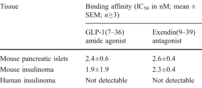
Table 1 Binding assay with the125I-BH-exendin(9–39) antagonist
Tissue Binding affinity (IC50in nM; mean ±
SEM; n≥3)
GLP-1(7–36)
amide agonist
Exendin(9–39)
antagonist
Mouse pancreatic islets 2.4±0.6 2.6±0.4
Mouse insulinoma 1.9±1.9 2.3±0.4
Human insulinoma Not detectable Not detectable
0 50 100
-10 -9 -8 -7 -6
125-I Exendin (9-39) spec. binding%
mouse insulinoma log[compound] (M) Exendin (9-39) GLP-1 -10 -9 -8 -7 -6 0 50 100 Exendin ( 9-39) GLP-1
125-I Exendin (9-39) spec. binding%
mouse pancreatic islets
log[compound] (M)
Fig. 1 Competition experiments in mouse pancreatic islets and mouse
insulinoma. In both examples, high affinity displacement of the125
I-BH-exendin(9–39) antagonist tracer by the GLP-1 receptor-selective
agonist GLP-1(7–36)amide (■) and exendin(9–39) antagonist (●) is
Wangen, Switzerland] or the antagonist 125 I-Bolton-Hunter-exendin(9–39) [74 TBq/mmol (2,000 Ci/mmol); Anawa, Wangen, Switzerland] as radioligands, used under identical experimental conditions. The GraphPad Prism program was used for curve fitting.
Results
Table1shows the excellent binding affinities of the GLP-1 receptor agonist GLP-1(7–36)amide and of the exendin (9–39) antagonist in mouse pancreatic islets and mouse insulinomas in competition experiments using 125 I-BH-exendin(9–39) antagonist as tracer. Figure 1 shows competition curves illustrating the almost identical high affinity displacement of the potent GLP-1 receptor agonist GLP-1(7–36)amide and the antagonist exendin(9–39) in these tissues. Table 1, however, also reveals that under identical conditions no measurable binding of 125 I-BH-exendin(9–39) was detected in the GLP-1 receptor-expressing human insulinomas.
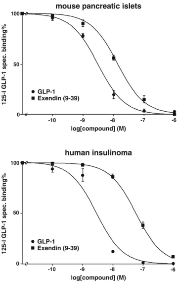
Table 2 shows the binding affinities of the GLP-1 receptor agonist GLP-1(7–36)amide and the exendin(9– 39) antagonist in mouse pancreatic islets and mouse insulinomas in competition experiments using the 125 I-GLP-1(7–36)amide agonist tracer. The data resemble the data obtained with the125I-BH-exendin(9–39) antagonist (Table 1), except that the IC50 values for the binding
affinity are found to be approximately five times higher for the antagonist exendin(9–39) than for the agonist GLP-1(7–36)amide. This is also illustrated in competi-tion experiments for mouse pancreatic islets in Fig.2.
More importantly, Table 2 also shows, surprisingly, that in human insulinomas the IC50 values for the
exendin(9–39) antagonist is more than 20 times higher than for the agonist GLP-1(7–36)amide. This is also illustrated in competition experiments for human insuli-nomas in Fig.2.
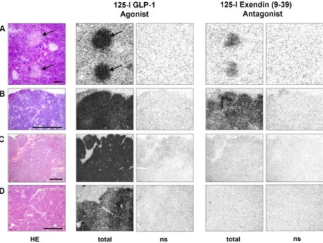
Figure 3 shows examples of GLP-1 receptor autoradi-ography which illustrate the above-mentioned observations on tissue sections. Using the agonist 125I-GLP-1(7–36)
amide as tracer, Fig. 3 shows that all tested tissues, namely mouse pancreatic islets, mouse Rip1Tag2 insu-linomas and two different human insuinsu-linomas express a very high density of GLP-1 receptors; the labelling is completely abolished in the presence of 100 nM GLP-1 (nonspecific binding), indicating the presence of specific GLP-1 receptors. Conversely, the125I-BH-exendin(9–39) antagonist tracer does label the GLP-1 receptors of the mouse pancreatic islets and mouse insulinomas, albeit with lower intensity than the agonist tracer. In line with the above-mentioned competition experiments, it does not or only very weakly label the GLP-1 receptors in the human insulinomas, despite the high density of GLP-1 receptors measured with the agonist tracer in these tissues. 0 50 100 125-I GLP-1 s pec. bindin g % log[compound] (M) -10 -9 -8 -7 -6 Exendin (9-39) GLP-1
mouse pancreatic islets
-10 -9 -8 -7 -6 0 50 100 125-I GLP-1 s pec. bindin g % Exendin (9-39) GLP-1 log[compound] (M) human insulinoma
Fig. 2 Competition experiments in mouse pancreatic islets and
human insulinoma tissues. High affinity displacement of the 125
I-GLP-1(7–36)amide tracer (125I-GLP-1) is observed by the GLP-1
receptor-selective agonist GLP-1(7–36)amide (●) in both tissues, while the antagonist exendin(9–39) (■) reveals a lower affinity, especially in the human insulinoma tissues. Mean ± SEM of > 3 independent experiments
Table 2 Binding assay with the125I-GLP-1(7–36)amide agonist
Tissue Binding affinity (IC50in nM; mean ±
SEM; n≥3) GLP-1(7–36) amide agonist
Exendin(9–39) antagonist
Mouse pancreatic islets 3.0±0.4 13±1.3
Mouse insulinoma 4.5±1.0 26±4.8
Discussion
The present data indicate that the 125I-BH-exendin(9–39) antagonist is a good tracer of GLP-1 receptors expressed in normal and neoplastic pancreatic β-cells in mice tissues. These data confirm under different conditions and extend to other systems the findings of Mukai et al. [8]. In addition, the antagonist exendin(9–39) shows a high affinity competi-tion comparable to that of the reference GLP-1 receptor agonist GLP-1(7–36)amide in mice tissues, with an excellent IC50 value of approximately 2 nM, when 125I-BH-exendin
(9–39) is used as tracer. Conversely, however, our data also show, most importantly, that the 125I-BH-exendin(9–39) antagonist is not a good tracer for the labelling of human GLP-1 receptor-expressing pancreatic tissues: this radio-labelled antagonist does not label the GLP-1 receptor-expressing human insulinomas, while under identical conditions it labels both mice pancreatic β-cells and insulinomas very well.
The comparison of binding affinities of the exendin (9–39) antagonist and the GLP-1 receptor agonist using
125I-GLP-1(7–36)amide tracer explains the above-mentioned
results obtained with the125I-BH-exendin(9–39) tracer. While the binding affinity values for exendin(9–39) tend to be lower than for GLP-1(7–36)amide in mice, this difference is markedly higher and reaches approximately a factor of 20
when tested in humans. The high IC50value of 63 nM for
exendin(9–39) in human insulinomas is therefore the likely explanation for the failure of in vitro visualization of GLP-1 receptors in this tissue with the antagonist tracer.
Our data indicate that the low binding affinity of the exendin(9–39) antagonist for human insulinomas is a sufficient reason to explain the absence of GLP-1 receptor visualization in these tissues. It gives, however, no clues as to whether, in general terms, a GLP-1 receptor antagonist is better or less adequate than a GLP-1 receptor agonist for GLP-1 receptor targeting in humans. For such a comparison, agonist and antagonist candidates with similar in vitro and in vivo binding characteristics should be compared, as was the case in the somatostatin and GRP receptor targeting studies [6,7].
The present data are a further example for species differences related to receptor binding characteristics of peptide analogs; this was shown previously for other peptides and more recently for GRP receptor analogs [10]. It indicates the need for including human tissues and human receptors in the preclinical testing of novel tracer candidates, either using cell lines expressing the human receptor or using resected human tumours, as shown in the present study. Hopefully, future studies will identify GLP-1 receptor antagonists with radiotargeting characteristics adequate for human tissue visualization.
Fig. 3 Comparative in vitro GLP-1 receptor autoradiography with the
125I-GLP-1(7–36)amide agonist tracer (125I-GLP-1) and with the125
I-BH-exendin(9–39) antagonist tracer in mouse tissue (A mouse
pancreatic islets, B mouse insulinoma) and human tissues (C and D two human insulinomas). Left column: haematoxylin and eosin (HE)-stained sections with pancreatic islets (arrows) in A (bar=0.1 mm) and insulinoma tumours in B–D (bars=1 mm). The “total binding”
column with the 125I-GLP-1 agonist shows high density of GLP-1
receptors in mouse pancreatic islets (arrows) and in each of the
insulinoma. The “ns” column represents nonspecific binding in the
presence of 100 nM GLP-1. Conversely, the“total binding” column
with the125I-BH-exendin(9–39) antagonist shows moderate density of
GLP-1 receptors in mouse pancreatic islets and in mouse insulinoma
but no binding at all in the two human insulinomas. The“ns” column
Conflicts of interest None.
References
1. Holst JJ. The physiology of glucagon-like peptide 1. Physiol Rev 2007;87:1409–39.
2. Reubi JC, Waser B. Concomitant expression of several peptide receptors in neuroendocrine tumours: molecular basis for in vivo multireceptor tumour targeting. Eur J Nucl Med Mol Imaging
2003;30:781–93.
3. Wild D, Mäcke H, Christ E, Gloor B, Reubi JC. Glucagon-like peptide 1-receptor scans to localize occult insulinomas. N Engl J
Med 2008;359:766–8.
4. Christ E, Wild D, Forrer F, Brändle M, Sahli R, Clerici T, et al. Glucagon-like peptide-1 receptor imaging for localization of
insulinomas. J Clin Endocrinol Metab 2009;94:4398–405.
5. Reubi JC. Peptide receptors as molecular targets for cancer
diagnosis and therapy. Endocr Rev 2003;24:389–427.
6. Ginj M, Zhang H, Waser B, Cescato R, Wild D, Wang X, et al. Radiolabeled somatostatin receptor antagonists are preferable to agonists for in vivo peptide receptor targeting of tumors. Proc Natl
Acad Sci U S A 2006;103:16436–41.
7. Cescato R, Maina T, Nock B, Nikolopoulou A, Charalambidis D, Piccand V, et al. Bombesin receptor antagonists may be preferable to agonists for tumor targeting. J Nucl Med 2008;49:318–26. 8. Mukai E, Toyoda K, Kimura H, Kawashima H, Fujimoto H, Ueda
M, et al. GLP-1 receptor antagonist as a potential probe for pancreatic beta-cell imaging. Biochem Biophys Res Commun
2009;389:523–6.
9. Wicki A, Wild D, Storch D, Seemayer C, Gotthardt M, Behe M, et al. [Lys40(Ahx-DTPA-111In)NH2]-Exendin-4 is a highly efficient radiotherapeutic for glucagon-like peptide-1
receptor-targeted therapy for insulinoma. Clin Cancer Res 2007;13:3696–
705.
10. Maina T, Nock BA, Zhang H, Nikolopoulou A, Waser B, Reubi JC, et al. Species differences of bombesin analog interactions with GRP-R define the choice of animal models in the development of