ORIGINAL PAPER
Triple pelvic osteotomy as treatment for osteoarthritis
secondary to developmental dysplasia of the hip
Dirk Janssen&Klaus Kalchschmidt&
Bernd-Dietrich Katthagen
Received: 17 November 2008 / Revised: 3 December 2008 / Accepted: 4 December 2008 / Published online: 12 February 2009
# Springer-Verlag 2009
Abstract Joint-preserving osteotomies are an established treatment for adult hip pain secondary to developmental dysplasia of the hip. However, their value for advanced osteoarthritis is unclear. Therefore this study addresses the question of long-term results of triple pelvic osteotomy in patients with second grade osteoarthritis. Thirty-two patients with second grade osteoarthritis secondary to developmental dysplasia of the hip before triple pelvic osteotomy were clinically and radiographically assessed 11.5 years postoper-atively. Five patients required conversion to total hip replacement. Kaplan-Meier survivorship analysis predicted a survival rate of 85.3%. The mean Harris hip score increased significantly with more than 56% good or very good results. A preoperative BMI > 25 and Harris hip score < 70 resulted in worse outcome or early conversion into total hip arthroplasty. The results indicate that developmental dysplasia of the hip even in second grade osteoarthritis can be treated with triple pelvic osteotomy.
Résumé dans les dysplasies douloureuses de la hanche, l’ostéotomie est un des traitements permettant de protéger l’articulation et de préserver l’avenir. Cependant, leur intérêt dans les arthroses évoluées n’est pas très clair. Cette étude a pour but d’évaluer les résultats à long terme d’une ostéotomie pelvienne chez des patients
présentant une arthrose de hanche de stade 2. 32 patients présentant une arthrose de hanche de stade 2 secondaire à une dysplasie de hanche ont été évalués cliniquement et radiographiquement à 11,5 ans post-opératoire. 5 patients ont nécessité la conversion de l’ostéotomie par une prothèse totale de hanche. La courbe de survie selon Kaplan-Meier permet de prédire un taux de survie de 85,3%. Le score moyen de Harris augmente de façon significative avec plus de 56% de bons et très bons résultats. Un BMI > 25 et un score de Harris < 70 sont des facteurs prédictifs de mauvais résultats ou d’une conversion vers une prothèse totale de hanche. ces résultats permettent de penser que les arthroses de grade II secondaires à une dysplasie de hanche peuvent être traitées par une triple ostéotomie pelvienne.
Introduction
Developmental dysplasia of the hip is known as a cause of secondary osteoarthritis [9,20]. As a treatment for adult hip pain due to dysplasia of the hip, especially for young patients with no or low grade osteoarthritis, pelvic osteotomies are performed to prevent the progression of osteoarthritis. The goal of the surgical approach is to improve the containment of the femoral head and to restore the congruity and stability of the joint to normalise joint contact pressure. Satisfactory intermediate [3,19,25] and long-term [2,10,11,13,14,22] results in patients with no or low grade osteoarthritis secondary to dysplasia of the hip are reported frequently. But the value of pelvic osteotomies for advanced osteoarthritis is controversial partly due to dearth of long-term results. Hence, it was the aim of this study to assess the long-term results of Toennis triple osteotomy in patients with advanced osteoarthritis of the hip defined by small cysts in the head or
D. Janssen (*)
Department of Pediatric Orthopaedic Surgery, University Hospital Basle,
Post Box, CH-4005 Basle, Switzerland e-mail: dirk.janssen@gmx.de
K. Kalchschmidt
:
B.-D. KatthagenDepartment of Orthopaedic Surgery, Staedtisches Klinikum Dortmund gGmbH,
Beurhausstr. 40, D-44137, Dortmund, Germany
acetabulum, increasing joint space narrowing and moderate loss of sphericity of the head. A subgroup analysis aimed to record the patients’ age at the time of the index operation, the body mass index (BMI) and the pre-operative Harris hip score as prognostic factors for long-term results.
Materials and methods
In 1992, 177 consecutive Toennis type triple pelvic osteotomies were performed at our clinic in 177 patients with various grades of osteoarthritis of the hip secondary to dysplasia. Osteoarthritis of the hip was classified into four radiographic stages according to Toennis [16] (Table 1). Thirty-five out of 177 patients were classified as having a second grade osteoarthritis and were included in this study. The mean age of the patients at the time of triple osteotomy was 38.6 years (range 23.9–57 years).The series comprised 30 women and five men.
Surgical technique
The pelvic osteotomy was performed in accordance with Toennis [16–18]. An arthrotomy was not routinely per-formed. All of the procedures were carried out by the same two senior surgeons. During the follow-up period the technique for osteosynthesis changed to fixation with screws. The temporarily implanted k-wires and wire cerclage were removed one year after osteotomy.
Postoperative management
During the early postoperative weeks, the following constraints were mandatory: (1) no weightbearing for 12 weeks, (2) joint movement only up to 30° for ab- and adduction, (3) no external rotation, and (4) a maximum flexion of 60°. Active internal rotation however was demanded. When radiographic signs of consolidation appeared six weeks after surgery, flexion up to 90° and abduction were permitted. All patients received thrombosis prophylaxis with low molecular weight heparin (Certo-parin) for the entire period of limited weightbearing.
Follow-up examination
All patients were included in a follow-up scheme. First, clinical examinations were carried out three and twelve months after the initial osteotomy; thereafter, patients presented themselves irregularly when required. The 35 patients with 35 hips classified as second grade osteoarthritis secondary to dysplasia of the hip were identified by preoperative radiographs and selected for the final study assessment. Three patients died during the follow-up period from causes unrelated to the osteotomy (two cases of lung cancer, one case of breast cancer). All remaining patients completed the study and returned for clinical and radiographic assessment be-tween September 2003 and March 2004. The mean duration of follow-up was 11.5 years (range 11– 12.2 years). The preoperative Harris hip score was routinely calculated prior to the index operation, and the preoperative body weight and body height were recorded. Clinical and radiographic examination were performed on all surviving patients (32 cases). Clinical follow-up was based on the Harris hip score and was performed by an independent physician. The radiograph-ic evaluation was based on anteroposterior and false-profile radiographs. The series of pre-, postoperative and final follow-up radiographs were analysed by an independent physician. The radiographs were evaluated for the grade of osteoarthritis, the centre-edge angle of Wiberg, the anterior centre-edge angle of Lequesne and de Sèze, the angle of the weight bearing zone of Bombelli and Aronson and the migration percentage of Reimers.
Statistical analysis
The statistical analyses of prognostic factors was based on the chi-square test, whereas the Wilcoxon matched pairs test was applied to compare the Harris hip score prior to and after intervention. Kaplan-Meier survival analysis, with conver-sion into a total hip replacement as end point, was used to analyse the survival rate. Statistical analyses were performed with Statistica software (version 6.0; StatSoft, Inc.). A p value of <0.05 was considered to be a significant result.
Table 1 Classification of osteoarthritis of the hip into four radiographic stages according to Toennis [16]
Grade 0 Grade I Grade II Grade III
No signs of osteoarthritis Increased sclerosis of the head and acetabulum, slight narrowing of the joint space, slight lipping at the joint margins
Small cysts in the head or acetabulum, increasing narrowing of the joint space, moderate loss of sphericity of the head
Large cysts in the head or acetabulum, severe narrowing or obliteration of the joint space, severe deformity of the head and necrosis
Results Clinical results
Prior to osteotomy the mean Harris hip score was 70.2 (range 35–91 points). During the follow-up five patients (15.6%) required a conversion to a total hip replacement (2.3, 2.9, 6, 7.7 and 10.3 years, respectively, after the index operation). In the remaining 27 patients the mean Harris hip score improved significantly to 81.2 (range 37–100 points) (p=0.02). Eighteen (66.7%) of the 27 surviving hips were rated good or very good, and six (22.2%) were rated bad. Five patients underwent triple osteotomy on the contralat-eral dysplastic hip in the following years.
Radiographic evaluation
The mean lateral centre-edge angle improved after the osteotomy from 10.2° (range 0–32°) to 34.6° (range18–49°) and the mean anterior centre-edge angle changed from 4.2° (range 0–32°) to 31° (range 12–51°). The migration percentage of Reimers improved from 39% (range 19.4– 56.7%) preoperatively to 13.1% (range 3.6–27.1%) in the postoperative evaluation. The weight bearing zone of Bombelli and Aronson changed from 24.8° (range 2–38°) to 5.7° (range 0–15°). Six radiographs (18.8%) showed radiographic signs of progression of osteoarthritis at the final follow-up.
Prognostic factors
To analyse the value of preoperative BMI and Harris hip score the study population was grouped according to their BMI and Harris hip score prior to surgery. The cut off was a BMI > 25 (defined as overweight) and a Harris hip score < 70 (defined as a bad score). The analyses revealed that preoperative overweight (p=0.003) and a bad Harris hip score (p=0.0025) prior to osteotomy correlated with bad Harris hip scores in the follow-up or even led to conversion to a total hip replacement. The age at the time of surgery did not relate to the long-term results.
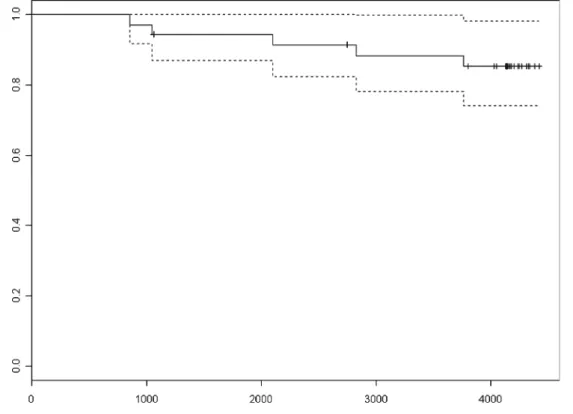
Survival analysis
Kaplan-Meier survival analysis (Table 2), with conversion to a total hip replacement as end point, revealed a survival rate of 85.3% (95% confidence interval 74.1–98.1%) at the time of the last follow-up examination (mean duration 11.5 years).
Complications
Double pseudarthrosis with nonunion of the pubis and the ischium occurred in one patient and was treated conserva-tively due to the lack of clinical signs and absence of pain. One patient developed an exostosis at the anterior inferior iliac spine which was surgically removed. Snapping of the
iliotibial band in one patient was resolved by surgical extension of the band. A thrombosis of the lower limb occurred perioperatively in one patient and was treated effectively. Six patients showed temporary postoperative irritation of the lateral femoral cutaneous nerve.
Discussion
Pelvic osteotomies are an established treatment for adult hip pain secondary to dysplasia of the hip, especially for young patients with no or low grade osteoarthritis. Long-term results for the triple osteotomy according to Toennis for this indication are good [7]. The role of pelvic osteotomies in advanced osteoarthritis is still controversial in particular because the results for total hip replacement as treatment for degenerative arthritis of the hip secondary to developmental dysplasia are favourable [6].
Early osteoarthritis and joint incongruency due to dysplasia of the hip are reported to be negative predictive factors [2,10,
19,22] for the osteotomy. De Kleuver et al. [2] report 20% good or excellent ratings in patients with evidence of osteoarthritis before triple acetabular osteotomy compared to 71% in patients without. Nakamura et al. [10] showed lower scores for pain, gait and particularly for mobility and hence poorer overall outcome in a group of patients with advanced osteoarthritis. Ito [4, 5] analysed the long-term effect of Chiari pelvic osteotomy for grade-3 osteoarthritis according to Toennis, although advanced osteoarthritis is reported to lead to unsatisfactory results [8, 12]. Despite a significant improvement of the Harris hip score the clinical results were inferior to those of total hip arthroplasty. The group consequently recommends the osteotomy for patients who prefer a joint-conserving procedure to a total hip arthroplasty, accepting a poorer clinical outcome.
On the other hand the literature reveals reports that support our hypothesis that osteotomies of the pelvic bone, even in second grade osteoarthritis secondary to dysplasia of the hip, can prevent progression of osteoarthritis. Yasunaga et al. [24] showed that result for rotational acetabular osteotomy in a group of elderly patients with early-stage osteoarthritis. They postulated that in older patients unsatisfactory medialisation of the femoral head is an important risk factor due to the inferior adaptability of the articular cartilage in comparison to younger patients. Takatori et al. [15] report long-term results for rotational acetabular osteotomy in patients with advanced coxarth-rosis (grades III and IV in the classification of the Japanese Orthopaedic Association). The majority of the patients had little or no pain at the recent follow-up and the condition of the hips was the same or better in most of the patients compared with the preoperative status. One patient under-went a secondary total hip arthroplasty. Furthermore,
Yasanuga et al. [21] showed a significant improvement in the system of Merle d’Aubigne and Postel in hips with advanced-staged osteoarthritis at a follow-up of eight years after rotational acetabular osteotomy. Aggravation com-pared with the preoperative score was reported for eight out of 29 joints. One total hip arthroplasty was performed in the fifth postoperative year.
In their recent publication Yasunaga et al. [23] report the results 8.5 years after rotational acetabular osteotomy in patients with advanced-stage osteoarthritis according to the Japanese Orthopaedic Association, which is equivalent to grade 2 of Toennis’s classification. Two out of 43 osteotomies had been converted to total hip arthroplasties and five hips showed end stage osteoarthritis in the radiographs. The mean Merle d’Aubigne clinical score improved significantly. A significant association with radiographic signs of progression of osteoarthritis after rotational acetabular osteotomy were fair postoperative joint congruency, a preoperative joint space of <2.2 mm, and a postoperative joint space of <2.5 mm.
Our study demonstrated the BMI and the preoperative Harris hip score are reliable predictors for the outcome. The ideal patient should have a BMI < 25 and a Harris hip score >70.
Therefore, triple osteotomy can be an alternative to total hip replacement. The duration of the operation and the postoperative treatment are indeed much longer. Patients have to be informed about that and especially about the fact that a progression of the osteoarthritis can occur and conversion to a total hip arthroplasty nevertheless can be necessary. The experiences of Ito [4, 5] with the Chiari pelvic osteotomy limit the prospects as a treatment for Toennis grade-3 osteoarthritis.
In the future, delayed gadolinium-enhanced magnetic resonance imaging of cartilage (dGEMRIC) could play an important role as a predictive factor. Cunningham [1] demonstrated that hips with a severely diminished joint space, higher Toennis grade (2 or 3), joint subluxation, or lower dGEMRIC index were less likely to benefit from the Bernese periacetabular osteotomy for hip dysplasia. There-fore, dGEMRIC could help to identify patients who would profit from a pelvic osteotomy despite the grade of osteoarthritis on the radiographs.
References
1. Cunningham T, Jessel R, Zurakowski D, Millis MB, Kim YJ (2006) Delayed gadolinium-enhanced magnetic resonance imag-ing of cartilage to predict early failure of Bernese periacetabular osteotomy for hip dysplasia. J Bone Joint Surg Am 88:1540–1548 2. de Kleuver M, Kooijman MA, Pavlov PW, Veth RP (1997) Triple osteotomy of the pelvis for acetabular dysplasia: results at 8 to 15 years. J Bone Joint Surg Br 79:225–229
3. Hsin J, Saluja R, Eilert RE, Wiedel JD (1996) Evaluation of the biomechanics of the hip following a triple osteotomy of the innominate bone. J Bone Joint Surg Am 78:855–862
4. Ito H, Matsuno T, Minami A (2004) Chiari pelvic osteotomy for advanced osteoarthritis in patients with hip dysplasia. J Bone Joint Surg Am 86:1439–1445
5. Ito H, Matsuno T, Minami A (2005) Chiari pelvic osteotomy for advanced osteoarthritis in patients with hip dysplasia. J Bone Joint Surg Am 87:213–225
6. Ito H, Matsuno T, Minami A, Aoki Y (2003) Intermediate-term results after hybrid total hip arthroplasty for the treatment of dysplastic hips. J Bone Joint Surg Am 85:1725–1732
7. Kuepper A, Kalchschmidt K, Katthagen BD (2003) 10-Jahres Ergebnisse der dreifachen Beckenosteotomie nach Toennis. Orthopaedische Praxis 39(7):412–419
8. Matsuno T, Ichioka Y, Kaneda K (1992) Modified Chiari pelvic osteotomy: a long term follow-up study. J Bone Joint Surg Am 74:470–478
9. Murphy SB, Kijewski PK, Millis MB, Harless A (1990) Acetabular dysplasia in the adolescent and young adult. Clin Orthop 261:214–23
10. Nakamura S, Ninomiya S, Takatori Y, Morimoto S, Umeyama T (1998) Long-term outcome of rotational acetabular osteotomy: 145 hips followed for 10–23 years. Acta Orthop Scand 69:259–265 11. Nozawa M, Shitoto K, Matsuda K, Maezawa K, Kurosawa H
(2002) Rotational acetabular osteotomy for acetabular dysplasia: a follow-up for more than ten years. J Bone Joint Surg Br 84:59–65 12. Reynolds DA (1986) Chiari innominate osteotomy in adults. Technique, indications and contra-indications. J Bone Joint Surg Br 68:45–54
13. Schramm M, Hohmann D, Radespiel-Troger M, Pitto RP (2003) Treatment of the dysplastic acetabulum with Wagner spherical osteotomy: a study of patients followed for a minimum of twenty years. J Bone Joint Surg Am 85:808–814
14. Schramm M, Pitto RP, Rohm E, Hohmann D (1999) Long-term results of spherical acetabular osteotomy. J Bone Joint Surg Br 81:60–66
15. Takatori Y, Ninomiya S, Nakamura S, Moromoto S, Moro T, Nagai I (2000) Long-term results of rotational acetabular osteotomy in young patients with advanced osteoarthrosis of the hip. J Orthop Sci 5:336–341
16. Toennis D, Arning A, Bloch M, Heinecke A, Kalchschmidt K (1994) Triple pelvic osteotomy. J Pediatr Orthop B 3:54–67 17. Toennis D, Behrens K, Tscharani F (1981) Eine neue Technik der
Dreifachosteotomie zur Schwenkung dysplastischer Hueftpfannen bei Jugendlichen und Erwachsenen. Z Orthop 119:253–265 18. Toennis D, Kalchschmidt K, Heinecke A (1999) Die
Hueftp-fannenschwenkung durch Dreifachosteotomie des Beckens – Stellenwert und Indikation in der Vielfalt operativer Korrek-turen der Dysplasiehuefte. Orthopaedische Praxis 35:607–620 19. Trousdale RT, Ekkernkamp A, Ganz R, Wallrichs SL (1995) Periacetabular and intertrochanteric osteotomy for the treatment of osteoarthrosis in dysplastic hips. J Bone Joint Surg Am 77:73–85 20. Weinstein SL (1987) Natural history of congenital hip dislocation
(CDH) and hip dysplasia. Clin Orthop 225:62–76
21. Yasunaga Y, Iwamori H, Ikuta Y, Yamamoto S, Harada A (1999) Rotational acetabular osteotomy for advanced osteoarthrosis secondary to dysplasia of the hip: results 6–11 years postopera-tively. Arch Orthop Trauma Surg 119:253–257
22. Yasunaga Y, Ochi M, Shimogaki K, Yamamoto S, Iwamori H (2004) Rotational acetabular osteotomy for hip dysplasia: 61 hips followed for 8–15 years. Acta Orthop Scand 75:10–15
23. Yasunaga Y, Ochi M, Terayama H, Tanaka R, Yamasaki T, Ishii Y (2006) Rotational acetabular osteotomy for advanced osteoarthri-tis secondary to dysplasia of the hip. J Bone Joint Surg Am 88:1915–1919
24. Yasunaga Y, Takahashi K, Ochi M, Ikuta Y, Hisatome T, Nakashiro J, Yamamoto S (2003) Rotational acetabular osteotomy in patients forty-six years of age or older: comparison with younger patients. J Bone Joint Surg Am 85:266–272
25. Yukiharu H, Toshiki I, Shinji K, Ken-Ichi Y, Shinji S, Hisashi I (2002) Eccentric rotational acetabular osteotomy for acetabular dysplasia: follow-up of one hundred and thirty-two hips for five to ten years. J Bone Joint Surg Am 84:404–410
![Table 1 Classification of osteoarthritis of the hip into four radiographic stages according to Toennis [16]](https://thumb-eu.123doks.com/thumbv2/123doknet/14861983.635639/2.892.74.818.945.1067/table-classification-osteoarthritis-hip-radiographic-stages-according-toennis.webp)