C L I N I C A L R E S E A R C H
A Useful Radiologic Method for Preoperative Joint-line
Determination in Revision Total Knee Arthroplasty
Jose Romero MD, Burkhardt Seifert PhD,Olaf Reinhardt MD, Oliver Ziegler MD, Oliver Kessler MD, MS
Received: 4 March 2009 / Accepted: 17 September 2009 / Published online: 5 November 2009
Ó The Association of Bone and Joint Surgeons1 2009
Abstract Intraoperative joint-line determination during revision TKA is difficult and no method exists to plan the position preoperatively. Two questions need to be answered: to which extent does the joint line differ from its anatomic position after revision TKA if it has only been determined intraoperatively, and can the joint line be cal-culated preoperatively based on the transepicondylar width. Of 22 consecutive patients with complete preoperative (before and after primary TKA) and postoperative (after revision TKA) radiograph documentation, the joint-line position was measured on plane radiographs using the medial epicondyle as a reference. On another set of 45 consecutive patients with no knee disorders other than meniscal lesions, the transepicondylar axis width (TEAW) and the perpendicular distance from the medial and lateral
epicondyles to the joint line were measured twice by two independent observers on plane AP radiographs of the knee. Significant joint-line alterations were observed after primary and revision TKA, implicating that a method for preoperative planning is needed. Because a linear correla-tion between the TEAW and the perpendicular distance from the epicondyles to the joint-line tangent was found, the ratio is useful to calculate the true joint-line position from the TEAW before revision TKA.
Level of Evidence: Level III, therapeutic study. See Guidelines for Authors for a complete description of levels of evidence.
Introduction
Alterations of the joint line in revision TKA have an impact on strength of the extensor mechanism, patellar pressure, patellar pain, and ROM [5,6].
No agreement exists regarding how the joint line is determined on plain radiographs. Some surgeons measure from the adductor tubercle to the joint line of the distal femur, whereas others measure from the lateral flare of the distal femur to the joint line. No consensus exists for which radiologic view should be used [3]. Some surgeons measure on the AP view using the epicondyles as a reference, whereas others use the tip of the fibular head or the lower pole of the patella to the proximal tibial surface on a lateral radiographic view. Alternatively, the contralateral knee, if it is not replaced, can be used to assess the joint-line position of the index knee. As a result of the variety of methods, measurement of the anatomic joint line and its appropriate restoration in revision TKA becomes difficult to reproduce. Intraoperatively, two basic methods exist for proper restoration of the joint line: the flexion-extension Each author certifies that he or she has no commercial associations
(eg, consultancies, stock ownership, equity interest, patent/licensing arrangements, etc) that might pose a conflict of interest in connection with the submitted article.
Each author certifies that his or her institution approved the human protocol for this investigation, that all investigations were conducted in conformity with ethical principles of research.
This work was performed at the University of Zurich, Balgrist, Switzerland.
J. Romero, O. Reinhardt, O. Ziegler, O. Kessler
Department of Orthopaedic Surgery, University of Zurich, Balgrist, Switzerland
B. Seifert
Biostatistics Unit, Institute of Social and Preventive Medicine, University of Zurich, Zurich, Switzerland
J. Romero (&)
Endoclinic Zurich, Klinik Hirslanden, Witellikerstrasse 40, 8032 Zu¨rich, Switzerland
e-mail: [email protected] DOI 10.1007/s11999-009-1114-1
gap-balancing technique, which first addresses the flexion gap, and the medial epicondylar referencing technique, which first addresses the extension gap. Griffin et al. [2] suggested using a ratio of the transepicondylar width to locate, intraoperatively, the position of the implant height in the coronal plane. His measurements are based on MRI findings and therefore are not applicable for revision cases. The aim of the first part of our study was to prove the hypothesis that in revision TKA, the joint line frequently is malpositioned compared with the original prearthroplasty anatomy, and that the radiograph from the primary TKA is not useful because it also may not reflect the true anatomic position before primary TKA. With this hypothesis in mind, we wanted to assess whether a routinely useful standardized method to reproducibly determine the joint line on radiographs before revision TKA could be devel-oped. The third aim was to determine if this method needs to be modified to address a gender-specific correlation between the TEAW and the distance from the medial and lateral epicondyles to the joint line.
Materials and Methods
Complete preoperative (before and after primary TKA) and postoperative (after revision TKA) radiographs of 22 consecutive patients who underwent revision TKA between December 1996 and May 2002 for failed primary TKA were included in the study. All patients had revision TKA using revision instrumentation that addresses stability in flexion first followed by adaptation of the extension gap. A posterior-stabilized revision system was used that allowed for joint-line adjustment by adding augments on the distal and posterior condyles.
The joint line was defined as a tangent composed of a straight line connecting the most distal points of the medial and lateral femoral condyles of the intact and replaced knee in extension on plane AP radiographs. In all cases, the perpendicular distance from the medial epicondyles to the described joint-line tangent was measured manually on the radiographs with an ordinary ruler. Because the dimension of the prosthesis is known from the manufac-turer and the size of the prosthesis was denoted in the medical records, we were able to calculate the magnifica-tion for each radiograph. The transepicondylar axis served as a reference to adjust for radiographic magnification between preoperative and postoperative radiographs.
We calculated changes of joint-line level after primary TKA and after revision TKA in reference to its anatomic level before primary TKA.
To assess the transepicondylar axis width and joint-line level in normal knees, the plane AP knee radiographs of 45 consecutive patients (25 males, 20 females) with a mean age
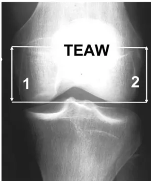
of 44.9 ± 17.8 years (range, 18–89 years) who were treated in our outpatient clinic for no knee disease other than men-iscal lesions were included in this study. A ruler with two plumb marks at a distance of 10 cm to each other, which is in routine use in our Radiologic Department, was attached to the distal thigh of every patient. It served as reference for calculation of the radiographic magnification on every xray. Radiographs were taken with the patient in the supine posi-tion with the knee in full extension. The TEAW, the distance from the medial and lateral epicondyles to the joint line, and the length of the radiographic marker were measured twice by two independent observers (a staff orthopaedic knee surgeon [JR] and a first-year orthopaedic resident [OR]) with an interval of 2 to 3 weeks between measurements by each observer. The measurements were done manually on the radiographs with an ordinary ruler. Interobserver and intra-observer reliabilities were analyzed in a random three-way ANOVA with factors rater, repetition, and subject. We estimated variance components using restricted maximum likelihood. Interrater reliability is the ratio of variance components not depending on the rater to the sum of all variance components. A value of 1 denotes an ideal reli-ability and a value of 0 denotes the worst possible relireli-ability. For each patient, we analyzed the relationships between measurements and gender differences using the means of four measurements. The epicondylar width was defined as the distance connecting the upper edge of the medial epicondylar sulcus and the most prominent edge of the lateral epicondyle (Fig. 1). The joint line was defined as the
Fig. 1 Distance 1 is the measurement from the medial epicondyle to the joint line. Distance 2 is the measurement from the lateral epicondyle to the joint line. TEAW = transepicondylar axis width.
tangent connecting the most distal points of the medial and lateral condyles. The medial joint-line level was defined as the perpendicular distance from the upper edge of the sulcus on the medial epicondyle to the joint-line tangent. The lateral joint-line level was defined as the perpendicular distance from the most prominent edge on the lateral epicondyle to the joint-line tangent.
We performed regression analysis to assess the rela-tionship between joint-line level and epicondylar width. Gender differences were analyzed using the unpaired t-test. We performed stepwise regression analyses for the analysis of dependencies between the medial and lateral joint line and epicondylar width and of gender differences in these relationships. Regression equations with and without intercept were calculated and compared with each other.
Results
Joint-line position usually is altered substantially from the native position after primary and revision TKA. Compared with preoperative measurements, the joint line after pri-mary TKA measured from the medial epicondyle shifted proximally in nine cases with a mean shift of 3.6 ± 2.4 mm and distally in 12 cases with a mean shift of 4.7 ± 1.8 mm. The joint line was at its anatomic position in one case. After revision TKA, the joint line measured from the medial epicondyle shifted proximally in 13 cases (mean, 6.1 ± 3.7 mm) and distally in seven cases (mean, 3.5 ± 2.4 mm) from its anatomic position before primary TKA. In two cases, the joint line was at its anatomic position.
Using the epicondyles as reference proved to be repro-ducible. The interobserver variability was 0.97 for the epicondylar width, 0.85 for the perpendicular distance from the medial epicondyle to the joint-line tangent, and 0.80 for the perpendicular distance from the lateral epicondyle to
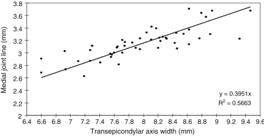
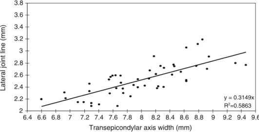
the joint-line tangent. The intraobserver variability was 0.98 for the epicondylar width, 0.92 for the perpendicular distance from the medial epicondyle to the joint-line tan-gent, and 0.86 for the perpendicular distance from the lateral epicondyle to the joint-line tangent. The measure-ments for adjustment of radiographic magnification showed an interrater variability of 0.82 and an intrarater variability of 0.76. The mean radiographic magnification was 9.2%, ranging from 4% to 14%. After specific adjustment for radiographic magnification for each case, the mean epic-ondylar width was 79.9 ± 6.5 mm (range, 66.0–94.7 mm), the mean perpendicular distance from the medial epicon-dyle to the joint-line tangent was 31.6 ± 2.5 mm (range, 26–37 mm), and the mean perpendicular distance from the lateral epicondyle to the joint-line tangent was 25.1 ± 2.7 mm (range, 21–32 mm). Regression analysis yielded a linear correlation (R2 = 0.65) between the epicondylar width and the perpendicular distance from the medial epicondyle to the joint-line tangent (y = 0.395x + 0.661). With an intercept of 0, the slope of the regression line (R2= 0.57) was 0.395 (Fig.2). If the coefficient is rounded up to 0.4, a difference of 1.0 mm to 1.7 mm exists to the regression line with intercept in the range of 65 mm to 95 mm of epicondylar width. Regression analysis also yielded a linear correlation (R2= 0.59) between the epic-ondylar width and the perpendicular distance from the lateral epicondyle to the joint-line tangent (y = 0.32x + 0.044). With an intercept of 0, the slope of the regression line (R2 = 0.58) was 0.32 (Fig.3). If the coefficient is rounded down to 0.3, a difference of 1.7 mm to 2.3 mm exists to the regression line with an intercept in the range of 65 mm to 95 mm of epicondylar width.
Gender yielded statistically significant differences for the mean epicondylar width (p \ 0.0001), for the mean perpendicular distance from the medial epicondyle to the joint-line tangent (p = 0.003), and for the mean
y = 0.3951x R2 = 0.5663 2 2.2 2.4 2.6 2.8 3 3.2 3.4 3.6 3.8 6.4 6.6 6.8 7 7.2 7.4 7.6 7.8 8 8.2 8.4 8.6 8.8 9 9.2 9.4 9.6
Transepicondylar axis width (mm)
Medial joint line (mm)
Fig. 2 Regression analysis of the perpendicular distance between the upper edge of the medial epicondylar prominence and the joint line (y = 0.395x; R2 = 0.5663).
perpendicular distance from the lateral epicondyle to the joint-line tangent (p = 0.007). For females, the mean epicondylar width was 75.7 ± 4.6 mm (range, 65.00– 85.5 mm), the mean perpendicular distance from the medial epicondyle to the joint-line tangent was 30.4 ± 0.2 mm (range, 26.8–33.0 mm), and mean perpendicular distance from the lateral epicondyle to the joint-line tan-gent was 24.0 ± 2.0 mm (range, 20.8–26.5 mm). For males, the mean epicondylar width was 83.3 ± 5.9 mm (range, 71.9–94.7 mm), the mean perpendicular distance from the medial epicondyle to the joint-line tangent was 32.7 ± 2.5 mm (range, 26.3–36.8 mm), and mean per-pendicular distance from the lateral epicondyle to the joint-line tangent was 26.1 ± 2.9 mm (range, 20.8–31.1 mm). Stepwise regression analysis yielded no gender-specific differences of either the slopes or the intercepts (all F-to-Enter \ 0.4) of the regression lines.
Discussion
The review of the 22 cases of revision knee arthroplasties of which complete radiographic documentation before and after primary TKA and after revision TKA was available showed that the anatomic joint-line position referenced from the epicondyles most frequently is not restored after revision TKA. A proximalisation of the joint line is more frequent and more pronounced than a distalisation after revision TKA. As determined by measuring the distance from the medial epicondyle to the joint line, 60% (13 of 22) of the knees had an elevated joint line on average of 6.1 ± 3.7 mm after revision knee arthroplasty. The joint line already was altered slightly from its anatomic position after primary TKA in these patients (elevated in nine cases on average by 2.4 mm and lowered in 12 cases on average by 3.2 mm). Therefore, the radiographs of the primary TKA cannot be used as a reference for joint line positioning at
revision TKA because the joint line established at primary TKA does not necessarily reflect the original position of the anatomic joint line. The resection level chosen at pri-mary TKA may have depended on degenerative bone wear, deformity, and flexion contracture. Determination of the joint line at revision TKA also may become difficult as a result of distal femoral bone loss during extraction of the femoral component or after two-stage revision for infection.
Therefore, we propose a radiologic method to reliably determine the joint-line position on radiographs after TKA before revision TKA if the original radiographs before primary TKA are not available or if the contralateral knee also has been replaced. The described method is based on the fact that we found a linear correlation between the epicondylar width and the perpendicular distance from the medial and lateral epicondyle of the joint-line tangent. This finding facilitates estimation of the joint level in revision knee arthroplasty, because regression analysis yielded a nongender-specific coefficient close to 0.4 (medial) and 0.3 (lateral), which needs to be multiplied with the TEAW to calculate the medial and lateral joint line, respectively. These measurements proved to be highly reliable and reproducible for preoperative planning on radiographs. This technique is applicable only to femurs in which the epicondyles can be accurately identified. One potential limitation of the measurements was that the radiographs were not taken with a device that would have held the knees in a consistent position. However, we were confident that all radiographs had been taken following the routine standard knee radiographic protocol for supine position of our institution. It consists of a support laterally to prevent external rotation of the lower extremity and upward posi-tion of the patella, which is checked by the xray technician. Griffin et al. [2] used an MRI technique to quantify a correlation between the width of the epicondyles and the distance to the joint line. They found a nongender-specific
y = 0.3149x R2=0.5863 2 2.2 2.4 2.6 2.8 3 3.2 3.4 3.6 3.8 6.4 6.6 6.8 7 7.2 7.4 7.6 7.8 8 8.2 8.4 8.6 8.8 9 9.2 9.4 9.6
Transepicondylar axis width (mm)
Lateral joint line (mm)
Fig. 3 Regression analysis of the distance between the upper edge of the lateral epicondylar prominence and the joint line (y = 0.3149x; R2= 0.5863).
ratio between the TEAW and the perpendicular distance to the joint line of 0.36 for the medial side and of 0.31 for the lateral side, which corresponded well with our findings. However, their study was based only on MRI. The clinical feasibility and validity of the measurements were not analyzed using conventional radiographs, which would be the preferred imaging for planning revision knee arthroplasty. An additional disadvantage for the clinical application might be the fact that they used the sulcus of the medial epicondyle, which might be less accurate to determine in a clinical situation with arthritic deformed knees [8].
The results of the current study may have an important clinical application, because the surgeon can easily evalu-ate the joint-line level on preoperative radiographs and can transfer that information to the intraoperative situation. However, it is not always easy to identify the medial or lateral epicondyle during surgery [7]. Other methods that rely solely on intraoperative determination of the joint-line level have shown severe potential error mechanisms. The flexion-extension gap balancing technique to restore the joint line [4] seems to be a valid method only if the surgeon is experienced. Laskin [5] reported on 45 revision cases in which he used the tip of the fibular styloid to the medial epicondylar sulcus and the inferior pole of the patella to determine the joint line on preoperative radiographs. However, as a consequence of patellar tendon fibrosis, patella baja often is encountered in failed TKA; therefore, it is impossible to use the patellar height as a reference [1]. The technique we presented, which uses a ratio of TEAW (factor 0.4 for the medial epicondyle or 0.3 for the
lateral epicondyle), is a highly reliable method to deter-mine the joint line on plane AP radiographs before revision knee arthroplasty because a linear correlation exists. The calculated joint-line level may be helpful during revision knee arthroplasty for proper placement of the implants with designated instrumentation.
References
1. Figgie HE III, Goldberg VM, Heiple KG, Moller HS III, Gordon NH. The influence of tibial-patellofemoral location on function of the knee in patients with the posterior stabilized condylar knee prosthesis. J Bone Joint Surg Am. 1986;68:1035–1040.
2. Griffin FM, Math K, Scuderi GR, Insall JN, Poilvache PL. Anatomy of the epicondyles of the distal femur: MRI analysis of normal knees. J Arthroplasty. 2000; 15:354–359.
3. Hoeffel DP, Rubash HE. Revision total knee arthroplasty: current rationale and techniques for femoral component revision. Clin Orthop Relat Res. 2000;380:116–132.
4. Krackow KA. Revision total knee replacement ligament balancing for deformity. Clin Orthop Relat Res. 2002;404:152–157. 5. Laskin RS. Management of the patella during revision total knee
replacement arthroplasty. Orthop Clin North Am. 1998; 29:355– 360.
6. Mahoney OM, Kinsey TL. Modular femoral offset stems facilitate joint line restoration in revision knee arthroplasty. Clin Orthop Relat Res. 2006;446:93–98.
7. Stoeckl B, Nogler M, Krismer M, Beimel C, de la Barrera JL, Kessler O. Reliability of the transepicondylar axis as an anatomical landmark in total knee arthroplasty. J Arthroplasty. 2006;21:878– 882.
8. Yoshino N, Takai S, Ohtsuki Y, Hirasawa Y. Computed tomog-raphy measurement of the surgical and clinical transepicondylar axis of the distal femur in osteoarthritic knees. J Arthroplasty. 2001;16:493–497.