N. S. Palhais D. Guntern A. Kagel M. Wettstein E. Mouhsine N. Theumann Received: 24 September 2008 Revised: 19 December 2008 Accepted: 31 January 2009 Published online: 7 April 2009
# European Society of Radiology 2009
Direct magnetic resonance arthrography
of the knee: utility of axial traction
Abstract The purpose of this study was to determine the impact of axial traction during acquisition of direct magnetic resonance (MR) arthrogra-phy examination of the knee in terms of joint space width and amount of contrast material between the cartilage surfaces. Direct knee MR arthrogra-phy was performed in 11 patients on a 3-T MR imaging unit using a T1-weighted isotropic gradient echo se-quence in a coronal plane with and without axial traction of 15 kg. Joint space widths were measured at the level of the medial and the lateral femorotibial joint with and without traction. The amount of contrast material in the medial and lateral femorotibial joint was assessed inde-pendently by two musculoskeletal radiologists in a semiquantitative manner using three grades (‘absence
of surface visualization,‘partial sur-face visualization or‘complete surface visualization’). With traction, joint space width increased significantly at the lateral femorotibial compartment (mean=0.55 mm,p=0.0105) and at the medial femorotibial compartment (mean=0.4 mm,p=0.0124). There was a trend towards an increased amount of contrast material in the femorotibial compartment with axial traction. Direct MR arthrography of the knee with axial traction showed a slight and significant increase of the width of the femorotibial compart-ment with a trend towards more con-trast material between the articular cartilage surfaces.
Keywords MR arthrography . Knee . Traction . Cartilage
Introduction
Direct magnetic resonance (MR) arthrography of the knee joint with injection of diluted gadolinium is widely used for the detection of intra-articular abnormalities, especially for evaluation of residual or recurrent meniscal tears following meniscal surgery [1–3]. MR arthrography also has the potential to identify intra-articular bodies [4], and chondral [5, 6] and osteochondral [7] lesions and is helpful after reconstruction of the anterior cruciate ligament [8].
The advantage of direct MR arthrography over standard MRI relates to joint distension and signal intensity differ-ences between contrast material and intra-articular struc-tures such as menisci and hyaline cartilage. Therefore it is crucial to have enough contrast material between the
cartilage surfaces so that contrast material extends into cartilage defects and meniscal tears. In our experience this is not always the case in conventional MR arthrography.
Traction has been used for joint distraction in hip arthroscopy, and subsequently introduced into arthrogra-phy. A 15-kg weight was used to approach the weight used at hip arthroscopy [9]. This is thought to allow a better penetration of contrast material between the articular surfaces and the adjacent structures and thus a better delineation of the intra-articular structures and their pathology. With regard to the shoulder and the hip, there are some reports that showed superior results when traction is applied during MR arthrography [10–12]. But studies evaluating the effects of traction during knee MR arthrog-raphy are lacking.
N. S. Palhais . D. Guntern (*) . A. Kagel . N. Theumann
Department of Radiology, University Hospital, University of Lausanne, Lausanne, Switzerland e-mail: [email protected] Tel.: +41-21-3144564 Fax: +41-21-3144554 M. Wettstein . E. Mouhsine Department of Musculoskeletal Medicine, University Hospital, University of Lausanne, Lausanne, Switzerland
The purpose of our study was to determine the feasibility of the leg traction combined with MR arthrography and the effect of this technique on visualization of cartilage and menisci surfaces.
Materials and methods
Between September and December 2006, 11 consecutive patients (5 female, 6 male; mean age 33.4 years; age range 14–59 years) referred for MR arthrography of the knee were included in a prospective study. The institutional review board of Centre Hospitalier Universitaire Vaudois approved this study. All patients gave informed consent.
Before the MR arthrography a mixture of 18 ml iodinated contrast material (Accupaque, GE Healthcare), 0.1 ml gadopentetate dimeglumine (Omniscan, GE Healthcare ) with one drop of epinephrine (Adrenalin, Sintetica) and 2 ml of Xylocain 1% was prepared. After sterile skin preparation a 20-gauge needle was inserted into the patellofemoral joint with a lateral approach under fluoro-scopic guidance with subsequent intra-articular injection of approximately 20 ml of the contrast material mixture. Following the injection of the contrast material mixture the patients were immediately transferred to the MR suite in wheelchairs.
The MR examination was performed on a 3-T MR imaging unit (Achieva, Philips Medical Systems, Best, NL) with a dedicated knee coil (8-channel SENSE; Philips Medical Systems). First a fat-suppressed T1-weighted three-dimensional high resolution isotropic volume examination (3D THRIVE ISO) sequence (repetition time (TR)/echo time (TE) 11 ms/4.9 ms, 0.75-mm section thickness, 180-mm field of view (FOV), 512×512 matrix, NEX 1.) in a coronal plane was acquired without traction.
Then the leg traction was applied on the MRI table with a standard MRI-compatible orthopedic skin traction device
(Fig.1). The traction device consists of two lateral adhesive straps fixed parallel to the leg from the ankle through the patellar level reinforced by a conventional bandage. A 5-cm distance between the sole of the foot and the traction plate is left to allow easy handling of the rope used for traction. A 15-kg traction weight consisting of a standard water bag was then applied. Under continuous traction, the fat-suppressed T1-weighted 3D THRIVE ISO sequence was repeated with the same parameters as without traction. Finally according to our standard protocol a fat-suppressed T2-weighted turbo spin echo (TSE) sequence (TR/TE 3.856 ms/80 ms, 3.50-mm section thickness, 150-mm FOV, 576×576 matrix, NEX 2.) was acquired in coronal and transverse planes.
For our study purposes only the coronal T1-weighted 3D THRIVE ISO sequences were analyzed.
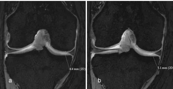
For a quantitative analysis the joint space width was measured by one musculoskeletal radiologist (DG) blinded to the presence or absence of traction. The joint space widths were measured at the level the medial and the lateral femorotibial joint with and without traction. The joint space width was defined as the minimal distance between the adjacent cortical surfaces (Fig.2).
The amount of contrast in the joint space was assessed independently by two musculoskeletal radiologists blinded to the presence or absence of traction in a semiquantitative manner. The spaces between the hyaline cartilage of the femoral and the tibial articular surfaces, between the femoral articular surface and the menisci and finally between the tibial articular surface and the menisci were assessed separately. The amount of contrast was graded as ‘absence of surface visualization’, ‘partial surface visual-ization’ or ‘complete surface visualization’. Absence of surface visualization was attributed when no contrast material was visible between the cartilage surfaces on all sections at any area of the joint space. Complete surface visualization was attributed when contrast material was visible on all slices covering the cartilage surfaces of the entire joint space. Partial surface visualization was attributed when the articular cartilages in one slice or in some area of the joint were not covered with contrast material.
Statistical analyses
The difference in joint space width with and without traction was analyzed using the t test for continuous variables. A p value less than 0.05 was considered statistically significant. The difference between the amount of contrast material in the joint space with and without traction was analyzed using the chi-square test for categorical variables. Again a p value less than 0.05 was considered statistically significant. Interobserver agree-ment for the semiquantitative analysis was tested by calculatingκ statistics. Agreement was rated, according to Fig. 1 A 20-year-old female has a dedicated coil (a) placed around
the examined knee, and the foot is connected to the specially manufactured traction system (b). Traction of 15 kg (bag of water) is applied via a single pulley on the patient’s leg
the method of Landis and Koch [13], as follows:κ values of 0–0.20 indicated slight agreement; 0.21–0.40, fair agreement; 0.41–0.60, moderate agreement; 0.61–0.80, substantial agreement; and 0.81–0.99, excellent agreement. Aκ value of 1.00 indicated absolute agreement.
All patients in the study group were instructed to inform the radiologist if they experienced any discomfort during application of the traction or during MRI data acquisition. All patients were given the option to request discontinua-tion of tracdiscontinua-tion at any point during the examinadiscontinua-tion.
Results
Quantitative analysis
The results are summarized in Table 1. With leg traction, joint space width increases significantly at the medial
femorotibial compartment (mean=0.4 mm, range−0.1 to 1.2 mm,p=0.0124) and at the lateral femorotibial compart-ment (mean=0.55 mm, range 0–1.6 mm, p=0.0105). The mean cortical surface separation of medial femorotibial compartment in 11 patients was 4.5 mm and increased to 4.9 mm after leg traction. Therefore the joint space width increased significantly with traction at the medial femoro-tibial compartment. For the lateral femorofemoro-tibial compartment the mean cortical surface separation was 5.9 mm without leg traction and increased to 6.4 mm with leg traction. Therefore the joint space width increased significantly with traction at the lateral femorotibial compartment.
Semiquantitative analysis
The results of the semiquantitative analysis including interobserver agreement are detailed in Table2. There was Fig. 2 Coronal T1-weighted
THRIVE MR arthrogram with-out (a) and with (b) traction (patient 11). Measurement of the medial femorotibial joint space width defined as the minimal distance between the two adja-cent cortical surfaces. Joint space width increases from 4.4 mm without traction to 5.1 mm with traction
Table 1 Measurement of the joint space width (mm)
Lateral femorotibial joint Medial femorotibial joint
Patients Without traction With traction Delta Without traction With traction Delta
1 5.1 6.5 1.4 2.8 3.7 0.9 2 6.5 6.5 0 4.8 4.7 −0.1 3 4.4 4.5 0.1 2.9 2.9 0 4 4.4 6 1.6 3 4.2 1.2 5 6.7 6.8 0.1 6.6 6.5 −0.1 6 7.6 7.9 0.3 5.4 5.4 0 7 4.2 5.4 1.2 4.2 4.6 0.4 8 9.3 9.7 0.4 4.7 5 0.3 9 4.1 4.1 0 6.5 6.9 0.4 10 6.6 6.9 0.3 4.2 4.9 0.7 11 5.8 6.5 0.7 4.4 5.1 0.7 Mean 5.88182 6.43636 0.555 4.5 4.9 0.4 t test 0.0105 0.0124
a trend towards a larger amount of contrast material between the articular cartilages with traction as compared without traction but it did not reach statistical significance (Fig.3). When all compartments are considered the number of compartments with complete cartilage surface visual-ization increased from 8 to 13 with the application of axial traction. The number of compartments with absence of cartilage surface visualization decreased from 20 to 17 and the number of compartments with partial cartilage surface visualization decreased from 38 to 36. Interobserver agreement between the two readers was moderate to absolute with values ranging from 0.47 to 1.
No patients experienced discomfort during traction application.
Discussion
The advantage of MR arthrography over standard MRI is related to the signal intensity differences between intra-articularly injected contrast material and intra-articular structures. In the knee intra-articular contrast material delineates menisci and hyaline cartilage and their defects. This is especially beneficial after meniscectomy, as tracking of contrast material within the substance of the operated meniscus is diagnostic of a re-tear or a residual tear (Fig. 4). But direct MR arthrography of the knee in diagnosing recurrent meniscal tear is not perfect with accuracies ranging from 85 to 87% [1,3]. Contrast material delineating cartilage defects may also enhance their
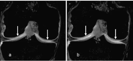
Fig. 3 Coronal T1-weighted THRIVE MR arthrogram in the same plane without (a) and with (b) traction. Note considerably more contrast material between the femoral and tibial articular cartilage surfaces with traction (arrows). The cartilage surfaces can be better evaluated. Note no contrast material between lateral and medial meniscus and femo-ral and tibial hyaline cartilage without and with traction
Table 2 Semiquantitative data for each joint compartment regarding surface visualization without and with traction
Without traction With traction
Contrast material between Absence of surface visualization Partial surface visualization Complete surface visualization Kappa Absence of surface visualization Partial surface visualization Complete surface visualization Kappa Chi-squared
Medial femoral condyle and meniscus
4 6 1 0.84 3 7 1 0.68 0.89
Medial tibial plateau and meniscus
6 5 0 0.47 4 7 0 1 0.39
Lateral femoral condyle and meniscus
5 5 1 0.84 4 5 2 1 0.8
Lateral tibial plateau and meniscus
4 7 0 0.81 5 4 2 0.85 0.23
Medial femorotibial carti-lage surfaces
1 7 3 0.8 1 7 3 0.8 1
Lateral femorotibial carti-lage surfaces
0 8 3 0.63 0 6 5 0.66 0.37
All compartments of knee joint
detection when natural intra-articular contrast, i.e. joint effusion, is lacking. MR arthrography seems to be superior to standard MRI in the detection of cartilage lesions at the knee especially for early lesions. But again MR arthrog-raphy is not perfect with reported sensitivities in detecting grade 1 lesions at the patellar cartilage ranging from 29 to 53% [5, 6]. To be able to benefit from the advantages of MR arthrography it is essential that enough contrast material enters between the articular surfaces. One of the advantages of direct versus indirect MR arthrography is that the former leads to joint distension enabling contrast material to enter between the articular surfaces; however, in our experience with direct knee arthrography this is not always the case and often there is no contrast material between hyaline cartilage and menisci probably due to inadequate joint distension. So we began to perform direct MR arthrography of the knee applying continuous traction of 15 kg to the examined knee during image acquisition. We think that this could lead to a better joint distension with more contrast material extending between the articular surfaces. There are three reported studies dealing with MR arthrography with image acquisition under continuous traction. The first study by Chan et al. evaluated the effect of traction during direct MR arthrography of the shoulder and concluded an improved accuracy in the evaluation of superior labral anteroposterior lesions [10]. In the second study Nishii et al. compared indirect MR arthrography of the hip with traction with standard MRI without traction in the evaluation of labral lesions and found a better accuracy of indirect MR arthrography with traction [12]; however, they did not compare indirect MR arthrography with and without traction. Recently Llopis et al. evaluated direct MR arthrography of the hip with leg traction [11] and found an improved visualization of the femoral and acetabular cartilage surfaces owing to a better separation of these structures. To our knowledge no studies dealing with knee MR arthrography with traction were performed. In our study knee distension with traction of 15 kg was possible. No patients experienced discomfort during the
application of the traction. We found a slight but significant increase of the femorotibial joint space width in both the medial and the lateral compartment. The only slight increase in joint space width could be due to muscle contraction by the patient counteracting the traction force. As Llopis et al. found a joint distension at the hip with only 6-kg traction [11], it could be possible, that 15 kg was too much traction leading to increased muscle contraction.
There was a trend of more contrast material entering between the articular cartilage surfaces when axial traction is applied that did not reach statistical significance. This could be due to the small patient number. Another explanation could be our grading system (absence of surface visualization, partial surface visualization or com-plete surface visualization), which perhaps was not fine enough and masked differences between both acquisitions. One could imagine that when the articular surfaces were partially covered with contrast material without traction, there was more contrast material with traction but the surfaces were still partially covered with contrast material. With our grading system this case would show no difference between the two acquisitions. Therefore we found partial surface visualization in the majority of compartments (Fig.5).
Despite the fact that the semiquantitative analysis did not show a statistically significant difference we think that traction knee MR arthrography could be useful at least in some patients. The amount of joint distension ranged from −0.1 to 1.6 mm. With a distension of 1.6 mm the cartilage surfaces are clearly separated which allows them to be better evaluated.
With a better separation of the femoral and tibial cartilages, cartilage surfaces are delineated by contrast material. This allows a better visualization of cartilage lesions especially surface lesions. When there is no contrast material between the cartilage surfaces, the visible cartilage layer is often a summation of the femoral and tibial cartilage and the interface between the two is difficult to see. In this situation surface cartilage lesions are difficult to Fig. 4 Sagittal reformation of a
coronal T1-weighted THRIVE MR arthrogram without (a) and with (b) traction. Without trac-tion (a), there is a nonspecific hyperintensity and irregularity of the posterior horn of the medial meniscus (arrow). With joint distraction (b), there is contrast material entering the substance of the posterior horn of the medial meniscus in a linear fashion diagnostic of a longitudinal vertical tear (arrow)
see and when they are visible it may be difficult to appreciate if the femoral or the tibial cartilage is involved. This study had several limitations. First, the number of subjects was small. Second, the effect of traction on visualization and the accuracy of diagnosis of lesions of intra-articular structures, such as the meniscus, were not
evaluated. But the aim of the study was primarily to describe a technical modification of direct knee MR arthrography and to give some indicators that with this technique direct knee MR arthrography may perform better with respect to assessment of hyaline cartilage and menisci.
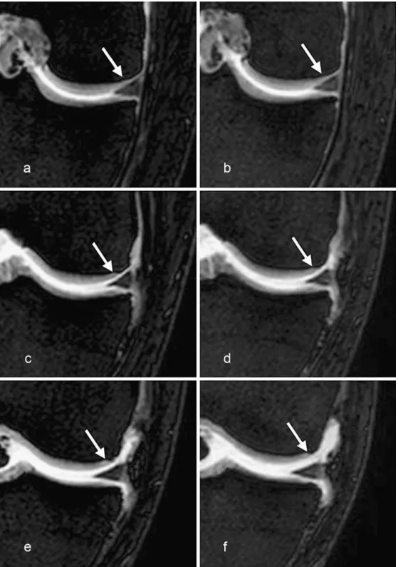
Fig. 5 Coronal T1-weighted THRIVE MR arthrograms without (a, c, e) and with (b, d, f) traction at three different levels of the medial aspect of the knee joint. a Posterior aspect of the knee without traction: there is no contrast material between the medial meniscus and the femoral hyaline cartilage (arrow). b Same level as a with traction: essentially no differ-ence to a, there is still no contrast material between the medial meniscus and the femo-ral hyaline cartilage (arrow). c More anterior level than a without traction: medial menis-cus and femoral hyaline carti-lage are partially separated with contrast material (arrow). d Same level as c with traction: medial meniscus and femoral hyaline cartilage are completely separated by contrast material (arrow). e More anterior level than c without traction: medial meniscus and femoral hyaline cartilage are completely sepa-rated by contrast material (arrow). f Same level as e with traction: separation of medial meniscus and femoral hyaline cartilage is accentuated (arrow). The final assessment of the compartment medial meniscus– medial femoral cartilage was no difference between no traction and traction as there was still no contrast between the meniscus and the femoral cartilage in the posterior aspect of the joint with traction i.e. partial surface visualization
Conclusion
Direct MR arthrography of the knee with axial traction of 15 kg showed a slight and significant increase of the width of the femorotibial compartment with a trend towards
more contrast material between the articular cartilage surfaces. Further studies have to evaluate if this slight joint distension improves accuracy in diagnosing recurrent and residual meniscal tears and cartilage lesions of the femorotibial compartment.
References
1. Applegate GR, Flannigan BD, Tolin BS, Fox JM, Del Pizzo W (1993) MR diagnosis of recurrent tears in the knee: value of intraarticular contrast material. AJR Am J Roentgenol 161:821–825 2. Magee T, Shapiro M, Rodriguez J,
Williams D (2003) MR arthrography of postoperative knee: for which patients is it useful? Radiology 229:159–163 3. White LM, Schweitzer ME, Weishaupt
D, Kramer J, Davis A, Marks PH (2002) Diagnosis of recurrent meniscal tears: prospective evaluation of con-ventional MR imaging, indirect MR arthrography, and direct MR arthrogra-phy. Radiology 222:421–429
4. Brossmann J, Preidler KW, Daenen B, Pedowitz RA, Andresen R, Clopton P, Trudell D, Pathria M, Resnick D (1996) Imaging of osseous and cartilaginous intraarticular bodies in the knee: com-parison of MR imaging and MR arthrography with CT and CT arthrography in cadavers. Radiology 200:509–517
5. Gagliardi JA, Chung EM, Chandnani VP, Kesling KL, Christensen KP, Null RN, Radvany MG, Hansen MF (1994) Detection and staging of chondromala-cia patellae: relative efficacies of conventional MR imaging, MR arthrography, and CT arthrography. AJR Am J Roentgenol 163:629–636 6. Harman M, Ipeksoy U, Dogan A,
Arslan H, Etlik O (2003) MR arthrog-raphy in chondromalacia patellae diag-nosis on a low-field open magnet system. Clin Imaging 27:194–199 7. Kramer J, Stiglbauer R, Engel A,
Prayer L, Imhof H (1992) MR contrast arthrography (MRA) in osteochondro-sis dissecans. J Comput Asosteochondro-sist Tomogr 16:254–260
8. McCauley TR, Elfar A, Moore A, Haims AH, Jokl P, Lynch JK, Ruwe PA, Katz LD (2003) MR arthrography of anterior cruciate ligament recon-struction grafts. AJR Am J Roentgenol 181:1217–1223
9. Sampson T (2005) The lateral ap-proach. In: Bryrd J (ed) Operative hip arthroscopy. Springer, New York, pp 129–144
10. Chan KK, Muldoon KA, Yeh L, Boutin R, Pedowitz R, Skaf A, Trudell DJ, Resnick D (1999) Superior labral an-teroposterior lesions: MR arthrography with arm traction. AJR Am J Roent-genol 173:1117–1122
11. Llopis E, Cerezal L, Kassarjian A, Higueras V, Fernandez E (2008) Direct MR arthrography of the hip with leg traction: feasibility for assessing artic-ular cartilage. AJR Am J Roentgenol 190:1124–1128
12. Nishii T, Nakanishi K, Sugano N, Naito H, Tamura S, Ochi T (1996) Acetabular labral tears: contrast-enhanced MR im-aging under continuous leg traction. Skeletal Radiol 25:349–356 13. Landis JR, Koch GG (1977) An
application of hierarchical kappa-type statistics in the assessment of majority agreement among multiple observers. Biometrics 33:363–374