Emerg Radiol (2005) 12: 19–29 DOI 10.1007/s10140-005-0435-y
O R I G I N A L R E S E A R C H
Thomas Schertler . Thomas Glücker . Simon Wildermuth . Karl-Peter Jungius . Borut Marincek . Thomas Boehm
Comparison of retrospectively ECG-gated and nongated MDCT
of the chest in an emergency setting regarding workflow, image
quality, and diagnostic certainty
Received: 15 March 2005 / Accepted: 15 June 2005 / Published online: 9 November 2005 # Am Soc Emergency Radiol 2005
Abstract Purpose: This study aims to assess the influence of ECG-gated acquisition on workflow and to compare image quality and diagnostic certainty for retrospectively ECG-gated and nongated multidetector computed tomog-raphy of the chest in the emergency suite. Materials and methods: Thirty-two consecutive patients were referred for both an ECG-gated and a nongated CT to rule out traumatic thoracic injury (n=15) or acute aortic dissection (n=17). The time from the start of the transportation from the emergency suite to the CT room until the start of the CT scan was recorded. Using a scoring system, the image quality of axial images and multiplanar reformats, the presence of disease, and the subjective diagnostic certainty were assessed with regard to the vascular structures, the bone structures, and the lung parenchyma. Results: The time needed for transportation and patient preparation was 12.1±1.7 min (8.1–14.5 min). The motion artifacts of the thoracic aorta and the supra-aortic vessels were signifi-cantly reduced in the ECG-gated data acquisition compared with the nongated technique (P<0.001). Subjective diag-nostic certainty for assessment of the aorta was significantly better using ECG gating. The image quality of the lung parenchyma (P<0.005), the spine (P<0.005), and the ribs
(P<0.002) was inferior in the ECG-gated data sets but did not compromise the detection rate of traumatic lesions and fractures. Conclusion: Performing ECG gating in the emergency room did not slow down the diagnostic workup. ECG-gated acquisition performed better in the assessment of the aorta, but image quality for lung and bone structures was slightly reduced. Further studies are required to assess the influence of the imaging technique on the diagnostic outcome.
Keywords ECG gating . Chest trauma . Aortic dissection . Blunt aortic injury
Introduction
Acute aortic pathology may be caused by blunt chest trauma or may be nontraumatic (e.g., aortic dissection or rupture of an aortic aneurysm). Blunt trauma is the leading cause of death in patients younger than 40 years of age in developed countries. Twenty to thirty percent of the injuries leading to death are located in the chest, and 10% are in the abdomen [1]. Blunt injuries of the aorta represent the most lethal condition among blunt chest injuries [2,3]. Only 20% of patients with acute aortic trauma reach the emergency room alive, and if properly treated, 70% of these patients will survive. However, the absolute number of aortic in-juries among patients with blunt chest trauma is low. Even nontraumatic aortic dissection is a rare condition, with an estimated incidence of 5 to 30 per 1 million people per year [4,5].
A fast and correct detection of aortic pathologies is one of the crucial aims of emergency imaging in patients with blunt chest trauma or suspected aortic dissection. Multi-detector computed tomography (MDCT) has been shown to be accurate in noninvasively assessing these disorders [6–8]. The optimal CT imaging technique to be used for traumatic aortic injury and aortic dissection is still a matter of debate. New options in ECG-synchronized data acqui-sition of the whole chest might improve diagnostic quality. Roos et al. [9] showed that image quality of the aorta was T. Schertler . S. Wildermuth . B. Marincek . T. Boehm
Department of Medical Radiology, Institute of Diagnostic Radiology, University Hospital Zurich, Zurich, Switzerland T. Glücker
Institute of Radiology, University Hospital Basel, Basel, Switzerland
K.-P. Jungius Institute of Radiology, Spital Brig, Switzerland T. Boehm (*)
Department of Radiology, Spitäler Chur AG, Loestrasse 170,
7000 Chur, Switzerland
e-mail: [email protected] Tel.: +41-81-2566452
superior using ECG-gated and ECG-triggered data acqui-sition compared to that using non-ECG-synchronized imaging. This positive effect of ECG synchronization was more pronounced in the region of the ascending aorta and less so in the aortic arch and proximal descending aorta [9]. Patients with blunt aortic injury and nontraumatic aortic dissection may, therefore, profit from ECG-synchronized data acquisition. However, diagnosis of bone and soft tissue injury should not be altered by application of the new technique, especially in trauma cases. Furthermore, ECG gating should not slow down the diagnostic workup in an emergency setting.
The aim of the present study was to assess the influence of ECG gating on workflow under emergency conditions in patients who underwent a CT examination within the first hour of their hospital stay and to assess the influence of the new technique on image quality in the emergency setting.
Materials and methods Patient selection
Between September 2003 and May 2004, 32 consecutive acute emergency patients (Table 1) with normal sinus rhythm were included in this study. All patients were re-ferred to our diagnostic radiology department/level I trauma center to rule out traumatic chest injuries (n=17) or aortic dissection (n=15). The CT examination was performed within the first hour of their stay in the emergency de-partment. The study was approved by the Regional Ethics Committee. For patients who were unable to consent before the examination (n=22), informed consent was acquired after the event but within the patient’s hospital stay. Training of the X-ray technicians
In preparation for this study, all technicians from our department performing emergency CT examinations under-went training in installing the ECG monitor within a time limit of 2 min. After passing this test, each technician performed at least three examinations with ECG gating under supervision but without direct help from the chief technician of the CT department within the normal clinical routine. Furthermore, they were trained to perform instal-lation of the monitor within the time frame that the staff from trauma surgery and anesthesiology uses for position-ing the patient on the CT table and for reinstallposition-ing the life-supporting equipment.
Assessment of the influence of ECG-gated acquisition on the workflow
Three parameters were recorded for every patient to assess the influence of ECG-gated data acquisition on workflow in the emergency setting. First, the CT technician recorded the time from the start of transportation from the emergency
suite to the neighboring CT room until the start of the CT scan (i.e., transfer and preparation time). The transfer and preparation time was later assessed in 32 victims with acute trauma receiving a CT examination of the chest without ECG gating to compare it with the study population. Sec-ond, the time needed for installing the ECG monitor and placing the electrodes on the patient’s chest (i.e., a subset of the transfer and preparation time) was recorded by the CT technician. Third, the trauma surgeon recorded whether or not the installation of the additional equipment resulted in a delay of the start of the CT acquisition. The department of trauma surgery cooperated with the study under the condition that the diagnostic workup was not delayed. In case of significant delays, the surgeon in charge had the option of terminating the study on that particular patient and ordering the CT technician to perform solely nongated acquisition.
Imaging technique
All CT scans were performed using a four-channel MDCT scanner (Siemens Somatom VolumeZoom VA41, Siemens Medical Systems, Forchheim, Germany). The scan was planned by acquiring a scout view starting at the level of the upper chest aperture and ending at the pelvic floor. The ECG-gated and nongated CT data sets were acquired with inspiratory breath-hold if the patients were able to respond to the breathing commands. Otherwise, the two series were acquired without breathing commands.
CT data acquisition consisted of an early arterial phase ECG-gated series and a consecutive late arterial phase non-ECG-gated series. In all patients, 150 ml nonionic, iodinated, low-osmolar contrast agent iodixanol (Visipa-que 320; Amersham Health, Buckinghamshire, UK) was injected into a cubital vein at a flow rate of 3 ml/s and subsequently flushed by 30 ml saline fluid at the same flow rate using a power injector (Ulrich Medical, Ulm, Germany). The first series was started using an automatic bolus track-ing option (Care Bolus, Siemens AG, Erlangen, Germany), measuring the time attenuation curve in the center of the ascending aorta. The threshold for starting the acquisition was set to 130 HU. After reaching the threshold value, a delay of 4 s for table feed and breathing instruction was applied. Data acquisition of the chest was performed in the Table 1 Patient characteristics
Patients Total number 32 Female 9 Male 23 Mean age±SD 51.3±20.8 Age range 24–83
Mean heart rate±SD 80±17
Heart rate, range 48–110
Reasons for referral
Blunt chest trauma 17
craniocaudal direction. The imaging volume extended from the apices of the lungs to the diaphragm. The acquisition parameters are shown in Table 2. The patient’s ECG was recorded simultaneously with the scan data. For image re-construction of the gated data sets in diastole, a fixed interval of −400 ms prior to the onset of the following R wave was used. This delay assures image reconstructions in mid-diastole [10]. For data reconstruction, the optional “segmented adaptive cardiac volume” (ACV) reconstruc-tion algorithm was used. With unchanged rotareconstruc-tion time (in this study 0.5 s per rotation), the temporal resolution can be improved using raw data from more than one cardiac cycle
for the reconstruction of an axial MDCT image [11]. In particular, for heart rates utmost 65 bpm, data from one heart cycle (i.e., single segment reconstruction) were used to reconstruct the axial images, and for heart rates >65 bpm, MDCT data from two adjacent heart cycles were used for image reconstruction [12].
The second, nongated late arterial phase was started immediately after the end of the early arterial ECG-gated phase, including the delay time for table feed (i.e., 35–40 s after the start of contrast media injection). It extended from the apices of the lung to the pelvic floor. The acquisition parameters are listed in Table2. Image reconstructions were
Table 3 Results of image assessment for the two readers (mean scores±SD) for ECG-gated and non-ECG-gated data acquisition and corresponding results of statistical assessment
Rating parameters ECG-gated Non-ECG-gated P value
Breathing artifacts 2.1±0.8 1.7±0.6 0.052 (ns)
Thoracic vascular structures: axial projection
Aortic valve 2.0±0.8 3.5±0.9 0.000 (s)
Ascending aorta
Level of the origin of the main coronary artery 1.5±0.6 3.3±0.9 0.000 (s)
Level of the pulmonary trunk 1.4±0.6 3.0±0.9 0.000 (s)
Thoracic vascular structures: oblique projection
Aortic valve 2.1±0.6 3.5±1.0 0.000 (s)
Ascending aorta 1.7±0.7 2.9±0.8 0.000 (s)
Supra-aortic vessels 1.3±0.6 2.1±0.4 0.000 (s)
Aortic arch 1.5±0.7 2.6±0.8 0.000 (s)
Descending aorta 1.2±0.4 1.9±0.4 0.000 (s)
Aorta: subjective diagnostic certainty 1.4±0.5 2.9±0.6 0.000 (s)
Lung parenchyma: axial projection 2.4±0.6 1.3±0.5 0.000 (s)a
Lung parenchyma: coronal projection 2.4±0.6 1.5±0.5 0.000 (s)a
Lung contusion detection 1.4±0.5 1.2±0.4 0.063 (ns)
Lung contusion: subjective diagnostic certainty 1.7±0.7 1.2±0.4 0.000 (s)a Bony thoracic structures: axial projection
Spine 1.7±0.5 1.4±0.5 0.014 (s)a
Ribs 1.8±0.4 1.5±0.5 0.002 (s)a
Bony thoracic structures: sagittal and oblique projection
Spine 1.9±0.3 1.5±0.5 0.000 (s)a
Ribs 2.0±0.3 1.6±0.5 0.003 (s)a
Spine fracture detection 1.1±0.3 1.1±0.2 0.5 (ns)
Spine fracture: subjective diagnostic certainty 1.1±0.4 1.2±0.2 1.0 (ns)
Radiation dose: thorax, female (mSv) 29.5 6.9 0.000 (s)a
Radiation dose: thorax, male (mSv) 23.6 5.7 0.000 (s)a
Statistically significant (s) and nonsignificant (ns) differences are marked
a
In favor of the non-ECG-gated data acquisition
Table 2 Scan parameters for retrospectively ECG-gated and non-ECG-gated MDCT
CT mode TP (kV) TC (eff. mA s) FR (mm) RT (s) C (row×mm) SW (mm) RI (mm)
ECG-gated MDCT
Thorax trauma/dissection protocol 120 300 3.8 0.5 4×2.5 3 2
Non-ECG-gated MDCT
Thorax/abdomen standard protocol 120 160 8.8 0.5 4×2.5 3 2
TP Tube potential (kV), TC tube current (mA s), FR feed/rotation (mm), RT rotation time (s), C collimation (row×mm), SW section width (mm), RI reconstruction increment (mm), MDCT multidetector computed tomography
performed using the BF30 (medium smooth) kernel for soft tissue imaging, the BF60 (sharp) kernel for lung imaging, and the BF70 (very sharp) kernel for bone imaging.
Image analysis
CT data for all patients were transferred to an Advantage Windows Workstation (GE Medical Systems, Advantage Workstation 4.0, Milwaukee, IL, USA) for soft copy image reading. Image analysis was performed in consensus by two experienced emergency radiologists who were blinded with respect to clinical information, patient data, and acquisition technique. In addition to soft copyreading of the axial im-ages, multiplanar reformations (MPRs) in oblique projec-tion for assessment of the thoracic aorta and in sagittal projection for assessment of the spine were created and assessed interactively by the two readers.
The assessment of the image quality was recorded according to the following rating scales:
Breathing artifacts
Breathing artifacts for all ECG-gated and nongated images were scored as follows: 1=no breathing artifacts; 2=minimal breathing artifacts, assessment unconfined; 3=moderate breathing artifacts, assessment slightly confined; 4=distinct breathing artifacts, but interpretable; and 5=severe breathing artifacts and not interpretable. Vascular structures
Image quality of thoracic vascular structures was rated for the aortic valve, the ascending aorta (at the level of the main coronary artery branches and at the level of the pulmonary trunk), the supra-aortic vessels, the aortic arch, and the descending aorta. All of these vascular structures were independently assessed axially and in oblique MPRs. The following rating scale was used: 1=no motion artifacts; 2=minimal motion artifacts, slight blurring of the vessel wall or the aortic valve leaflets, unconfined assessment; 3=moderate motion artifacts, severe blurring of the vessel wall or the aortic valve leaflets, slightly confined assessment; 4=distinct
motion artifacts, double contours of the vessel wall or the aortic valve leaflets, confined assessment but in-terpretable; and 5=severe motion artifacts, severely confined assessment of the vessel wall or the aortic valve leaflets.
For assessment of subjective diagnostic certainty with respect to aortic injury or dissection, the following scores were used: 1=distinctly assessable, 2=slight in-certitude, 3=strong inin-certitude, and 4=not assessable. Fig. 1 Axial CT image showing
the ascending aorta at the level of the origin of the left main coronary artery in a patient with suspected aortic dissection. The gated image (a) shows a well-defined aortic wall without dis-section. The nongated image (b) reveals a double contour of the vessel wall (arrow) in the ascending aorta. Consulting several slices above and below this particular nongated slice revealed a pulsation-caused double contour of the aortic wall and no dissection
Fig. 2 Axial CT image showing an aneurysma of the ascending aorta. The ECG-gated image (a) correctly shows the aneurysma without a dissection in contrast to the nongated image (b) which shows a double contour (arrow) of the vessel wall caused by motion artifacts
Lung parenchyma
The image quality of the lung parenchyma was rated in axial source images and in coronal MPRs. The axial
and coronal images were scored as follows: 1=excel-lent image quality of the lung parenchyma with sharp demarcation of the interlobar fissures, the pulmonary vessels, the perilobular septae, and the centrilobular Fig. 3 Axial (a, b), coronal
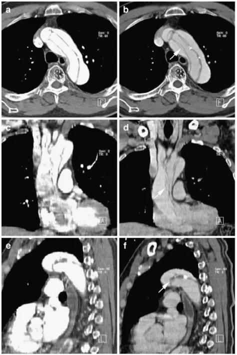
(c, d), and oblique (e, f) MPRs of an ascending aorta dissection with distention in the aortic arch, brachiocephalic trunk, and descending aorta. The dissection membrane is slightly blurred and shows a double contour (arrows) in the nongated images (b, d, f) compared with the gated images (a, c, e). However, the correct diagnosis of an aortic dissection could be made with both acquisition techniques
Fig. 4 Axial CT image in lung window settings showing a lung contusion (arrow) on the left side in a patient after blunt chest trauma. No significant differ-ence in detection of lung con-tusion was revealed between the ECG-gated (a) and the nongated (b) images
complex anywhere in the image stack; 2=slight blurring of the interlobar fissures, pulmonary vessels, perilob-ular septae, and the centrilobperilob-ular complex; 3= clearly detectable motion artifacts with severe blurring or double contours anywhere in the image stack with slightly confined assessment of the lung parenchyma; and 4=motion artifacts in more than 50% of the image stack with distinctly confined assessment of the lung parenchyma.
Detection of parenchymal injuries of the lung was assessed using the following five-point scale: 1=lung contusion definitely not present; 2=lung contusion possible, but most likely not present; 3=indeterminate; 4=lung contusion most likely present; and 5=lung con-tusion definitely present. The following parenchymal changes in patients with acute chest trauma were con-sidered as lung contusions: 1=ground glass attenuation, 2=focal consolidation, 3=parenchymal tear with visible air-filled cavitation, and 4=parenchymal tear with ac-tive bleeding. Subjecac-tive diagnostic certitude based on overall image quality with respect to injuries of the lung parenchyma were rated with the following scores: 1=distinctly assessable, 2=slight incertitude, 3=strong incertitude, and 4=not assessable.
Bony chest
With respect to interpretability, bony thoracic struc-tures, especially with regard to the spine and the ribs,
were separately scored for ECG-gated and nongated data sets. For the assessment of the ribs in axial and oblique MPRs, the following scores were used: 1=no motion artifacts with excellent assessment of the cor-tical bone; 2=moderate motion artifacts, blurring of the bone contours, unconfined assessment (double con-tours of the cortical bone) of not more than one or two ribs; 3=severe motion artifacts with confined assess-ment of more than two ribs; and 4=severe motion ar-tifacts, bone chest structures not assessable.
For evaluating the spine in axial images, coronal MPRs, and sagittal MPRs, the following scores were used: 1=trabecular structures well defined, no corti-cal double contours; 2=trabecular structures slightly blurred; 3=trabecular structures distinctly blurred and cortical double contours detected, fractures and luxa-tions firmly ruled out; and 4=severe motion artifacts with distinctly confined assessment.
The two radiologists rated the ability to detect the fracture of the spine with the following scores: 1= fracture definitely not present; 2=fracture most likely not present; 3=indifferent, not sure if a fracture is present; 4=fracture most likely present; and 5=fracture definitely present.
For assessment of subjective diagnostic certainty with respect to spine fractures, the following scores were used: 1=distinctly assessable, 2=slight incertitude, 3=strong incertitude, and 4=not assessable.
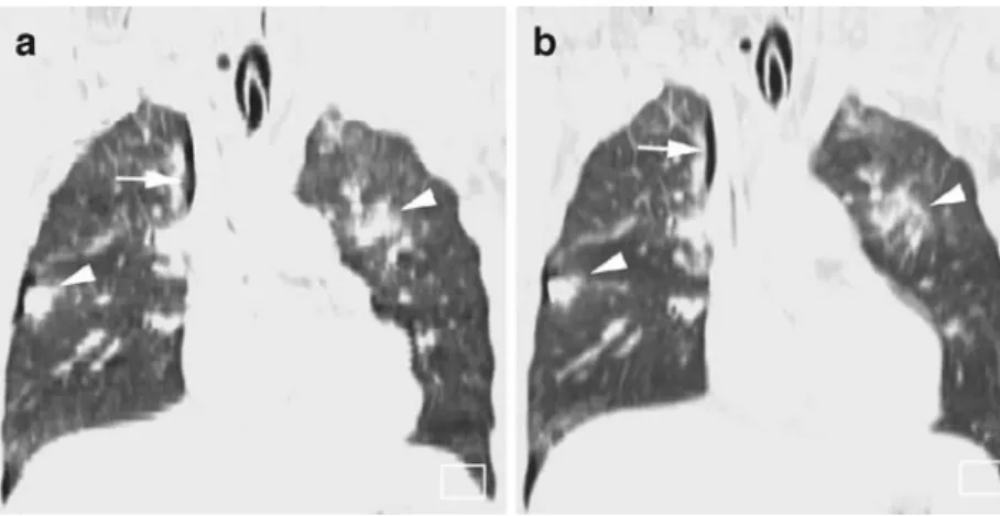
Fig. 5 Coronal MPRs showing lung contusions (arrowhead) in both lungs and a right-sided pneumothorax (arrow) in a me-chanically ventilated 38-year-old male patient after blunt chest trauma. The nongated image (b) shows a sharper and clearer demarcation of the lung paren-chyma and of the diaphragm compared with the gated image (a). Furthermore, the pathologic image findings such as consoli-dations and the right-sided pneumothorax are better shown in the nongated image (b)
Fig. 6 Coronal MPRs at the level of the carina. The nongated image (b) shows a sharp de-marcation of the carina and the lung parenchyma. In contrast, stair step artifacts (arrowheads) in the carina and blurring of the lung parenchyma (arrow) can be detected in the ECG-gated im-ages (a) and decrease the image quality of the gated images
Radiation dose
To compare the radiation exposure, effective radiation doses were calculated using a commercially available computer program (WinDose, version 2.1a; Scandi-tronix-Wellhöfer Dosimetrie, Schwarzenbruck, Ger-many) [13]. Effective radiation dose calculations in this software program are based on Monte Carlo calcula-tions for anthropomorphic mathematic phantoms that were obtained by the GSF National Research Center for Environment and Health (Neuherberg, Germany) [13]. By entering different scan parameters, including colli-mation, pitch, kerma, tube current, tube voltage, scan-ning range, anatomic area, and the patient’s gender, the software program provides an estimation of the ef-fective radiation dose [13].
Statistical analysis
For statistical analysis, commercially available software was used (SSPS 11.5 for Windows, SPSS Inc., Chicago, IL, USA). The Wilcoxon signed rank test was used to compare the parameters between ECG-gated and non-ECG-gated MDCT. P values less than 0.05 were considered, indicating statistically significant differences.
Results
Impact of retrospectively ECG-gated acquisition on workflow in the trauma emergency center
All CT examinations were performed as planned. The average time for the installation of the additional equipment for ECG gating was 1.8±0.5 min (range, 1.1–2.5 min). This time was integrated into the usual time frame necessary for the preparation of a trauma patient for a CT examination. A significant delay of the workflow was not encountered in any patient. The average transfer and preparation time was 12.1±1.7 min (8.1–14.5 min) for the 32 patients of the study population with ECG-gated acquisition and 12.3±1.7 min
(8.4–14.7 min) for the 32 patients with acute trauma and with nongated acquisition. The latter represented the con-trol group for this time measurement. The trauma surgeon in charge never took the option of terminating an ECG-gated study on a particular patient. Qualitative assessment of the workflow performed by the trauma surgeon in charge did not show delays caused by the installation of the ECG-gating equipment.
Assessment of image quality
Breathing artifacts The mean scores and standard devia-tions (SD) for rating of breathing artifacts for the ECG-gated and nonECG-gated series were 2.1±0.8 and 1.7±0.6, respectively, and were not significantly different (P=0.052) (Table 3).
Assessment of thoracic vascular structures Assessment of the aortic valve in both the axial and the oblique projections revealed significantly reduced motion artifacts when using ECG-gated data sets compared with the nongated series (P<0.001) (Table3). The ascending aorta at the level of the origin of the left main coronary artery and at the level of the pulmonary trunk in ECG-gated axial images showed significantly fewer motion artifacts compared with non-ECG-gated data (P<0.001). Even in an oblique projection of the ascending aorta, pulsation artifacts were less pro-nounced in the ECG-gated series (P<0.001) (Table3). The rating of motion artifacts of the supra-aortic vessels, the aortic arch, and the descending aorta in the oblique re-format was significantly better for the ECG-gated series (P<0.001) (Table3), and the subjective diagnostic certainty regarding the entire aorta was significantly better with ECG gating (Table3). Figures1and2show non-gated and ECG-gated images of the aortic root with typical motion artifacts in the non-ECG-gated series. Figure 3shows an example of an aortic dissection with double contour artifacts of the dissection membrane with non-gated technique.
Lung parenchyma The rating of image quality of the lung parenchyma in axial as well as in coronal projections revealed significantly better results for the nongated series
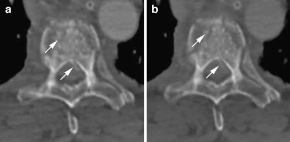
Fig. 7 Axial CT image showing a compression fracture of the sixth thoracic vertebra after blunt chest trauma. No signifi-cant difference in detection of bone injuries was seen between the ECG-gated series (a) and the nongated series (b). However, the image quality with respect to the sharpness of the cortical bone structures (arrow) between the two acquisition techniques is more in favor of the nongated technique (b)
(P<0.001) (Table 3). The detectability of lung contusions was rated slightly better with the non-ECG-gated series, but no significant difference between both data sets was seen (P=0.063). Figure 4 shows examples of lung contusions with both techniques in comparable image quality. Figure5 shows examples of lung contusions with both techniques in coronal multiplanar reformations with slightly better image quality with non-gated technique. Enlarged views of the carina (Fig. 6) reveal stair step artifacts occuring with the ECG-gated technique which are not seen with non-gated acquisition. Consequently, the subjective diagnostic cer-tainty with respect to parenchymal injuries of the lung was rated significantly better for the non-ECG-gated data (P<0.001) (Table3).
Bony chest structures The rating of the image quality of the thoracic spine in axial images, sagittal MPR, and coronal MPR showed significantly better results in the non-ECG-gated series (P<0.05), whereas the rating of spine fracture detection revealed no significant difference
between ECG-gated and non-ECG-gated data (Table3). In Fig.7the sharper demarcation of the bone structures with non-gated acquisition is shown. However, the subjective diagnostic certainty with respect to spine fractures was not significantly different between the two data sets (Table3). The assessment of the ribs in axial and coronal projections showed significantly better results for the non-ECG-gated data (P≤0.003) (Table3).
Radiation dose The mean effective dose was 29.5 mSv for females and 23.6 mSv for males in the ECG-gated series and 6.9 mSv for females and 5.7 mSv for males in the non-ECG-gated series, with a mean thoracic scan length of 30.7 cm (P<0.005) (Table3).
Comparison of diagnostic concordance
The final clinical diagnoses are summarized in Table 4. These diagnoses included the results of all clinical exam-Table 4 Overview over the
final clinical diagnoses in the patient population
Patients with suspected nontraumatic dissection
Patients with blunt chest trauma
Total 15 17
No pathology detected 4 0
Pathology detected 11 17
Traumatic thoracic injuries of the bony structures
Fractures of thoracic vertebrae 4
Fractures of one or more ribs 10
Traumatic injuries of the lung parenchyma
Lung contusions 7
Pneumothoraces 6
Pathologic changes of the great thoracic vessels
Stanford type A dissections 3 Stanford type B dissections 2 Ectasia or aneurysma
of the ascending aorta
3 Ectasia or aneurysma
of the descending aorta
2
Aortic elongation 1
Other thoracic pathologies
Pulmonary embolism 1
Struma 1
Bronchogenic tumor 1
Pericarditis calcarea 1
Mediastinal hematoma 3
Other extrathoracic pathologies
Presternal hematoma 1
Fractures of upper lumbar vertebrae 5
Laceration of the spleen 2
Laceration of the liver 3
Extraluminal subphrenic air of the gastrointestinal tract
inations and imaging studies performed during the patient’s hospital stay and were considered as the reference standard for the presence of disease. Compared with this reference standard, ECG-gated and nongated acquisition performed equally well in the detection of disease, despite the dif-ferences in image quality. All fractures, lung contusions, and vascular abnormalities in the study population were detected correctly with both imaging techniques.
Discussion
Acute traumatic injury of the thoracic aorta and acute nontraumatic aortic dissection are rare but clinically im-portant. They may be confined to the intima and media layer only or may occur as a transmural rupture (e.g., af-fecting intima, media, and adventitia of the aorta). In both cases, a fast and correct diagnosis of the injury may be lifesaving. Introduction of a new CT acquisition technique for assessment of patients with acute aortic disease in the emergency setting has to fulfill two conditions: It must not slow down the diagnostic workup, and it must provide significantly better image quality regarding the vascular injury. Image quality with respect to bone and soft tissue injuries should be unaffected or only reduced to a level that does not compromise detection of traumatic injury.
Influence of ECG gating on the diagnostic workup Negative effects on workflow can be prevented mainly by adequate training of the CT technicians. This was per-formed in the above-mentioned training program. Ten CT technicians (five permanent technicians from the CT de-partment and five additional technicians) were trained to adequately perform ECG-gated CT examinations in an emergency setting. Our results show that given adequate training and workflow design, cardiac gating does not delay the diagnostic workup. One might argue that our data are inadequate for assessing the influence of the new acquisi-tion technique on workflow. However, we were forced to limit data acquisition for study purposes in the emergency setting to the most relevant data. One principal interest of the trauma surgeon was aimed at staying in the so-called “golden hour” [14], e.g., to finish the whole diagnostic workup of the patient within 1 h after admission. It can be assumed that the involved clinicians were meticulous in observing and assessing the time frame and the workflow to avoid any delays. Thus, the acquired data are likely to be objective regarding the clinicians’ assessment of the clinical workflow.
Patient selection
The patient population in the current study was heteroge-neous. It consisted of patients assessed after blunt trauma and patients with suspected aortic dissection. This could be considered a limitation of the study. However, CT
tech-nique is the same for assessment of both patient groups, and assessment of the image quality was the principal endpoint of the study. Following the strategy of including both trau-ma patients as well as patients with suspected nontrautrau-mat- nontraumat-ic aortnontraumat-ic dissections, we were able to recruit a population with a high percentage of aortic pathology as well as other posttraumatic pathologies of the chest.
Critical issues of ECG-gated data acquisition in the emergency setting
The current study was designed to assess whether ECG-gated MDCT of the chest in critically ill patients is robust enough for routine use in an emergency setting. Roos et al. [9] showed that cardiac-gated MDCT was superior than nongated MDCT for the assessment of the thoracic aorta. However, their study was performed on nonemergency patients. Data acquisition for cardiac-gated MDCT is based on a pitch well below one to acquire raw data over the whole cardiac cycle for selective reconstruction in an arbitrary part of the cardiac cycle [10]. The small table feed increases susceptibility for motion artifacts other than car-diac motion, such as breathing artifacts. Furthermore, pa-tients cannot be preselected or premedicated to adjust the heart rate as typically done in the nonemergency situation with cardiac-gated acquisition.
Breathing artifacts
Performing ECG-gated data acquisition in critically ill pa-tients in the emergency suite is much more challenging than that in nonemergency patients. To test the robustness of the acquisition technique, we chose a patient population that required a CT examination within the first hour after admission to the emergency department. Such patients are much less prone to follow breathing commands or to perform a breath-hold during the entire data acquisition. Furthermore, a certain subpopulation of patients will be mechanically ventilated during data acquisition. In this study, this was the case with 15 patients, and mechanical ventilation was not stopped during data acquisition. The scan protocol was tailored for critically ill patients such that breath-hold times were minimized. This was achieved by finding a compromise between axial resolution and ac-quisition time. A collimation of 4 × 2.5 mm was used for both the gated and the nongated series. This scan protocol permits the reconstruction of axial images in 3-mm slice thickness. Roos et al. [15] demonstrated that a slice thick-ness of 3 mm is sufficient to rule out spinal fractures even under the condition that no enlarged (e.g., dedicated small field of view) reconstruction of the spine is performed. Furthermore, acquisition of the whole chest in ECG-gated mode in a single breath-hold is possible with this colli-mation, and tube-cooling times are less frequently occur-ring compared with 1-mm collimation.
Patients with sedation and mechanical ventilation could obviously not follow breathing commands. Thus,
respira-tory artifacts and compromise of the data quality of the lung parenchyma were likely to be encountered. In this subgroup of patients, image quality reduction related to the absence of breath-hold was likely to occur.
Heart rate
In critically ill patients, an increased heart rate is a chal-lenging factor for ECG-gated acquisition. The optimal heart rate (e.g., the heart rate that allows for the maximum temporal resolution on a particular CT scanner) depends on the minimal gantry rotation time the scanner offers and on the reconstruction algorithm. The Siemens VolumeZoom VA41 is a 4-row scanner with a minimal rotation time of 0.5 ms. The minimal temporal resolution is 250 ms when one heart cycle is used for image reconstruction. This time will be reduced to 125 ms when the ACV (segmented ACV reconstruction) is used. If this option is enabled, one heart cycle will be used in case of a heart rate of utmost 65 bpm. Two heart cycles will be used when the heart rate increases to more than 65 bpm. However, the best image quality in coronary artery imaging is achieved with heart rates below 60 bpm [16]. In patients referred for MDCT angiography of the coronary arteries, beta receptor blockers are typically administered before data acquisition to reduce heart rate. This is of course not an option for traumatic patients in the emergency setting. The imaging technique used in such patients should cope with higher heart rates and deliver a sufficient image quality irrespective of the heart rate. The mean heart rate in the current patient population was 80±17 (mean±SD; range, 48–110) and was, therefore, far from the optimum.
Assessment of image quality and diagnostic concordance
Image quality of the thoracic aorta was far better when using the ECG-gated technique compared with that when using the nongated technique for the aortic valve, the as-cending aorta, the aortic arch, and the aortic isthmus. These anatomic regions are responsible for about 98% of the traumatic aortic injuries [2,3,17–23] and for about 90% of the nontraumatic aortic dissections. We may, therefore, suppose that patients with traumatic aortic injury and aortic dissection may profit from ECG-gated acquisition, espe-cially when the intimal lesions and intramural hematoma of the aortic wall are small. Such decent lesions might be overlooked when using the nongated technique in the pres-ence of motion artifacts. In the present series, all five aortic dissections were correctly diagnosed with the ECG-gated as well as the nongated series. However, assessment of the ascending aorta (18 patients with double contour of the vessel wall or a severely blurred aortic wall), of the aortic arch (4 patients with severe blurring of the vessel wall), and of the proximal descending aorta (4 patients with se-vere blurring of the vessel wall) was sese-verely confined in the nongated images.
In the ECG-gated images, this was the case in only one patient. This caused a much better subjective diagnostic certainty when using ECG gating. The detection of aortic pathology was not impaired when using non-ECG gating, but the study population did not contain patients with in-tramural hematomas or other faint pathologic changes of the ascending aorta, which are only detectable with a pul-sation artifact-free series. This may legitimize the signif-icantly higher estimated radiation dose for ECG-gated ac-quisition compared with that for nongated data acac-quisition. However, we cannot draw definitive conclusions regarding the impact of the better image quality achieved by cardiac gating on improvement of the diagnostic outcome because the number of patients with aortic pathology is far too low in this preliminary study. Further studies on a much larger patient population are required to assess the diagnostic purpose of ECG gating on detection of faint pathologic changes of the aorta in cases of acute traumatic or non-traumatic aortic disease.
Despite the high heart rates and insufficient cooperation of the patients with regard to breath-holding, the image quality of the aorta in the ECG-gated series was significantly better than that of nongated CT examinations. Thus, ECG-gated acquisition can be considered to be robust enough to work well even under these unfavorable conditions.
Reduced image quality without diagnostic compromise to the cardiac-gated images compared to nongated acqui-sition was encountered in this study for soft tissue and bone. Reduced image quality might have occurred for the fol-lowing two reasons: the longer acquisition time for cardiac gating might increase the frequency of breathing artifacts, or the reduced image quality may be caused by the dif-ferences in the reconstruction algorithm itself. Breathing artifacts were rated for both methods, and the scores did not differ significantly. Supposing that the breathing artifact score discriminates well between the methods, the recon-struction algorithm is causing the differences of image quality between ECG-gated and nongated acquisition. However, despite the significantly lower image quality of cardiac-gated acquisition regarding bone and lung imaging, all pathologies (spine fractures, rib fractures, lacerations of upper abdominal parenchymal organs, and lung contu-sions) were correctly detected using both techniques.
Limitations
We acknowledge the following limitations of this study. First, one drawback is the limited possibility to blind the studies regarding acquisition technique. The readers were not told directly what imaging technique was used in a certain CT series. However, the severe differences in mo-tion artifact reducmo-tion in the aortic root as well as the dif-ferences in contrast enhancement might have revealed the imaging technique to the readers and caused a certain bias. Second, we were not able to recruit a large number of patients with aortic disease and certain posttraumatic changes of the chest. Therefore, our data may not be used to assess the influence of the imaging technique on
diagnostic quality regarding detection of aortic injury, spine fractures, etc.
Finally, this study was performed using a 4-row MDCT scanner. Scanners which use the 16- or 64-row technique may provide better image quality due to faster scanning with higher axial resolution (i.e., 1-mm slice width or lower). Fast scanning may reduce pulsation artifacts of the aorta without application of cardiac gating. The gain in image quality achieved by cardiac gating compared with a nongated study may, therefore, be lower in a 64-row scanner compared with that in a 4-row scanner. The dif-ferences may even be inconsiderable.
However, if cardiac gating is performed on a 64-row scanner in the emergency setting, its influence on workflow remains unchanged, as compared with a 4-row scanner. The principal differences of the image reconstruction algorithms remain unchanged as well. A definite conclusion regarding the impact of more recent scanner hardware on image quality requires additional studies.
Conclusion
The design of the study and the patient selection strategy permitted the assessment of the robustness of ECG-gated acquisition in the emergency setting in comparison with nongated acquisition. After adequate training, ECG gating could be performed without delaying the workflow in an emergency setting and yielded an improved image quality of the aorta. Image quality of the lung parenchyma and bone structures was reduced but without diagnostic compromise. Further studies are required to assess the impact of cardiac gating on diagnostic outcome.
Acknowledgements This research has been supported by the NCCR CO-ME of the Swiss National Science Foundation. We thank Ulrich Helfenstein, MD, and Burkhardt Seifert, PhD, for their contributions to the statistical analyses.
References
1. Rieger M, Sparr H, Esterhammer R et al (2002) Modern CT diagnosis of acute thoracic and abdominal trauma. Radiologe 42: 556–563
2. Feczko JD, Lynch L, Pless JE, Clark MA, McClain J, Hawley DA (1992) An autopsy case review of 142 nonpenetrating (blunt) injuries of the aorta. J Trauma 33:846–849
3. Wintermark M, Wicky S, Schnyder P (2002) Imaging of acute traumatic injuries of the thoracic aorta. Eur Radiol 12:431–442
4. Khan IA, Nair CK (2002) Clinical, diagnostic, and manage-ment perspectives of aortic dissection. Chest 122:311–328 5. Hagan PG, Nienaber CA, Isselbacher EM et al (2000) The
International Registry of Acute Aortic Dissection (IRAD): new insights into an old disease. JAMA 283:897–903
6. Mirvis SE, Shanmuganathan K, Miller BH, White CS, Turney SZ (1996) Traumatic aortic injury: diagnosis with contrast-enhanced thoracic CT—five-year experience at a major trauma center. Radiology 200:413–422
7. Gavant ML, Flick P, Menke P, Gold RE (1996) CT aortography of thoracic aortic rupture. AJR Am J Roentgenol 166:955–961 8. Fishman JE (2000) Imaging of blunt aortic and great vessel
trauma. J Thorac Imaging 15:97–103
9. Roos JE, Willmann JK, Weishaupt D, Lachat M, Marincek B, Hilfiker PR (2002) Thoracic aorta: motion artifact reduction with retrospective and prospective electrocardiography-assisted multi-detector row CT. Radiology 222:271–277
10. Ohnesorge B, Flohr T, Becker C et al (2000) Cardiac imaging by means of electrocardiographically gated multisection spiral CT: initial experience. Radiology 217:564–571
11. Kachelriess M, Ulzheimer S, Kalender WA (2000) ECG-correlated image reconstruction from subsecond multi-slice spiral CT scans of the heart. Med Phys 27:1881–1902 12. Vogl TJ, Abolmaali ND, Diebold T et al (2002) Techniques for
the detection of coronary atherosclerosis: multi-detector row CT coronary angiography. Radiology 223:212–220
13. Kalender WA, Schmidt B, Zankl M, Schmidt M (1999) A PC program for estimating organ dose and effective dose values in computed tomography. Eur Radiol 9:555–562
14. McLaughlin JS, Shama Z, Hirsch E, Khazei AH, Attar S, Cowley A (1969) Cardiovascular dynamics in human shock. Am Surg 35:166–176
15. Roos JE, Hilfiker P, Platz A et al (2004) MDCT in emergency radiology: is a standardized chest or abdominal protocol suf-ficient for evaluation of thoracic and lumbar spine trauma? AJR Am J Roentgenol 183:959–968
16. Nieman K, Rensing BJ, van Geuns RJ et al (2002) Non-invasive coronary angiography with multislice spiral computed tomography: impact of heart rate. Heart 88:470–474
17. Ben-Menachem Y (1993) Rupture of the thoracic aorta by broadside impacts in road traffic and other collisions: further angiographic observations and preliminary autopsy findings. J Trauma 35:363–367
18. Creasy JD, Chiles C, Routh WD, Dyer RB (1997) Overview of traumatic injury of the thoracic aorta. Radiographics 17:27–45 19. Cheng I, McLellan BA, Joyner C, Christakis G (2000) Aortic root trauma: serious injuries requiring early recognition and management. J Trauma 48:525–529
20. Bashar AH, Kazui T, Washiyama N et al (2002) Stanford type a aortic dissection after blunt chest trauma: case report with a reflection on the mechanism of injury. J Trauma 52:380–381 21. Ono M, Yagyu K, Furuse A, Kotsuka Y, Kubota H (1998) A
case of Stanford type A acute aortic dissection caused by blunt chest trauma. J Trauma 44:543–544
22. Perchinsky M, Gin K, Mayo JR (1998) Trauma-associated dissection of the thoracic aorta. J Trauma 45:626–629 23. Gammie JS, Katz WE, Swanson ER, Peitzman AB (1996)
Acute aortic dissection after blunt chest trauma. J Trauma 40: 126–127