HAL Id: hal-01434808
https://hal.archives-ouvertes.fr/hal-01434808
Submitted on 12 Jan 2021HAL is a multi-disciplinary open access
archive for the deposit and dissemination of sci-entific research documents, whether they are pub-lished or not. The documents may come from teaching and research institutions in France or abroad, or from public or private research centers.
L’archive ouverte pluridisciplinaire HAL, est destinée au dépôt et à la diffusion de documents scientifiques de niveau recherche, publiés ou non, émanant des établissements d’enseignement et de recherche français ou étrangers, des laboratoires publics ou privés.
Oxidative DNA damage and repair in the radioresistant
archaeon Thermococcus gammatolerans
Ewa Barbier, Arnaud Lagorce, Amine Hachemi, Murielle Dutertre, Aurore
Gorlas, Lucie Morand, Christine Saint-Pierre, Jean-Luc Ravanat, Thierry
Douki, J. Armengaud, et al.
To cite this version:
Ewa Barbier, Arnaud Lagorce, Amine Hachemi, Murielle Dutertre, Aurore Gorlas, et al.. Ox-idative DNA damage and repair in the radioresistant archaeon Thermococcus gammatolerans. Chemical Research in Toxicology, American Chemical Society, 2016, 29 (11), pp.1796-1809. �10.1021/acs.chemrestox.6b00128�. �hal-01434808�
Oxidative DNA Damage and Repair in the Radioresistant Archaeon
Thermococcus gammatolerans
Ewa Barbier,
†,‡Arnaud Lagorce,
§,∥Amine Hachemi,
§Murielle Dutertre,
§Aurore Gorlas,
§Lucie Morand,
†,‡Christine Saint-Pierre,
†,‡Jean-Luc Ravanat,
†,‡Thierry Douki,
†,‡Jean Armengaud,
⊥Didier Gasparutto,
†,‡Fabrice Confalonieri,
§and Jean Breton
*
,†,‡†University of Grenoble Alpes, INAC, LCIB, F-38000 Grenoble, France ‡CEA, INAC, SyMMES, F-38000 Grenoble, France
§University of Paris-Sud, Institute for Integrative Biology of the Cell (I2BC), Université Paris Saclay, CEA, CNRS, Orsay, France ∥University of Perpignan, IHPE− UMR 5244 CNRS/IFREMER/Univ. Montpellier, Montpellier, F-34095, France
⊥CEA, DSV-Li2D, Laboratory“Innovative Technologies for Detection and Diagnostics”, BP 17171, Bagnols-sur-Cèze, F-30207,
France
*
S Supporting InformationABSTRACT: The hyperthermophilic archaeon Thermococcus gammatolerans can resist huge doses of γ-irradiation, up to 5.0 kGy, without loss of viability. The potential to withstand such harsh conditions is probably due to complementary passive and active mechanisms, including repair of damaged chromosomes. In this work, we documented the formation and repair of oxidative DNA lesions in T. gammatolerans. The basal level of the oxidized nucleoside, 8-oxo-2′-deoxyguanosine (8-oxo-dGuo), was estab-lished at 9.2 (± 0.9) 8-oxo-dGuo per 106nucleosides, a higher level than those usually measured in eukaryotic cells or bacteria. A significant increase in oxidative damage, i.e., up to 24.2 (± 8.0)
8-oxo-dGuo/106nucleosides, was measured for T. gammatolerans exposed to a 5.0 kGy dose ofγ-rays. Surprisingly, the yield of
radiation-induced modifications was lower than those previously observed for human cells exposed to doses corresponding to a few grays. One hour after irradiation, 8-oxo-dGuo levels were significantly reduced, indicating an efficient repair. Two putative base excision repair (BER) enzymes, TGAM_1277 and TGAM_1653, were demonstrated both by proteomics and transcriptomics to be present in the cells without exposure to ionizing radiation. Their transcripts were moderately upregulated after gamma irradiation. After heterologous production and purification of these enzymes, biochemical assays based on electrophoresis and MALDI-TOF (matrix-assisted laser desorption ionization−time of flight) mass spectrometry indicated that both have a β-elimination cleavage activity. TGAM_1653 repairs 8-oxo-dGuo, whereas TGAM_1277 is also able to remove lesions affecting pyrimidines (1-[2-deoxy-β-D-erythro-pentofuranosyl]-5-hydroxyhydantoin (5-OH-dHyd) and 1-[2-deoxy-β-D
-erythro-pentofuranosyl]-5-hydroxy-5-methylhydantoin (5-OH-5-Me-dHyd)). This work showed that in normal growth conditions or in the presence of a strong oxidative stress, T. gammatolerans has the potential to rapidly reduce the extent of DNA oxidation, with at least these two BER enzymes as bodyguards with distinct substrate ranges.
■
INTRODUCTIONExtremophile microorganisms have developed a wide range of mechanisms to survive in harsh environmental conditions. Thermococcus gammatolerans, a hyperthermophilic prokaryote described as the most radioresistant archaeon identified to date, was isolated from samples obtained in a hydrothermal vent located in the Gulf of California.1 This obligatory anaerobic heterotrophic organism has a genome of 2.04 megabases (Mb), shows an optimal growth temperature of 88 °C, and fully withstands a 5.0 kiloGrays (kGy) dose of gamma irradiation without loss of viability.2 The molecular and biological mechanisms explaining this remarkable resistance of T. gammatolerans have not yet been defined. This microorganism probably combines different and complementary strategies to
resist such hostile conditions, as is the case for other radiotolerant organisms.3 Ionizing radiation is associated with the production of reactive oxygen species (ROS), passive resistance mechanisms consist of the modification of the Mn/ Fe intracellular ion ratio and the production of “compatible solutes” composed of chemical agents such as polyols, sugars, and amino acids allowing the stabilization of macromolecules.4 A more active mechanism involves combating oxidative stress using antioxidant enzymes. Finally, another way to resist ionizing radiation is to repair shattered chromosomal DNA as efficiently as possible. Pulsed field gel electrophoresis has
Received: April 18, 2016
Published: September 27, 2016
allowed time course analysis of the archaeon chromosomal repair after irradiation. Such experiments have indicated that T. gammatolerans chromosomes are fully degraded after a 5.0 kGy dose ofγ-rays. DNA strand breaks are fully repaired after 5 h by cells in stationary phase, while 6 h are necessary for cells in exponential growth phase.5 Proteomic approaches have demonstrated that T. gammatolerans produces key factors of the main DNA repair systems such as base excision repair (BER) and double-strand break repair.2It is worth noting that some of these proteins are present in the absence of stress, which may be useful for survival in the harsh conditions that constantly surround these extremophiles or during drastic environmental changes that are especially unfavorable to life. Despite a growing amount of data regarding radiotolerant organisms, DNA repair mechanisms in Archaea, and partic-ularly in T. gammatolerans, remain poorly understood.
Most of the DNA alterations resulting from high doses of radiation have never been analyzed in radioresistant archaea. Among these, 8-oxo-2′-deoxyguanosine (8-oxo-dGuo) is a classic biomarker of oxidative stress. Its chemical structure is presented inFigure 1. The main mechanism of formation of 8-oxo-dGuo upon exposure to ionizing radiation depends on an indirect effect: the radiolysis of water generates the hydroxyl radical ̇OH which reacts with C8 of the guanine.6 Mutations (mainly G→ T transversions) can occur in the genome if 8-oxo-dGuo is not repaired prior to replication.7 Sample preparation and measurement of 8-oxo-dGuo are technically challenging to avoid DNA artifactual oxidation, and as a consequence misleading results can be found in the literature.6 DNA lesions 5′,8-cyclonucleosides (Figure 1) are radiation-induced, tandem base-sugar alterations usually produced in
lower yields than 8-oxo-dGuo but with a more severe biological impact resulting from a drastic alteration of the DNA structure.8 The formation of these complex DNA lesions is known to be favored at low oxygen concentrations.9The large radiation dose that can be applied to T. gammatolerans cells and their natural anaerobic growth conditions makes the quantitation of 5′,8-cyclonucleosides highly relevant.
In the present study, we first quantified oxidative DNA damage in T. gammatolerans, both at basal levels and after exposure to high doses of γ-irradiation, by measuring 8-oxo-dGuo and 5′,8-cyclonucleosides by HPLC coupled to tandem mass spectrometry (HPLC-MS/MS). We also documented the molecular mechanisms of two putative BER enzymes identified in the genome: TGAM_1277, initially annotated as a putative type III endonuclease (endoIII), and TGAM_1653, a predicted DNA glycosylase,2 phylogenetically close to the 8-oxoguanine glycosylase from Pyrobaculum aerophilum.10 Transcriptomics and proteomics were used to assess their presence in T. gammatolerans with and without exposure to irradiation. After their heterologous production, TGAM_1277 and TGAM_1653 proteins were assayed for their cleaving activities on different oxidative DNA lesions by electrophoresis and matrix-assisted laser desorption ionization−time of flight (MALDI-TOF) mass spectrometry.
■
EXPERIMENTAL PROCEDUREST. gammatolerans Growth Conditions. The T. gammatolerans EJ3 strain was cultivated in serum bottles or in 1 L Schott bottles, under anaerobic conditions at 85 °C in complex organic medium (VSM), as described in ref2. Cell culture densities were measured by Figure 1.Chemical structures of the DNA lesions mentioned in this work. 8-oxo-dGuo, 5-OH-dHyd, and 5-OH-5-Me-dHyd represent single oxidation products of 2′-deoxyguanosine, 2′-deoxycytidine, and thymidine, respectively. 5′R-cyclo-dAdo represents an example of 5′,8-cyclonucleosides that are considered as a family of complex oxidative DNA damage. 8-oxo-dGuo and 5′,8-5′,8-cyclonucleosides were measured in T. gammatolerans DNA at basal levels and after irradiation. 8-oxo-dGuo, 5-OH-dHyd, 5-OH-5-Me-dHyd, and 5′,8-cyclonucleosides were tested as potential substrates for the T. gammatolerans enzymes, TGAM_1653 and TGAM_1277.
optical microscopy (Olympus BH-29) using a Thoma counting chamber (Microgravure Precis).
Gamma Irradiation and DNA Extraction. Gamma Irradiation. For 8-oxo-dGuo quantification after irradiation, large volumes of exponentially growing cells (600 mL in 1 L Schott bottles, 7× 10⁷ cells/mL) werefiltered (MicraCloth Calbiochem, La Jolla, CA, USA) to remove sulfur compounds from the VSM-S° medium. In anaerobic conditions, cells were concentrated ×10 in Hungate tubes for γ-irradiation. Cells were then exposed on ice to a dose of 2.5 kGy or 5.0 kGy (100% survival)5using a137Csγ-ray source (40 Gy/min, IBL637
CisBio International, Institut Curie, Orsay, France). As a control, the same number of nonirradiated cells was kept on ice during irradiation. Irradiated and nonirradiated cells were either collected immediately or incubated for 60 min at 85°C for recovery in fresh VSM-S° medium on a reciprocal shaker before centrifugation (4000g, 20 min at 4°C). Cells were immediately frozen and stored at −80 °C until further processing. Each culture was performed in independent duplicates.
For gene expression assays, 600 mL of exponentially growing cells werefiltered (MicraCloth Calbiochem, La Jolla, CA, USA) to remove sulfur compounds from the VSM-S° medium and concentrated 10-fold by centrifugation. Cells were exposed on ice to a dose of 2.5 kGy or 5.0 kGy using a137Csγ-ray source (19 Gy/min, GSR D1 GSM, Institut
Curie, Orsay, France). As a control, the same number of nonirradiated cells was kept on ice during irradiation. Irradiated and nonirradiated cells were either collected immediately or incubated for 60 or 120 min at 85°C for recovery in fresh VSM-S° medium on a reciprocal shaker before centrifugation (4000g, 20 min at 4°C). Cells were immediately frozen and stored at−80 °C until further processing. Each culture was repeated twice.
DNA Extraction and Digestion. T. gammatolerans cells (10 mg) were supplemented with 300μL of lysis buffer containing 100 mM Tris-HCl at pH 8.3, 50 mM EDTA, 100 mM NaCl, and 0.15 mM deferoxamine. Thirty microliters of 10% SDS (v/v) and 30μL of 10% sarkosyl (v/v) were then added to the cell suspension. The samples were incubated with 150μg of RNase A (Sigma) and 3.5 U RNase T1 (Sigma) for 15 min at 50°C. Six hundred micrograms of protease (Qiagen) was added, and the incubation was pursued for 1.5 h at 37 °C. DNA was precipitated with 600 μL of 20 mM EDTA-Na2, 7.6 M
NaI, 40 mM Tris-HCl, 0.3 mM deferoxamine at pH 8, and 1 mL of 100% isopropanol. The pellets were washed with 40% isopropanol (v/ v) and subsequently with 70% ethanol (v/v), andfinally suspended in 50μL of 0.1 mM deferoxamine.
The DNA digestion was performed in two steps. First, 0.025 U phosphodiesterase II, 2.5 U DNase II, 0.5 U nuclease P1 (Sigma), and 2.5μL of buffer (200 mM succinic acid and 100 mM CaCl2, pH 6)
were added, and the samples were incubated for 2 h at 37°C. Then, 6 μL od 500 mM Tris-HCl, 1 mM EDTA at pH 8, 0.015 U phosphodiesterase I (Worthington), and 2 U alkaline phosphatase (Sigma) were added, and the samples were incubated for 2 h at 37°C. The reaction was stopped by adding 3.5 μL of 0.1 N HCl. For cyclonucleoside detection, thefirst step of the digestion was repeated before adding HCl, as optimized previously.9
HPLC-MS/MS Quantification of 8-Oxo-dGuo and 5′,8-Cyclo-nucleosides. 8-Oxo-dGuo. Digested DNA samples were resolved on an Uptisphere 2× 150 mm octadecylsilyl silica gel column (Interchim) operated with an Agilent series 1100 HPLC system. The mobile phase was a gradient of methanol in 2 mM aqueous ammonium formate (2 to 10% (v/v) in 25 min and 10 to 25% (v/v) in the following 10 min). Undamaged nucleosides were monitored with a UV detector set at 260 nm, while 8-oxo-dGuo was measured in positive mode with an API 3000 triple quadrupolar mass spectrometer (PerkinElmer/SCIEX) equipped with electrospray ionization (ESI). The system was operated in multiple reaction monitoring mode using a specific transition (m/z 284→ 168). DNA damage rates measured in different experimental conditions were compared using the Mann−Whitney test (p < 0.05). For each condition,five measurements were performed: two technical replicates for thefirst biological sample and three technical replicates for the second biological sample.
5′,8-Cyclonucleosides. Four lesions were measured simultaneously: 5′R and 5′S diastereomers of 5′,8-cyclo-2′-deoxyadenosine
(5′,8-cyclodAdo) and 5′,8-cyclo-2′-deoxyguanosine (5′,8-cyclodGuo). Di-gested DNA samples were resolved on an Uptispher 2× 150 mm column of silica octadecylsilyle (Interchim) operated with an Accela HPLC (Thermo Corporation). Nucleosides were resolved by a linear gradient of acetonitrile (0 to 15% (v/v) gradient in 30 min) in ammonium formate. Undamaged nucleosides were detected at 260 nm with a UV detector, while cyclonucleosides were specifically monitored as previously described9with a TSQ Quantum Ultra mass detector equipped with ESI.
Transcriptomic and Proteomic Analysis of TGAM_1277 and TGAM_1653. For expression profiling by real-time RT-PCR (reverse transcription PCR), total RNAs were extracted with TriReagent solution (Sigma), and genomic DNA was digested by Turbo DNase (Ambion) according to the manufacturer’s instructions. The absence of genomic DNA in RNA samples was checked by PCR using 16S rRNA primers (Table S1) and 2μg of total RNA as template. Reverse transcription was performed in a total volume of 20μL in the presence of 5μg of total RNA, 2 pmol gene-specific reverse primers (Table S1), 0.5 mM each dNTP, and 200 units of SuperScriptII (Invitrogen, Carlsbad, CA). After a 30 min of incubation at 42°C, 200 units of SuperScriptII was added, and the samples were incubated at 42°C for 30 min. The hydrolysis of the RNA was performed in the presence of 11μL of 1 N NaOH for 10 min at 70 °C, and the solution was neutralized with 11 μL of 1 N HCl. cDNAs were desalted with Microcon-G30 (Millipore), dried under vacuum, and resuspended in 10μL of water. cDNA templates were diluted 1/100 with water for expression measurements. Standard conditions were used with LightCycler FastStart DNA MasterPLUS SYBR green I (Roche), i.e., DNA denaturation at 95°C for 10 min followed by 40 cycles of 95 °C for 10 s, 60°C for 10 s, and 72 °C for 15 s, using the LightCycler System (Roche Diagnostics). Gene expression was calculated relative to the geometric average expression of four reference genes using the formula 2−ΔCt.11,12 The reference genes used in this work, namely, TGAM_0829, TGAM_01054, TGAM_1165, and TGAM_1926, encode a TATA box-binding protein (tbp), an elongation factor 1-alpha (tuf), a CDP-alcohol phosphatidyltransferase, and a DNA-directed RNA polymerase subunit B (rpoB),13respectively. Expression of these selected reference genes remains particularly stable in T. gammatolerans cells challenged with heavy metal (i.e., cadmium)13or
with ionizing radiation, as revealed by genome-wide transcriptomic data (unpublished data). Proteomics information for TGAM_1277 and TGAM_1653 were extracted from data we had previously obtained through a shotgun proteomic analysis, now available on the ProteomeXchange Consortium (data set identifier PXD000402).
Heterologous Production and Purification of BER Enzymes. Bacterial Strains and Expression Plasmids. E. coli strain DH5α (Life Technologies) was used in all cloning experiments and for plasmid amplifications. The E. coli Rosetta (DE3) strain (Novagen) was used for protein production. The pET30a(+) plasmid (Novagen) was used for bacterial expression of N-terminal His6-tagged proteins.
Cloning and Expression of TGAM_1277 and TGAM_1653 Glycosylase Genes. TGAM_1277 (230 aa) and TGAM_1653 (263 aa) coding DNA sequences (CDS) werefirst amplified by PCR with genomic DNA as the template using a highfidelity DNA polymerase (Fusion high fidelity, ThermoFisher Scientific), according to the manufacturer’s instructions (DNA denaturation at 95 °C for 2 min followed by 25 cycles of 95°C for 10 s, 55 °C for 20 s and 72 °C for 30 s) and using primer pairs containing EcoRV and XhoI restriction sites at their 5′ and 3′ ends, respectively, as described inTable S2. The PCR fragments were digested with the corresponding restriction enzymes and cloned in pET30a (+) at the same unique restriction sites found in the polylinker. The amplified CDS and their fusion in-frame with the series of tags located in the N-terminal of the recombinant proteins, including a His6-tag, were confirmed by double-stranded
sequencing using T7P and T7TERM primers (Beckman Coulter Genomics). Because of the codon usage bias in archaea, both proteins were expressed in the E. coli Rosetta (DE3) strain encoding rare tRNA genes, a strategy which has proved successful for numerous archaeal proteins.14,15E. coli Rosetta (DE3) strain cells were transformed with the individual expression plasmid and were grown overnight at 37°C
with shaking in 5 mL of LB media containing 25μg/mL kanamycin and 30μg/mL chloramphenicol. The cultures were transferred into 500 mL (1:100 dilution) of LB media supplemented with 25μg/mL kanamycin and 30μg/mL chloramphenicol in 2 L Erlenmeyer flasks until the OD600reached 0.5−0.6. The His6-tagged TGAM_1277 and
TGAM_1653 recombinant proteins were expressed for 4 h at 37°C after induction with 1 mM IPTG (isopropylβ-D
-1-thiogalactopyrano-side). Cells were harvested by centrifugation and resuspended in 8 mL of buffer A (20 mM Tris-HCl, pH 7.5, 1 mM NaCl, 1 mM DTT, and 20 mM imidazole), supplemented with 300μL of a protease inhibitor cocktail for bacterial cells (Sigma-Aldrich), andfinally stored at −80 °C overnight.
Purification of TGAM_1277 and TGAM_1653 Recombinant Glycosylases. After thawing on ice, E. coli cells were lyzed with an Eaton Press at a pressure of 10,000 psi, and the resulting extracts were sonicated for 2 × 15 s. The protein extracts were centrifuged at 15,000g for 30 min at 4°C. The soluble proteins were loaded onto His-trap columns (Macherey Nagel) previously equilibrated with buffer A and washed in the same buffer. The recombinant proteins were purified by affinity FPLC (fast protein liquid chromatography) (Amersham, GE Healthcare) with increasing steps of imidazole (50 mM, 150 mM, and 250 mM) in the same buffer and dialyzed twice (Spectrumlabs, MWCO 15 kDa) overnight at 4°C against 500 mL of buffer B (20 mM Tris-HCl, pH 7.5, 50% Glycerol v/v, 200 mM NaCl, and 1 mM DTT). The purified recombinant proteins were almost 95% pure as judged by SDS−PAGE analysis (Figure 2). The concentrations of TGAM_1653 and TGAM_1277 were 0.33 and 0.22 mg/mL, respectively, as estimated by Bradford assays.
DNA Glycosylase Activity Assays. Oligonucleotides (ODNs). In order to perform activity tests, ODNs of the following sequences were used: 5′-CAC TTC GG1A T2CG TGA CTG ATC T-3′ (control),
where G1 was replaced by 8-oxo-dGuo, and T2 by 1-[2-deoxy-β-D -erythro-pentofuranosyl]-5-hydroxyhydantoin (5-OH-dHyd) or 1-[2-deoxy-β-D-erythro-pentofuranosyl]-5-hydroxy-5-methylhydantoin
(5-OH-5-Me-dHyd). All ODNs, except those containing 8-oxo-dGuo (Eurogentec), were synthesized in-house as previously described.16,17 In order to form double stranded ODNs, sequences fully complementary to the control ODN were obtained from Eurogentec, along with three other ODNs presenting G, A, or T mismatches opposite the G1position.
Radiolabeling of ODNs. ODNs (10 pmol) were [5′-32P]-labeled at
their 5′-ends with 10 μCi [γ-32P] ATP (10 mCi/mL, PerkinElmer
France) and 1 unit of T4 polynucleotide kinase (10 U/μL, Life Technologies/Invitrogen) in the supplied “forward” reaction buffer.
The labeling reaction was performed at 37°C for 1 h. Unincorporated [γ-32P] ATP was then removed using Illustra MicroSpin G-25
Columns (GE Healthcare), and the [5′-32P]-labeled strands were
finally hybridized with their complementary sequences by incubation at 90°C for 3 min followed by slow cooling until room temperature was reached.
Polyacrylamide Gel Electrophoresis. The [5′-32P]-labeled ODN
duplexes (100 fmol) were incubated with 2 pmol TGAM_1653 or TGAM_1277 proteins in the reaction buffer consisting of 20 mM Tris-HCl, buffered at pH 7.8, and containing 1 mM EDTA and 150 mM KCl. After 30 min of incubation at 37°C, the temperature was raised progressively to 65 °C, and the reaction was maintained at this temperature for 10 min. In parallel, control experiments with E. coli formamidopyridine DNA N-glycosylase (Fpg) (a gift from Dr. B. Castaing, CNRS, Orléans, France) were performed. An incubation of 1 h at 37°C was carried out in the reaction buffer for Fpg. The samples were then denatured by heating for 3 min at 90°C in formamide solution and resolved by electrophoresis on polyacrylamide gel in denaturing conditions. Signal detection was performed by phosphor-imaging with the Personal Molecular Imager system (Bio-Rad).
MALDI-TOF-MS. For MALDI-TOF-MS measurements, ODNs were not radioactively labeled but directly hybridized with their complementary sequence. These duplexes (10 pmol) were incubated with 20 pmol TGAM_1653, TGAM_1277, Fpg, or Endo III proteins, in conditions as described above for TGAM_1653 and TGAM_1277. The samples were then desalted with the 50WX8 100−200 Dowex resin (Sigma-Aldrich) and analyzed with a Microflex instrument (Bruker).
■
RESULTSBasal Levels of Oxidative DNA Damage and Quantitation after Irradiation. We first wanted to obtain quantitative data about oxidative DNA damage in T. gammatolerans with and without exposure to ionizing radiation. DNA damage was measured in cells that were neither irradiated nor incubated on ice (“basal levels”,Figure 3). Cells were also exposed to aγ-ray dose of either 2.5 or 5.0 kGy. The exposure lasted 62.5 min (2.5 kGy) or 125 min (5.0 kGy) and was performed on ice to avoid a significant increase of temperature. Cells were then either immediately collected after irradiation or
Figure 2.Purification of recombinant TGAM_1277 and TGAM_1653 by FPLC affinity chromatography on His-Trap columns after expression in E. coli. TGAM_1277 (4.8μg) and TGAM_1653 (5.1 μg) were resolved on a 10% SDS−PAGE gel and stained with Instant Blue (Expedeon).
Figure 3.Levels of 8-oxo-dGuo in T. gammatolerans. The“basal level” of 8-oxo-dGuo was determined with cells that were neither irradiated nor kept on ice. For gamma irradiation experiments performed at 2.5 kGy or 5.0 kGy, cells were harvested immediately after treatment (0 h) or after 1 h of recovery in culture media (1 h). Nonirradiated controls were stored on ice, while the other cells were irradiated. Vertical bars: ± SD. For each condition, five measurements were performed: two technical replicates for thefirst biological replicate and three technical replicates for the second biological replicate.*: statistically significant difference, according to the Mann−Whitney test, with p < 0.05.
incubated in fresh medium for 1 h before being processed. For each condition, nonirradiated controls were prepared in parallel and stored on ice during irradiation. Extraction protocols were carefully optimized, on the basis of a protocol developed for mammalian cells,18 to obtain DNA solutions suitable for HPLC-MS/MS measurement of nucleosides and to avoid artifactual oxidation of DNA, which may lead to an over-estimation of damage levels.
In these conditions, 8-oxo-dGuo basal levels in T. gammatolerans (9.2 ± 0.9 8-oxo-dGuo/106 nucleosides) were
more abundant than that in nonirradiated cells stored on ice, which presented a minimum of 4.2 ± 0.5 8-oxo-dGuo/106 nucleosides for the 5.0 kGy/0 h control and a maximum of 8.4 ± 1.4 8-oxo-dGuo/106nucleosides for the 2.5 kGy/1 h control.
Irradiated cells exhibited higher levels of DNA lesions than the corresponding controls (Figure 3), particularly at 5.0 kGy, with an increase from 4.2 ± 0.5 8-oxo-dGuo/106 nucleosides for controls to 24.2 ± 8.0 8-oxo-dGuo/106 nucleosides for
irradiated cells (p < 0.05, Mann−Whitney). The 1 h incubation of irradiated cells in a fresh medium was associated with a decrease in 8-oxo-dGuo mean values, which was only significant after 5.0 kGy treatment, falling to 9.7± 2.9 8-oxo-dGuo/106
nucleosides. In control cells, incubation in fresh medium was associated with a slight but statistically significant increase of oxidized purines: from 5.1± 1.2 8-oxo-dGuo/106nucleosides
to 8.4± 1.4 8-oxo-dGuo/106nucleosides for 2.5 kGy controls and from 4.2± 0.5 8-oxo-dGuo/106nucleosides to 6.5± 1.1
8-oxo-dGuo/106nucleosides for 5.0 kGy controls.
The 5′,8-cyclonucleosides (5′R and 5′S diastereomers of 5′,8-cyclodAdo and 5′,8-cyclodGuo) were also measured in the same experimental conditions, but chromatograms resulting from these analyses did not show detectable peaks, indicating the absence of these DNA alterations or rates below the limit of detection of our methods, which, during this experiment, was estimated at 1.0 lesion/106nucleosides.
P r o t e o m i c D e t e c t i o n o f T G A M _ 1 2 7 7 a n d TGAM_1653, and Transcriptional Expression of the Corresponding Genes. We focused on two putative BER enzymes identified in the genome of T. gammatolerans: TGAM_1277 and TGAM_1653, which have been annotated as a putative endoIII and a predicted 8-oxoG glycosylase, respectively. We analyzed their occurrence and possible regulation in cells by combining proteomic and transcriptomic approaches. A recent shotgun proteomic survey has been conducted for T. gammatolerans cells subjected to a 5.0 kGy γ-irradiation dose and the corresponding control without irradiation.19 We extracted from the data deposited to the ProteomeXchange Consortium (data set identifier PXD000402) the spectral counts for the TGAM_1277 and TGAM_1653 proteins in order to evaluate the presence of both proteins. The former was detected and validated through two proteotypic peptides ([134-CANIVLAYGFGR-145] and [215-ELCPYAKGLVR-225]), while the latter was detected by means of a unique peptide ([217-AEETGIPPLHIDSILWPVLGGK-239]) in this shotgun survey. As estimated by the normalized spectral abundance factor (NSAF) obtained by normalizing spectral counts with the molecular weight of each protein,20 TGAM_1277 (NSAF 0.22) and TGAM_1653 (NSAF 0.10) were slightly less abundant compared to TGAM_1679, the reverse gyrase (NSAF 0.95), and in any case present in fairly low amounts compared to the whole set of detected proteins. We monitored by real-time RT-PCR the expression of their corresponding genes, TGAM_1277 and TGAM_1653, in
control conditions and following irradiation. For γ-irradiation, exponentially growing cells were exposed to 2.5 or 5.0 kGy and collected immediately (T0) or incubated postirradiation in fresh VSM medium at 85°C for 60 or 120 min (T1 and T2, seeExperimental Procedures). At both doses, 100% survival had previously been observed.5 Expression profiles of both genes in these conditions are shown inFigure 4. For TGAM_1277, we observed a comparable relative
expression in control conditions and in cells immediately collected after irradiation (T0; 0.03). A slight increase (2-fold) of the relative expression could be detected 1 and 2 h (T1, T2) after a dose of 2.5 kGy. Interestingly, exposure to a higher dose (5.0 kGy) led to a significant upregulation (3-fold) as measured 2 h after irradiation (T2). For TGAM_1653, a similar relative expression was revealed when nonirradiated cells and cells collected immediately after irradiation were compared (0.11− 0.12). Our data show that the basal relative expression of TGAM_1653 (0.11−0.12) was higher than that with TGAM_1277 (0.03). However, TGAM_1653 relative expres-sion remained stable followingγ-irradiation at a 2.5 kGy dose, and only a weak upregulation could be measured after 2 h (T2) for 5.0 kGy irradiation.
Figure 4.Expression profiles of TGAM_1277 and TGAM_1653 in T. gammatolerans cells exposed toγ-rays at a dose of 2.5 and 5.0 kGy. TGAM_1277 and TGAM_1653 expression were quantified by real-time RT-PCR using RNA extracted from control cells (not irradiated: NI), cells collected immediately after irradiation (T0), and cells incubated for 1 or 2 h post-irradiation (T1; T2). TGAM_1277 and TGAM_1653 expression were calculated relative to the transcript levels of constitutively expressed genes (TGAM_0829, TGAM_01054, TGAM_1165, and TGAM_1926) according to the formula: 2−ΔCt= 2−(CtgeneX‑CtRefGenes). Each data point is the average of two biological replicates. The standard deviation is indicated by error bars. The TGAM_1277 and TGAM_1653 genes were expressed in all conditions tested.
A c t i v i t y o f R e c o m b i n a n t T G A M _ 1 6 5 3 a n d TGAM_1277. To characterize their biochemical activities, the two glycosylases were produced in Escherichia coli in fusion with a His6-tag in the N-terminal domain of the protein, as several
glycosylases contain a zinc finger motif in their C-terminal domain. We decided to not remove the 48-amino-acid N-terminal extension by proteolysis as our data showed that it does not disrupt protein activities (see hereafter). The excision activities of the recombinant TGAM_1653 and TGAM_1277 proteins were assessed at a temperature increasing from 37 to 65 °C by means of an electrophoretic cleavage assay using double-stranded and32P-labeled ODNs. DNA lesions inserted into the substrates were (i) 8-oxo-dGuo, which is a model of oxidized purine, (ii) 5-OH-dHyd, and (iii) 5-OH-5-Me-dHyd, the latter two being models of oxidized pyrimidines. For this experimental setup, the bacterial Fpg enzyme, which is known to have a glycosylase activity toward 8-oxo-dGuo, 5-OH-dHyd, and 5-OH-5-Me-dHyd, was used as a positive control.
TGAM_1653 was only able to cleave probes containing 8-oxo-dGuo, whereas TGAM_1277 showed a broader substrate specificity by reacting with the three lesions (Figure 5A). The effect of the opposite base on glycosylase activity toward 8-oxoG (8-oxo-7,8-dihydroguanine, the oxidized base) was also evaluated using four probes with the four different bases in front of the altered position (Figure 5B). As already shown by Tchou et al.,21Fpg activity is partially inhibited by an 8-oxoG:A mismatch. Once again, TGAM_1277 presented a broad activity in contrast to TGAM_1653, the latter being less efficient when confronted with DNA containing 8-oxo-G:A and 8-oxoG:G pairs, whereas TGAM_1277 activity was only weakly affected by the opposite base of the lesion.
The gels inFigure 5A display different sizes of short excision products. For a given enzyme tested for different lesions, this phenomenon is simply explained by the different localizations of DNA modifications within the probe sequences. However, this difference in migration was also noted for one given lesion cleaved by different enzymes: fragments generated by TGAM_1277 with hydantoin lesions exhibited higher molec-ular weights than others produced with Fpg. Regarding 8-oxo-dGuo, fragments obtained with TGAM_1653 and TGAM_1277 were also of higher molecular weight than the cleavage products of Fpg. As this observation could probably be explained by differences in excision mechanisms, we used MALDI-TOF MS to obtain further insight. This methodology allows a precise determination of the size of the cleavage products, hence providing complementary details about repair mechanisms.22 Regarding the excision process of both T. gammatolerans enzymes, our hypothesis was that a β-elimination occurs, while in contrast Fpg is known to act as a β,δ-elimination enzyme.23
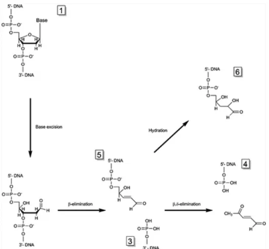
Both possible elimination processes are described inFigure 6.
In our experiments, Fpg and endoIII were used as controls forβ,δ-elimination of 8-oxo-dGuo and β-elimination of 5-OH-5-Me-dHyd, respectively. Experimental MALDI-TOF MS spectra acquired on the products of Fpg and recombinant TGAM_1653 and TGAM_1277 excision are shown inFigures 7and 8. 8-oxo-dGuo repair by Fpg wasfirst characterized by the absence of peak 1 corresponding to 8-oxo-dGuo-containing strands. This was indicative of a total cleavage of the substrate by the enzyme. The presence of peaks 3 ([M− H]−= 4332.9 Da; calculated = 4332.8 Da) and 4 ([M− H]−= 2135.6 Da; calculated = 2135.4 Da) is characteristic of theβ,δ-elimination process. For TGAM_1653 tested on 8-oxo-dGuo substrates,
the absence of peak 1 proves a complete cleavage, whereas the presence of peaks 3 ([M− H]− = 4332.5 Da) and 5 ([M − H]− = 2233.9 Da; calculated = 2233.4 Da) confirms a β-elimination mechanism. The MALDI-TOF MS spectrum of TGAM_1277 activity revealed that peak 1 was clearly visible, indicating that in our experimental conditions, this enzyme did not cut all the available substrate. TGAM_1277 was therefore considered as less active than Fpg and TGAM_1653, confirming the results obtained with electrophoretic gels
(Figure 5). Considering the cleavage products, we were able
to detect peak 3 but not peak 4, thus characterizing a β-elimination process. Actually, the presence of peak 6 ([M −
Figure 5. (A) Base excision activities of Fpg, TGAM_1653, and TGAM_1277 assessed with three oxidative DNA lesions: 8-oxo-dGuo, 5-OH-dHyd, and 5-OH−Me-dHyd. 22-mer ODN sequence: 5′-CAC TTC GG1A T2CG TGA CTG ATC T-3′ (control). In modified
ODNs, G1was replaced by 8-oxo-dGuo and T2by OH-dHyd or 5-OH-5-Me-dHyd. 100 fmol ODNs were incubated with 2 pmol enzyme. For Fpg, the cleavage was carried out at 37 °C. For TGAM_1653 and TGAM_1277, the incubation was initiated at 37°C, and the temperature was progressively increased to 65 °C. The different sizes of the cleavage products are explained by the positions of the damaged bases in the ODN sequences and by the enzymes presenting different excision processes. (B) Base excision activities of Fpg, TGAM_1653, and TGAM_1277 on probes containing 8-oxo-dGuo. The influence of the opposite nucleoside was evaluated by using, for each enzyme, a set of four ODNs with dC, dT, dA, or dG paired with the oxidative lesion.
Figure 6.Excision reaction catalyzed by bifunctional glycosylases viaβ or β,δ-elimination mechanisms. Successive cleavages of 3′ and 5′-strands on each side of the abasic site lead to deoxyribose residue excision. Numbers 1 to 6 were attributed to each reaction product to match with fragments obtained by MALDI-TOF mass spectrometry analysis (Figures 7and8). Number 2 (not represented) stands for the complementary strand.
Figure 7.MALDI-TOF MS spectra resulting from the excision by Fpg, TGAM_1653, or TGAM_1277 of ODNs containing 8-oxo-dGuo. Ten pmol ODNs were incubated with 20 pmol enzyme. The incubation was initiated at 37°C, and the temperature was progressively increased to 65 °C. The nature of the detected fragments (from 1 to 6) is described inFigure 6.“bis” indicates a secondary ion presenting two charges.
H]−= 2251.9 Da; calculated = 2251.5 Da) probably reveals a hydration product of the α−β-unsaturated ketone, which should have been represented by fragment 5.24 We can therefore assume that TGAM_1277 uses a β-elimination process.
The same principle was applied to the cleavage of ODNs containing 5-OH-5-Me-dHyd (Figure 8). The MALDI-TOF MS profile corresponding to our control endoIII confirms its β-elimination activity with the presence of peak 3 ([M− H]−= 3714.9 Da; calculated = 3715.4 Da) and hydrated product 6 ([M − H]− = 2893.7 Da; calculated = 2893.9 Da). The β,δ-elimination exhibited by Fpg is demonstrated again by the presence of peaks 3 ([M − H]− = 3715.6 Da; calculated = 3715.4 Da) and 4 ([M− H]−= 2776.5 Da; calculated = 2777.8 Da). The MS profile of TGAM_1277 is very close to that obtained with endoIII: product 3 at 3714.8 Da and product 6 at 2893.5 Da indicatingβ-elimination. The only difference with endoIII is the absence of peak 1, indicating a more efficient cleavage. TGAM_1653 is not associated with short fragments, a
characteristic that again indicates its absence of activity on 5-OH-5-Me-dHyd.
In conclusion, TGAM_1653 and TGAM_1277 both exhibit aβ-elimination cleavage activity. TGAM_1653 is only able to cleave 8-oxo-dGuo, whereas TGAM_1277 is also able to cut ODNs containing pyrimidine lesions (dHyd and 5-OH-5-Me-dHyd). It is worth noting that ODN substrates containing 8-oxodA and 5′,8-cyclonucleosides were also tested but did not show detectable excisions (data not shown).
■
DISCUSSIONT. gammatolerans Shows a Basal Level of 8-Oxo-dGuo That Is Enhanced by High Doses of γ-Irradiation but Rapidly Decreases Postexposure. Measurement of oxida-tive base damage in cellular DNA is a rather commonly used biomarker of oxidative stress but has for a long time been associated with questionable results due to possible over-estimation related to technical artifacts. By using the most appropriate preanalytical precautions, as well as sensitive and
Figure 8.MALDI-TOF MS spectra resulting from the excision by Fpg, Endo III, TGAM_1653, or TGAM_1277 of ODNs containing 5-OH-5-Me-dHyd. Ten pmol ODNs were incubated with 20 pmol enzyme. The incubation was initiated at 37°C, and the temperature was progressively increased to 65°C. The nature of the detected fragments (from 1 to 6) is described inFigure 6.“bis” indicates a secondary ion presenting two charges.
specific methods such as HPLC-electrochemical detection and, more recently, HPLC-MS/MS, it is now accepted that background levels of 8-oxo-dGuo in human cells such as lymphocytes are below the threshold of 1 lesion per 106
nucleosides (4 lesions per 106 guanines).25,26 This level corresponds to approximately 10,000 8-oxo-dGuo per single nucleus.27Using HPLC-MS/MS, 8-oxo-dGuo measurements in E. coli DNA carried out by our team showed basal rates of 2.6 (± 0.9) 8-oxo-dGuo per 106nucleosides (unpublished data). In the present study, basal levels measured in T. gammatolerans grown in laboratory experimental conditions were 9.2 (± 0.9) 8-oxo-dGuo per 106 nucleosides, which represents approx-imatively 18 8-oxo-dGuo per chromosome (T. gammatolerans is a polyploid organism for which the mean number of chromosome copies has not been determined yet). While an overwhelming proportion of published studies reporting 8-oxo-dGuo rates have been performed using mammalian cells, the only relevant work comparable with ours was carried out using the archaeon Halobacterium salinarum.28 In this halophilic organism, a basal level of 2.1 8-oxoG (oxidized base) per 106
bases was observed by means of gas chromatography coupled to mass spectrometry. Two other oxidative purine lesions were assessed in the same study: 4,6-diamino-5-formamidopyrimi-dine (FapyAde) and 2,6-diamino-4-hydroxy-5-formamidopyr-imidine (FapyGua), which showed concentrations of 0.3 and 0.9 lesion per 106bases, respectively. Elevated rates of 8-oxo-dGuo found in T. gammatolerans could be related to the growth conditions and more specifically to the temperature. Indeed, the optimal growth temperature for this hyperthermophilic organism (85 °C) was chosen for the experimental work presented here, while lower temperatures were used in other studies: 37 °C for human cells and 42 °C for H. salinarum. Heat-induced formation of oxidized guanine, at least in vitro, has been reported.29Because nonirradiated cells stored on ice presented only 4.2 8-oxo-dGuo lesions per 106nucleosides, this hypothesis of heat-induced damage formation is reinforced. This was confirmed by the result obtained with nonirradiated cells incubated for 1 h at 85°C in culture medium, which also showed a significant increase in their 8-oxo-dGuo content. It can be highlighted here that another type of DNA damage, abasic sites, was also found at elevated levels in Pyrococcus abyssi: between 25 (exponential phase) and 42 (stationary phase) abasic sites per 100,000 base pairs in the genome of this hyperthermophilic archaeon versus 2 and 4, respectively, abasic sites per 100,000 base pairs in E. coli.30
In order to study DNA damage in the context of exposure to ionizing radiation, T. gammatolerans samples were subjected to high doses of γ-radiation. Levels of 8-oxo-dGuo measured in cells harvested immediately after treatment significantly increased. The corresponding yields of radiation-induced modifications are 2.8 8-oxo-dGuo/1012 nucleosides/Gy at 2.5 kGy and 4.0 8-oxo-dGuo/1012 nucleosides/Gy at 5.0 kGy. Again, measurements of 8-oxo-dGuo after irradiation have previously been measured mainly using eukaryotic cells25,31−33 and with doses lower than 500 Gy. In the THP1 malignant monocyte cell line, HPLC-MS/MS analysis indicated a damage yield reaching 20 8-oxo-dGuo/109 nucleosides/Gy (∼200 to
300 8-oxo-dGuo/cell/Gy).31Kish et al.28analyzed the oxidized base, 8-oxoG, after exposure of the archaeon H. salinarum at 0, 2.5, 5.0, and 7.5 kGy. A plateau was reached at 2.5 kGy, for which the levels were 2.1 8-oxoG/106bases and 3.1 8-oxoG/ 106bases at 0 and 7.5 kGy, respectively (60Co gamma source),
corresponding to less than 1 lesion/1012 bases/Gy. Therefore,
this archaeon also showed yields of oxidized guanine formation far below those observed in human cells. These very low yields may be explained by a cellular redox environment and low O2
levels that prevent the formation of oxidative DNA damage, or by detoxification systems.4 In the same halophilic organism, Robinson et al.34observed an increase of FapyGua levels at 2.5 kGy but no significant progression of 8-oxoG.
Apart from a potential role of DNA repair enzymes, low levels of oxidative DNA damage observed in T. gammatolerans after irradiation may be linked to mechanisms such as highly constitutively expressed detoxification systems. For example, the genome of this archaeon is containing sequences related to proteins known to decrease oxidative stress such as a thioredoxin reductase (TGAM_0180), a glutaredoxin-like protein (TGAM_1302), and 2 peroxiredoxins (TGAM_1253 and TGAM_1220).2,4
T. gammatolerans cells exposed to 5.0 kGy radiation showed a significant decrease of 8-oxo-dGuo after 1 h recovery in culture medium, whereas this phenomenon was not significant at the lower levels of oxidative DNA damage observed at 2.5 kGy. Studies performed with H. salinarum indicated a decrease of FapyGua, FapyAde, and 8-oxoG rates after a 2.5 kGy irradiation and 2 h recovery compared with nonexposed organisms (rates after 1 h recovery were not measured).28This decrease of 8-oxo-dGuo 1 h after the end of irradiation may contribute to the resistance of T. gammatolerans. It may be explained by DNA repair systems or other factors limiting the extent of DNA damage.
Cyclonucleoside Formation in T. gammatolerans DNA Is Questionable. Cyclonucleosides (in this study, cyclo-purines) are classified as complex oxidative DNA lesions presenting, in the same nucleotide, a covalent bond between the C8 of the base and the C5′ of the 2-deoxyribose. In vitro studies show that these bulky lesions are mutagenic35 and interfere with replication and transcription.36−38 Contrary to small oxidative DNA damage such as oxidized bases, it also appears that cyclopurines are repaired by the nucleotide excision repair system and not by BER enzymes. In parallel to these mechanistic results, measurements of cyclopurine rates in DNA extracted from eukaryotic cultured cells and tissues have been performed by a limited number of research teams. In spite of similar methods, i.e., isotope dilution techniques in combination with HPLC coupled to mass spectrometry, discrepancies can be noted among the concentrations reported in the literature. In THP1 human monocytes, Belmadoui et al.9 did not detect 5′R and 5′S diastereomers of 5′,8-cyclodAdo above the detection limit of the method, which was 0.1 lesion per 109 nucleosides. In rodent tissues, the team of Wang
measured basal levels of 5′R and 5′S diastereomers of 5′,8-cyclodAdo and 5′,8-cyclodGuo superior to 0.1 lesion per 106
nucleosides.39−41In the work reported here, we did not show the presence of detectable cyclopurines in T. gammatolerans cells in any of the conditions tested. Indeed, even exposure to 5.0 kGy radiation did not lead to a signal above our limit of detection, estimated at 1 lesion/106 nucleosides for this
experiment (the limit of detection is dependent on the quantity of DNA available for the analysis). In our case, another favorable factor for the detection of cyclonucleosides should be the anaerobic growth conditions of T. gammatolerans. Cyclo-nucleosides are indeed more prone to formation at low O2
concentrations.9 Taken together, these data indicate that cyclonucleoside formation in living cells, T. gammatolerans in our case, may occur at very low levels, if at all.
TGAM_1653 and TGAM_1277 Are Slightly Upregu-lated after Heavy Radiation Exposure. Data extracted from proteomic results indicated the presence at low levels of BER enzymes TGAM_1653 and TGAM_1277 in nonirradiated and irradiated cells. No difference between these two conditions was noted in terms of protein abundance, but the sensitivity of the MS shotgun approach is not sufficient to detect moderate variations. The expression of the corresponding genes was confirmed by RT-PCR. For TGAM_1277, the main difference appeared 2 h after a 5.0 kGy irradiation, with an increase of the relative expression from 0.03 (T0) to 0.09. TGAM_1653 basal mRNA levels were higher than those of TGAM_1277, but the regulation of the transcription of this gene was less sensitive to irradiation. The expression of BER enzymes before exposure to ionizing radiation is not surprising since extremophiles need constitutive protective mechanisms to deal with their harsh growth conditions. The DNA damage analysis reported here shows the presence of an elevated amount of oxidized bases in basal conditions that may require a sufficient amount of DNA repair enzymes. The weak or absent upregulation of several DNA repair genes after exposure to genotoxic stress has already been observed in other archaea. Pyrococcus f uriosus subjected to 2.5 kGy60Coγ-rays did not show changes in mRNA levels for BER glycosylases but presented a moderate upregulation of radA (DNA repair by homologous recombination) and of a DNA repair gene cluster possibly involved in translesion synthesis.42 UV irradiation of hyperthermophilic Sulfolobus archaea did not lead to overexpression of DNA repair genes, including BER genes.43
It is also recognized that a majority of DNA repair factors are constitutively expressed in eukaryotic cells.44 At the protein level, the expression of BER factors is not massively induced, even after significant exposure to genotoxic agents.45We also observed that 8-oxo-dGuo produced after γ-ray exposure are repaired within the first hour of recovery, whereas maximum levels of TGAM_1277 and TGAM_1653 transcripts were measured at 2 h. We can suggest that newly synthesized enzymes are produced (i) to repair other types of more
persistent oxidative DNA damage or (ii) to reconstitute the pool of constitutive enzymes that has probably been, at least partly, altered by irradiation. This degradation of BER factors byγ-rays has already been shown to occur in human cells46at much lower doses (up to 60 Gy) compared to those in the present study. Finally, it is worth mentioning that our observations are mainly based on mRNA levels. It would be interesting to complement them with further data related to proteins, as well as their post-translational modifications, which are known to play an important role in the regulation of BER in eukaryotes.47
TGAM_1653 Excises OxoG, TGAM_1277 Excises 8-OxoG and Hydantoin Lesions, and Both Possess β-Elimination Activity. According to our study, TGAM_1277 and TGAM_1653 are bifunctional BER enzymes: they are able (i) to recognize and remove oxidized bases by cleaving the N-glycosyl bond between the deoxyribose and the damaged base, and (ii) to cleave the resulting abasic site leading to a single-strand break (lyase activity). More precisely, we demonstrated that both enzymes cleave the abasic site via a β-elimination process resulting in a 3′-phospho-α,β-unsaturated aldehyde and in a 5′-phosphate extremity (Figure 6).
TGAM_1277 was annotated as a putative type III endonuclease.2 Indeed, we have shown here that this enzyme shares the common features of the bacterial BER enzyme, i.e., bifunctionality, a β-elimination process and the capacity to repair hydantoin lesions (5-OHdHyd and 5-OH-5-Me-dHyd).22,48By contrast with bacterial EndoIII, we found that TGAM_1277 is also able to cleave 8-oxo-dGuo, particularly when mispaired with T or G. This variety of substrates is also found for the E. coli enzyme Fpg, which presents a β,δ-elimination mechanism.
When compared with TGAM_1277, TGAM_1653 shows a more restrictive panel of substrates as it does not cleave oxidized pyrimidines and is not as efficient as TGAM_1277 in cleaving 8-oxo-dGuo when mispaired, particularly to A and G. This profile of substrates is close to that noted in other studies
Figure 9. Sequence alignment of Pyrobaculum aerophilum 8-oxoG DNA glycosylase (Pa-AGOG), TGAM_1653, and TGAM_1277. Identical residues are in red, and residues shared by two proteins are in blue. Residues playing an important role in catalytic activity or 8-oxo-dGuo recognition in Pa-AGOG (Q31, K140, K147, D172, D218, and W222) are indicated in green. The alignment was performed using the MultAlin tool62followed by manual editing.
for the human BER enzyme, hOGG1 (human 8-oxoG DNA Glycosylase I).49,50
As for TGAM_1277 and TGAM_1653, the 8-oxoG DNA glycosylase from the sulfate-reducing archaeon Archeoglobus f ulgidus (Afogg) presents a bifunctional and β-elimination mechanism.51This protein is able to repair 8-oxo-dGuo when associated with C and G, and to a lesser extent when mispaired with T and A. Pyrobaculum aerophilum 8-oxoG DNA glycosylase (Pa-AGOG), a formative member of a new DNA glycosylase family, also demonstrated a bifunctional and β-elimination process.10 Structural studies indicated that this enzyme has an HhH-GPD motif (helix-hairpin-helix associated with a glycine-proline-rich sequence, followed by a conserved aspartate) that is significantly different in terms of amino acid sequence compared to that of hOGG1 but similar in terms of tridimensional structure.
Alignment of the Pa-AGOG, TGAM_1277, and TGAM_1653 polypeptides is shown in Figure 9. Pa-AGOG and TGAM_1653 display only 25% sequence identity and 43% similarity, but P. aerophilum and T. gammatolerans belong to two separate phyla of Archaea (Crenarcheota vs Euryarcheota). However, residues playing an important role for catalytic activity (K140, D172) or substrate recognition (Q31, K147, D218, and W222) in Pa-AGOG10,52 are conserved in TGAM_1653 and not in TGAM_1277 (Figure 9). This suggests that Pa-AGOG and TGAM_1653 may belong to the same glycosylase family, even if some discrepancies are revealed. Pa-AGOG is able to repair 8-oxo-dGuo when paired with C and A but presented a stronger efficiency for 8-oxoG:G mismatches.52 We showed here that this strong capacity to cleave 8-oxoG:G pairs was also shared by TGAM_1277, TGAM_1653 and Afogg. It could be interesting for hyper-thermophilic organisms to cleave 8-oxoG:G lesions generated by the incorporation of free 8-oxodGMP opposite to a G, this phenomenon probably being favored by high temperatures.10,53 Neither Afogg nor Pa-AGOG was tested for its capacity to repair damaged pyrimidines, making the comparison with T. gammatolerans enzymes difficult to extend further. We also tested cyclonucleosides as potential substrates: TGAM_1277 and TGAM_1653 did not show cleavage activity. Previously published experiments, based on eukaryote whole-cell extracts or purified enzymes, have already demonstrated that BER does not deal with these complex oxidative DNA lesions.54−57
In order to obtain a comprehensive picture of BER in T. gammatolerans, complementary experiments have to be performed on other putative BER factors highlighted by our previous genomic and proteomic screenings.2 At least seven candidates, annotated as “uracil-DNA glycosylase”, “base excision DNA repair protein, HhH-GPD superfamily”, “AP endonuclease family 2”, or “endonuclease V”, have been identified. Study of these archaeal enzymes can lead to the discovery of original DNA repair activities, as revealed by the recent characterization of EndoQ found in Pyrococcus f uriosus and T. kodakarensis.58 This endonuclease is able to repair deaminated bases generated, for example, by ionizing radiation or elevated temperatures. Glycosylases and lyases are the very first enzymes involved in the BER system. It may also be interesting to study other BER proteins involved in the later stages of the pathway. For example, it would be relevant to focus attention on BER factors processing single strand breaks as these latter lesions are also generated by ionizing radiation.47 Finally, a further characterization of enzymatic activities including parameters such as thermostability or influence of
pH, would be interesting, particularly for a potential use of these proteins as new tools for research and applied biotechnologies laboratories.59
■
CONCLUSIONSThis work established that the basal level of the oxidative DNA damage, 8-oxo-dGuo, in T. gammatolerans is higher than that reported for mammalian cells. As expected, this level of damage increased with high doses of ionizing radiation. Remarkably, the yield of lesions generated was much lower than those observed in human cell lines. One hour postirradiation, the rate of oxidative DNA damage was the same as the control background, indicating that the repair process is rather rapid. TGAM_1277 and TGAM_1653 BER enzymes were slightly upregulated after heavy radiation exposure. As shown here, these enzymes are able to initiate DNA repair of oxidized pyrimidines (TGAM_1277) and guanines (TGAM_1277 and TGAM_1653). Therefore, T. gammatolerans is at any time prepared to counteract DNA oxidation with this protein arsenal. While the search for cyclonucleosides showed no significant levels above technical background, other original DNA lesions such as clusters of damage33,60 would be interesting to identify in radiotolerant organisms such as T. gammatolerans. Regarding DNA repair proteins such as TGAM_1277 and TGAM_1653, structural studies with the enzymes alone and with their DNA substrates would provide additional data about recognition and cleaving mechanisms.
Further molecular studies on proteins from T. gammatoler-ans, one of the most radiotolerant organisms currently known, should bring valuable information on DNA repair in archaea. Moreover, deciphering the mechanisms explaining its extra-ordinary resistance to extreme conditions may have potential applications in biotechnology.61
■
ASSOCIATED CONTENT*
S Supporting InformationThe Supporting Information is available free of charge on the
ACS Publications website at DOI:
10.1021/acs.chemres-tox.6b00128.
List of oligonucleotides used for quantitative real-time RT-PCR and cloning and expression of TGAM_1277 and TGAM_1653-primer pairs used to amplify coding sequences from total genomic DNA (PDF)
■
AUTHOR INFORMATIONCorresponding Author
*CEA Grenoble, INAC, Service Systèmes Moléculaires et nanoMatériaux pour l’Energie et la Santé (UMR E_3 CEA/ Université Grenoble Alpes), 17 rue des Martyrs, 38054 Grenoble cedex 09, France. Tel: +33-0-4-38-78-56-01. E-mail:
jean.breton@cea.fr.
Funding
This study was supported by the Agence Nationale de la Recherche (ANR-12-BSV6-0012-01).
Notes
The authors declare no competingfinancial interest.
■
ACKNOWLEDGMENTSWe thank Dr. B. Castaing (Orléans, France) for providing Fpg and advice. We also thank Dr. V. Favaudon (Institut Curie, Orsay) for access to the137Cs gamma irradiation source.
■
ABBREVIATIONS5′,8-cyclodAdo, 5′,8-cyclo-2′-deoxyadenosine; 5′,8-cyclodGuo, 5′,8-cyclo-2′-deoxyguanosine; 5-OH-5-Me-dHyd,
1-[2-deoxy-β-D-erythro-pentofuranosyl]-hydroxy-methylhydantoin;
5-OH-dHyd, 1-[2-deoxy-β-D
-erythro-pentofuranosyl]-5-hydroxy-hydantoin; 8-oxo-dGuo, 8-oxo-2′-deoxyguanosine; oxoG, 8-oxo-7,8-dihydroguanine; Afogg, 8-oxoG DNA glycosylase from the sulfate-reducing archaeon Archeoglobus f ulgidus; BER, base excision repair; CDS, coding DNA sequence; Endo III, type III endonuclease; ESI, electrospray ionization; FapyAde, 4,6-diamino-5-formamidopyrimidine; FapyGua, 2,6-diamino-4-hy-droxy-5-formamidopyrimidine; Fpg, formamidopyridine DNA N-glycosylase; FPLC, fast protein liquid chromatography; HhH-GPD motif, helix-hairpin-helix associated with a gly-cine−proline-rich sequence, followed by a conserved aspartate; hOGG1, human 8-oxoG DNA glycosylase 1; HPLC-MS/MS, HPLC coupled to tandem mass spectrometry; kGy, kiloGray; MALDI-TOF, matrix-assisted laser desorption ionization−time offlight; Mb, megabase; NSAF, normalized spectral abundance factor; ODN, oligodeoxynucleotide; Pa-AGOG, Pyrobaculum aerophilum 8-oxoG DNA glycosylase; radA, DNA repair by homologous recombination; ROS, reactive oxygen species; rpoB, DNA-directed RNA polymerase subunit B; RT-PCR, reverse transcription PCR; Tbp, TATA box-binding protein
■
REFERENCES(1) Jolivet, E., L’Haridon, S., Corre, E., Forterre, P., and Prieur, D. (2003) Thermococcus gammatolerans sp. nov., a hyperthermophilic archaeon from a deep-sea hydrothermal vent that resists ionizing radiation. Int. J. Syst. Evol. Microbiol. 53, 847−851.
(2) Zivanovic, Y., Armengaud, J., Lagorce, A., Leplat, C., Guerin, P., Dutertre, M., Anthouard, V., Forterre, P., Wincker, P., and Confalonieri, F. (2009) Genome analysis and genome-wide proteomics of Thermococcus gammatolerans, the most radioresistant organism known amongst the Archaea. Genome Biology 10, R70.
(3) Confalonieri, F., and Sommer, S. (2011) Bacterial and archaeal resistance to ionizing radiation. Cost Chemistry Cm0603-Melusyn Joint Meeting: Damages Induced in Biomolecules by Low and High Energy Radiations 261, 012005.
(4) Webb, K. M., and DiRuggiero, J. (2012) Role of Mn2+ and compatible solutes in the radiation resistance of thermophilic bacteria and archaea. Archaea 2012, 1.
(5) Tapias, A., Leplat, C., and Confalonieri, F. (2009) Recovery of ionizing-radiation damage after high doses of gamma ray in the hyperthermophilic archaeon Thermococcus gammatolerans. Extremo-philes 13, 333−343.
(6) Cadet, J., Douki, T., and Ravanat, J. L. (2011) Measurement of oxidatively generated base damage in cellular DNA. Mutat. Res., Fundam. Mol. Mech. Mutagen. 711, 3−12.
(7) Michaels, M. L., and Miller, J. H. (1992) The GO system protects organisms from the mutagenic effect of the spontaneous lesion 8-hydroxyguanine (7,8-dihydro-8-oxoguanine). J. Bacteriol. 174, 6321− 6325.
(8) Chatgilialoglu, C., Ferreri, C., and Terzidis, M. A. (2011) Purine 5′,8-cyclonucleoside lesions: chemistry and biology. Chem. Soc. Rev. 40, 1368−1382.
(9) Belmadoui, N., Boussicault, F., Guerra, M., Ravanat, J. L., Chatgilialoglu, C., and Cadet, J. (2010) Radiation-induced formation of purine 5′,8-cyclonucleosides in isolated and cellular DNA: high stereospecificity and modulating effect of oxygen. Org. Biomol. Chem. 8, 3211−3219.
(10) Sartori, A. A., Lingaraju, G. M., Hunziker, P., Winkler, F. K., and Jiricny, J. (2004) Pa-AGOG, the founding member of a new family of archaeal 8-oxoguanine DNA-glycosylases. Nucleic Acids Res. 32, 6531− 6539.
(11) Pfaffl, M. W. (2001) A new mathematical model for relative quantification in real-time RT-PCR. Nucleic Acids Res. 29, 45e.
(12) Vandesompele, J., De Preter, K., Pattyn, F., Poppe, B., Van Roy, N., De Paepe, A., and Speleman, F. (2002) Accurate normalization of real-time quantitative RT-PCR data by geometric averaging of multiple internal control genes. Genome Biol. 3, research0034.1.
(13) Lagorce, A., Fourcans, A., Dutertre, M., Bouyssiere, B., Zivanovic, Y., and Confalonieri, F. (2012) Genome-wide transcrip-tional response of the archaeon Thermococcus gammatolerans to cadmium. PLoS One 7, e41935.
(14) Armengaud, J., Fernandez, B., Chaumont, V., Rollin-Genetet, F., Finet, S., Marchetti, C., Myllykallio, H., Vidaud, C., Pellequer, J. L., Gribaldo, S., Forterre, P., and Gans, P. (2003) Identification, purification, and characterization of an eukaryotic-like phosphopante-theine adenylyltransferase (coenzyme A biosynthetic pathway) in the hyperthermophilic archaeon Pyrococcus abyssi. J. Biol. Chem. 278, 31078−31087.
(15) Gabant, G., Auxilien, S., Tuszynska, I., Locard, M., Gajda, M. J., Chaussinand, G., Fernandez, B., Dedieu, A., Grosjean, H., Golinelli-Pimpaneau, B., Bujnicki, J. M., and Armengaud, J. (2006) THUMP from archaeal tRNA:m22G10 methyltransferase, a genuine autono-mously folding domain. Nucleic Acids Res. 34, 2483−2494.
(16) Gasparutto, D., Ait-Abbas, M., Jaquinod, M., Boiteux, S., and Cadet, J. (2000) Repair and coding properties of 5-hydroxy-5-methylhydantoin nucleosides inserted into DNA oligomers. Chem. Res. Toxicol. 13, 575−584.
(17) Muller, E., Gasparutto, D., Lebrun, C., and Cadet, J. (2001) Site-specific insertion of the (5R*) and (5S*) diastereoisomers of 1-[2-deoxy-beta-D-erythro-pentofuranosyl]-5-hydroxyhydantoin into oligo-deoxyribonucleotides. Eur. J. Org. Chem. 2001, 2091−2099.
(18) Ravanat, J. L., Douki, T., Duez, P., Gremaud, E., Herbert, K., Hofer, T., Lasserre, L., Saint-Pierre, C., Favier, A., and Cadet, J. (2002) Cellular background level of 8-oxo-7,8-dihydro-2′-deoxyguanosine: an isotope based method to evaluate artefactual oxidation of DNA during its extraction and subsequent work-up. Carcinogenesis 23, 1911−1918. (19) Yang, Y. S., Fernandez, B., Lagorce, A., Aloin, V., De Guillen, K. M., Boyer, J. B., Dedieu, A., Confalonieri, F., Armengaud, J., and Roumestand, C. (2015) Prioritizing targets for structural biology through the lens of proteomics: the archaeal protein TGAM_1934 from Thermococcus gammatolerans. Proteomics 15, 114−123.
(20) Christie-Oleza, J. A., Fernandez, B., Nogales, B., Bosch, R., and Armengaud, J. (2012) Proteomic insights into the lifestyle of an environmentally relevant marine bacterium. ISME J. 6, 124−135.
(21) Tchou, J., Kasai, H., Shibutani, S., Chung, M. H., Laval, J., Grollman, A. P., and Nishimura, S. (1991) 8-oxoguanine (8-hydroxyguanine) DNA glycosylase and its substrate-specificity. Proc. Natl. Acad. Sci. U. S. A. 88, 4690−4694.
(22) D’Ham, C., Romieu, A., Jaquinod, M., Gasparutto, D., and Cadet, J. (1999) Excision of dihydroxy-dihydrothymine, 5,6-dihydrothymine, and 5-hydroxycytosine from defined sequence oligonucleotides by Escherichia coli endonuclease III and Fpg proteins: kinetic and mechanistic aspects. Biochemistry 38, 3335−3344.
(23) Bhagwat, M., and Gerlt, J. A. (1996) 3′- and 5′-strand cleavage reactions catalyzed by the Fpg protein from Escherichia coli occur via successive beta- and delta-elimination mechanisms, respectively. Biochemistry 35, 659−665.
(24) Darwanto, A., Farrel, A., Rogstad, D. K., and Sowers, L. C. (2009) Characterization of DNA glycosylase activity by matrix-assisted laser desorption/ionization time-of-flight mass spectrometry. Anal. Biochem. 394, 13−23.
(25) Frelon, S., Douki, T., Ravanat, J. L., Pouget, J. P., Tornabene, C., and Cadet, J. (2000) High-performance liquid chromatography– tandem mass spectrometry measurement of radiation-induced base damage to isolated and cellular DNA. Chem. Res. Toxicol. 13, 1002− 1010.
(26) Gedik, C. M., and Collins, A. (2005) Establishing the background level of base oxidation in human lymphocyte DNA: results of an interlaboratory validation study. FASEB J. 19, 82−84.
(27) Ohno, M., Miura, T., Furuichi, M., Tominaga, Y., Tsuchimoto, D., Sakumi, K., and Nakabeppu, Y. (2006) A genome-wide distribution of 8-oxoguanine correlates with the preferred regions for recombina-tion and single nucleotide polymorphism in the human genome. Genome Res. 16, 567−575.
(28) Kish, A., Kirkali, G., Robinson, C., Rosenblatt, R., Jaruga, P., Dizdaroglu, M., and DiRuggiero, J. (2009) Salt shield: intracellular salts provide cellular protection against ionizing radiation in the halophilic archaeon, Halobacterium salinarum NRC-1. Environ. Micro-biol. 11, 1066−1078.
(29) Bruskov, V. I., Malakhova, L. V., Masalimov, Z. K., and Chernikov, A. V. (2002) Heat-induced formation of reactive oxygen species and 8-oxoguanine, a biomarker of damage to DNA. Nucleic Acids Res. 30, 1354−1363.
(30) Palud, A., Villani, G., L’Haridon, S., Querellou, J., Raffin, J. P., and Henneke, G. (2008) Intrinsic properties of the two replicative DNA polymerases of Pyrococcus abyssi in replicating abasic sites: possible role in DNA damage tolerance? Mol. Microbiol. 70, 746−761. (31) Pouget, J. P., Frelon, S., Ravanat, J. L., Testard, I., Odin, F., and Cadet, J. (2002) Formation of modified DNA bases in cells exposed either to gamma radiation or to high-LET particles. Radiat. Res. 157, 589−595.
(32) Douki, T., Ravanat, J. L., Pouget, J. P., Testard, I., and Cadet, J. (2006) Minor contribution of direct ionization to DNA base damage inducedby heavy ions. Int. J. Radiat. Biol. 82, 119−127.
(33) Regulus, P., Duroux, B., Bayle, P. A., Favier, A., Cadet, J., and Ravanat, J. L. (2007) Oxidation of the sugar moiety of DNA by ionizing radiation or bleomycin could induce the formation of a cluster DNA lesion. Proc. Natl. Acad. Sci. U. S. A. 104, 14032−14037.
(34) Robinson, C. K., Webb, K., Kaur, A., Jaruga, P., Dizdaroglu, M., Baliga, N. S., Place, A., and Diruggiero, J. (2011) A major role for nonenzymatic antioxidant processes in the radioresistance of Halobacterium salinarum. J. Bacteriol. 193, 1653−1662.
(35) Yuan, B., Wang, J., Cao, H., Sun, R., and Wang, Y. (2011) High-throughput analysis of the mutagenic and cytotoxic properties of DNA lesions by next-generation sequencing. Nucleic Acids Res. 39, 5945− 5954.
(36) Abraham, J., and Brooks, P. J. (2011) Divergent effects of oxidatively induced modification to the C8 of 2′-deoxyadenosine on transcription factor binding: 8,5′(S)-cyclo-2′-deoxyadenosine inhibits the binding of multiple sequence specific transcription factors, while 8-oxo-2′-deoxyadenosine increases binding of CREB and NF-kappa B to DNA. Environ. Mol. Mutagen. 52, 287−295.
(37) Jasti, V. P., Das, R. S., Hilton, B. A., Weerasooriya, S., Zou, Y., and Basu, A. K. (2011) (5′S)-8,5′-Cyclo-2′-deoxyguanosine is a strong block to replication, a potent pol V-dependent mutagenic lesion, and is inefficiently repaired in Escherichia coli. Biochemistry 50, 3862−3865.
(38) Walmacq, C., Wang, L., Chong, J., Scibelli, K., Lubkowska, L., Gnatt, A., Brooks, P. J., Wang, D., and Kashlev, M. (2015) Mechanism of RNA polymerase II bypass of oxidative cyclopurine DNA lesions. Proc. Natl. Acad. Sci. U. S. A. 112, E410−419.
(39) Wang, J., Clauson, C. L., Robbins, P. D., Niedernhofer, L. J., and Wang, Y. (2012) The oxidative DNA lesions 8,5′-cyclopurines accumulate with aging in a tissue-specific manner. Aging Cell 11, 714−716.
(40) Tilstra, J. S., Robinson, A. R., Wang, J., Gregg, S. Q., Clauson, C. L., Reay, D. P., Nasto, L. A., Croix, C. M., St, Usas, A., Vo, N., Huard, J., Clemens, P. R., Stolz, D. B., Guttridge, D. C., Watkins, S. C., Garinis, G. A., Wang, Y., Niedernhofer, L. J., and Robbins, P. D. (2012) NF-kappaB inhibition delays DNA damage-induced sen-escence and aging in mice. J. Clin. Invest. 122, 2601−2612.
(41) Mitra, D., Luo, X., Morgan, A., Wang, J., Hoang, M. P., Lo, J., Guerrero, C. R., Lennerz, J. K., Mihm, M. C., Wargo, J. A., Robinson, K. C., Devi, S. P., Vanover, J. C., D’Orazio, J. A., McMahon, M., Bosenberg, M. W., Haigis, K. M., Haber, D. A., Wang, Y., and Fisher, D. E. (2012) An ultraviolet-radiation-independent pathway to melanoma carcinogenesis in the red hair/fair skin background. Nature 491, 449−453.
(42) Williams, E., Lowe, T. M., Savas, J., and DiRuggiero, J. (2007) Microarray analysis of the hyperthermophilic archaeon Pyrococcus f uriosus exposed to gamma irradiation. Extremophiles 11, 19−29.
(43) Dorazi, R., Gotz, D., Munro, S., Bernander, R., and White, M. F. (2007) Equal rates of repair of DNA photoproducts in transcribed and non-transcribed strands in Sulfolobus solfataricus. Mol. Microbiol. 63, 521−529.
(44) Christmann, M., and Kaina, B. (2013) Transcriptional regulation of human DNA repair genes following genotoxic stress: trigger mechanisms, inducible responses and genotoxic adaptation. Nucleic Acids Res. 41, 8403−8420.
(45) Parsons, J. L., and Dianov, G. L. (2013) Co-ordination of base excision repair and genome stability. DNA Repair 12, 326−333.
(46) Jeong, H. G., Youn, C. K., Cho, H. J., Kim, S. H., Kim, M. H., Kim, H. B., Chang, I. Y., Lee, Y. S., Chung, M. H., and You, H. J. (2004) Metallothionein-III prevents gamma-ray-induced 8-oxoguanine accumulation in normal and hOGG1-depleted cells. J. Biol. Chem. 279, 34138−34149.
(47) Dianov, G. L., and Hubscher, U. (2013) Mammalian base excision repair: the forgotten archangel. Nucleic Acids Res. 41, 3483− 3490.
(48) Gasparutto, D., Muller, E., Boiteux, S., and Cadet, J. (2009) Excision of the oxidatively formed 5-hydroxyhydantoin and 5-hydroxy-5-methylhydantoin pyrimidine lesions by Escherichia coli and Saccharomyces cerevisiae DNA N-glycosylases. Biochim. Biophys. Acta, Gen. Subj. 1790, 16−24.
(49) Morland, I., Rolseth, V., Luna, L., Rognes, T., Bjoras, M., and Seeberg, E. (2002) Human DNA glycosylases of the bacterial Fpg/ MutM superfamily: an alternative pathway for the repair of 8-oxoguanine and other oxidation products in DNA. Nucleic Acids Res. 30, 4926−4936.
(50) Leipold, M. D., Workman, H., Muller, J. G., Burrows, C. J., and David, S. S. (2003) Recognition and removal of oxidized guanines in duplex DNA by the base excision repair enzymes hOGG1, yOGG1, and yOGG2. Biochemistry 42, 11373−11381.
(51) Chung, J. H., Suh, M. J., Park, Y. I., Tainer, J. A., and Han, Y. S. (2001) Repair activities of 8-oxoguanine DNA glycosylase from Archaeoglobus f ulgidus, a hyperthermophilic archaeon. Mutat. Res., DNA Repair 486, 99−111.
(52) Lingaraju, G. M., Prota, A. E., and Winkler, F. K. (2009) Mutational studies of Pa-AGOG DNA glycosylase from the hyper-thermophilic crenarchaeon Pyrobaculum aerophilum. DNA Repair 8, 857−864.
(53) Thiviyanathan, V., Somasunderam, A., Hazra, T. K., Mitra, S., and Gorenstein, D. G. (2003) Solution structure of a DNA duplex containing 8-hydroxy-2′-deoxyguanosine opposite deoxyguanosine. J. Mol. Biol. 325, 433−442.
(54) Brooks, P. J., Wise, D. S., Berry, D. A., Kosmoski, J. V., Smerdon, M. J., Somers, R. L., Mackie, H., Spoonde, A. Y., Ackerman, E. J., Coleman, K., Tarone, R. E., and Robbins, J. H. (2000) The oxidative DNA lesion 8,5′-(S)-cyclo-2′-deoxyadenosine is repaired by the nucleotide excision repair pathway and blocks gene expression in mammalian cells. J. Biol. Chem. 275, 22355−22362.
(55) Kuraoka, I., Bender, C., Romieu, A., Cadet, J., Wood, R. D., and Lindahl, T. (2000) Removal of oxygen free-radical-induced 5′,8-purine cyclodeoxynucleosides from DNA by the nucleotide excision-repair pathway in human cells. Proc. Natl. Acad. Sci. U. S. A. 97, 3832−3837. (56) Corne, C., Fiche, J. B., Gasparutto, D., Cunin, V., Suraniti, E., Buhot, A., Fuchs, J., Calemczuk, R., Livache, T., and Favier, A. (2008) SPR imaging for label-free multiplexed analyses of DNA N-glycosylase interactions with damaged DNA duplexes. Analyst 133, 1036−1045.
(57) Pande, P., Das, R. S., Sheppard, C., Kow, Y. W., and Basu, A. K. (2012) Repair efficiency of (5′S)-8,5′-cyclo-2′-deoxyguanosine and (5′S)-8,5′-cyclo-2′-deoxyadenosine depends on the complementary base. DNA Repair 11, 926−931.
(58) Shiraishi, M., Ishino, S., Yamagami, T., Egashira, Y., Kiyonari, S., and Ishino, Y. (2015) A novel endonuclease that may be responsible for damaged DNA base repair in Pyrococcus f uriosus. Nucleic Acids Res. 43, 2853−2863.
(59) Gogos, A., and Clarke, N. D. (1999) Characterization of an 8-oxoguanine DNA glycosylase from Methanococcus jannaschii. J. Biol. Chem. 274, 30447−30450.
(60) Ravanat, J. L., Breton, J., Douki, T., Gasparutto, D., Grand, A., Rachidi, W., and Sauvaigo, S. (2014) Radiation-mediated formation of complex damage to DNA: a chemical aspect overview. Br. J. Radiol. 87, 20130715.
(61) Grasso, S., and Tell, G. (2014) Base excision repair in Archaea: back to the future in DNA repair. DNA Repair 21, 148−157.
(62) Corpet, F. (1988) Multiple sequence alignment with hierarchical-clustering. Nucleic Acids Res. 16, 10881−10890.