HAL Id: cea-02402414
https://hal-cea.archives-ouvertes.fr/cea-02402414v2
Submitted on 23 Nov 2020
HAL is a multi-disciplinary open access
archive for the deposit and dissemination of
sci-entific research documents, whether they are
pub-lished or not. The documents may come from
teaching and research institutions in France or
abroad, or from public or private research centers.
L’archive ouverte pluridisciplinaire HAL, est
destinée au dépôt et à la diffusion de documents
scientifiques de niveau recherche, publiés ou non,
émanant des établissements d’enseignement et de
recherche français ou étrangers, des laboratoires
publics ou privés.
Two new polymorphic structures of human full-length
alpha-synuclein fibrils solved by cryo-electron
microscopy
Ricardo Guerrero-Ferreira, Nicholas Taylor, Ana-Andreea Arteni, Pratibha
Kumari, Daniel Mona, Philippe Ringler, Markus Britschgi, Matthias Lauer,
Ali Makky, Joeri Verasdonck, et al.
To cite this version:
Ricardo Guerrero-Ferreira, Nicholas Taylor, Ana-Andreea Arteni, Pratibha Kumari, Daniel Mona,
et al.. Two new polymorphic structures of human full-length alpha-synuclein fibrils solved by
cryo-electron microscopy. eLife, eLife Sciences Publication, 2019, 8, pp.e48907. �10.7554/eLife.48907�.
�cea-02402414v2�
*For correspondence: [email protected] (ABo¨); [email protected] (LB); [email protected] (HS) Present address:†Robert P Apkarian Integrated Electron Microscopy Core, Emory, University School of Medicine, Atlanta, United States Competing interest:See page 17
Funding:See page 17 Received: 30 May 2019 Accepted: 30 November 2019 Published: 09 December 2019 Reviewing editor: Sjors HW Scheres, MRC Laboratory of Molecular Biology, United Kingdom
Copyright Guerrero-Ferreira et al. This article is distributed under the terms of theCreative Commons Attribution License,
which permits unrestricted use and redistribution provided that the original author and source are credited.
Two new polymorphic structures of
human full-length alpha-synuclein fibrils
solved by cryo-electron microscopy
Ricardo Guerrero-Ferreira
1†, Nicholas MI Taylor
2, Ana-Andreea Arteni
3,4,
Pratibha Kumari
5, Daniel Mona
6,7, Philippe Ringler
1, Markus Britschgi
6,7,
Matthias E Lauer
8, Ali Makky
9, Joeri Verasdonck
5, Roland Riek
5, Ronald Melki
4,
Beat H Meier
5, Anja Bo¨ckmann
10*, Luc Bousset
4*, Henning Stahlberg
1*
1
Center for Cellular Imaging and NanoAnalytics (C-CINA), Biozentrum, University of
Basel, Basel, Switzerland;
2Structural Biology of Molecular Machines Group, Protein
Structure & Function Programme, Novo Nordisk Foundation Center for Protein
Research, Faculty of Health and Medical Sciences, University of Copenhagen,
Copenhagen, Denmark;
3Institut de Biologie Inte´grative de la Cellule (I2BC), CEA,
CNRS, Universite´ Paris Sud, Universite´ Paris-Saclay, Gif-sur-Yvette, France;
4Institut
Fancois Jacob (MIRCen), CEA and Laboratory of Neurodegenerative Diseases,
CNRS, Fontenay-Aux-Roses, France;
5Laboratory of Physical Chemistry, ETH Zurich,
Zurich, Switzerland;
6Roche Pharma Research and Early Development, Neuroscience
and Rare Diseases Discovery and Translational Medicine Area, Roche Innovation
Center Basel, Basel, Switzerland;
7Roche Pharma Research and Early Development,
Neuroscience Discovery, Roche Innovation Center Basel, Basel, Switzerland;
8Roche
Pharma Research and Early Development, Chemical Biology, Roche Innovation
Center Basel, Basel, Switzerland;
9Institut Galien Sud, CNRS, Universite´
Paris-Sud, Universite´ Paris-Saclay, Chaˆtenay-Malabry, France;
10Molecular Microbiology
and Structural Biochemistry, Labex Ecofect, UMR 5086 CNRS, Universite´ de Lyon,
Lyon, France
Abstract
Intracellular inclusions rich in alpha-synuclein are a hallmark of severalneuropathological diseases including Parkinson’s disease (PD). Previously, we reported the structure of alpha-synuclein fibrils (residues 1–121), composed of two protofibrils that are
connected via a densely-packed interface formed by residues 50–57 (Guerrero-Ferreira, eLife 218;7: e36402). We here report two new polymorphic atomic structures of alpha-synuclein fibrils termed polymorphs 2a and 2b, at 3.0 A˚ and 3.4 A˚ resolution, respectively. These polymorphs show a radically different structure compared to previously reported polymorphs. The new structures have a 10 nm fibril diameter and are composed of two protofilaments which interact via intermolecular salt-bridges between amino acids K45, E57 (polymorph 2a) or E46 (polymorph 2b). The non-amyloid component (NAC) region of alpha-synuclein is fully buried by previously non-described interactions with the N-terminus. A hydrophobic cleft, the location of familial PD mutation sites, and the nature of the protofilament interface now invite to formulate hypotheses about fibril formation, growth and stability.
Introduction
Lewy bodies (LB) and Lewy neurites (LN) are neuropathological hallmarks of Parkinson’s disease and other Lewy body disorders. These intracellular neuronal features contain a cytoplasmic enrichment
of the protein alpha-synuclein (a-Syn), thereby defining these diseases as synucleinopathies (Spillantini et al., 1998a;Spillantini et al., 1997). Apart from this finding in the postmortem brain, the central role of a-Syn in Parkinson’s disease (PD) is highlighted by the fact that certain mutations in the a-Syn gene (SNCA) cause familial forms of PD and other synucleinopathies ( Appel-Cresswell et al., 2013; Kru¨ger et al., 1998; Lesage et al., 2013; Pasanen et al., 2014;
Polymeropoulos et al., 1997;Zarranz et al., 2004) and that duplication or triplication of the SNCA gene lead to either a sporadic or early-onset PD, respectively, in affected families ( Chartier-Harlin et al., 2004; Flagmeier et al., 2016; Fujioka et al., 2014; Iba´n˜ez et al., 2004;
Singleton et al., 2003). The 14 kDa protein a-Syn is known to readily form fibrils in vitro (Conway et al., 1998;Hashimoto et al., 1998) and induce a-Syn inclusions when injected in model animals (Mougenot et al., 2012;Peelaerts et al., 2015).
Shahmoradian et al. have recently investigated the ultrastructure of LBs, using identification by fluorescence light microscopy and then correlative imaging by electron microscopy (CLEM). They found that a slight minority of analyzed LBs contained filamentous or dense proteinaceous struc-tures, while the vast majority of LBs was primarily composed of membrane fragments (Lewis et al., 2019;Shahmoradian et al., 2019). However, even in those cases, LBs had been identified due to their enrichment of a-Syn (antibody LB509, recognizing residues 115–122 of a-Syn).Moors et al. (2018)recently studied the ultrastructure of LBs by super-resolution light microscopy, revealing an onion-like distribution of different forms of a-Syn in nigral LBs and LNs. Their work suggests LBs to be structured encapsulations of aggregated proteins and lipids. While the structure and building blocks of LBs and the putative involvement of aggregated forms of a-Syn in the formation of LBs or in toxicity towards neurons and glia appear to be more complex than previously thought, it is clear that the protein a-Syn plays a pivotal role in PD, and therefore in LB formation. The conformational state and impact of a-Syn protein may also differ strongly between synucleinopathies.Peng et al. (2018)reported a-Syn preparations from glial cytoplasmic inclusions from multiple system atrophy patients being much more potent in seeding a-Syn aggregation into cell cultures than a-Syn prepa-rations than PD patients. A detailed understanding of the conformational space of structural poly-morphs of a-Syn is important to move forward the discovery of diagnostic tools and therapeutics for synucleinopathies, including PD.
a-Syn protein consists of 140 amino acids. The N-terminus (residues 1–60) is rich in lysine residues and contains KTK lipid-binding motif repeats associated with vesicle binding (George, 2002;
George et al., 1995;Perrin et al., 2000). It is also the region which contains all known SNCA famil-ial PD mutations: A30P (Kru¨ger et al., 1998), E46K (Zarranz et al., 2004), H50Q ( Appel-Cresswell et al., 2013;Proukakis et al., 2013), G51D (Lesage et al., 2013), A53E (Pasanen et al., 2014), and A53T (Polymeropoulos et al., 1997). The central region (residues 61–95) is the non-amy-loid-ß component (NAC region) (Giasson et al., 2001;Ue´da et al., 1993), which is essential for a-Syn aggregation (Li et al., 2002). The related protein ß-synuclein (ß-syn) (Jakes et al., 1994; Stefa-nis, 2012) lacks a stretch of 12 aminoacid residues within the NAC region (residues 71–82) and is unable to form fibrils.
The highly unstructured C-terminus of a-Syn (residues 96–140) can bind calcium and it is popu-lated by negatively charged residues (Li et al., 2007;Post et al., 2018;Vilar et al., 2008). Trunca-tion of this domain may play a role in a-Syn pathology by promoting fibril formaTrunca-tion (Crowther et al., 1998;Li et al., 2005;Liu et al., 2005;Wang et al., 2016) and being involved in Lewy body formation (Dufty et al., 2007;Mahul-Mellier et al., 2019;Prasad et al., 2012). Inhibi-tion of C-terminal truncaInhibi-tion has also been shown to reduce neurodegeneraInhibi-tion in a transgenic mouse model of Multiple System Atrophy (MSA) (Bassil et al., 2016).
Amyloid fibrils, even within a single sample, may exhibit heterogeneous conformations, referred to as polymorphism. Different polymorphs can be distinguished based on their diameter, their twist, how many protofilaments form a fibril (Riek, 2017), their behavior under limited proteolysis, their appearance under fiber diffraction (Bousset et al., 2013), or their structure using NMR or cryo- EM (Close et al., 2018;Fa¨ndrich et al., 2018; Meier and Bo¨ckmann, 2015). The polymorphs can be structurally different at the level of the protofibrils, or in the way the protofilament assemble. Specifi-cally for a-Syn fibrils, structural heterogeneity has been observed by solid-state NMR
(Bousset et al., 2013;Comellas et al., 2012;Comellas et al., 2011;Gath et al., 2012;Gath et al., 2014a; Gath et al., 2014b; Heise et al., 2005; Lv et al., 2012; Verasdonck et al., 2016;
Vilar et al., 2008), quenched hydrogen/deuterium (H/D) exchange data (Vilar et al., 2008) and cryo-EM (Guerrero-Ferreira et al., 2018;Li et al., 2018a;Li et al., 2018b). A detailed understand-ing of the conformational space that structural polymorphs of a-Syn do access is central to not only understand how the same polypeptide chain can fold into different structures, but also to thoroughly characterize material used for in vitro and in vivo experiments.
Our recent work on the structure of recombinant a-Syn(1-121), using cryo-electron microscopy (cryo-EM) (Guerrero-Ferreira et al., 2018) and other investigations on full-length a-Syn (Li et al., 2018a;Li et al., 2018b) revealed a-Syn fibrils in a structure composed of two protofilaments that buried the sites associated with familial PD in the interface region between the two protofilaments. This is here termed a-Syn polymorph 1a. Li et al. (Li et al., 2018a) reported an additional poly-morph, here termed a-Syn polymorph 1b, in which the interface between virtually identical protofila-ments is different. Previous reports on the structure of a-Syn fibrils by micro-electron diffraction (microED;Rodriguez et al., 2015) or solid-state NMR (Tuttle et al., 2016) either focused on small peptides, or did not describe the two protofilaments and the interface region.
Currently, no high-resolution structures of patient derived a-Syn have been obtained. Previous studies looking at the overall morphology of these types of samples, offered a first glance to fibril variability within a sample and among synucleinopathies. Filaments immunolabeled by a-Syn anti-bodies have been described as straight, unbranched fibrils, with widths of 5 nm or 10 nm and with varying patterns of conjugated gold depending on the specific antibody used (Crowther et al., 2000). Filaments extracted from cingulate cortex of patients with Dementia with Lewy Bodies (DLB) were also positive for a-Syn immunolabeling and revealed comparable morphologies (Spillantini et al., 1998a). In contrast, filaments extracted from Multiple System Atrophy (MSA) brains exhibited larger diameter spanning from 5 nm to 18 nm with two morphologies described by Spillantini et al. (Spillantini et al., 1998b) as ‘straight’’ and ‘twisted’.
Here, we report new cryo-EM polymorphic structures of in vitro generated amyloid fibrils of a-Syn (denoted polymorphs 2a and 2b). Our structures reveal remarkable differences to the previously solved a-Syn polymorphs, and inform new hypotheses to explain the mechanism of and factors involved in in vitro amyloid fibril assembly, the potential role of familial PD mutations on fibril struc-ture, and contribute to our understanding of amyloid fibril polymorphism.
Results and discussion
The structure of a-Syn fibril polymorphs
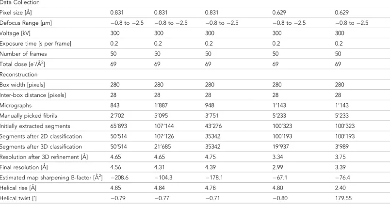
Fibrils of recombinant, full-length, human a-Syn were prepared using the conditions summarized in
Table 1. Preformed fibrils (PFFs) were quick-frozen in holey carbon-coated copper grids and imaged with a Titan Krios electron microscope at 300kV, equipped with a Quantum-LS energy filter. Micro-graphs were acquired with a K2 Summit direct electron detector, drift-corrected and dose-weighted through the FOCUS interface (Biyani et al., 2017).
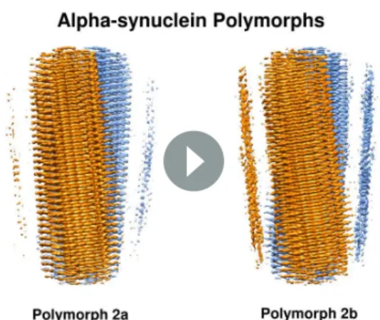
Helical image processing of 100’323 fibril segments extracted from 1’143 micrographs, revealed the presence of two distinct fibril polymorphs at the step of 3D classification. These polymorphs, termed a-Syn polymorph 2a and a-Syn polymorph 2b to distinguish them from the previously described a-Syn fibrils (a-Syn polymorphs 1a and 1b; Guerrero-Ferreira et al., 2018; Li et al., 2018a;Li et al., 2018b), were separately refined, resulting in 3D reconstructions at overall resolu-tions of 3.1 A˚ and 3.5 A˚ respectively (Table 2, Figure 1, Figure 1—figure supplement 1 and
Video 1). The left-twisting handedness of the fibrils was confirmed by AFM imaging, as done previ-ously (Guerrero-Ferreira et al., 2018). The maps show clear side-chain densities and ß-strand sepa-ration along the helical axis, and indicate that both fibril types are formed by two protofilaments of approximately 5 nm diameter, which are composed of distinct rungs of density.
Refined atomic models of the fibril cores indicate that polymorph 2a and polymorph 2b in terms of their local atomic structure share a common protofilament kernel, and which is clearly distinct from the one described previously in polymorphs 1a and 1b. However, the packing between the two protofilaments is different. Therefore, a-Syn fibrils exhibit assembly polymorphism as defined by
filaments (SFs) (Fitzpatrick et al., 2017). In each protofilament of the new a-Syn polymorph 2a, suc-cessive rungs of ß-strands are related by helical symmetry with a rise of 4.8 A˚ and a twist of !0.80˚ with the subunits within the two protofilaments packed in the same plane, facing each other (Figure 1D,Figure 1—figure supplement 2C) in two-fold symmetry. Distinctively, the two protofila-ments in a-Syn polymorph 2b are offset by 2.4 A˚ in height between each other, related by an approximate 21screw symmetry with a twist of 179.55˚ (Figure 1E,Figure 1—figure supplement
2D). In a-Syn polymorph 2a, residue K45 of one protofilament forms a salt-bridge with residue E57 of the other protofilament, and vice-versa (Figure 1B,Figure 1—figure supplement 2A). In a-Syn
Table 1. Growth conditions for a-Syn fibrils.
Study Buffer composition pH
Temp–
erature Time
Concen–
tration Method a-Syn type
Poly–
morph PDB
This study 50 mM Tris-HCl 150 mM KCl 7.5 37˚C 1 week (600 r.p.m.) 700 mM Cryo-EM + NMR Full-length (‘named fibrils’) 2a, 2b 6ssx 6sst ( Guerrero-Ferreira et al., 2018) DPBS (Gibco) 2.66 mM KCl, 1.47 mM KH2PO4, 137.93 mM NaCl, 8.06 mM Na2HPO4 7 to 7.3 37˚C 5 days (1000 r.p.m.) 360 mM (5 mg/ mL) Cryo-EM Truncated (1-121) 1a 6h6b
(Li et al., 2018a) 50 mM Tris, 150 mM KCl, 0.05% NaN3 7.5 37˚C 3 days (900 r. p.m.) 500 mM Cryo-EM Full-length, N-terminal acetylated 1a 6a6b (Li et al., 2018b) 15 mM tetrabutyl– phosphonium bromide Not speci– fied Room tempe– rature 14–30 days (quiescent)
300 mM Cryo-EM Full-length 1a,b 6cu7 6cu8 (Tuttle et al., 2016) 50 mM sodium
phosphate 0.12 mM EDTA 0.02% sodium azide (w/ v) 7.4 37˚C 3 weeks (200 r.p.m.) 1000 mM (15 mg/ mL) NMR Full-length 1a 2n0a (Rodriguez et al., 2015) 5 mM lithium hydroxide 20 mM sodium phosphate 0.1 M NaCl
7.5 37˚C 72 hr 500 mM Micro-ED Peptides: SubNACore, NACore, PreNAC 4rik 4ril 4znn (Rodriguez et al., 2015) 50 mM Tris 150 mM KCl 7.5 37˚C 72 hr 500 mM No structure Full-length (Gath et al., 2014a) 50 mM Tris-HCl
150 mM KCl 7.5 37˚C 4 days (600 r. p.m.) 300 mM NMR secondary structure Full-length 2
(Gath et al., 2012) 5 mM Tris-HCl 7.5 37˚C 7 days (600 r. p.m.) 300 mM NMR secondary structure Full-length Different from 1, 2 (Verasdonck et al., 2016) 5 mM NaPO4 9 37˚C 4 days (600 r. p.m.) 300 mM NMR secondary structure Full-length 1
This study DPBS (Gibco): 2.66 mM KCl, 1.47 mM KH2PO4, 137.93 mM NaCl, 8.06 mM Na2HPO4 7 to 7.3 37˚C 5 days (1000 r.p.m.) 360 mM (5 mg/ mL)
Cryo-EM Full-length, E46K 2a
This study DPBS (Gibco): 2.66 mM KCl, 1.47 mM KH2PO4, 137.93 mM NaCl, 8.06 mM Na2HPO4 7 to 7.3 37˚C 5 days (1000 r.p.m.) 360 mM (5 mg/ mL) Cryo-EM Full-length, N-terminal acetylated 2a
This study DPBS (Gibco): 2.66 mM KCl, 1.47 mM KH2PO4, 137.93 mM NaCl, 8.06 mM Na2HPO4 7 to 7.3 37˚C 5 days (1000 r.p.m.) 360 mM (5 mg/ mL) Cryo-EM Full-length, Phosphorylation at position S129 2a
Table 2. Cryo-EM structure determination statistics.
E46K mutant Phosphorylated N-terminal acetylated a-Syn polymorph 2a a-Syn polymorph 2b
Data Collection
Pixel size [A˚] 0.831 0.831 0.831 0.629 0.629
Defocus Range [mm] !0.8 to !2.5 !0.8 to !2.5 !0.8 to !2.5 !0.8 to !2.5 !0.8 to !2.5
Voltage [kV] 300 300 300 300 300
Exposure time [s per frame] 0.2 0.2 0.2 0.2 0.2
Number of frames 50 50 50 50 50
Total dose [e-/A˚2] 69 69 69 69 69
Reconstruction
Box width [pixels] 280 280 280 280 280
Inter-box distance [pixels] 28 28 28 28 28
Micrographs 843 1’887 948 1’143 1’143
Manually picked fibrils 2’702 5’095 3’751 5’233 5’233
Initially extracted segments 65’893 107’144 43’276 100’323 100’323 Segments after 2D classification 50’514 107’126 35342 100’193 100’193 Segments after 3D classification 50’514 21’685 35342 19’937 3’989
Resolution after 3D refinement [A˚] 4.65 4.65 4.75 3.34 3.75
Final resolution [A˚] 4.56 4.31 4.39 2.99 3.39
Estimated map sharpening B-factor [A˚2]
!208.6 !104.3 !178.1 !67.1 !76.4
Helical rise [A˚] 4.85 4.84 4.78 4.80 2.40
Helical twist [˚] !0.79 !0.77 !0.71 !0.80 179.55
Table 3. Model building statistics.
a-Syn polymorph 2a a-Syn polymorph 2b
Initial model used [PDB code] 6ssx 6sst
Model resolution [A˚] (FSC = 0.143)
2.98 3.4
Model resolution range [A˚] 2.98 3.1
Map sharpening B-factor [A˚2] !67.1 !76.4
Model composition Non-hydrogen atoms Protein residues Ligands 4900 730 0 4900 730 0 B-factors [A˚2] (non-hydrogen atoms)
Protein Ligand 48.18 -70.32 -R.m.s. deviations
Bond lengths [A˚] Bond angles [˚] 0.008 0.938 0.009 0.941 Validation MolProbity score Clashscore Poor rotamers [%] 2.43 6.14 4.26 2.11 5.73 2.13 Ramachandran plot Favored [%] Allowed [%] Disallowed [%] 87.39 12.61 0.00 89.86 10.14 0.00
polymorph 2b, the interaction between protofilaments occurs only through salt-bridges between residues K45 and E46 from adjacent protofilaments (Figure 1C,Figure 1—figure supplement 2B).
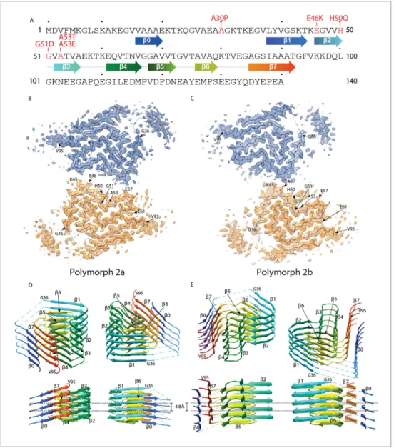
For polymorph 2a (PDB ID 6SSX) and polymorph 2b (PDB ID 6SST), each a-Syn molecule within a protofilament is composed of eight in-register parallel ß-strands (ß0–7;Figure 1A,D and E): residues 16–20 (ß0), 38–44 (ß1), 46–50 (ß2), 52–56 (ß3), 61–66 (ß4), 68–72 (ß5), 76–79 (ß6), and 85–92 (ß7). These are separated by either a lysine (L45) between ß1 and ß2, glycine residues (that is, G51 between ß2 and ß3, and G67 between ß4 and ß5) or arches (that is, E57-K60 between ß3 and ß4, G73-T75 between ß5 and ß6, and K80-G84 between ß6 and ß7). The NAC region encompasses
ß-Figure 1. Cross-sections of the a-Syn polymorph 2a and 2b cryo-EM structures. (A) Sequence of human a-Syn with familial PD mutation sites indicated in red. ß strands are indicated by arrows colored from blue to orange. Cryo-EM densities and atomic models of polymorph 2a (B) and polymorph 2b (C) of a-Syn. Each cryo-Cryo-EM map shows two protofilaments (blue and orange) forming a fibril. PD-associated mutations sites, and first and last residues of the NAC regions are indicated. (D and E) Rainbow rendering views of the secondary structure elements in five successive rungs of both polymorphs. A view perpendicular to the helical axis is shown to illustrate the height differences in a single a-Syn fibril. Colors correspond to the arrows in the sequence displayed in panel (A). The online version of this article includes the following figure supplement(s) for figure 1:
Figure supplement 1. Local resolution estimation and FSC curves.
Figure supplement 2. Interface regions between two protofilaments of the a-Syn polymorph 2a and 2b. Figure supplement 3. NMR identification of the residues forming the N-terminal beta strand.
strands ß4 to ß7 and it appears entirely sur-rounded by densities in our cryo-EM map with ß1 to ß3 on one side of the fibril and additional den-sities on the other side. Minor differences between polymorphs 2a and 2b are observed in the turn between ß3 and ß4 (Figure 1D and E,
Figure 2A and C).
As described for a-Syn polymorph 1a ( Guer-rero-Ferreira et al., 2018), and also shown in patient-derived Tau filaments (Fitzpatrick et al., 2017), there are considerable changes in the height of the a-Syn monomer along the helical axis, with the highest point being ß3 and the low-est ß4 (Figure 1D and E).
a-Syn fibrils in polymorph 1a are formed by ß-stands that are arranged in bent b-arches, run-ning along the length of each protofilament, pre-viously described as a Greek key-like architecture (Guerrero-Ferreira et al., 2018;Li et al., 2018a;
Li et al., 2018b; Tuttle et al., 2016). Also, the polymorphs 2a and 2b reported here present bent b-arch motifs. However, the components and the orientation of ß-strands contributing to the motifs are now radically different. In poly-morphs 2a and 2b, two bent b-arches orient back-to-back, one showing a single, and the other showing two bends. The first b-arch is formed by strands 1/6, 2/5 and 3/4, with bends located at res-idues K45/V74, G51/G67, and the tip formed by K58. The strands interact through mainly hydropho-bic, but also polar clusters. The second bent b-arch shares b-strands 4, 5 and 6 with the first one,
Video 1. Comparison of cryo-EM maps of a-Syn fibril polymorphs. Cryo-EM reconstructions of a-Syn fibrils at 3.1 A˚ (polymorph 2a) and 3.5 A˚ (polymorph 2b) resolution detailing the interaction between two protofilaments (blue and orange) in each fibril, the 4.9 A˚ spacing between ß-strands and the topology of a-Syn monomers within a single protofilament. https://elifesciences.org/articles/48907#video1
Figure 2. Structure and distribution of amino acids in the new a-Syn fibril polymorphs. Amino acids are colored in blue for positively charged, in red for negatively charged, in green for polar (including glycine), and in white for hydrophobic residues. Even and odd numberings are given on one monomer each. (A and C) Backbone structure. (B) and (D) Surface view.
and comprises in addition b-strand 7; again, interactions within the arch are of hydrophobic and polar type. This arch shows only one bend, at residues G67/G84.
The structure shares the extended use of glycine residues to form turns, and the hydrophobic and polar clusters forming the fibril core with other fibril structures (as for instance amyloid-b;
Gremer et al., 2017;Schu¨tz et al., 2015;Wa¨lti et al., 2016), which contribute to protofilament sta-bility, as also previously shown for a-Syn polymorph 1a (Guerrero-Ferreira et al., 2018;Li et al., 2018a;Li et al., 2018b).
Similar to a-Syn polymorph 1a, hydrophilic clusters are found at the periphery of the fibril ( Fig-ure 2). However, in contrast to a-Syn polymorph 1a, where the protofilament interface is formed by a hydrophobic steric zipper-geometry, in polymorphs 2a and 2b the interface is formed by salt-bridges.
Interestingly, residue I88 marks the beginning of a hydrophobic area composed of residues A89, A90, A91, F94 and V95, which contribute to the stabilization of an additional beta-strand density that is clearly visible in the cryo-EM maps of both polymorphs (Figure 1B and C). We propose that this interacting region corresponds in polymorph 2a to a hydrophobic stretch formed by residues V16 to E20, and here use for it the term b0. This region was previously shown to be an isolated ß-strand identified in the N-terminus of a-Syn fibrils by solid-state NMR on equivalent fibril prepara-tions (Bousset et al., 2013;Gath et al., 2014a) (Figure 1—figure supplement 3A). The localization of the stretch can be derived from cross peaks present in NMR 2D PAR (De Pae¨pe et al., 2008) spectra that connect S87 to A17, A18 and A19 (Figure 1—figure supplement 3B). Cross peaks in PAR spectra are indicative of proximities smaller than 6–7 A˚ between the spins, as illustrated in Fig-ure 1—figFig-ure supplement 3D, which shows other meaningful structural restraints identified. This localizes A17, A18 and A19 in proximity to S87 (Figure 1—figure supplement 3C). The orientation of the N-terminal b0-strand is then given by both the absence in the spectra of cross peaks between S87 and V16, as well as clear side chain density fitting K21 in the EM map. Interestingly, no signals were observed in the NMR spectra for residues M1-V15, K21-V37 and V95-A140 in the a-Syn fibril structure. These regions correspond precisely to the stretches in our cryo-EM structure for which model building is not possible due to lower resolution, suggesting that these regions are indeed dis-ordered in this polymorph. Also, no NMR peak doubling, although present for a subset of resonan-ces (Gath et al., 2014a), was observed for residues at the filament interface, and no distinction could be made between polymorphs 2a and 2b, indicating that the structures of the monomers in the polymorphs 2a and 2b are very close to each other. The interruption of the polypeptide chain between strands ß0 and ß1 in polymorph 2a and 2b, and between ß3 and ß4 in polymorph 2b due to lack of clear density, is compatible with a connection to the following strands of the same layer or alternatively to the neighboring layer.
For both a-Syn polymorph 2a and 2b, differences in height within residues of a protofilament reveal a hydrophobic area (Figure 2) akin to the hydrophobic cleft described for a-Syn polymorph 1a (Guerrero-Ferreira et al., 2018). It is composed of residues Q62 to V74 (ß4-ß5) (Figure 1D and E), and located between ß-strands ß2/ß3 and ß6/ß7. These residues correspond to a stretch of exclusively hydrophobic or polar residues, completely devoid of charged amino acids. Consistent with these results and with the concept that the hydrophobic core is essential for assembly, is the finding byEl-Agnaf et al. (1998) that, within the NAC region, residues E61-A78 are the amyloido-genic component. The hydrophobic cleft in a-Syn polymorphs 2a and 2b contrasts with the one found in a-Syn polymorph 1a, where intermolecular interactions involving residues V74-V82 may be the initial binding event responsible for fibril elongation (Guerrero-Ferreira et al., 2018).
Our structural cryo-EM analysis of a-Syn fibrils prepared with E46K mutant or phosphorylated and N-terminally acetylated protein shows these also to be composed of two protofilaments and having an overall diameter of 10 nm (Figure 3). The lower order of these fibrils prevented a separation of the a-Syn rungs along the fibril axis, but still allowed discerning the separation of individual ß-sheets. As with polymorphs 1a, 2a and 2b, ß-sheets in a-Syn fibrils from E46K mutant, phosphorylated or N-terminally acetylated protein are arranged forming the characteristic bent b-arch like shape, and an arrangement closely resembling that of polymorph 2a (Figure 3).
Comparison with previous structures
At first glance, the back-to-back arranged b-arches of polymorphs 2a and 2b appear similar to that of polymorph 1a, wrongly proposing that a mere protofilament rearrangement defines the
difference between them. However, closer inspection and comparison with secondary structure ele-ments identified in previous NMR studies on equivalent fibril preparations (Bousset et al., 2013;
Gath et al., 2014a) revealed that polymorphs 2a and 2b radically differ from the construction of pol-ymorphs 1.Figure 4shows the backbones of the polymorphs with different color codes for N-termi-nus, NAC region, and C-termiN-termi-nus, andFigure 4—figure supplement 1shows each 10 residues in a different color. When compared to polymorphs 1, the new structures of polymorphs 2a and 2b show an inverted bend of the first b-arch motif, comprising mainly the black and cyan segments in Fig-ure 4—figFig-ure supplement 1. This motif is largely conserved between polymorphs 1a and 1b (Li et al., 2018a) (Figure 4,Figure 4—figure supplement 1 andVideo 2), where it forms part of the interfilament interface, once via the black segment in polymorph 1a, and once via the cyan/light green segments in polymorph 1b. In polymorphs 2, this b-arch is formed by an inverted amino-acid sequence: when the two motifs are superimposed, the chain runs from cyan to black in one case, in the other from black to cyan. This inversion profoundly changes the amino acid distribution within the arch between polymorphs 1 and 2. Also, the b-arch in polymorph two is extended with a second bend followed by a b-strand region, comprising part of the orange segment largely unobserved in polymorphs 1.
In polymorph 1, the light green chain segment forms, together with parts of the above-described first b-arch, a second bent b-arch, completed by the red segment. This motif again exists in poly-morphs 2, but is located on the outside of the first arch, while in polymorph 1a it is located at the inside. Also, the arch is inverted, and the amino acid distribution again differs, as shown by the dif-ferent arrangement of the colored segments. Polymorph 1b is devoid of this motif. A hydrophilic cavity identified in polymorph 1a (located in the bend between the black and light green segments) is also present in polymorphs 2, but there between the partly disordered pink/orange segments, and the red segment.
The different arrangements result in a radically changed interface between the protofilaments in polymorphs 1 and 2. Unlike the hydrophobic interaction between protofilaments in polymorph 1a by the black segment, and in 1b by the cyan and light green segments, the interface in the new poly-morphs 2a and 2b is formed by electrostatic interactions in the green segment, through salt-bridges between residues K45 and E57, or K45 and E46, respectively in 2a and 2b polymorphs (Figure 1— figure supplement 2C and F,Figure 1—figure supplement 3,Figure 4—figure supplement 1).
Furthermore, the presence of an additional ß-strand ß0 on the periphery of the fibril (in the blue segment) is a remarkable difference between polymorphs 1 and polymorphs 2. This stretch of resi-dues, V16-E20, forms a hydrophobic steric-zipper geometry with residues S87-A91, which are part of ß7, at the end of the amyloidogenic NAC region. In polymorphs 2a and 2b, this results in the N-ter-minus wrapping around the fibril and enclosing the NAC region (cyan, light green and red seg-ments). This buries the serine residue at position 87, a phosphorylation site located within the NAC region, inside this interface. This is in contrast to a-Syn polymorph 1a, where approximately 40
Figure 3. Cryo-EM cross-sections of fibrils, formed by E46K, p-S129 phosphorylated, and N-terminally acetylated a-Syn protein. Fibrils formed by E46K mutant a-Syn protein (A), Ser129 phosphorylated a-Syn protein (B), and N-terminally acetylated a-Syn protein (C) were analyzed by cryo-EM. Image processing did not allow reaching sufficient resolution for model building, but the cross-sections of the obtained 3D reconstructions are compatible with polymorph 2a for all three forms.
residues on both ends of the a-Syn fibril are flexible and surround the fibril with a fuzzy coat, leaving part of the NAC region (that is, K80 to V95, red and magenta segments), including S87, exposed (Figure 1B and C,Figure 4—figure supplement 1B).
Origin of distinct a-Syn polymorphs
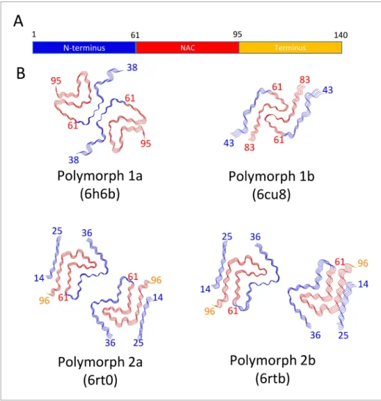
Presently, there are four different polymorphs of a-Syn fibrils known at atomic resolution, including the structures presented here. They can be classified into two groups with each group having a dis-tinct fold (folds 1 and 2) (that is, at the protofilament level). Within each group there are two disdis-tinct protofilament packings or assembly polymorphisms (polymorphs a and b;Figure 4).
In an attempt to rationalize the origin of the distinct polymorphs, we first concentrate on the dis-tinct folds at the protofilament level. All fold one structures (polymorphs 1a and 1b) were grown
Figure 4. Schematic representation of a-Syn polymorphs. (A) Diagram representing a-Syn regions with the N-terminus in blue, the NAC region in red and the C-terminus in yellow. (B) Representation of a-Syn fibril polymorphs 1a (PDB ID 6h6b;Guerrero-Ferreira et al., 2018), 1b (PDB ID 6cu8;Li et al., 2018a), 2a (PDB ID 6rt0, this work), and 2b (PDB ID 6rtb, this work), highlighting the striking differences in protofilament folding in a-Syn polymorphs 1a and 1b, compared to Syn polymorphs 2a and 2b. The atomic models obtained by cryo-EM of a-Syn polymorph 1a, polymorph 1b (Li et al., 2018a;Li et al., 2018b) and a-Syn polymorphs 2a and 2b (this work). Protein Data Bank (PDB) accession numbers are indicated.
The online version of this article includes the following figure supplement(s) for figure 4: Figure supplement 1. Comparison between polymorphs 1 and 2.
under buffer conditions comprising either a poly-anion (that is, phosphate with three negative charges and N3- with having locally two negative
charges) or a big chaotropic negative ion (that is Br-), while polymorphs 2a and 2b of wildtype
a-Syn were grown under phosphate-free conditions with the only anion being Cl-(Table 1). Interest-ingly, there are adjacent to the salt-bridge H50-E57 three lysine residues in polymorph 1a at posi-tions K43, K45 and K58 (the latter from the other protofilament) that are close in space. Between them, Guerrero-Ferreira et al. (2018) observed a density in the cryo-EM map, presumably a phosphate ion of poly-anionic nature that neutral-izes the repulsion of the three positive charged residues (Figure 4). Albeit still of polymorph 1a type, in presence of N3-the structure is distinct in
this area with K58 flipping inward into the cavity forming a salt-bridge with E61 attributed to the less poly-anion-like character of N3-when compared
with phosphate. In the presence of the chaotropic Br-, K58 is facing more the solvent with a closer distance between H50 and K45 (Figure 4). In the absence of a neutralizing poly-anion in this area of positively charged side chains or Br- exerting its chaotropic property, it is unlikely that polymorph
one can be obtained, indicating that in the presence of Cl- as the only anion, a distinct polymorph
must be formed.
The fibrils of the familial variant E46K (Figure 3) were also grown under phosphate conditions but adopt polymorph 2a fold. The polymorph 1a fold we obtained for the wildtype 1–121 form, introdu-ces an electrostatic repulsion between K46 and K80, instead of the 46–80 salt-bridge stabilizing this polymorph. The structural features thus provide a rationale how changes corresponding to only a few kcal (or less) in the stability of polymorph 1a (such as absence/presence of phosphate, single point mutation), can lead to the protein adopting an entirely different polymorph. This finding also highlights the postulated flat energy landscape of fibril formation and the conformational promiscu-ity that comes along with it.
The fact that the polymorphs found both by Li et al. (Li et al., 2018a) and here by us are distinct, further accentuates this remark as they grew under similar conditions (except for the salt composi-tion and additives, see Table 1). While the packing interfaces between polymorphs 1a and 1b are both hydrophobic, they still substantially differ, as in polymorph 1a residues V66-A78 form the inter-face, and in polymorph 1b residues H50-E57 form the interface (Figure 4). The difference is smaller in the case of polymorphs 2, where the protofilament interactions are of inter-protofilament salt-bridge character but are packed differently. It is evident that the energy differences between the two polymorphs are therefore very minute, but still the structures are distinct.
Familial PD mutation sites in the new a-Syn polymorphs
A series of familial mutations have been identified in families with a history of PD. Based on
Fujioka et al. (2014)andFlagmeier et al. (2016), these mutations may lead to sporadic PD (A30P, E46K, A53E), early-onset (G51D, A53T) or late-onset (H50Q) forms of Parkinson’s disease. The locali-zation of the different sites in the structures is compared inFigure 4—figure supplement 1. Funda-mentally different folds between polymorphs 1 and 2 place the familial PD mutation sites into an entirely different environment.
The E46K mutation has been found to promote a-Syn phosphorylation in mice (Dettmer et al., 2017), and in neuronal cells it showed to be toxic, with toxicity being enhanced by simultaneously mutating E35 and E61 to lysines (Mbefo et al., 2015). E46 holds a central role in polymorphs 1a and 2b. In polymorph 1a, where this motif resides at the beginning of one bent b-arch, E46 forms a stabi-lizing salt-bridge with K80. In contrast, in a-Syn polymorph 2b this residue is part of the protofila-ment interface, where the K45-E46 salt-bridge interface to E46-K45 from the other protofibril appears to be critical to the protofilament interface as these residues are the only interaction point between the protofilaments (Figure 1—figure supplement 2). Thus, protofilament interaction in this
Video 2. Structural differences between a-Syn polymorphs. Atomic models of a-Syn fibrils
represented as rounded ribbons with the N-terminus in blue and the NAC region in red.
polymorph 2b manner would be unlikely since the mutation would induce a charge repulsion between lysines K45 and E46K from the two protofilaments. And indeed, the mutant E46K adopts polymorph 2a, which result in two lysines (that is, K45 and K46) from one protofilament interacting with E57 in the other. Interestingly, the a-Syn E46K mutant fibril investigated here by cryo-EM, resulted in protofilament assembly corresponding to a-Syn polymorph 2a, confirming that the inter-action between K45/K46 and E57 can indeed be maintained (Figure 3).
Heterozygous mutations in residues H50, G51, and A53 are associated with familial forms of PD (Appel-Cresswell et al., 2013;Lesage et al., 2013;Pasanen et al., 2014;Polymeropoulos et al., 1997). As we reported previously, in a-Syn polymorph 1a, these sites are an integral part of the interface region, contributing to the steric-zipper architecture and fibril stability, so that the muta-tions G51D, A53E and A53T are not compatible with polymorph 1a (Guerrero-Ferreira et al., 2018) (Figure 4—figure supplement 1). In contrast, in a-Syn polymorph 2a, these residues lie in the cavity formed between the two protofilaments, and in polymorph 2b they are surface-exposed where the two protofilaments interact (Figure 3—figure supplement 1). While these mutations are not in direct conflict with the formation of a-Syn fibril polymorphs 2a and 2b, the structures of fibrils formed with these mutations remain to be determined.
The A30P mutation leads to a form of PD with an age of onset between 54 and 76 years (Flagmeier et al., 2016;Fujioka et al., 2014;Kru¨ger et al., 1998). In our structures of polymorphs 2a and 2b, a steric-zipper interface between V16-E20 and I88-A91 causes the a-Syn N-terminus to wrap around the NAC region. Under these conditions, residue A30 might be surface-exposed in a defined manner, as it forms part of the fibrillar core in the sense that it is on both sides linked to nearby structured regions. The disordered nature of the region between K21 and G37 results in weak density in the cryo-EM map, and makes accurate model building in this region difficult. Never-theless, in a-Syn polymorph 1a, A30 is not part of the fibril core but instead is found in the disor-dered region corresponding to residues M1 to V37 (Guerrero-Ferreira et al., 2018).
Post translational modifications (PTMs) and fibrillization in a-Syn
polymorphs
Certain post-translational modifications of a-Syn associated with neuropathology inhibit the process of a-Syn fibril formation in vitro, suggesting that they are late events rather than occurring before protein aggregation (Oueslati et al., 2010). Most of these modifications take place at the C-terminal region (Mahul-Mellier et al., 2019), with the exception of acetylation and ubiquitination, which mostly affect residues in the N-terminal region. Ubiquitination alters mostly N-terminal lysines between residues 1 and 36, with K21, K23, K32 and K34 being the major sites for ubiquitin conjuga-tion (Nonaka et al., 2005). The phosphorylated and acetylated forms determined here fold into polymorph 2a (Figure 3). In this polymorph, the amino-acid stretch, which appears disordered in polymorph 1a fibrils, is more distinct, as the interaction of ß-strand ß0 with the fibril core brings the N-terminal region back to the fibril. While the involvement of the N-terminal region has been sug-gested by NMR for two different polymorphs (Bousset et al., 2013;Gath et al., 2012;Gath et al., 2014a;Gath et al., 2014b), our structure for the first time shows how this N-terminal region that is important in the context of post-translational modifications, can be positioned in the protofilament.
Implications for fibril preparation protocols
When pre-formed fibrils (PFFs) are to be studied, the protocol used for their preparation is crucial. Conditions used to prepare such samples vary (Table 1). It has been previously shown that a-Syn polymorphism may arise when different fibrillization methods are used (Lv et al., 2012;
Verasdonck et al., 2016). NMR has shown to be able to distinguish fibrils in polymorphic mixtures in samples, when the monomer fold differs substantially (Bousset et al., 2013;Gath et al., 2012;
Gath et al., 2014a;Gath et al., 2014b;Verasdonck et al., 2016). Still, NMR could for instance not distinguish between the two assembly polymorphs 2a and 2b. The here reported fibril polymorphism raises questions regarding the structural consistency of recombinant fibrils generated to study a-syn in vitro and calls to investigate the structures resulting from different fibril preparation conditions, or to screen fibril polymorphism in samples prepared by a single preparation protocol, in order to com-pile a library of a-Syn polymorphs to inform studies using PFFs on cell culture or animal models.
Recently, fibrillar aggregates of Tau protein were purified from human postmortem brain from Alzheimer’s, Pick’s disease, and chronic traumatic encephalopathy patients, showing variations in Tau fibril conformations between diseases (Falcon et al., 2018; Falcon et al., 2019;
Fitzpatrick et al., 2017), and these fibrils all differed from the in-vitro generated, heparin-induced tau fibrils (Zhang et al., 2019). However, to our knowledge, purification of a-Syn fibrils from human brain of Parkinson’s disease patients is more challenging. Existing protocols so far have only been able to produce filamentous material that co-fractionated with numerous contaminants (e.g., lipofus-cin, amyloid, etc.), including membranes (Iwatsubo et al., 1996). Light microscopy and electron microscopy approaches to analyze the ultrastructural composition of LBs show location maps by fluo-rescence or structural features at the nanometer scale (Lewis et al., 2019; Moors et al., 2018;
Shahmoradian et al., 2019). But methods to study the presence and polymorph of a-Syn fibrils in the diseased human brain and cerebrospinal fluid, and the mechanisms by which a-Syn may be caus-ing Parkinson’s disease and contribute to progression of the disease, remain to be developed.
Conclusion
We present here two new structures of a-Syn fibril polymorphs (polymorph 2a (PDB ID 6rt0), and polymorph 2b (PDB ID 2rtb)). These differ in their protofilament interfaces but are formed by the same protofibril subunit structure, which is distinct from previously described a-Syn folds.
Our results describe 3D structures very different from previous work and demonstrate how a-Syn amyloid fibrils can reach different cross-ß architectures in spite of having the same amino acid sequence. The structural information from the various a-Syn polymorphs allowed informed hypothe-ses on how amyloid fibrils may form and how their formation may be related to pathogenicity. More importantly, these structures add to the knowledge of the conformational space of this protein, which is central for structure-based design of imaging tracers or inhibitors of amyloid formation. In this context, a large scale, in vitro study, investigating the structure of a-Syn fibrils produced under different aggregation conditions would prove very informative.
Determination of the structural space of fibril polymorphs, including those of a-Syn carrying ease-relevant mutations, and of a-Syn states purified from diseased human brain, is pivotal to dis-cover whether and how fibrils might form or could be dissolved, and if and how they may interact with affected neurons and contribute to disease.
Materials and methods
Key resources table
Reagent type
(species) or resource Designation Source or reference Identifiers
Additional information
Strain, strain
background (E.coli)
BL21(DE3) Stratagene Agilent Technology #200131 Expression performed in LB medium Transfected
construct (pET-3a)
pET3a Novagen https://www.addgene.
org/vector-database/2637/
Encoding full length human alpha synuclein asyn (UniProtKB - P37840) with a silent mutagenesis of codon 136 (TAC to TAT) Transfected
construct (pRT21)
pRT21 (Masuda et al., 2006) Full length human alpha
synuclein asyn (UniProtKB - P37840) with a silent mutagenesis of codon 136 (TAC to TAT) Transfected
construct (pNatB)
pNatB (Johnson et al., 2010) http://www.addgene. org/53613/
Expression of the fission yeast NatB complex -chloramphenicol marker Chemical compound, drug DEAE sepharose fast flow GE Healthcare, #17-0709-01
Continued
Reagent type
(species) or resource Designation Source or reference Identifiers
Additional information Other Copper/ carbon grids https://www.quantifoil.com/ R 2/2 grids Software, algorithm
UCSF Chimera (Pettersen et al., 2004) https://www.cgl. ucsf.edu/chimera
RRID:SCR_004097 Software,
algorithm
SerialEM (Mastronarde, 2005) https://bio3d.colorado. edu/SerialEM/
RRID:SCR_017293 Software,
algorithm
FOCUS (Biyani et al., 2017) http://focus-em.org Software,
algorithm
MotionCor2 (Zheng et al., 2017) https://emcore.ucsf.edu/ ucsf-motioncor2 RRID:SCR_016499 Software, algorithm RELION 2, 3 (Scheres, 2012; Zivanov et al., 2018) http://www2.mrc-lmb. cam.ac.uk/relion RRID:SCR_016274 Software, algorithm
COOT (Emsley and
Cowtan, 2004) https://www2.mrc-lmb.cam.ac.uk/ personal/pemsley/coot/ RRID:SCR_014222 Software, algorithm
Molprobity (Williams et al., 2018) http://molprobity. biochem.duke.edu
RRID:SCR_014226
a
-Syn expression and purification
The fibrillary polymorph or WT full length unmodified a-Syn polymorph 2a and 2b were assembled from monomeric a-Syn expressed and purified as described inBousset et al. (2013)andGath et al. (2014a). Briefly recombinant, wild-type a-Syn was expressed in E. coli strain BL21(DE3), transformed with the expression vector pET3a (Novagen) encoding wild-type, full-length a-Syn. Expression was induced by 0.5 mM IPTG for 2 hr, when the bacteria grown in LB medium at 37˚C had reached an optical density of 1.0 at 660 nm. Soluble, monomeric a-Syn was purified from the bacteria lysate as previously described (Ghee et al., 2005). a-Syn concentration was determined spectrophotometri-cally using an extinction coefficient of 5960 M!1*cm!1 at 280 nm. Pure a-Syn (0.7 mM) in 50 mM
Tris-HCl, pH 7.5, 150 mM KCl was filtered through sterile 0.22 mm filters and stored at !80˚C. For preparation of fibrils carrying post-translationally modifications, full-length a-Syn was expressed in competent Escherichia coli BL21(DE3) (Stratagene, La Jolla, CA, USA) from the pRT21 expression vector. To acetylate the N-terminus, cells were pre-transfected by pNatB vector coding for the N-terminal acetylase complex (plasmid kindly provided by Daniel Mulvihill, School of Bio-sciences, University of Kent, Canterbury, UK) (Johnson et al., 2010). The various a-Syn forms were purified by periplasmic lysis, ion exchange chromatography, ammonium sulfate precipitation, and gel filtration chromatography as previously described (Guerrero-Ferreira et al., 2018;Huang et al., 2005; Luk et al., 2009). Purified a-Syn was phosphorylated using polo like kinase 2 (PLK2) expressed in E. coli BL21-DE3-pLysS, and isolated via its His-tag. Phosphorylated from non-phos-phorylated a-Syn was then separated using standard ion exchange and gel filtration chromatogra-phy. N-terminally acetylated and phosphorylated a-Syn strains were cleared from endotoxins using one run of Detoxi-Gel Endotoxin Removing Gel (Thermo Scientific) or until endotoxins were below detection level. Protein sequences were verified by tryptic digestion and MALDI mass spectrometry (MS). Alternatively, HPLC/ESI tandem MS was performed to determine total mass. Coomassie blue or silver staining of the SDS PAGE gel and analytical ultracentrifugation were used to determine purity and monodispersity. Protein concentration was measured using the bicinchoninic acid (BCA) assay (Thermo Scientific) with bovine serum albumin as a standard. Purified a-Syn was dialyzed in a 2 kDa Slide-A-Lyzer unit (Thermo Scientific, for max. 3 ml) against HPLC-water (VWR). Aliquots (500 mg) were dispensed into 1.5 ml tubes, frozen on dry ice, and lyophilized for 2 hr in an Eppendorf concentrator (Eppendorf) and stored at !80˚C until use.
Disease-linked E46K mutant a-Syn was expressed and purified using a periplasmic purification protocol as described earlier (Campioni et al., 2014;Huang et al., 2005). Briefly, plasmid pRK172 was co-expressed with N-terminal acetyltransferase B (NatB) complex as described earlier (Johnson et al., 2010). Colonies containing both plasmids (NatB and pRK172) were selected using two different antibiotics and grown in 1 liter of lysogeny broth at 37˚C. After reaching an optical
density of 1.0 at 600 nm, expression was induced with 1 mM IPTG for 5 hr. Cells were harvested and a-Syn collected from the periplasmic space of the cells, using osmotic shock methods described ear-lier (Huang et al., 2005). Protein was further purified using ion exchange chromatography and hydrophobic interaction chromatography (Campioni et al., 2014).
Fibrillization
Full-length, wildtype unmodified a-Syn was incubated at 37˚C for one week under continuous shak-ing in an Eppendorf Thermomixer set at 600 r.p.m., to assemble into fibrillar form. 700 mM a-Syn was assembled in 50 mM Tris-HCl, pH 7.5, 150 mM KCl buffer (Table 1).
To prepare fibrils of full-length, phosphorylated, N-terminally acetylated, N-terminally acetylated and the E46K mutant a-Syn, recombinant protein (dialyzed and lyophilized) was diluted to 5 mg/mL in 200 mL of Dulbecco’s phosphate buffered saline (DPBS) buffer (Gibco; 2.66 mM KCL, 1.47 mM KH2PO4, 137.93 mM NaCl, 8.06 mM Na2HPO4-7H2O pH 7.0–7.3). After 5 days of incubation at 37˚C
with constant agitation (1,000 rpm) in an orbital mixer (Eppendorf), reactions were sonicated for 5 min in a Branson 2510 water bath, aliquoted, and stored at !80˚C. All fibrils were created in the presence of an air-water interface. The presence of amyloid fibrils was confirmed by thioflavin T fluorimetry and high molecular weight assemblies were visualized by gel electrophoresis.
Electron microscopy
Cryo-EM grids were prepared using a Leica EM GP automatic plunge freezer (Leica Microsystems) with 80% humidity at 20˚C. 3 mL aliquots were applied onto 60 s glow-discharged, 300 mesh, copper Quantifoil grids (R2/1). After blotting, grids were plunge frozen in liquid ethane cooled by liquid nitrogen.
Micrographs were acquired on a Titan Krios (ThermoFisher Scientific) transmission electron micro-scope, operated at 300 kV and equipped with a Gatan Quantum-LS imaging energy filter (GIF, 20 eV zero loss energy window; Gatan Inc). Images were recorded on a K2 Summit electron counting direct detection camera (Gatan Inc) in dose fractionation mode (50 frames) using the Serial EM soft-ware (Mastronarde, 2005) at pixel sizes of 0.831 A˚ or 0.629 A˚, and a total dose of ~69 electrons per square Angstrom (e-/A˚2) for each micrograph. Micrographs were processed and analyzed during data collection with FOCUS (Biyani et al., 2017), applying drift-correction and dose-weighting using MotionCor2 (Zheng et al., 2017). Specific data collection parameters for the various datasets are detailed inTable 2.
Image processing
Computer image processing and helical reconstruction was carried out with RELION 2.1 (Scheres, 2012) and RELION 3.0 ß (Zivanov et al., 2018), using the methods described in
Zheng et al. (2017). Filament selection per micrograph was done manually in RELION 2.1. Segments were extracted with a box size of 280 pixels and an inter-box distance of 28 pixels. A summary of the number of micrographs and segments that went into the various steps of processing are pre-sented inTable 2. After 2D classification with a regularization value of T = 2, 2D class averages with a visible separation of individual rungs were selected for further processing. For class averages formed by segments from polymorph 2, the calculated power spectra showed a meridional peak intensity (Bessel order n = 0) at the layer line of 1/ (4.9 A˚). For polymorph 2b, power spectra showed peak intensities on both sides of the meridian (Bessel order n = 1). This is the result of an approxi-mate 21screw symmetry between a-Syn subunits on the two protofilaments (Figure 1—figure
sup-plement 1). The best 2D classes were selected for a round of 3D classification with T = 8 and optimization of the helical twist and rise using a helical_z_percentage parameter (He and Scheres, 2017) of 10%. A cylinder generated via the helix toolbox tool was used as initial model. This resulted in three classes where ß-sheets perpendicular to the fibril axis were clearly separated. Segments con-tributing to classes 1 and 2, which corresponded to a-Syn polymorphs 2a and 2b, respectively, were then processed separately. The 3D maps from their respective classes were used as initial models after applying a low-pass filter to 30 A˚. Then, a 3D classification with a single class (K = 1) and T = 20, which has allowed the successful reconstruction of amyloid filaments, was carried out (Falcon et al., 2018;Fitzpatrick et al., 2017;Guerrero-Ferreira et al., 2018).
The 3D auto-refine procedure in RELION 3.0, with optimization of helical twist and rise, resulted in structures with overall resolutions of 3.34 A˚ (a-Syn polymorph 2a) and 3.75 A˚ (a-Syn polymorph 2b). Post-processing with soft-edge masks and estimated map sharpening B-factors of !67.1 and !76.4 A˚
2
, respectively, gave maps with resolutions of 3.0 A˚ (a-Syn polymorph 2a) and 3.3 A˚ (a-Syn polymorph 2b) (by the FSC 0.143 criterion). Local resolution values and local-resolution-filtered maps were obtained in RELION 3.0. (Table 2,Figure 1—figure supplement 1).
Model building and refinement
A model for the a-Syn polymorph 2a fibril was built into the RELION local resolution-filtered map with COOT (Emsley and Cowtan, 2004), by conserving as many secondary structure elements as possible from our previous a-Syn polymorph one model (PDB ID 6h6b) (Guerrero-Ferreira et al., 2018), together with the use of secondary structure information derived from ssNMRs served to establish an initial model which served as an initial model. The structure was then refined against the same map with PHENIX real space refine (Afonine et al., 2013) using rotamer and Ramachandran restraints, and non-crystallographic symmetry and beta strand geometry constraints. The building and refinement of the a-Syn polymorph 2b model was more challenging due to the lower resolution of the map. An initial structure was built into the a-Syn polymorph 2b local resolution-filtered map by fitting of the a-Syn polymorph 2a model using COOT. To successfully refine the structure, it was necessary to generate secondary structure restraints in Phenix, based on CA backbone and intermo-lecular beta sheets to refine the structure. All structures were validated using Molprobity (Williams et al., 2018). Figures were prepared using UCSF Chimera (Pettersen et al., 2004).
NMR spectroscopy
NMR spectra were recorded at 20.0 T static magnetic field using 3.2 mm rotors and a triple-reso-nance probe. The reproducibility of the sample preparation was previously verified with 20 ms DARR fingerprint spectra (Gath et al., 2014a). The secondary chemical-shift analysis was based on the sequential assignments (BMRB accession code 18860) and was presented before (Gath et al., 2014a;Gath et al., 2014b). The PAR spectrum (De Pae¨pe et al., 2008;Lewandowski et al., 2007) was recorded using a mixing time of 8 ms. The assignments for the S87/A17-19 cross peaks are unambiguous within a range of 0.15 ppm, corresponding to about ½ of the13C line width.
Assign-ments of restraints given for reference on the full aliphatic region of the PAR spectrum in the Fig-ure 1—figFig-ure supplement 3 are mostly ambiguous, which prevented structure determination by NMR. Ambiguities were lifted by comparison to the here determined cryo-EM structure.
Data availability
Raw cryo-EM micrographs are available in EMPIAR, entry numbers EMPIAR-10323. The 3D maps are available in the EMDB, entry numbers EMD-10307 (a-Syn polymorph 2a) and EMD-10305 (a-Syn-polymorph 2b). Atomic coordinates are available at the PDB with entry numbers PDB 6SSX (a-Syn polymorph 2a) and PDB 6SST (a-Syn polymorph 2b).
Acknowledgements
We thank Liz Spycher, Jana Ebner, Alexandra Kronenberger, Daniel Schlatter, Daniela Huegin, Ralph Thoma, Christian Miscenic, Martin Siegrist, Sylwia Huber, Arne Rufer, Eric Kusznir, Peter Jakob, Tom Dunkley, Joerg Hoernschmeyer, and Johannes Erny at Roche for their technical support to clone, express, purify and characterize the different forms of a-Syn; Kenneth N Goldie, Lubomir Kovacik and Ariane Fecteau-Lefebvre for support in cryo-EM. Calculations were performed using the high-performance computing (HPC) infrastructure administered by the scientific computing center at the University of Basel (sciCORE;http://scicore.unibas.ch). The Novo Nordisk Foundation Center for Pro-tein Research is supported financially by the Novo Nordisk Foundation (NNF14CC0001). NMIT is a member of the Integrative Structural Biology Cluster (ISBUC) at the University of Copenhagen. This work was in part supported by the Synapsis Foundation Switzerland, the Heidi-Seiler Stiftung Foun-dation, and the Swiss National Science Foundation (grants CRSII3_154461 and CRSII5_177195 and 20020_178792), and the French ANR (ANR-12-BS08-0013-01), the LABEX ECOFECT (ANR-11-LABX-0048) within the Universite´ de Lyon program Investissements d’Avenir (ANR-11-IDEX-0007). AAA, LB and RM received funding from the European Union’s Horizon 2020 research and innovation
programme under grant agreement No. 116060 (IMPRiND), the Swiss State Secretariat for Educa-tion, Research and Innovation (SERI) under contract number 17.00038, the Fondation Bettencourt Schueller, The Fondation pour la Recherche Me´dicale (Contract DEQ 20160334896),The Fondation Simone et Cino Del Duca of the Institut de France and the EC Joint Program on Neurodegenerative Diseases (TransPathND, ANR-17-JPCD-0002–02 and Protest-70, ANR-17-JPCD-0005–01), by the French Infrastructure for Integrated Structural Biology (FRISBI) [ANR-10-INSB-05–01]. The opinions expressed and arguments employed herein do not necessarily reflect the official views of these fund-ing bodies.
Additional information
Competing interests
Daniel Mona, Markus Britschgi, Matthias E Lauer: employee at Roche and may additionally hold Roche stock/stock options. The other authors declare that no competing interests exist.
Funding
Funder Grant reference number Author
Novo Nordisk NNF14CC0001 Nicholas MI Taylor Synapsis Foundation –
Alzhei-mer Research Switzerland
Ricardo Guerrero-Ferreira Henning Stahlberg Heidi-Seiler Stiftung Ricardo Guerrero-Ferreira
Henning Stahlberg Swiss National Science
Foun-dation
CRSII3_154461 Ricardo Guerrero-Ferreira Nicholas MI Taylor Henning Stahlberg Swiss National Science
Foun-dation
CRSII5_177195 Ricardo Guerrero-Ferreira Henning Stahlberg Swiss National Science
Foun-dation
20020_178792 Ricardo Guerrero-Ferreira Henning Stahlberg Agence Nationale de la
Re-cherche
ANR-12-BS08-0013-01 Ronald Melki Luc Bousset Labex ANR-11-LABX-0048 Anja Bo¨ckmann EU Framework Programme for
Research and Innovation H2020
116060 IMPRiND Ana-Andreea Arteni Ronald Melki Luc Bousset
SERI 17.00038 Ana-Andreea Arteni
Beat H Meier Luc Bousset Fondation Bettencourt
Schuel-ler
Ana-Andreea Arteni Beat H Meier Luc Bousset Fondation pour la Recherche
Me´dicale
Contract DEQ 20160334896 Ana-Andreea Arteni Beat H Meier Luc Bousset Fondation Simone et Cino Del
Duca
TransPathND ANR-17-JPCD-0002-02
Beat H Meier Luc Bousset EU Joint Programme –
Neuro-degenerative Disease Re-search Protest-70 ANR-17-JPCD-0005-01 Ana-Andreea Arteni Beat H Meier Luc Bousset French Infrastructure for
Inte-grated Structural Biology
ANR-10-INSB-05-01 Ana-Andreea Arteni Beat H Meier Luc Bousset University of Lyon Investissements d’Avenir
ANR-11-IDEX-0007 Anja Bo¨ckmann Agence Na)onale de la Recherche Scien)fique Agence Na)onale de la Recherche Scien)fique
The funders had no role in study design, data collection and interpretation, or the decision to submit the work for publication.
Author contributions
Ricardo Guerrero-Ferreira, Conceptualization, Data curation, Formal analysis, Validation, Investiga-tion, VisualizaInvestiga-tion, Methodology, Writing - original draft, Writing - review and editing; Nicholas MI Taylor, Data curation, Formal analysis, Validation, Investigation, Visualization, Writing - review and editing, Structural calculation, building of the structural model; Ana-Andreea Arteni, Validation, Investigation, Screen the assemblies for optimization, optimize cryo-EM freezing conditions and set up experimental conditions for particle imaging. Acquire the first high resolution Cryo-EM images of the "fibril" type of assemblies; Pratibha Kumari, Validation, Investigation, data interpretation and discussion; Daniel Mona, Matthias E Lauer, Investigation, Methodology, expressed, purified and ana-lyzed recombinant human alpha-synuclein (wt, N-acetylated, pSer129 aSyn, C-terminally truncated forms) and performed initial biochemical and biophysical characterization and quality control of gen-erated fibrils; Philippe Ringler, Data curation, Formal analysis, Investigation, data interpretation and discussion; Markus Britschgi, Resources, Data curation, Investigation, Methodology, expressed, puri-fied and analyzed recombinant human alpha-synuclein (wt, N-acetylated, pSer129 aSyn, C-terminally truncated forms) and performed initial biochemical and biophysical characterization and quality con-trol of generated fibrils; Ali Makky, Formal analysis, Investigation, Methodology, acquire and inter-pret AFM data on alpha-synuclein; Joeri Verasdonck, Data curation, Formal analysis, Investigation, acquire and interpret Solid state NMR data; Roland Riek, Investigation, Methodology, data interpre-tation and discussion; Ronald Melki, Resources, Formal analysis, Supervision, Funding acquisition, Investigation, Visualization, Methodology, Writing - review and editing; Beat H Meier, Conceptuali-zation, Resources, Supervision, Investigation, Methodology, Writing - review and editing; Anja Bo¨ck-mann, Conceptualization, Supervision, Investigation, Writing - review and editing, solid-state NMR studies, supervision, building of the structural model; Luc Bousset, Conceptualization, Resources, Formal analysis, Validation, Investigation, Visualization, Methodology, Writing - original draft, Writ-ing - review and editWrit-ing, expressed, purified and analyzed recombinant human alpha-synuclein (wt, unmodified form) generated fibrils; Henning Stahlberg, Conceptualization, Resources, Software, Supervision, Funding acquisition, Validation, Investigation, Methodology, Writing - original draft, Project administration, Writing - review and editing
Author ORCIDs
Ricardo Guerrero-Ferreira https://orcid.org/0000-0002-3664-8277
Nicholas MI Taylor http://orcid.org/0000-0003-0761-4921
Ana-Andreea Arteni http://orcid.org/0000-0001-6462-905X
Philippe Ringler http://orcid.org/0000-0003-4346-5089
Markus Britschgi https://orcid.org/0000-0001-6151-4257
Matthias E Lauer http://orcid.org/0000-0003-3252-8718
Beat H Meier http://orcid.org/0000-0002-9107-4464
Anja Bo¨ckmann https://orcid.org/0000-0001-8149-7941
Henning Stahlberg https://orcid.org/0000-0002-1185-4592 Decision letter and Author response
Decision letterhttps://doi.org/10.7554/eLife.48907.sa1
Author responsehttps://doi.org/10.7554/eLife.48907.sa2
Additional files
Supplementary files
.Transparent reporting form
Data availability
Raw cryo-EM micrographs are available in EMPIAR, entry numbers EMPIAR-10323. The 3D maps are available in the EMDB, entry numbers EMD-10307 (a-Syn polymorph 2a) and EMD-10305
(a-Syn-polymorph 2b). Atomic coordinates are available at the PDB with entry numbers PDB 6SSX (a-Syn polymorph 2a) and PDB 6SST (a-Syn polymorph 2b).
The following datasets were generated:
Author(s) Year Dataset title Dataset URL
Database and Identifier Ricardo
Guerrero-Ferreira
2019 aSyn polymorph 2a https://www.ebi.ac.uk/ pdbe/entry/emdb/EMD-10307
Electron Microscopy Data Bank, EMD-1030 7
Ricardo Guerrero-Ferreira
2019 aSyn polymorph 2a https://pdbe.org/6ssx Electron Microscopy Data Bank, 6SSX Ricardo
Guerrero-Ferreira
2019 aSyn polymorph 2b https://www.ebi.ac.uk/ pdbe/entry/emdb/EMD-10305
Electron Microscopy Data Bank, EMD-1030 5
Ricardo Guerrero-Ferreira
2019 aSyn polymorph 2b https://pdbe.org/6sst Electron Microscopy Data Bank, 6SST Ricardo
Guerrero-Ferreira
2019 aSyn polymorph 2a and 2b electron microscopy images
https://www.ebi.ac.uk/ pdbe/emdb/empiar/en-try/10323
Electron Microscopy Data Bank, EMPIAR-10323
References
Afonine PV, Headd JJ, Terwilliger TC, Adams PD. 2013. New tool: phenix_real_space_refine. Comp. Crystal 4: 43–44.
Appel-Cresswell S, Vilarino-Guell C, Encarnacion M, Sherman H, Yu I, Shah B, Weir D, Thompson C, Szu-Tu C, Trinh J, Aasly JO, Rajput A, Rajput AH, Jon Stoessl A, Farrer MJ. 2013. Alpha-synuclein p.h50q, a novel pathogenic mutation for parkinson’s disease. Movement Disorders 28:811–813.DOI: https://doi.org/10.1002/ mds.25421,PMID: 23457019
Bassil F, Fernagut PO, Bezard E, Pruvost A, Leste-Lasserre T, Hoang QQ, Ringe D, Petsko GA, Meissner WG. 2016. Reducing C-terminal truncation mitigates synucleinopathy and neurodegeneration in a transgenic model of multiple system atrophy. PNAS 113:9593–9598.DOI: https://doi.org/10.1073/pnas.1609291113,PMID: 274 82103
Biyani N, Righetto RD, McLeod R, Caujolle-Bert D, Castano-Diez D, Goldie KN, Stahlberg H. 2017. Focus: the interface between data collection and data processing in cryo-EM. Journal of Structural Biology 198:124–133. DOI: https://doi.org/10.1016/j.jsb.2017.03.007
Bousset L, Pieri L, Ruiz-Arlandis G, Gath J, Jensen PH, Habenstein B, Madiona K, Olieric V, Bo¨ckmann A, Meier BH, Melki R. 2013. Structural and functional characterization of two alpha-synuclein strains. Nature
Communications 4:2575.DOI: https://doi.org/10.1038/ncomms3575,PMID: 24108358
Campioni S, Carret G, Jordens S, Nicoud L, Mezzenga R, Riek R. 2014. The presence of an air-water interface affects formation and elongation of a-Synuclein fibrils. Journal of the American Chemical Society 136:2866– 2875.DOI: https://doi.org/10.1021/ja412105t,PMID: 24460028
Chartier-Harlin M-C, Kachergus J, Roumier C, Mouroux V, Douay X, Lincoln S, Levecque C, Larvor L, Andrieux J, Hulihan M, Waucquier N, Defebvre L, Amouyel P, Farrer M, Deste´e A. 2004. a-synuclein locus duplication as a cause of familial parkinson’s disease. The Lancet 364:1167–1169.DOI: https://doi.org/10.1016/S0140-6736(04) 17103-1
Close W, Neumann M, Schmidt A, Hora M, Annamalai K, Schmidt M, Reif B, Schmidt V, Grigorieff N, Fa¨ndrich M. 2018. Physical basis of amyloid fibril polymorphism. Nature Communications 9:699.DOI: https://doi.org/10. 1038/s41467-018-03164-5
Comellas G, Lemkau LR, Nieuwkoop AJ, Kloepper KD, Ladror DT, Ebisu R, Woods WS, Lipton AS, George JM, Rienstra CM. 2011. Structured regions of a-synuclein fibrils include the early-onset parkinson’s disease mutation sites. Journal of Molecular Biology 411:881–895.DOI: https://doi.org/10.1016/j.jmb.2011.06.026, PMID: 21718702
Comellas G, Lemkau LR, Zhou DH, George JM, Rienstra CM. 2012. Structural intermediates during a-Synuclein fibrillogenesis on phospholipid vesicles. Journal of the American Chemical Society 134:5090–5099.
DOI: https://doi.org/10.1021/ja209019s
Conway KA, Harper JD, Lansbury PT. 1998. Accelerated in vitro fibril formation by a mutant alpha-synuclein linked to early-onset parkinson disease. Nature Medicine 4:1318–1320.DOI: https://doi.org/10.1038/3311, PMID: 9809558
Crowther RA, Jakes R, Spillantini MG, Goedert M. 1998. Synthetic filaments assembled from C-terminally truncated alpha-synuclein. FEBS Letters 436:309–312.DOI: https://doi.org/10.1016/S0014-5793(98)01146-6, PMID: 9801138
Crowther RA, Daniel SE, Goedert M. 2000. Characterisation of isolated alpha-synuclein filaments from substantia nigra of Parkinson’s disease brain. Neuroscience Letters 292:128–130.DOI: https://doi.org/10.1016/S0304-3940(00)01440-3,PMID: 10998565