HAL Id: cea-02378779
https://hal-cea.archives-ouvertes.fr/cea-02378779
Submitted on 7 Nov 2020HAL is a multi-disciplinary open access archive for the deposit and dissemination of sci-entific research documents, whether they are pub-lished or not. The documents may come from teaching and research institutions in France or abroad, or from public or private research centers.
L’archive ouverte pluridisciplinaire HAL, est destinée au dépôt et à la diffusion de documents scientifiques de niveau recherche, publiés ou non, émanant des établissements d’enseignement et de recherche français ou étrangers, des laboratoires publics ou privés.
Magnetoreception in Microorganisms
Caroline Monteil, Christopher Lefèvre
To cite this version:
Caroline Monteil, Christopher Lefèvre. Magnetoreception in Microorganisms. Trends in Microbiology, Elsevier, 2019, 28 (4), pp.266-275. �10.1016/j.tim.2019.10.012�. �cea-02378779�
1
Magnetoreception in microorganisms 1
2 3
Caroline L. Monteil & Christopher T. Lefevre*
4 5 6 7
Aix-Marseille University, CNRS, CEA, UMR7265 Institute of Biosciences and
8
Biotechnologies of Aix-Marseille, Saint-Paul-lez-Durance, France
9 10 11 12 13 14 15 16 17 18
*Correspondence: [email protected] (C.T. Lefevre).
19 20
Keywords : magnetoreception; biomineralization; magnetosomes; magnetotactic bacteria;
21
protists; holobionts; symbiosis; grazing
2
Magnetoreception is the sense whereby organisms geolocate and navigate in response to 23
the Earth’s magnetic field lines. For decades, magnetotactic bacteria have been the only 24
known magnetoreceptive microorganisms. The magnetotactic behaviour of these aquatic 25
prokaryotes is due to the biomineralization of magnetic crystals. While an old report 26
alleged microbial algae with a similar behaviour, recent discoveries have demonstrated 27
the existence of unicellular eukaryotes able to sense the geomagnetic field and revealed 28
different mechanisms and strategies involved in such a sensing. Some ciliates can be 29
magnetically guided after predation of magnetotactic bacteria, while some flagellates 30
acquired this sense through symbiosis with magnetic bacteria.A report has even suggested 31
that some magnetotactic protists could biomineralize magnetic crystals. 32
33
Sensing the Earth’s magnetic field lines 34
Microorganisms can sense environmental features and respond to their fluctuations through
35
different systems of receptors dedicated to, for example, temperature, light, pressure, gravity,
36
energy sources and all sorts of biogenic chemical signals [1–4]. The Earth’s magnetic field is
37
no exception and some microorganisms elaborated sensory structures or ecological strategies
38
to exploit its variations. The Earth’s core is a permanent magnet whose field vectors point the
39
south magnetic pole away from the north magnetic at every location on the planet but at the
40
equator [5]. The geomagnetic field lines resulting from horizontal and vertical components,
41
every organism able to measure such vector may differentiate the deepness from the surface
42
and the four cardinal directions. During the 11th century, humans learned how to artificially
43
exploit the Earth’s magnetic field for navigation with needle compasses [6], while many
44
animals such as birds, fish and insects, naturally have this ability for long-distance migration
45
[7–9]. Magnetoreception emerged in the microbial world too, in which this sense may represent
46
a selective advantage for microorganisms living in spatially fluctuating niches like those of
47
chemically stratified environments. Indeed, the field being locally unchanged, it limits
48
environmental prospection for energy sources and electron acceptors to bidirectional transects
49
instead of a volume [10]. This trait may thus compensate unstable disturbances of vertical
50
chemical gradients and help microorganisms to move toward their optimal local niche. All
51
magnetically sensitive microorganisms described to date associate this sense to a
chemo-52
aerotaxis system to swim along chemical and oxygen gradients towards attractants or away
53
repellents. This behaviour, called magnetotaxis, was until recently, observed in magnetotactic
54
bacteria only. However, recent discoveries revealed that some unicellular eukaryotes in
55
different phylogenetic groups are magnetotactic or at least become transiently
3
magnetoreceptive by different mechanisms and interactions [11,12]. Here, we propose to
57
review the diversity and different strategies microbes have developed to obtain their magnetic
58
sense.
59 60
Bacterial magnetoreception and biomineralization of nanomagnets 61
Magnetoreception in bacteria and vertebrates was clearly proven for both at the same period in
62
the late 1970s [13–17]. For vertebrates, elucidating genetic, cellular and biophysical processes
63
involved in magnetoreception remains a challenge in sensory biology for many decades [18,19].
64
Several mechanisms are putatively involved in vertebrates magnetoreception, among which a
65
magnetite-based magnetoreceptor, a light-driven electron transfer reaction in photoreceptors
66
and an electromagnetic induction by electroreceptors [19]. In microorganisms however, only
67
one mechanism is known: the biomineralization of permanent nanomagnets by bacteria within
68
specific organelles called magnetosomes (Figure 1). The mechanistic and genetic basis of
69
magnetic sensing in magnetotactic bacteria (MTB) is the best described so far and was
70
extensively reviewed this last decade [20,21]. Most of our knowledge on magnetosome
71
synthesis comes from genetic and biochemical studies performed on two model strains MSR-1
72
and AMB-1 of the Magnetospirillum genus. Genomics and genetics identified one cluster of
73
genes of variable size and organization responsible for magnetotaxis, within which an operon,
74
namely the mamAB operon, that is essential for magnetosome formation in all MTB described
75
so far [22,23]. Major progress has been made in the understanding of the genetic basis and
76
biochemical mechanisms involved in magnetosome membrane biogenesis [24],
77
biomineralization [25] and alignment [26]. The alignment in chains of several magnetosomes
78
gives a magnetic moment to the cell along its motility axis, which ensures the parallel
79
orientation with the Earth’s magnetic field. Recent biodiversity studies, guided by the
80
development of “omics” technologies, increased our knowledge on the diversity of these
81
bacteria [27–29] and the mechanisms involved in their magnetotactic behaviour [30–32]. While
82
the majority of the lineages described belong to several classes of Proteobacteria, MTB are also
83
distributed in other phyla like the Nitrospirae and the Omnitrophica [33]. Metagenomic data
84
also suggest that some bacteria within the Latescibacteria and Planctomycetes phyla could also
85
form magnetosomes [27,29]. Magnetotactic species produce a diversity of chains with different
86
crystalline structures or chemical composition (magnetite Fe3O4 or greigite Fe3S4) that seem to
87
be clade-specific [34]. The processes that led to the polyphyletic distribution of
88
magnetoreception in bacteria in such a wide range of phyla nourished a debate as soon as the
89
first greigite producers were discovered [35]. The timing of magnetosome emergence and the
4
history of its diversification over bacteria evolution are a permanent matter of investigation that
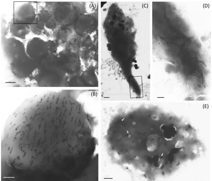
91
new genomes and MTB species regularly feed and will fully resolve [27,28,36–38]. The
92
evolutionary steps that led to the emergence of such complex organelle itself and magnetic
93
sensing are so far unknown. MTB evolved different magnetotactic behaviours according to the
94
species [30] with different motility strategies [39] and specific dividing processes [40]. These
95
behaviours represent a selective advantage for these bacteria that find more efficiently their
96
habitat thanks to the inclination of the magnetic field lines [41]. Despite the diversity of the
97
morphologies, motility behaviours, magnetosomes chains and genetic groups, the bacterial
98
magnetic sensing is always associated to bacteria sharing the same niche [29]. This niche is
99
located at the anoxic-microoxic interface of aquatic sediments where the equilibrium of
100
environmental conditions, notably redox, is fragile [30,42]. This zone is the seat of important
101
geochemical processes [42], to which some MTB species were shown to participate in [43].
102 103
Magnetically responsive protozoan grazers 104
Benthic microbial communities are not only composed of prokaryotes, but also of microbial
105
eukaryotes among which protists and fungi that share the same habitat and need to adapt to the
106
same environmental pressures. After the discovery of the first MTB, arose the hypothesis that
107
magnetotaxis could have evolved in protists thriving in anoxic/microoxic aquatic habitats. In
108
the early years of MTB research, magnetotactic aggregates formed by tens of cells were
109
discovered in a marine lagoon in Brazil [44]. Their multicellularity was rather supporting that
110
these organisms were unlikely bacteria. In fact, those aggregates were the so-called
111
multicellular magnetotactic prokaryotes (MMPs) that represent one of the most interesting and
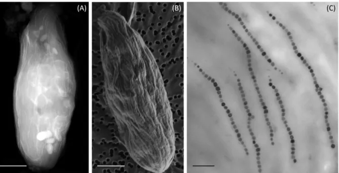
112
unusual examples of prokaryotic morphology (Figure 1D and E) [45,46]. Later, an
Americano-113
Brazilian research network opened the way to a new field of research on magnetoreception by
114
observing the first magnetotactic microbial eukaryote [47]. The difficulties to find and collect
115
them have been a huge barrier to the characterization of their magnetoreception for years.
116
Efforts to find them again failed and their study was still limited by the light and electron
117
microscopy analyses carried out in the 80’s.
118
Fifteen years later, populations of diverse magnetic unicellular eukaryotes were observed in the
119
chemocline of the seasonally chemically stratified, coastal Salt Pond, Massachusetts, USA 120
[48]. Biflagellates, dinoflagellates and ciliates were observed among magnetically concentrated
121
microbial communities. A similar observation, from the same environment was reported later
122
[49]. Like magnetotactic bacteria, they migrated and accumulated at the edge of a hanging water
123
droplet in a magnetic field but displayed a different swimming compared to MTB. In these
5
single-celled eukaryotes, magnetite particles with morphologies and dimensions similar to
125
those of magnetosome-producing bacteria were visualized. However, the origin of these
126
particles was difficult to determine. Based on their observation, endosymbiosis could not be
127
excluded as well as biomineralization by the protist itself, but ingestion of MTB was suggested
128
to be responsible for the magnetic response of some protists, known to be bacterivorous
129
organisms. As many aquatic protists, MTB grazers (i.e. protists that predate MTB) are
130
facultative anaerobes capable of both aerobic and anaerobic growth, which should allow them
131
to easily predate MTB. In Salt Pond, it was shown that the magnetically responsive protists
132
were more abundant during the early and late season of stratification, when the chemocline
133
narrows and MTB concentrate in a smaller volume [49], indicating a correlation between
134
concentration of MTB and that of magnetically responsive protists. Moreover, when the
135
magnetotactic protists were magnetically concentrated at the edge of a droplet where MTB
136
aggregate, they appeared to exhibit a slow looping swimming motion that could be associated
137
to a predatory behaviour [49].
138
By feeding a predatory ciliate with magnetically purified Ca. Magnetoglobus multicellularis, it
139
has been shown that greigite magnetosomes could be dissolved within the acidic vacuoles of
140
the ciliate [50]. In this study, it was not possible to state if this behaviour occurs in
141
environmental conditions and if magnetoreception could arise from the interaction. Recently,
142
this behaviour was investigated in natural populations of protists and MTB, and it was shown
143
that ciliates affiliated to the genus Uronema (Stramenopiles-Alveolates-Rhizaria, SAR group)
144
were able to ingest hundreds of MTB into acidic vacuoles to progressively become sensitive to
145
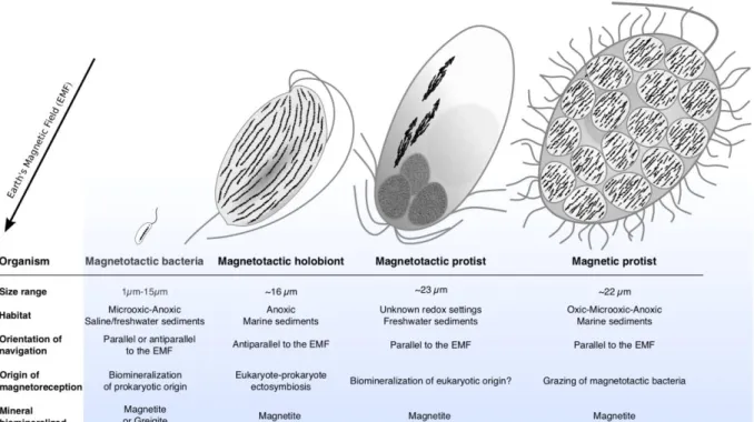
the variations of the magnetic fields in the same way than MTB (Figure 2A and B) [11]. The
146
magnetic response in MTB-grazers is certainly not encoded in the eukaryote genome, but this
147
behaviour seems to facilitate movement of grazers towards the prey biomass. It is still unclear
148
if a chemotaxis is also involved and if this interaction has been selected over the course of the
149
protist evolution. However, the directed grazing towards these preys specifically and the
150
magnetic response suggest that this interaction is widespread among marine and freshwater
151
heterotrophic flagellates and ciliates (Figure 2). MTB-grazers seem to have evolved different
152
strategies for the internalization of MTB and their magnetosome chains: (i) sequestration in
153
food vacuoles (Figure 2A and B), (ii) accumulation in a specific location in the cell (Figure 2C
154
and D), and (iii) an apparently random storage in the cell (Figure 2E) [11,48,50]. These
155
magnetically responsive protists also appear to have evolved different strategies to deal with
156
iron toxicity with (i) the egestion of magnetic inclusions where magnetosomes accumulated
157
[48] and (ii) the progressive dissolution of magnetosomes until the colloidal iron would be
6
expelled from the protistan cell via the cytoproct [11,50,51]. Protists that feed on MTB have
159
been proposed to play a significant role in iron cycling [11,48,49]. It was shown that digestion
160
of colloidal iron in the food vacuoles of protists during grazing of particulate and colloidal
161
matter could generate more bioavailable iron for other species, such as phytoplankton [52,53].
162
Thus, MTB grazing could be involve in recycling particulate iron back to a bioavailable form
163
in the environment through dissolution [54].
164 165
Symbiotic origin of magnetoreception in protists 166
Long neglected, mutualistic symbiosis and cooperation are today recognized as major
167
diversification forces in the same way as competition for resources and natural selection have
168
been for centuries [55]. These concepts even became central in our understanding of the
169
mechanisms and strategies governing species interactions and adaptation in innumerable
170
biological systems. Mutualistic symbioses between prokaryotes – macroscopic eukaryotes are
171
the best examples. They have been particularly well documented for insects, plants or corals,
172
for which studies have shown the importance of certain highly specialized algae, bacteria and
173
fungi on host nutrition and reproduction [56–58]. These symbioses are also extremely diverse
174
and abundant between unicellular eukaryotes and prokaryotes, even though less characterized
175
[59]. Some of them are even the foundation of the evolutionary scenarios on organelle
176
acquisition and eukaryogenesis [60]. Symbiotic associations between protists and bacteria or
177
archaea are common in anoxic marine sediments [61]. Prokaryotic symbionts can be associated
178
with their protist hosts as ectosymbionts (attached to the host surface) or as endosymbionts
179
(located beneath the host cell membrane). These symbionts can expand the host niche by
180
complementing host metabolism, improving its motility or adaptation to new conditions.
181
The recent discovery of magnetotactic holobionts changed our vision of magnetoreception in
182
unicellular eukaryotes. A magnetotactic protist belonging to the Euglenozoa, Excavates, was
183
observed in marine sediments worldwide [12,62]. Its magnetoreception was the result of a
184
cooperation with ectosymbiotic bacteria with whom they live in a mutualistic symbiosis (Figure
185
3) [12]. Again, magnetoreception is magnetite-based and originates from prokaryotes
186
biomineralizing magnetosomes chains. Unlike MTB-grazers, the sensing and geolocalization
187
in this biological system benefits to both organisms thanks to the long-term cooperation
188
established with the host. Microscopy and genomic analyses indicate these ectosymbiotic
189
bacteria are not magnetotactic like MTB in the sense they do not move by means of their own
190
flagella and do not sense chemical gradients; they can be considered only as magnetic. With
191
the protistan host, magnetic ectosymbiotic bacteria (MEB) form a microbial holobiont acting
7
as a supramicroorganism and an ecological unit. Hundreds of these MEB, all belonging to a
193
single strain of the Deltaproteobacteria class, are aligned parallel to the host motility axis
194
(Figure 3A and B) and magnetically orient it. Surprisingly, it was experimentally proven that
195
this magnetic guidance was influenced by oxygen concentrations while genome sequencing
196
showed that MEB could not sense chemical gradients. It is thus likely that holobiont
197
magnetotaxis was collectively ensured thanks to the biflagellate chemotaxis. The partners’
198
interdependency relies also on metabolic exchanges, among which some of them could be
199
identified based on the host ultrastructure and MEB genome. Their syntrophy is based on the
200
transfer of molecular hydrogen from the host to the MEB that use it to reduce sulphate. Protists
201
are bacteria predators and ferment organic matter into energy thanks to mitochondrion-like
202
organelles called hydrogenosomes [63]. This process generates ATP and by products like CO2,
203
acetate and H2 that is diffused through the membrane plasma. Other chemicals might be
204
exchanged but need to be identified through isotope probing and comparative genomics. For
205
example, genomic data indicate that acetate and CO2 are likely used by the MEB as well [12].
206 207
Magnetic biomineralization in protists? 208
The search for biomineralizing magnetic microbial eukaryotes has become an endeavour for
209
many researchers as soon as Torres de Ajaujo and coworkers reported in 1986 the magnetotactic
210
algae with many magnetite particles organized in chains [47]. It was clear that the
211
magnetoreception of these microorganisms was magnetite-based similarly to MTB. Although
212
the chains appeared to be located in or near the cell wall, authors could not identify their
213
organization relative to each other and from the flagellate motility axis. With the limitation of
214
the imaging techniques and DNA typing at that time, many questions remained unanswered,
215
making it hard to conclude about the origin of their magnetoreception. No ultrastructural feature
216
could inform on the presence or the absence of ecto- or endo-symbiotic bacteria even though
217
bullet-shaped magnetosomes looked like those of Deltaproteobacteria [34]. These flagellated
218
protists, tentatively identified as Anisonema platysomum (Euglenophyceae, Euglenozoa,
219
Excavata) based on morphological criteria, were not observed again in decades after their first
220
isolation in a coastal mangrove swamp in northeastern Brazil.
221
In 2019, a new report revivals the hypothesis of a biomineralization-based magnetoreception in
222
unicellular eukaryotes [64]. Uncultured single-celled eukaryotic flagellates from two different
223
Brazilian freshwater sites were observed harbouring anisotropic bullet-shaped magnetite
224
magnetosomes aligned in complex aggregations of multiple chains within the cell. Light and
225
transmission electron microscopy (TEM) images of these magnetotactic microbes were not
8
decisive enough to allow their identification and no genotyping or genome sequencing was
227
performed. Their size, morphology and the multiple flagella at the anterior pole of the cells
228
attested that the protist belongs to another group of that previously observed 30 years before.
229
Because of the magnetosomes size two times longer than bacterial magnetosomes and the
230
absence of specific micro-compartments typical of a bacterial cell or digestive vacuoles,
231
observations rather supported that the biomineralization is performed by the eukaryote itself.
232
However, contrasted TEM cross sections are missing to validate the ultrastructure of the
233
magnetotactic protist and the relative position of the magnetosomes chains. If such result was
234
validated, magnetoreception could have arose from an ancient endosymbiosis event with an
235
MTB, but also from a secondary lateral gene transfer of the whole magnetosome genes cluster.
236
Even if these scenarios are those the most plausible and parsimonious, it is not to exclude that
237
magnetic biomineralization emerged independently a second time in eukaryotes and converged
238
toward the same function. Future research, such as the identification of the genetic determinants
239
involved in magnetosomes formation, will help to validate that magnetosome magnetite
240
biomineralization occurs in this protist.
241 242
Concluding Remarks and Future Prospects 243
The latest discoveries have unravelled a broader diversity of magnetoreceptive microorganisms
244
in the Eukarya domain than previously thought. Advances in single-cell sorting and genomics
245
are opening up a path to study their ultrastructural, phylogenetic and biological characterization.
246
So far, microbial magnetoreception seems to be restricted to aquatic microorganisms and to be
247
systematically coupled to magnetotaxis, which includes biomineralization of magnetic
248
particles, chemo-aerotaxis and a motility response. But two other ecological strategies were
249
shown to be associated indirectly with magnetoreception in microorganisms: predation of
250
magnetotactic bacteria and symbiosis with magnetic bacteria (Figure 4). The distribution of this
251
function among microorganisms inhabiting anoxic/microoxic habitats strengthens the idea that
252
it represents a key adaptation to these habitats. Although our knowledge on the ecology,
253
diversity and evolution of magnetoreceptive microorganisms have dramatically increased these
254
recent years [29,37,65], many important questions remain unanswered and should be addressed
255
in future work (see Outstanding Questions). Only one prokaryotic origin has been formally
256
demonstrated, and a possible independent emergence in eukaryotes needs to be investigated.
257
The discovery of magnetotactic protists specifically, paves the way to a new field of research
258
at the interface of many disciplines. It is unclear yet in which extent this trait is widespread
259
among benthic protists since the classical isolation protocols and methods still need to be
9
adapted to these organisms. Furthermore, all magnetoreceptive microorganisms identified to
261
date are those coupling magnetoreception and geolocalization to navigation. But other
262
biological systems showed that geolocalization may be used for a purpose other than navigation,
263
as some immobile fungi that use gravity instead to sense verticality and direct the spread of
264
spores [66]. Thus, beyond exploring the diversity of magnetotactic organisms, alternative
265
protocols should be developed to isolate magnetoreceptive microorganisms that are not
266
magnetotactic. Exploration of new habitats along with the application of new material and
267
methods for the observation of magnetically responsive organisms will certainly answer this
268
question and many others that the scientific community is currently enthusiastic about.
269
The discovery of microorganisms able of magnetoreception is also particularly appealing for
270
some scientists that search for the origin of magnetoreception in macroorganisms like migratory
271
birds or fish. Indeed, for non-specialists in evolutionary biology, it is easy to mistakenly think
272
that modern lineages of microbial eukaryotes reflect ancestral forms of these. As a consequence,
273
it was speculated that ancient prokaryotic or eukaryotic forms of magnetosomes or even
274
endosymbiotic MTB could be at the origin of magnetoreception in animals [67,68]. The
275
possibility of a symbiosis between a magnetotactic bacterium that would be at the origin of
276
magnetoreception remains very speculative, as there is no evidence of the presence of MTB in
277
symbiosis with such animal. For example, magnetite biomineralization mechanisms have been
278
identified for some macroorganisms such as chitons or honeybees and would involve ferritin
279
deposition [71,72], not magnetosome formation. Then, despites the report of the presence of
280
magnetotactic symbionts [69], closely related to the free-living MTB strain SS-5 [70], within
281
the marine bivalve Thyasira cf. gouldi, this association is likely more related to predation.
282
Indeed, the magnetotactic cells in their host lose the integrity of their magnetosome chain and
283
possibly their flagellum. Moreover, the bivalves appeared to not use magnetotactic bacteria for
284
magnetoreception while bacteria do not seem to take any advantage to be in the host. Thus,
285
there is no evidence of a connexion between magnetotactic bacteria and magnetoreception in
286
animals so far. We believe that advanced methods in imaging and genomics will contribute in
287
the future to decipher some of the enigma about the diversity of the magnetoreceptive
288
mechanisms, its emergence and evolution in micro- and macroorganisms.
289 290
Acknowledgments 291
We thank the French National Research Agency (ANR-16-TERC-0025-01 and
ANR-18-CE31-292
0003), the New Zealand Ministry for Business, Innovation and Employment (grant no.
10
LVLX1703) and all our collaborators and other researchers that contribute to make this field of
294
research so exciting and in perpetual progression.
295 296
References 297
1 Bryant, D.A. and Frigaard, N.-U. (2006) Prokaryotic photosynthesis and phototrophy
298
illuminated. Trends Microbiol. 14, 488–496
299
2 Walsby, A.E. (1994) Gas vesicles. Microbiol. Rev. 58, 94–144
300
3 Wadhams, G.H. and Armitage, J.P. (2004) Making sense of it all: bacterial chemotaxis.
301
Nat. Rev. Mol. Cell Biol. 5, 1024–1037
302
4 Shivaji, S. and Prakash, J.S.S. (2010) How do bacteria sense and respond to low
303
temperature? Arch. Microbiol. 192, 85–95
304
5 Guyodo, Y. and Valet, J.P. (1999) Global changes in intensity of the Earth’s magnetic field
305
during the past 800 kyr. Nature 399, 249–252
306
6 Merrill, R.T. and McElhinny, M.W. (1983) The Earth’s Magnetic Field: Its History, Origin,
307
and Planetary Perspective, Academic Press.
308
7 Wiltschko, R. and Wiltschko, W. (2012) Magnetoreception. Adv. Exp. Med. Biol. 739, 126–
309
141
310
8 Clites, B.L. and Pierce, J.T. (2017) Identifying Cellular and Molecular Mechanisms for
311
Magnetosensation. Annu. Rev. Neurosci. 40, 231–250
312
9 Begall, S. et al. (2014) Magnetoreception in Mammals. In Advances in the Study of
313
Behavior, Vol 46 46 (Naguib, M. et al., eds), pp. 45–88, Elsevier Academic Press Inc
314
10 Klumpp, S. et al. (2019) Swimming with magnets: From biological organisms to synthetic
315
devices. Phys. Rep. 789, 1–54
316
11 Monteil, C.L. et al. (2018) Accumulation and Dissolution of Magnetite Crystals in a
317
Magnetically Responsive Ciliate. Appl. Environ. Microbiol. 84, pii: e02865-17
318
12 Monteil, C.L. et al. (2019) Ectosymbiotic bacteria at the origin of magnetoreception in a
319
marine protist. Nat. Microbiol. DOI: 10.1038/s41564-019-0432-7
320
13 Blakemore, R. (1975) Magnetotactic bacteria. Science 190, 377–379
321
14 Walcott, C. et al. (1979) Pigeons have magnets. Science 205, 1027–1029
322
15 Gould, J.L. et al. (1978) Bees have magnetic remanence. Science 201, 1026–1028
323
16 Frankel, R. et al. (1979) Magnetite in Freshwater Magnetotactic Bacteria. Science 203,
324
1355–1356
325
17 Bellini, S. (2009) On a unique behavior of freshwater bacteria. Chin. J. Oceanol. Limnol.
326
27, 3–5
11
18 Mouritsen, H. (2018) Long-distance navigation and magnetoreception in migratory
328
animals. Nature 558, 50–59
329
19 Nordmann, G.C. et al. (2017) Magnetoreception—A sense without a receptor. PLOS Biol.
330
15, e2003234
331
20 Uebe, R. and Schüler, D. (2016) Magnetosome biogenesis in magnetotactic bacteria. Nat.
332
Rev. Microbiol. 14, 621–637
333
21 Grant, C.R. et al. (2018) Organelle Formation in Bacteria and Archaea. Annu. Rev. Cell
334
Dev. Biol. 34, 217–238
335
22 Lohsse, A. et al. (2011) Functional analysis of the magnetosome island in Magnetospirillum
336
gryphiswaldense: the mamAB operon is sufficient for magnetite biomineralization. PloS
337
One 6, e25561
338
23 Lefèvre, C.T. et al. (2013) Comparative genomic analysis of magnetotactic bacteria from
339
the Deltaproteobacteria provides new insights into magnetite and greigite magnetosome
340
genes required for magnetotaxis. Environ. Microbiol. 15, 2712–2735
341
24 Raschdorf, O. et al. (2016) Genetic and Ultrastructural Analysis Reveals the Key Players
342
and Initial Steps of Bacterial Magnetosome Membrane Biogenesis. PLOS Genet. 12,
343
e1006101
344
25 Hershey, D.M. et al. (2016) MamO Is a repurposed serine protease that promotes magnetite
345
biomineralization through direct transition metal binding in magnetotactic bacteria. PLoS
346
Biol. 14, e1002402
347
26 Toro-Nahuelpan, M. et al. (2019) MamY is a membrane-bound protein that aligns
348
magnetosomes and the motility axis of helical magnetotactic bacteria. Nat. Microbiol. DOI:
349
10.1038/s41564-019-0512-8
350
27 Lin, W. et al. (2018) Genomic expansion of magnetotactic bacteria reveals an early
351
common origin of magnetotaxis with lineage-specific evolution. ISME J. 12, 1508–1519
352
28 Lin, W. et al. (2017) Origin of microbial biomineralization and magnetotaxis during the
353
Archean. Proc. Natl. Acad. Sci. U. S. A. 114, 2171–2176
354
29 Lin, W. et al. (2017) Diversity and ecology of and biomineralization by magnetotactic
355
bacteria. Environ. Microbiol. Rep. 9, 345–356
356
30 Lefèvre, C.T. et al. (2014) Diversity of magneto-aerotactic behaviors and oxygen sensing
357
mechanisms in cultured magnetotactic bacteria. Biophys. J. 107, 527–538
358
31 González, L.M. et al. (2015) Sudden motility reversal indicates sensing of magnetic field
359
gradients in Magnetospirillum magneticum AMB-1 strain. ISME J. 9, 1399–1409
12
32 Popp, F. et al. (2014) Polarity of bacterial magnetotaxis is controlled by aerotaxis through
361
a common sensory pathway. Nat. Commun. 5, 5398
362
33 Kolinko, S. et al. (2016) Single-cell genomics of uncultivated deep-branching
363
magnetotactic bacteria reveals a conserved set of magnetosome genes. Environ. Microbiol.
364
18, 21–37
365
34 Pósfai, M. et al. (2013) Phylogenetic significance of composition and crystal morphology
366
of magnetosome minerals. Front. Microbiol. 4, 344
367
35 DeLong, E.F. et al. (1993) Multiple evolutionary origins of magnetotaxis in bacteria.
368
Science 259, 803–806
369
36 Lefèvre, C.T. et al. (2013) Monophyletic origin of magnetotaxis and the first
370
magnetosomes. Environ. Microbiol. 15, 2267–2274
371
37 Lefèvre, C.T. and Wu, L.F. (2013) Evolution of the bacterial organelle responsible for
372
magnetotaxis. Trends Microbiol. 21, 534–543
373
38 Monteil, C.L. et al. (2018) Genomic study of a novel magnetotactic Alphaproteobacteria
374
uncovers the multiple ancestry of magnetotaxis. Environ. Microbiol. 20, 4415–4430
375
39 Murat, D. et al. (2015) Opposite and coordinated rotation of amphitrichous flagella governs
376
oriented swimming and reversals in a magnetotactic spirillum. J. Bacteriol. 197, 3275–3282
377
40 Lefèvre, C.T. et al. (2015) Positioning the Flagellum at the Center of a Dividing Cell To
378
Combine Bacterial Division with Magnetic Polarity. mBio 6, e02286
379
41 Bennet, M. et al. (2014) Influence of Magnetic Fields on Magneto-Aerotaxis. PLoS ONE
380
9, e101150
381
42 Brune, A. et al. (2000) Life at the oxic-anoxic interface: microbial activities and
382
adaptations. FEMS Microbiol. Rev. 24, 691–710
383
43 Rivas-Lamelo, S. et al. (2017) Magnetotactic bacteria as a new model for P sequestration
384
in the ferruginous Lake Pavin. Geochem. Perspect. Lett. 5, 35–41
385
44 Esquivel, D. et al. (1983) Magnetotactic Microorganisms in the Rio-De-Janeiro Region.
386
Biol. Cell 47, 227–233
387
45 Abreu, F. et al. (2013) Cell adhesion, multicellular morphology, and magnetosome
388
distribution in the multicellular magnetotactic prokaryote Candidatus Magnetoglobus
389
multicellularis. Microsc. Microanal. Off. J. Microsc. Soc. Am. Microbeam Anal. Soc.
390
Microsc. Soc. Can. 3, 1–9
391
46 Qian, X.-X. et al. (2019) Juxtaposed membranes underpin cellular adhesion and display
392
unilateral cell division of multicellular magnetotactic prokaryotes. Environ. Microbiol.
393
DOI: 10.1111/1462-2920.14710
13
47 Torres de Araujo, F.F. et al. (1986) Magnetite and magnetotaxis in algae. Biophys. J. 50,
395
375–378
396
48 Bazylinski, D.A. et al. (2000) Occurrence and distribution of diverse populations of
397
magnetic protists in a chemically stratified coastal salt pond. Chem. Geol. 169, 319–328
398
49 Simmons, S.L. and Edwards, K.J. (2006) Geobiology of magnetotactic bacteria. In
399
Magnetoreception and Magnetosomes in Bacteria pp. 77–102, Springer, Berlin, Heidelberg
400
50 Martins, J.L. et al. (2007) Grazing protozoa and magnetosome dissolution in magnetotactic
401
bacteria. Environ. Microbiol. 9, 2775–2781
402
51 Allen, R. (1974) Food vacuole membrane growth with microtubule-associated
membrane-403
transport in Paramecium. J. Cell Biol. 63, 904–922
404
52 Barbeau, K. et al. (1996) Role of protozoan grazing in relieving iron limitation of
405
phytoplankton. Nature 380, 380061a0
406
53 Pernthaler, J. (2005) Predation on prokaryotes in the water column and its ecological
407
implications. Nat. Rev. Microbiol. 3, 537–546
408
54 Sherr, E.B. and Sherr, B.F. (2002) Significance of predation by protists in aquatic microbial
409
food webs. Antonie Van Leeuwenhoek 81, 293–308
410
55 Margulis, L. and Fester, R. (1991) Symbiosis as a Source of Evolutionary Innovation:
411
Speciation and Morphogenesis, MIT Press.
412
56 Rowan, R. and Knowlton, N. (1995) Intraspecific diversity and ecological zonation in coral
413
algal symbiosis. Proc. Natl. Acad. Sci. U. S. A. 92, 2850–2853
414
57 Engel, P. and Moran, N.A. (2013) The gut microbiota of insects - diversity in structure and
415
function. Fems Microbiol. Rev. 37, 699–735
416
58 Vandenkoornhuyse, P. et al. (2015) The importance of the microbiome of the plant
417
holobiont. New Phytol. 206, 1196–1206
418
59 López-García, P. et al. (2017) Symbiosis in eukaryotic evolution. J. Theor. Biol. 434, 20–
419
33
420
60 López-García, P. and Moreira, D. (2015) Open questions on the origin of eukaryotes.
421
Trends Ecol. Evol. 30, 697–708
422
61 Bernhard, J.M. et al. (2000) The Santa Barbara Basin is a symbiosis oasis. Nature 403, 77–
423
80
424
62 Yubuki, N. and Leander, B.S. (2018) Diversity and Evolutionary History of the
425
Symbiontida (Euglenozoa). Front. Ecol. Evol. 6, 100. doi: 10.3389/fevo.2018.00100
426
63 Müller, M. et al. (2012) Biochemistry and evolution of anaerobic energy metabolism in
427
eukaryotes. Microbiol. Mol. Biol. Rev. MMBR 76, 444–495
14
64 Leão, P. et al. Magnetosome magnetite biomineralization in a flagellated protist: evidence
429
for an early evolutionary origin for magnetoreception in eukaryotes. Environ. Microbiol.
430
doi: 10.1111/1462-2920.14711
431
65 Lefèvre, C.T. and Bazylinski, D.A. (2013) Ecology, diversity, and evolution of
432
magnetotactic bacteria. Microbiol Mol Biol Rev 77, 497–526
433
66 Nguyen, T.A. et al. (2018) Evolutionary novelty in gravity sensing through horizontal gene
434
transfer and high-order protein assembly. PLOS Biol. 16, e2004920
435
67 Kirschvink, J.L. et al. (2001) Magnetite-based magnetoreception. Curr. Opin. Neurobiol.
436
11, 462–467
437
68 Natan, E. and Vortman, Y. (2017) The symbiotic magnetic-sensing hypothesis: do
438
Magnetotactic Bacteria underlie the magnetic sensing capability of animals? Mov. Ecol. 5,
439
22. doi: 10.1186/s40462-017-0113-1
440
69 Dufour, S.C. et al. (2014) Magnetosome-containing bacteria living as symbionts of
441
bivalves. ISME J. 8, 2453–2462
442
70 Lefèvre, C.T. et al. (2012) Novel magnetite-producing magnetotactic bacteria belonging to
443
the Gammaproteobacteria. ISME J. 6, 440–450
444
71 Shaw, J.A. et al. (2009) Ultrastructure of the epithelial cells associated with tooth
445
biomineralization in the chiton Acanthopleura hirtosa. Microsc Microanal, 15, 154–65
446
72 Hsu, C.Y. and Chan, Y.P. (2011) Identification and localization of proteins associated with
447
biomineralization in the iron deposition vesicles of honeybees (Apis mellifera). PLoS One.
448
6, e19088
449 450 451
15 Glossary
452
Biomineralization: biological process by which eukaryotes and prokaryotes are able to 453
produce minerals.
454
Chemocline: horizontal layer formed in aquatic habitats by a strong vertical chemistry gradient 455
that marks the boundary between two contrasted chemical environments.
456
Cytoproct: special pore in the pellicle of ciliates used for exocytosis and membrane recycling. 457
Eukaryogenesis: evolutionary transition at the origin of the eukaryotic cell. 458
Grazing: feeding strategy that some protozoa evolved to feed on microorganisms. 459
Hydrogenosomes: double membrane-bounded organelles of mitochondrial ancestry found in 460
some anaerobic eukaryotes. They are involved in catabolic processes that produce molecular
461
hydrogen, acetate, carbon dioxide and ATP.
462
Holobiont: Assemblage of different species living in symbiosis that forms an ecological unit. 463
Magnetosomes: prokaryotic organelles composed of nano-sized, magnetic, iron-mineral 464
crystals, enveloped by a biological membrane. Usually arranged in chains within the cell, they
465
provide the cell with a permanent magnetic dipole and allow magnetic sensing.
466
Magnetotaxis: behaviour of some motile aquatic bacteria that align passively along Earth’s 467
magnetic field lines while they swim to facilitate their navigation towards their preferred
468
habitat.
469
Mutualistic symbiosis: long-term relationship between different species living in symbiosis in 470
which all partners benefit from the biological interaction.
471
Protists: unicellular eukaryotic microorganisms (cells containing a nucleus). They do not refer 472
to a taxonomic unit and are polyphyletically distributed into the Eukarya domain.
473
Protozoa: heterotrophic unicellular eukaryotes that feed on organic matter such as other 474
microorganisms.
475
Polyphyletic distribution: Distribution of a trait or organisms in different taxonomic groups 476
that are not related by a direct common ancestor.
477
Symbiosis: defines a long-term physical coexistence of two or more species, in which at least 478
one partner is dependent on the others, no matter what the effect of the interaction on the
479
partner’s fitness.
480
Syntrophy: trophic interdependency of two symbiotic species. Also referred as a mutualistic 481
metabolism in which partner’s metabolisms rely on the exchange of metabolic products of each
482
other’s.
483 484 485
16 Figures and figure legends
486
487
Figure 1. Transmission electron microscope images of magnetotactic bacteria. 488
Magnetotactic spirillum (A), cocci (B) and curved rod (C) isolated from the freshwater Lake
489
Pavin, France. Intact (D) and disaggregated (D) multicellular magnetotactic prokaryote (MMP)
490
isolated from the Mediterranean Sea in Carry-le-Rouet, France. Magnetotactic cells in (A-C)
491
produce elongated prismatic (A and B) or bullet-shaped (C) magnetite magnetosomes while the
492
MMP (E) produce octahedral greigite magnetosomes. Scale bars represent 0.5 µm.
493 494 495
17 496
Figure 2. Transmission electron microscope images of magnetic ciliates that grazed 497
magnetotactic bacteria isolated from the Mediterranean Sea. (A-B) Ciliate that sequestrates 498
magnetosomes in digestive vacuoles. The black frame in (A) shows where the higher
499
magnification image in (B) was taken. (C-D) Ciliate that accumulates magnetosomes in a
500
specific location within its cytoplasm. The black frame in (C) shows where the higher
501
magnification image in (D) was taken. (E) Ciliate that randomly stores grazed magnetotactic
502
bacteria inside its cell. Scale bars represent 2 µm (A, C and E) and 0.5 µm (B and D).
503 504
18 505
Figure 3. Electron microscope images of magnetotactic holobionts isolated from the 506
Mediterranean Sea. High angle annular dark field scanning transmission electron microscope 507
(A) and scanning transmission electron microscope (B) images of single magnetotactic
508
holobionts showing the presence of hundreds of bacteria surrounded a protistan cell. (C)
509
Transmission electron microscope image of several magnetosome chains produced by magnetic
510
ectosymbiotic bacteria allowing magnetoreception of the protist. Scale bars represent 2 µm (A
511
and B) and 0.5 µm (C). (A and B) Image courtesy of N. Menguy and K. Benzerara, respectively.
512 513
19 514
Figure 4. Schematic comparison of magnetoreceptive microorganisms morphologies and 515
magnetic behaviours. Magnetosomes chains are represented in black. Organism’s anterior-516
posterior orientation is given relative to the Earth Magnetic Field (EMF) direction in the
517
Northern Hemisphere. These orientations are reverse for microorganisms of the Southern
518
Hemisphere.
519 520