HAL Id: hal-02339207
https://hal.sorbonne-universite.fr/hal-02339207
Submitted on 30 Oct 2019
HAL is a multi-disciplinary open access
archive for the deposit and dissemination of sci-entific research documents, whether they are pub-lished or not. The documents may come from teaching and research institutions in France or abroad, or from public or private research centers.
L’archive ouverte pluridisciplinaire HAL, est destinée au dépôt et à la diffusion de documents scientifiques de niveau recherche, publiés ou non, émanant des établissements d’enseignement et de recherche français ou étrangers, des laboratoires publics ou privés.
Pooled Analysis of external-beam RADiotherapy
parameters in phase II and phase III trials in
radiochemotherapy in Anal Cancer (PARADAC)
Eleonor Rivin del Campo, Oscar Matzinger, Karin Haustermans, Didier
Peiffert, Robert Glynne-Jones, Kathryn Winter, Andre Konski, Jaffer Ajani,
Jean-François Bosset, Jean-Michel Hannoun-Lévi, et al.
To cite this version:
Eleonor Rivin del Campo, Oscar Matzinger, Karin Haustermans, Didier Peiffert, Robert Glynne-Jones, et al.. Pooled Analysis of external-beam RADiotherapy parameters in phase II and phase III trials in radiochemotherapy in Anal Cancer (PARADAC). European Journal of Cancer, Elsevier, 2019, 121, pp.130-143. �10.1016/j.ejca.2019.08.022�. �hal-02339207�
1
Pooled Analysis of external-beam RADiotherapy parameters in phase II and phase III trials in radiochemotherapy in Anal Cancer (PARADAC).
Eleonor Rivin del Campo1, Oscar Matzinger2, Karin Haustermans3, Didier Peiffert4, Robert Glynne-Jones5, Kathryn A. Winter6, Andre A. Konski7, Jaffer A. Ajani8, Jean-François Bosset9, Jean-Michel Hannoun-Levi10, Marc Puyraveau11, A. Bapsi Chakravarthy12, Helen Meadows13, John Northover14, Laurence Collette15, Melissa Christiaens3, Philippe Maingon16.
1 Department of Radiation Oncology, Tenon University Hospital, Sorbonne University, Paris, France. 2 Department of Radiation Oncology, Genolier Clinic, Genolier, Switzerland.
3 Department of Radiation Oncology, UZ Leuven University Hospital, Leuven, Belgium. 4Department of Radiation Oncology, Institut de Cancérologie de Lorraine, Nancy, France. 5Department of Radiation Oncology, Mount Vernon Cancer Centre, Northwood, United Kingdom.
6 NRG Oncology Statistics and Data Management Center, American College of Radiology, Philadelphia, Pennsylvania, United
States of America.
7 Department of Radiation Oncology, University of Pennsylvania Perelman School of Medicine. Leonard Davis Institute of
Health Economics. Department of Radiation Oncology The Chester County Hospital, West Chester, Pennsylvania, United States of America.
8Department of Radiation Oncology, MD Anderson Cancer Center, Houston, Texas, United States of America. 9 Department of Radiation Oncology, Jean Minjoz University Hospital, Besançon, France.
10 Department of Radiation Oncology, Centre Antoine Lacassagne, Nice, France. 11 Department of Statistics, Jean Minjoz University Hospital, Besançon, France.
12Department of Radiation Oncology, Vanderbilt University Medical Center, Nashville, Tennessee, United States of America. 13Cancer Research UK & UCL Cancer Trials Centre, London, United Kingdom.
14 Department of Surgery, The London Clinic and St Marks Hospital, London, United Kingdom. 15 Department of Statistics, EORTC headquarters, Brussels, Belgium.
16Department of Radiation Oncology, La Pitié Salpêtrière - Charles Foix University Hospital, Sorbonne University, Paris, France.
Acknowledgments of research support for the study: The Kom Op Tegen Kanker supported the development of this project through the “Emmanuel van der Schueren Fellowship for Quality Assurance in Radiotherapy” EORTC fellow grants (Oscar Matzinger, Melissa Christiaens). UK Co-ordinating Committee for Cancer Research (UKCCCR), Radiation Therapy Oncology Group (RTOG), French Federation Nationale des Centres de Lutte Contre le Cancer (FNLCC), European Organization for the Research and Treatment of Cancer (EORTC) and the ECOG-ACRIN Cancer Research Group (ECOG-ECOG-ACRIN). ECOG-ECOG-ACRIN grants U10CA180820 and UG1CA233270.
Corresponding author: Eleonor Rivin del Campo, MD, PhD Department of Radiation Oncology,
Tenon University Hospital- Hôpitaux Universitaires Est Parisien, Sorbonne University 4 rue de la Chine, 75020, Paris, France
Ph.: (33) 156016210 ; FAX: (33) 156016599
Email: eleonor.rivindelcampo@aphp.fr
Running head: PARADAC – Meta-analysis: radiotherapy parameters in anal cancer
Keywords: anal cancer, radiation, overall treatment time, chemoradiation
2
Abstract
PURPOSE: Concomitant external-beam radiochemotherapy (5-Fluorouracil-Mitomycin C) has
become the standard of care in anal cancer since the ‘90s. A pooled analysis of individual patient
data from 7 major trials was performed quantifying the effect of RT-related parameters on the
outcome of patients with anal cancer.
MATERIALS AND METHODS: Pooling databases from combined modality trials, the impact
of RT parameters (total dose, gap duration, OTT: overall treatment time) on outcome including
locoregional failure (LRF), 5-year progression-free-survival (PFS) and toxicities were
investigated. Individual patient data was received for 10/13 identified published studies
conducted from 1987-2008(n=3031). A Cox-regression model was used (landmark=3 months
post-RT for first follow-up).
RESULTS: After data inspection indicating severe heterogeneity between trials, only 1343
patients from 7/10 studies received were analyzed (the most recent ones, since 1994; median
follow-up=4.1 years). A higher overall 5-year LRF rate [22.8% (95%CI 22.3-27.3%)]
significantly correlated with longer OTT (p=0.03), larger tumor size (p<0.001) and borderline
significantly with male gender (p=0.045). Though significant differences were not observed,
subset analyses for LRF (dose range:50.4-59Gy) seemed to favor lower doses (p=0.412), and
when comparing a 2 week gap versus 3 (dose:59.4Gy), results suggested 3 weeks might be
detrimental (p=0.245). For a 2 week gap versus none (dose range:55-59.4Gy) no difference was
observed (p=0.89). Five-year PFS was 65.7% (95%CI: 62.8-68.5%)]. Higher PFS rates were
Five-3
year OS [76.7%(95%CI: 73.9%-79.3%)] correlated positively with female gender (p<0.001),
small tumor size(p=0.027) and short OTT(p=0.026). Descriptive toxicity data is presented.
CONCLUSION: For patients receiving concurrent external-beam doublet chemoradiation
a longer OTT seems detrimental to outcome. Further trials involving modern techniques may
4
Pooled Analysis of external-beam RADiotherapy parameters in
phase II and phase III trials in Anal Cancer (PARADAC).
Introduction
Anal cancer is a rare tumor arising from the gastrointestinal tract. Over the past decades
an increase in incidence has been observed for in situ and squamous cell anal carcinoma[1,2].
The most important risk factors are: previous gynaecological or blood related cancer, smoking,
sexually transmitted diseases (HPV, HIV, herpes, etc.), sexual partners exceeding 10, long-term
immunosuppression and receptive anal intercourse[2].
Until the seventies abdominoperineal resection remained the standard of care, with an
operative mortality rate of about 6-8 percent[3]. Five-year overall survival (OS) rates of 40 to
70% were reported, with 5-year recurrence rates of approximately 40%[4,5]. After radiation
therapy (RT), whether or not followed by surgery, similar disappointing results were reported[4].
The concomitant use of chemotherapy, 5-Fluorouracil (5-FU) with Mitomycin C (MMC), with
usually split course RT dramatically improved local control (LC) and colostomy free survival
(CFS). Thus, radical surgery was replaced with primary chemoradiation as the standard of
care[3,6,7]. Almost two thirds of patients could be cured and their sphincter preserved with this
approach[3,6,7]. Nevertheless, the prognosis remained poor for patients with advanced disease or
lymph node involvement. Despite attempts to improve this treatment, the standard treatment for
locally advanced disease remained unchanged for years with the most recent contributions
confirming the efficicacy of RT with 5-FU and MMC[7–10]. The most concrete improvement is
5
(IMRT) and Volumetric Modulated Arc Therapy (VMAT), allowing a better toxicity profile with
comparable outcomes[11–16]. However, many questions remain a matter of debate.
A pooled analysis of individual patient data from 7 major prospective trials was
performed with the aim to quantify the effect of RT-related parameters on the outcome of
patients with anal cancer to improve guidelines for future RT combined modality studies.
Materials and methods
Selection criteria:
A consensus meeting was held at the European Organization for the Research and
Treatment of Cancer (EORTC) Headquarters on March 6th, 2008, with representatives of
European countries and collaborative groups active in the field of radiochemotherapy for
advanced anal cancer: the Italian group, Spanish Group, Nordic Group, UK Co-ordinating
Committee for Cancer Research (UKCCR) and the French Federation Nationale des Centres de
Lutte Contre le Cancer (FNCLCC). The aim of the meeting was to agree on the design of the
next phase III randomized trial testing improved radio-therapeutic regimes. To prepare, the group
reviewed available evidence from phase II and III studies for anal cancer, including the then
recently closed phase III trials, such as ACCORD-03[17]. The studies identified at the time are
detailed in Table 1.
Studies:
ACCORD-03, examined the impact of therapeutic intensification by induction
chemotherapy and/or high dose RT[17]. Several studies (EORTC 22011, RTOG 98-11 and ACT
6
without induction or maintenance chemotherapy[8,9,18,19]. The EORTC 22953 trial
investigated the effect of a reduction of the gap (16-days) in combination RT and 5-FU-MMC
treatment for anal cancer versus a standard 6 weeks gap[20]. In the UKCCCR EXTRA trial
intravenous 5-FU was replaced by Capecitabine[21]. ECOG-ACRIN 42-92 studied the feasibility
of Cisplatin replacing MMC with a 2 week gap[22].
A consensus emerged that it was not timely to start a new trial, given the number of
existing projects in various countries that were still maturing, and the difficulty to finance such
academic trials. Considering the rarity of the disease, an international European or worldwide
collaboration was needed for a successful phase III study.
A proposal was made for pooling databases from existing trials to explore the impact of
specific RT parameters and toxicities on outcome. It was acknowledged that to address these
questions would entail between-trial protocol comparisons, resulting in a need to obtain a more
homogeneus patient group (Figure 1, 2). The questions that emerged revolved around the impact
of overall treatment time (OTT, affected by the duration of the gap) and RT dose on local
recurrence. The relationships between irradiated pelvic volume and pelvic recurrence rates, the
total dose to the elective inguinal area and inguinal control, of prognostic factors for long term
outcome in relation with the baseline patient and disease characteristics, as well as long term
toxicity were assessed.
Data collection and extraction:
For each included study, the protocol and publication of results were obtained. The
following individual patient data were requested for each patient:
7
- Tumor-related: Date of diagnosis; cT; cN; cM; maximum tumor size; tumor localization
(i.e. anal margin involved); inguinal node involvement (if involved, number and size);
pelvic nodal involvement (if involved, number and size).
- Treatment-related: Treatment arm; RT parameters [date of start, total dose to the primary
tumor (number of fractions), total dose to positive lymph nodes (number of fractions),
total dose to the elective pelvic area (number of fractions), Total dose to the elective
inguinal area (number of fractions), OTT (tumor; positive nodes; elective), gap duration];
chemotherapy (if given, date of start, drugs used (5-FU, MMC, Cisplatin, other).
- Outcomes: Last follow up date; survival status; local recurrence and date (if any); pelvic
recurrence status and date; inguinal recurrence status and date; distant recurrence status
and date; date of colostomy (if performed); date of lymph node resection and procedure
(if performed).
- Late Toxicity: Bladder; bone; skin; intestine; anus (worst grade, date of start, scale).
No specific format for data collection was specified. Since many trials were historical,
not all data could be obtained for all studies, depending on the data collection system used (see
results).
Statistical analysis:
Recognizing that treatment effects of interest in the analysis required assessments across
studies, but were essentially fixed by each trial protocol, a Cox model was developed to adjust
for patient specific factors in a subgroup of the data selected to minimize systematic
8
representing RT dose and OTT. The model included adjustment for age (continuous linear
effect), gender, tumor localization (anal margin, anal canal or both), N stage (pelvic N0/N+
without inguinal involvement/ pelvic N+ with inguinal involvement/ pelvic N+ with unknown
inguinal status) and tumor size (modeled as an effect nested within combinations of N status and
tumor location). The latter was to accommodate for interaction effects between T stage and N
stage and tumor location, and to fulfill the proportional of hazard assumptions. The OTT was
modelled as a continuous effect, whereas the total dose was assessed in categories (≤50.5 Gy, >50.5 to 55 Gy, >55 to 59 Gy, >59 to 59.4 Gy and >59.4 Gy). The categories were created to
accommodate the intrinsically discrete set of total doses planned in the study protocols
(Appendix A: Figure 1).
For the analysis of locoregional failure (LRF), our primary endpoint, a landmark of 3
months was used to homogenize the timing of first disease assessment during follow-up in the
studies, and the time was censored at 5 years to harmonize the duration of follow-up. The
covariate effects were summarized by their hazard ratio and its associated 95% confidence
interval.
The adequacy of the Cox regression model was evaluated by performing the graphical
and numerical methods of Lin, Wei and Ying [23]. Based on intermediate study results that
showed a single model could not fit the early trials that used RT alone ± one chemotherapeutic
drug and the more recent studies that used RT ± a doublet of drugs, the primary analysis set was
restricted to patients treated with doublet chemotherapy after 01/01/1994.
The analysis was exploratory and two-sided tests were used with a 5% significance level.
9
Merging data
Data was provided by 10/13 trials, amounting to 3031 patients. Assessment of data
received and of study protocols revealed severe heterogeneity in patient selection criteria, the
staging system used and the capture of the RT treatment data. An attempt was made to
harmonize the data from the trials, to allow merging of the databases while excluding as few
patients as possible (Figure 1).
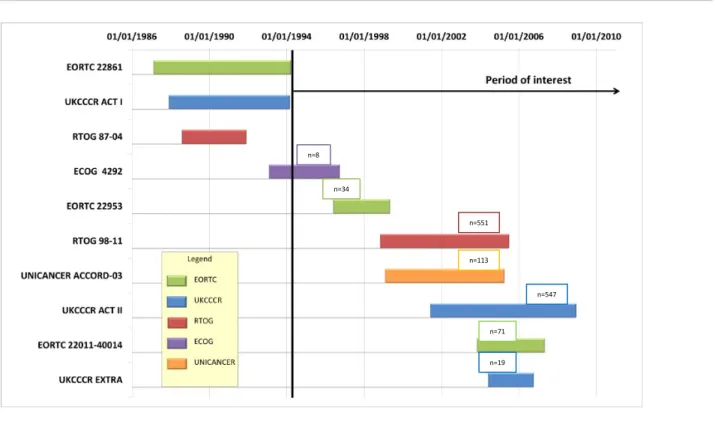
Patients were recruited during a two decade period (1987-2008) (Figure 2). The
difference in terms of data collection, patient staging, treatments and patient follow-up was
particularly evident for the first 3 trials (EORTC 22861, ACT I and RTOG 87-04). The first two
were conducted with exclusive RT versus chemoradiotherapy with two chemotherapeutic drugs,
while the RTOG 87-04 trial evaluated chemoradiotherapy with one-agent versus doublet
chemotherapy. The later trials assessed RT + doublet chemotherapy[6,24–26]. Heterogeneity
was such that no model could fit both groups of studies together. Since current treatment
involves doublet chemotherapy with RT, the analysis was restricted to the 7 more recent studies
(Figure 1; Figure 2).
Certain parameters (e.g.: planned RT dose, OTT) were specified inside a trial, but varied
between trials. Thus it was essential to minimize any systematic differences in other factors that
would also be confounded with the trial effect in our models. We assessed the eligibility criteria
of the studies and selected patients for the analysis fitting the most stringent criteria across
protocols, excluding patients >75 years or patients with unknown age, T1N0 and M1 or Mx
disease from the analysis.
For many patients important data [e.g. N-stage, dose to the anal canal, OTT, tumor
10
particular, OTT and total dose could not be calculated for patients who received brachytherapy.
Since these factors were key to the analysis, these patients were excluded. Also, very short
treatment times (<30 days) or very low dose (<36Gy) could result from excess toxicity, very
poor prognosis or performance status (30 patients) or from protocol planned dose adaptations
triggered by intermediate assessment of the response to treatment (21 patients). We applied a
landmark of 90 days to not overestimate the association between treatment and outcome (due to
the exclusion of the 21 patients during the landmark) and harmonize the timing of the first visit
in the studies.
Toxicity was also scored heterogeneously between trials. Some trials systematically
reported late toxic events at each follow-up, whereas others only reported one event (the worst
grade) per patient in the received data files. The scales used for grading were also very
heterogeneous (RTOG/EORTC, LENT-SOMA, NCI CTC different versions) (Appendix A.
Table 1). A common scale for toxicity was applied and was based on the ‘RTOG/EORTC Late
RT Morbidity Scoring Scheme’ as it was the most used[27]. The toxicities were classified into
groups and scored into mild/moderate vs. severe. Late toxicity was considered as starting more
than 90 days after the start of treatment.
Results pooled analysis:
A total of 1343/3031 patients were eligible for the pooled analysis (Figure 1). Their
characteristics and RT-related parameters are presented in Table 2. The overall median duration
of follow-up within these trials was 4.1 years (Appendix A. Figure 2).
In total, 419 patients (31.2%) developed clinical failure during follow-up (local, inguinal
11
An inguinal recurrence was recorded in 4 studies (ACCORD-03, EORTC 22011,
EXTRA and ACT II), occurring in 54 of the 741 patients evaluable in these studies (7.3%). At
treatment initiation; 21 of the 54 patients had N0 disease, while 33 had positive nodes, of which
6 had no positive inguinal nodes.
LRF was described in 295 patients (22%). LRF was defined as a local recurrence, a
regional recurrence or local surgery (abdominoperineal resection or colostomy). The 5-year
cumulative rate of LRF was 22.8% [(95%CI 22.3-27.3%) Appendix A. Figure 3]. Lymph node
resection surgery was not considered as an event (this data was only available on the 3 EORTC
trials, only 3 patients had a lymph node resection before documented LFR).
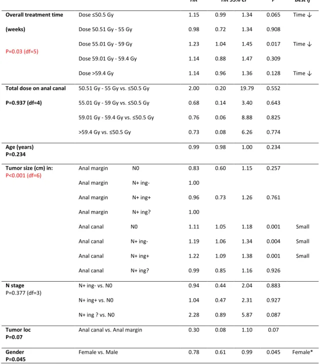
LRF was significantly correlated with OTT (p=0.03), tumor size (p<0.001) and
borderline significantly associated with gender (p=0.045). OTT and total dose were not markedly
different between males and females (Table 3).
As the dose and the gap duration were very closely related by design of the protocols
with the lowest (50.4 Gy, except for the good prognosis patients in RTOG) and intermediate
doses (55-59 Gy) given without a gap and the higher doses (>59.4 Gy) given with a 2 or 3 week
gap, no model was able to study the effect of the dose and the gap duration separately. A
comparison between the lower and intermediate doses, without a gap, could be made. To avoid
bias in favor of low doses, N0 patients treated at 45Gy were excluded. Only patients from the
RTOG 98-11 treated at intermediate doses versus patients treated in ACT II and EXTRA at a
planned dose of 50.4Gy were considered in this analysis (also adjusted for gender, age, tumor
location, N-stage and tumor size). Based on 220 locoregional relapse events (n=977), no
difference between dose levels could be seen. However, results seemed to favor a lower dose
12
to receive a dose of 59.4Gy with a 2 week gap versus a 3 week gap (i.e. patients in EORTC
studies versus ACCORD-03 arms 1 and 3). In this group of 332 patients 68 locoregional relapses
were registered. The analysis suggests that a 3 week gap might be detrimental, although the
effect was not statistically significant (Appendix A: Table 3)
A 2 week gap was compared to no gap in patients intended to receive a dose of 55 to
59.4Gy (EORTC studies versus RTOG 98-11). In this group, 113 events were observed among
521 patients. A supplementary sensitivity analysis was performed in this group by restricting the
analysis to patients who had effectively received a dose of >56.5Gy and <60.0Gy (n=214). Only
74 events were seen. Neither analysis showed a significant difference between a 2-week gap and
no gap (Appendix A: Table 4).
The 5-year progression free survival (PFS) rate was 65.7% (95% CI: 62.8-68.5%). The
occurrence of a clinical event was less frequent in women (p<0.001) or in patients with small
tumor size (p<0.001) or shorter OTT (p=0.025). The 5-year OS was 76.7% (95%CI:
73.9%-79.3%) and it correlated with gender (P<0.001), with a higher OS for women, small tumor size
(p=0.027) and short OTT (p=0.026) (Appendix A. Figure 3, Table 3).
Late toxicities (90 days or more from the start of treatment), were divided into
gastro-intestinal, skin or subcutaneous tissue and bladder-genito-urinary late toxicities (Table 4).
Discussion
This meta-analysis of individual patient data from 10 trials evaluating results of anal
cancer patients receiving combined modality treatment confirmed the effect of RT-related factors
on patient outcomes. OTT and tumor size were found to have a significant impact on LRF, PFS
13
LRF was the predominant pattern of relapse, with a 5-year LRF rate in accordance with
recent trials[28–30]. Shorter OTTs were significantly associated with less LRF, as were small
tumor sizes for tumors of the anal canal and female gender. For patients receiving intermediate
doses (55-59 Gy, RTOG 98-11) versus 50.4 Gy (ACT II and EXTRA), no major difference of
LRF was observed between dose levels. Paradoxically, patients who received the lower dose
seemed to have better outcomes. This may have not reached significance due to possible
remaining biases in the meta-analysis (after adjusting for patient characteristics) and to
differences between protocol guidelines. It is noteworthy that of the three trials included in this
analysis, only ACT II required RT quality assurance, although the results have not been reported.
If indeed the RT in ACT II was administered following quality assurance guidelines more
closely, this may be a factor favoring the lower dose level used in this trial. These findings
support current guidelines from ESMO-ESSO-ESTRO guidelines which suggest no benefit in
administering RT doses >50 Gy without a gap during combined modality treatment, especially
for good responders[31]. The duration of the gap on LRF was assessed by comparing patients
planned to receive 59.4 Gy with a 2 or a 3 week gap from the EORTC studies versus
ACCORD-03 arms 1 and 3, finding that a 3 week gap could be potentially detrimental. The lack of a
significant difference may be attributed the small number of patients in this analysis. However,
when a 2 week gap was compared to no gap for doses between 57 and 59.4 Gy (EORTC studies
versus RTOG 98-11) there were no significant differences between groups. Our data suggests
that if the gap is detrimental, it is for gaps ≥ 3 weeks, which also increase OTT (significantly associated with LRF). This may explain the lack of benefit in ACCORD-03 for doses >59.4Gy,
as a 3 week gap was used[17]. In 2011, Glynne-Jones et al. advocated to avoid “split course”
14
planned, it should be done as late as possible in the treatment course, and on the other hand,
non-randomized retrospective data seems to indicate a worse outcome with gaps of >38 or >63
days[32–36].
With regards to survival rates, PFS at 5 years was similar to a recent phase II study
evaluating the addition of Cetuximab to 5-FU-Cisplatin concurrent chemoradiotherapy, and
approximately 10% lower than in two other recent studies[28–30]. Five-year OS (76.7%) was
also similar to the results of the Cetuximab association trial, and 4-7% lower than the other two
recent studies[28–30]. Shorter OTTs, smaller tumors and female gender, again, were found to be
significantly associated with a better PFS and OS. OTT is a known prognostic factor in other
squamous cell carcinomas, such as cervical cancer, where every day >52 days causes a reduction
of LC by 1%, as well as in head and neck cancer[37,38]. Although there is a paucity of data on
the characteristics of proliferation of anal cancer, they seem similar to cervical cancer, with a
potential median doubling-time of 4.1 days[39]. It is noteworthy, that clinical results of
combined modality therapy in anal cancer show OTT might have less of an impact as
chemotherapy may increase the interval of growth delay of clonogenic cells[40]. An OTT >41
days was found to compromise 5-year LC, whether the treatment included a gap or not[41]. An
analysis performed on pooled data from the RTOG 87-04 and 98-11 trials found a significant
impact of OTT on local and LRF (in agreement with the present meta-analysis), colostomy
failure and time to failure without a correlation with CFS or OS (conversely, it correlated with
OS in the present meta-analysis)[42].
Considering the impact of OTT on outcomes of combined therapy for anal cancer,
strategies have been developed to shorten OTT while attempting to reduce toxicity. IMRT and
15
gastro-intestinal toxicity with comparable outcomes to results from 3D-conformal RT
treatments[12–14,16,43]. These results should be interpreted with caution. A recent analysis of
the national Veterans Affairs database compared 779 patients receiving either IMRT or
3D-conformal RT[15]. Indeed, they found a decrease of treatment breaks, resulting in shorter OTT in
patients receiving IMRT, but without significant improvement of severe gastro-intestinal or
hematological toxicity. The authors hypothesize this may be due to incorrect planning or
delivery, which highlights the need to follow international guidelines and homogenize
contouring practices to improve these results, not only in terms of toxicity, but also in terms of
outcomes[14,15,31,44]. A single center study which reviewed 45 patients treated by
3D-conformal RT or IMRT found OTT was an independent predictor of OS and PFS[45].
Additionally, simultaneous integrated boost during IMRT or VMAT treatment can considerably
shorten OTT. A multicenter retrospective study evaluated 190 patients receiving either a
simultaneous integrated boost (SIB) or a sequential boost for combined modality treatment for
anal cancer[46]. With a median follow up of 34 and 31 months for each group, median OTT was
significantly lower (43 vs. 60 days) in the SIB group, as was the cumulative incidence of
colostomy[13].
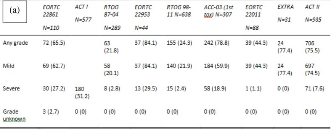
Toxicity results from this meta-analysis were difficult to interpret due to heterogeneous
toxicity scoring and reporting. When observing the descriptive data from the pooled analysis,
higher severe gastro-intestinal toxicity appears for the EORTC 22861 and EORTC 22953 trials,
higher severe skin toxicity for the EORTC 22953 trial and higher severe genito-urinary toxicity
for the EORTC 22953 and ACCORD-03 trials.
A limitation of this meta-analysis is that individual patient data from only 10 of the 13
16
patients) may have influenced the meta-analysis. Nevertheless, data from the large phase III
trials were received and analyzed, which is a strength. For the sake of homogeneity
(heterogeneous patient selection criteria, staging systems and capture of RT data), patient data
from 3 of the early trials as well as 44% of the patients from the rest of the studies had to be
excluded from the analysis. Also, the intrinsic design of the studies analyzed did not allow the
dose and the gap duration to be evaluated separately. Nor was the relationship between OTT and
gap duration examined. Approximately 25% of patients in the final analysis received
neoadjuvant chemotherapy (patients from RTOG 98-11 and ACCORD-03), which might have
caused an accelerated repopulation at the time of combined RT-doublet chemotherapy,
eventually impacting the relevance of OTT in these patients[8,17]. Furthermore, this analysis did
not evaluate the impact of which concurrent chemotherapy scheme was used (the hypothesis
considered them as equivalent). Last, but not least, we were not able to stratify according to HPV
status or smoking.
Ongoing trials may shed more light these questions, such as the PLATO protocol
(ISRCTN88455282), integrating 3 trials: ACT3 for small tumors (phase II, non-randomized,
local excision), ACT4 for intermediate-risk tumors (phase II, randomized, standard-dose 50.4 Gy
vs. reduced-dose 41.4 Gy combined modality treatment) and ACT5 for locally advanced tumors
(phase II/III, randomized, standard dose 53.2 Gy vs. 58.8 Gy and 61.6 Gy combined modality
treatment) with the primary outcome of 3-year LRF. Thus, the question of dose-escalation is
being tested in larger tumors, which is coherent with our results, as smaller tumors had better
outcomes. A recent pooled analysis of two prospective trials including locally advanced anal
17
Treatment outcomes may not only benefit from changes in RT schemes, but also from
changes in the drugs used for combined therapy. In the aforementioned study, although the
addition of cetuximab to Cisplatin/5-FU decreased local failure rates, it came at the cost of
increased toxicity[30]. Another study testing this association had to close prematurely due to
serious adverse events[48]. Future directions might include combinations with immunotherapy,
such as pembrolizumab[49]. Ongoing trials are investigating the role of immunotherapy in the
adjuvant setting, EA2165: Nivolumab After Combined Modality Therapy in Treating Patients
With High Risk Stage II-IIIB Anal Cancer (NCT03233711).
As MR-Linacs become more widely available, newer techniques such as MRI-guided RT
(MRgRT) are being evaluated[50]. Online adaptive treatment strategies may be developed using
this technology, considering the clear superiority of MRI imaging for soft tissues. This technique
could allow real time tumor tracking, taking into account intra-fractional motion[51]. Daily
adaptive re-planning would allow effective treatment while potentially reducing toxicity[52].
Recent progress in the validation of a specific EORTC quality of life questionnaire for
anal cancer will allow better assessment of toxicity in patients included in future trials[53]. Such
patient-related outcomes are essential to provide more robust evidence on toxicity outcomes.
Conclusions
This meta-analysis suggests that for patients treated with a combination of external-beam
RT with 2 chemotherapeutic drugs, a longer OTT is detrimental. Further analyses suggest that in
the dose range of 50.4-59 Gy lower doses seem to be preferred. Treatment gaps longer than 2
weeks appear to be detrimental. When comparing a 2 week gap to no gap (dose range: 55-59.4
18
and a somewhat lower dose given with no gap. Further prospective trials involving
IMRT/VMAT techniques or even with MRI-guided RT may better define optimal OTT and dose
escalation in schedules with no gap.
Conflict of interest statement: The authors declare no conflict of interest.
Acknowledgments: Research support for the study: The Kom Op Tegen Kanker supported the
development of this project through the “Emmanuel van der Schueren Fellowship for Quality
Assurance in Radiotherapy” EORTC fellow grants (Oscar Matzinger, Melissa Christiaens). UK
Co-ordinating Committee for Cancer Research (UKCCCR), Radiation Therapy Oncology Group
(RTOG), French Federation Nationale des Centres de Lutte Contre le Cancer (FNLCC),
European Organization for the Research and Treatment of Cancer (EORTC) and the
ECOG-ACRIN Cancer Research Group (ECOG-ECOG-ACRIN). ECOG-ECOG-ACRIN grants CA180820 and
19
Reference List
[1] Nelson RA, Levine AM, Bernstein L, Smith DD, Lai LL. Changing patterns of anal canal
carcinoma in the United States. J Clin Oncol 2013;31:1569–75.
doi:10.1200/JCO.2012.45.2524.
[2] Nelson VM, Bowen Benson III A. Epidemiology of Anal Canal Cancer. Surg Oncol Clin
2017;26:9–15. doi:10.1016/j.soc.2016.07.001.
[3] Papillon J, Mayer M, Montbarbon JM, Gerard JP, Chassard JL, Bailly C. A new approach
to the management of epidermoid carcinoma of the anal canal. Cancer 1983:1830–7.
[4] Ryan DP, Compton CC, Mayer RJ. Carcinoma of the anal canal. N Engl J Med 2000:792–
800. doi:10.1016/j.calphad.2008.12.005.
[5] Lin D, Gold HT, Schreiber D, Leichman LP, Sherman SE, Becker DJ. Impact of
socioeconomic status on survival for patients with anal cancer. Cancer 2018;124:1791–7.
doi:10.1002/cncr.31186.
[6] Bartelink BH, Roelofsen F, Eschwege F, Rougier P, Bosset JF, Gonzalez DG, et al.
Concomitant Radiotherapy and Chemotherapy Is Superior to Radiotherapy Alone in the
Treatment of Locally Advanced Anal Cancer: Results of a Phase III Randomized Trial of
the European European Organization for Research and Treatment of Cancer Radiotherapy
a. J Clin Oncol 1997;15:2040–9. doi:10.1200/JCO.1997.15.5.2040.
[7] Spithoff K, Cummings B, Jonker D, Biagi JJ. Chemoradiotherapy for Squamous Cell
Cancer of the Anal Canal: A Systematic Review. Clin Oncol 2014;26:473–87.
20
[8] Ajani J a. Fluorouracil, mitomycin, and radiotherapy vs. fluorouracil, cisplatin, and
radiotherapy for carcinoma of the anal canal: a randomized controlled trial. Dis Colon
Rectum 2008;51:1914–21. doi:10.1007/s10350-008-9429-7.
[9] James RD, Glynne-Jones R, Meadows HM, Cunningham D, Myint AS, Saunders MP, et
al. Mitomycin or cisplatin chemoradiation with or without maintenance chemotherapy for
treatment of squamous-cell carcinoma of the anus (ACT II): a randomised, phase 3,
open-label, 2x2 factorial trial. Lancet Oncol 2013;14:516–24.
doi:10.1016/S1470-2045(13)70086-X.
[10] Lim F, Glynne-Jones R. Chemotherapy/chemoradiation in anal cancer: A systematic
review. Cancer Treat Rev 2011;37:520–32. doi:10.1016/j.ctrv.2011.02.003.
[11] Kachnic L, Winter K, Myerson R, Goodyear M, Abitbol A, Koenig J, et al. NRG
Oncology/RTOG 0529: Long-Term Outcomes of Dose-Painted Intensity Modulated
Radiation Therapy, 5-Fluorouracil, and Mitomycin-C in Anal Canal Cancer. Int J Radiat
Oncol Biol Phys 2017;99:S64–5. doi:10.1016/j.ijrobp.2017.06.159.
[12] Call JA, Prendergast BM, Jensen LG, Ord CB, Goodman KA, Jacob R, et al.
Intensity-modulated Radiation Therapy for Anal Cancer Results From a Multi-Institutional
Retrospective Cohort Study. Am J Clin Oncol 2016;39:8–12.
doi:10.1097/COC.0000000000000009.
[13] Franco P, Arcadipane F, Ragona R, Mistrangelo M, Cassoni P, Munoz F, et al. Volumetric
modulated arc therapy (VMAT) in the combined modality treatment of anal cancer
21
[14] Klausner G, Blais E, Jumeau R, Biau J, De Meric De Bellefon M, Ozsahin M, et al.
Management of locally advanced anal canal carcinoma with intensity-modulated
radiotherapy and concurrent chemotherapy. Med Oncol 2018;35:134.
doi:10.1007/s12032-018-1197-1.
[15] Bryant AK, Huynh-Le M-P, Simpson DR, Mell LK, Gupta S, Murphy JD. Clinical
Investigation Intensity Modulated Radiation Therapy Versus Conventional Radiation for
Anal Cancer in the Veterans Affairs System Radiation Oncology. Int J Radiat Oncol Biol
Phys 2018;102:109–15. doi:10.1016/j.ijrobp.2018.05.044.
[16] De Bari B, Lestrade L, Franzetti-Pellanda A, Jumeau · Raphael, Biggiogero M, Kountouri
M, et al. Modern intensity-modulated radiotherapy with image guidance allows low
toxicity rates and good local control in chemoradiotherapy for anal cancer patients. J
Cancer Res Clin Oncol 2018;144:781–9. doi:10.1007/s00432-018-2608-6.
[17] Peiffert D, Tournier-Rangeard L, Gérard JP, Lemanski C, François E, Giovannini M, et al.
Induction chemotherapy and dose intensification of the radiation boost in locally advanced
anal canal carcinoma: Final analysis of the randomized UNICANCER ACCORD 03 trial.
J Clin Oncol 2012;30:1941–8. doi:10.1200/JCO.2011.35.4837.
[18] Matzinger O, Roelofsen F, Mineur L, Koswig S, Van Der Steen-Banasik EM, Van Houtte
P, et al. Mitomycin C with continuous fluorouracil or with cisplatin in combination with
radiotherapy for locally advanced anal cancer (European Organisation for Research and
Treatment of Cancer phase II study 22011-40014) for the EORTC Radiation Oncology
and Gastr. Eur J Cancer 2009;45:2782–91. doi:10.1016/j.ejca.2009.06.020.
22
intergroup anal carcinoma trial: Tumor diameter predicts for colostomy. J Clin Oncol
2009;27:1116–21. doi:10.1200/JCO.2008.19.6857.
[20] Bosset JF, Roelofsen F, Morgan DAL, Budach V, Coucke P, Jager JJ, et al. Shortened
irradiation scheme, continuous infusion of 5-fluorouracil and fractionation of mitomycin
C in locally advanced anal carcinomas. Results of a phase II study of the European
Organization for Research and Treatment of Cancer Radiotherapy and Gastro. Eur J
Cancer 2003;39:45–51.
[21] Glynne-Jones R, Meadows H, Wan S, Gollins S, Leslie M, Levine E, et al. Extra—a
multicenter phase II study of chemoradiation using a 5 day per week oral regimen of
Capecitabine and intravenous Mitomycin C in anal cancer. Int J Radiat Oncol Biol Phys
2008;72:119–26. doi:10.1016/j.ijrobp.2007.12.012.
[22] Meropol NJ, Niedzwiecki D, Shank B, Colacchio TA, Ellerton J, Valone F, et al.
Induction therapy for poor-prognosis anal canal carcinoma: A phase II study of the Cancer
and Leukemia Group B (CALGB 9281). J Clin Oncol 2008;26:3229–34.
doi:10.1200/JCO.2008.16.2339.
[23] Lin DY, Wei LJ, Ying Z. Checking the Cox Model with Cumulative Sums of
Martingale-Based Residuals. Biometrika 1993;80:557–72.
[24] UKCCCR Anal Cancer Trial Working Party U. Epidermoid anal cancer: Results from the
UKCCCR randomised trial of radiotherapy alone versus radiotherapy, 5-fluorouracil, and
mitomycin: UKCCCR Anal Cancer Trial Working Party. UK Co-ordinating Committee
on Cancer Research. UKCCCR Anal Cancer Trial Work. Lancet 1996;348:1049–54.
23
[25] Northover J, Glynne-Jones R, Sebag-Montefiore D, James R, Meadows H, Wan S, et al.
Chemoradiation for the treatment of epidermoid anal cancer: 13-year follow-up of the first
randomised UKCCCR Anal Cancer Trial (ACT I). Br J Cancer 2010;102:1123–8.
doi:10.1038/sj.bjc.6605605.
[26] Flam M, John M, Pajak TF, Petrelli N, Myerson R, Doggett S, et al. Role of Mitomycin in
Combination With Fluorouracil and Radiotherapy, and of Salvage Chemoradiation in the
Definitive Nonsurgical Treatment of Epidermoid Carcinoma of the Anal Canal: Results of
a Phase III Randomized Intergroup Study. J Clin Oncol 1996;14:2527–39.
doi:10.1200/JCO.1996.14.9.2527.
[27] Cox JD, Stetz J, Pajak TF. Toxicity criteria of the radiation therapy oncology group
(RTOG) and the european organization for research and treatment of cancer (EORTC).
vol. 3. 1995.
[28] Delhorme J-B, Antoni D, Mak KS, Severac F, Freel KC, Schumacher C, et al. Treatment
that follows guidelines closely dramatically improves overall survival of patients with anal
canal and margin cancers. Crit Rev Oncol / Hematol 2016;101:131–8.
doi:10.1016/j.critrevonc.2016.03.001.
[29] Hosni A, Han K, Le LW, Ringash J, Brierley J, Wong R, et al. The ongoing challenge of
large anal cancers: prospective long term outcomes of intensity-modulated radiation
therapy with concurrent chemotherapy. Oncotarget 2018;9:20439–50.
[30] Garg MK, Zhao F, Sparano JA, Palefsky J, Whittington R, Mitchell EP, et al. Cetuximab
Plus Chemoradiotherapy in Immunocompetent Patients With Anal Carcinoma: A Phase II
24
Cancer Research Group Trial (E3205). J Clin Oncol 2017;35:718–26.
doi:10.1200/JCO.2016.69.1667.
[31] Glynne-Jones R, Nilsson PJ, Aschele C, Goh V, Peiffert D, Cervantes A, et al. Anal
cancer : ESMO-ESSO-ESTRO clinical practice guidelines for diagnosis , treatment and follow-up *. Eur J Surg Oncol 2014;40:1165–76. doi:10.1016/j.ejso.2014.07.030.
[32] Glynne-Jones R, Sebag-Montefiore D, Adams R, Mcdonald A, Gollins S, James R, et al. ’
“Mind the gap”’—the impact of variations in the duration of the treatment gap and overall
treatment time in the first UK anal cancer trial (ACT I). Int J Radiat Oncol Biol Phys
2011;81:1488–94. doi:10.1016/j.ijrobp.2010.07.1995.
[33] Matthews J, on behalf of TROG 99.02 participants -. Early anal canal carcinoma — the
Trans-Tasman Radiation Onocology Group (TROG) experience in TROG 99.02 study.
55th Annu. Sci. Meet. R. Aust. New Zeal. Coll. Radiol., 2005, p. A3.
[34] Weber DC, Kurtz JM, Allal AS. The impact of gap duration on local control in anal canal
carcinoma treated by split-course radiotherapy and concomitant chemotherapy. Int J
Radiat Oncol Biol Phys 2001;50:675–680.
[35] Deniaud-Alexandre E, Touboul E, Tiret E, Sezeur A, Houry S, Gallot D, et al. Results of
definitive irradiation in a series of 305 epidermoid carcinomas of the anal canal. Int J
Radiat Oncol Biol Phys 2003;56:1259–1273. doi:10.1016/S0360-3016(03)00417-6.
[36] Peiffert D, Bey P, Pernot M, Guillemin F, Luporsi E, Hoffstetter S, et al. Conservative
treatment by irradiation of epidermoid cancers of the anal canal: prognostic factors of
25
[37] Eun Lee J, Jae Huh S, on Park W, Hoon Lim D, Chan Ahn Y, Soo Park C, et al. Radical
radiotherapy for locally advanced cancer of uterine cervix. Cancer Res Treat
2004;36:222–7.
[38] Hendry JH, Bentzen SM, Dale RG, Fowler JF, Wheldon TE, Jones B, et al. A Modelled
Comparison of The Effects of Using Different Ways to Compensate for Missed Treatment
Days in Radiotherapy. Clin Oncol 1996;8:297–307.
[39] Wong CS, Tsang RW, Cummings BJ, Fyles AW, Couture J, Brierley JD, et al.
Proliferation parameters in epidermoid carcinomas of the anal canal. Radiother Oncol
2000:349–53.
[40] Cummings BJ, Keane TJ, O’sullivan B, Wong CS, Catton CN. Epidermoid anal cancer:
treatment by radiation alone or by radiation and sfluorouracil with and without Mitomycin
C. Radiat Oncol Biol Phys 1991;21:1115–25.
[41] Graf R, Wust P, Hildebrandt B, Gögler H, Ullrich R, Herrmann R, et al. Clinical study
impact of overall treatment time on local control of anal cancer treated with
radiochemotherapy. Oncology 2003;65:14–22. doi:10.1159/000071200.
[42] Ben-Josef E, Moughan J, Ajani JA, Flam M, Gunderson L, Pollock J, et al. Impact of
Overall Treatment Time on Survival and Local Control in Patients With Anal Cancer : A Pooled Data Analysis of Radiation Therapy Oncology Group Trials 87-04 and 98-11. J
Clin Oncol 2010;28:5061–6. doi:10.1200/JCO.2010.29.1351.
[43] Kachnic LA, Winter K, Myerson RJ, Goodyear MD, Willins J, Esthappan J, et al. Clinical
26
Intensity Modulated Radiation Therapy in Combination With 5-Fluorouracil and
Mitomycin-C for the Reduction of Acute Morbidity in Carcinoma of the Anal Canal Ra.
Int J Radiat Oncol Biol Phys 2013;86:27–33. doi:10.1016/j.ijrobp.2012.09.023.
[44] Rouard N, Peiffert D, Rio E, Mahé M-A, Delpon G, Marchesi V, et al.
Intensity-modulated radiation therapy of anal squamous cell carcinoma: Relationship between
delineation quality and regional recurrence. Radiother Oncol 2019;131:93–100.
doi:10.1016/j.radonc.2018.10.021.
[45] Bazan JG, Hara W, Hsu A, Kunz PA, Ford J, Fisher GA, et al. Intensity-modulated
radiation therapy versus conventional radiation therapy for squamous cell carcinoma of
the anal canal. Cancer 2011;117:3342–51. doi:10.1002/cncr.25901.
[46] Franco P, Bari B De, Arcadipane F, Lepinoy A, Ceccarelli M, Furfaro G, et al. Comparing
simultaneous integrated boost vs sequential boost in anal cancer patients: results of a
retrospective observational study. Radiat Oncol 2018:1–8.
doi:10.1186/s13014-018-1124-9.
[47] Faivre J-C, Peiffert D, Vendrely V, Lemanski C, Hannoun-Levi J-M, Mirabel X, et al.
Prognostic factors of colostomy free survival in patients presenting with locally advanced
anal canal carcinoma: A pooled analysis of two prospective trials (KANAL 2 and
ACCORD 03). Radiother Oncol 2018. doi:10.1016/j.radonc.2018.08.008.
[48] Levy A, Azria D, Pignon J-P, Delarochefordiere A, Martel-Lafay I, Rio E, et al. Low
response rate after cetuximab combined with conventional chemoradiotherapy in patients
with locally advanced anal cancer: Long-term results of the UNICANCER ACCORD 16
27
[49] Ott PA, Piha-Paul SA, Munster P, Pishvaian MJ, Van Brummelen EMJ, Cohen RB, et al.
Safety and antitumor activity of the anti-PD-1 antibody pembrolizumab in patients with
recurrent carcinoma of the anal canal. Ann Oncol 2017:1036–41.
doi:10.1093/annonc/mdx029.
[50] Corradini S, Alongi F, Andratschke N, Belka C, Boldrini L, Cellini F, et al. MR-guidance
in clinical reality: current treatment challenges and future perspectives. Radiat Oncol
2019;14. doi:10.1186/s13014-019-1308-y.
[51] Masson I, Delpon G, Vendrely V. Revue générale Apport de la radiothérapie guidée par
l’image et repositionnement du patient dans le cancer anorectal Image-guided
radiotherapy contribution and patient setup for anorectal cancer treatment.
Cancer/Radiothérapie 2018;22:622–30. doi:10.1016/j.canrad.2018.06.019.
[52] Glynne-jones R, Tan D, Hughes R, Hoskin P. Squamous-cell carcinoma of the anus:
progress in radiotherapy treatment. Nat Publ Gr 2016;13:447–59.
doi:10.1038/nrclinonc.2015.218.
[53] Sodergren SC, Johnson CD, Gilbert A, Tomaszewski KA, Chu W, Chung HT, et al.
EORTC QLQ-ANL27 Phase I-III development of the EORTC QLQ-ANL27, a
health-related quality of life questionnaire for anal cancer. Radiother Oncol 2018;126:222–8.
doi:10.1016/j.radonc.2017.11.018.
[54] Konski A, Garcia M, John M, Krieg R, Pinover W, Myerson R, et al. Evaluation of
planned treatment breaks during radiation therapy for anal cancer: update of RTOG 92-08.
28
[55] Peiffert D, Giovannini M, Ducreux M, Michel P, Francois E, Lemanski C, et al.
High-l-dose radiation therapy and neoadjuvant plus concomitant chemotherapy with
5-fluorouracil and cisplatin in patients with locally advanced squamous-cell anal canal
cancer: Final results of a phase lI study. Ann Oncol Oncol 2001;12:397–404.
[56] Chakravarthy AB, Catalano PJ, Martenson JA, Mondschein JK, Wagner H, Mansour EG,
et al. Long-term follow-up of a phase II trial of high-dose radiation with concurrent
5-Fluorouracil and Cisplatin in patients with anal cancer (ECOG E4292). Int J Radiat Oncol
29
Figure legends:
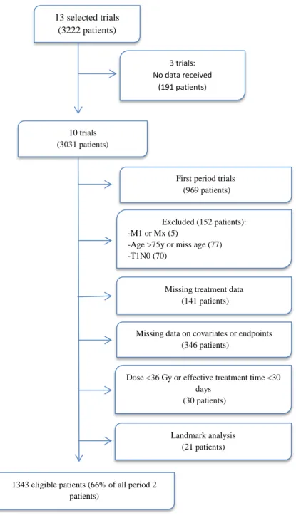
Figure 1: PRISMA flowchart of selection of eligible patients for the pooled analysis to get a more homogenous group of patients.
Figure 2: Summary of Trials. n= number of patients included in the final analysis per trial.
30
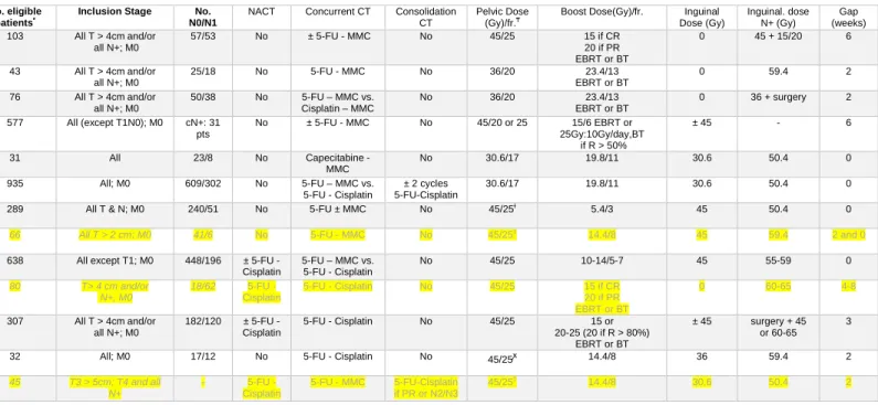
Table 1. Phase II and III studies in anal cancer. Individual patient data was received for all studies in bold. No data was received for those in light gray.
Group Study Name Phase No. Patients No. eligible patients*
Inclusion Stage No. N0/N1
NACT Concurrent CT Consolidation CT
Pelvic Dose (Gy)/fr.₸
Boost Dose(Gy)/fr. Inguinal Dose (Gy) Inguinal. dose N+ (Gy) Gap (weeks) EORTC
22861[6] III 110 103 All T > 4cm and/or
all N+; M0 57/53 No ± 5-FU - MMC No 45/25 15 if CR 20 if PR EBRT or BT 0 45 + 15/20 6 22953[20] II 44 43 All T > 4cm and/or all N+; M0 25/18 No 5-FU - MMC No 36/20 23.4/13 EBRT or BT 0 59.4 2 22011[18] II 88 76 All T > 4cm and/or all N+; M0 50/38 No 5-FU – MMC vs. Cisplatin – MMC No 36/20 23.4/13 EBRT or BT 0 36 + surgery 2 UKCCCR ACT I[24,25]
III 585 577 All (except T1N0); M0 cN+: 31
pts No ± 5-FU - MMC No 45/20 or 25 15/6 EBRT or 25Gy:10Gy/day,BT if R > 50% ± 45 - 6 EXTRA[2 1] II 31 31 All 23/8 No Capecitabine - MMC No 30.6/17 19.8/11 30.6 50.4 0
ACT II[9] III 940 935 All; M0 609/302 No 5-FU – MMC vs.
5-FU - Cisplatin
± 2 cycles 5-FU-Cisplatin
30.6/17 19.8/11 30.6 50.4 0
RTOG
87-04[26] III 310 289 All T & N; M0 240/51 No 5-FU ± MMC No 45/25ɫ 5.4/3 45 50.4 0
92-08[54] II 46 and 20 66 All T > 2 cm; M0 41/6 No 5-FU - MMC No 45/25‡
14.4/8 45 59.4 2 and 0
98-11[8,19]
III 682 638 All except T1; M0 448/196 ± 5-FU -
Cisplatin 5-FU – MMC vs. 5-FU - Cisplatin No 45/25 10-14/5-7 45 55-59 0 FNLCC [55]** II 80 80 T> 4 cm and/or N+, M0 18/62 5-FU - Cisplatin 5-FU - Cisplatin No 45/25 15 if CR 20 if PR EBRT or BT 0 60-65 4-8 ACCORD -03 [17]**
III 307 307 All T > 4cm and/or
all N+; M0 182/120 ± 5-FU - Cisplatin 5-FU - Cisplatin No 45/25 15 or 20-25 (20 if R > 80%) EBRT or BT ± 45 surgery + 45 or 60-65 3 ECOG-ACRIN
42-92[56] II 32 32 All; M0 17/12 No 5-FU - Cisplatin No 45/25ꭙ 14.4/8 36 59.4 2
CALGB 9281[22] II 45 45 T3 > 5cm; T4 and all N+ - 5-FU - Cisplatin 5-FU - MMC 5-FU-Cisplatin if PR or N2/N3 45/25‡ 14.4/8 30.6 50.4 2
* No. of eligible patients with analyzable data ** published in 2012, ongoing at the time of the meeting
₸in fractions of 1.8 Gy for all studies, except for ACT 1 in fractions of 1.8 Gy or 2.25 Gy. RTOG 98-11 advises the boost to be given in fractions of 2 Gy.
ɫ field reduction at 30.6 Gy and at 36 Gy. If after the full treatment there was a residual histologically confirmed primary or inguinal lymph node, addition of a second boost (9 Gy). ‡ field reduction at 30.6 Gy.
ꭙ field reduction at 30.6 Gy and at 36 Gy.
Abbreviations: No., number; Gy, NACT, neoadjuvant chemotherapy; CT, chemotherapy; fr., fractions; Gy, Gray; EORTC, European Organization for the Research and Treatment of Cancer; UKCCCR, UK Co-ordinating Committee for Cancer Research; RTOG, Radiation Therapy Oncology Group; FNCLCC, French Federation Nationale des Centres de Lutte Contre le Cancer; ECOG-ACRIN, ECOG-ACRIN Cancer Research Group; CALGB, Cancer and Leukemia Group B; 5-FU, 5-fluouracil; MMC, Mitomycin C; CR, complete response; PR, partial response; el., electrons; ph., photons; BT, brachytherapy; R > 50%, response > 50%; EBRT, external
31
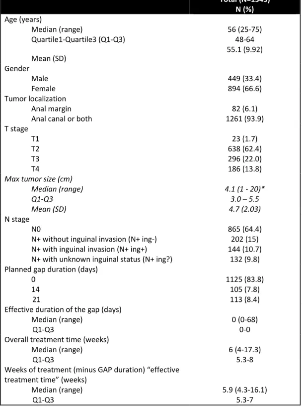
Table 2: Patient characteristics and radiotherapy-related parameters of the group of patients included in the pooled analysis.
Total (N=1343) N (%) Age (years) Median (range) Quartile1-Quartile3 (Q1-Q3) Mean (SD) 56 (25-75) 48-64 55.1 (9.92) Gender Male Female 449 (33.4) 894 (66.6) Tumor localization Anal margin
Anal canal or both 1261 (93.9) 82 (6.1)
T stage T1 T2 T3 T4 23 (1.7) 638 (62.4) 296 (22.0) 186 (13.8)
Max tumor size (cm) Median (range) Q1-Q3 Mean (SD) 4.1 (1 - 20)* 3.0 – 5.5 4.7 (2.03) N stage N0
N+ without inguinal invasion (N+ ing-) N+ with inguinal invasion (N+ ing+)
N+ with unknown inguinal status (N+ ing?)
865 (64.4) 202 (15) 144 (10.7)
132 (9.8) Planned gap duration (days)
0 14 21 1125 (83.8) 105 (7.8) 113 (8.4) Effective duration of the gap (days)
Median (range)
Q1-Q3 0 (0-68) 0-0
Overall treatment time (weeks) Median (range)
Q1-Q3 6 (4-17.3) 5.3-8
Weeks of treatment (minus GAP duration) “effective treatment time” (weeks)
Median (range)
32 Total dose to Pelvis (Gy)
Median (range) Q1-Q3
N non missing observations
39.6 (0.9-60) 30.6-45
1329 Total dose on anal canal (Gy)
Median (range) Q1-Q3 <50.5Gy >50.5-<55Gy >55-<59Gy >59Gy-<59.4Gy >59.4Gy 50.4 (38.6-98) 50.4-59 716 (53.3) 169 (12.6) 198 (14.7) 152 (11.3) 108 (8)
33
Table 3: Models of overall treatment time and dose of radiation therapy for (a)
Locoregional failure (b) Progression free survival (c) Overall survival and (d) Treatment parameters by gender.
(a) Locoregional failure
HR HR 95% CI P Best if
Overall treatment time (weeks) P=0.03 (df=5) Dose ≤50.5 Gy Dose 50.51 Gy - 55 Gy Dose 55.01 Gy - 59 Gy Dose 59.01 Gy - 59.4 Gy Dose >59.4 Gy 1.15 0.98 1.23 1.14 1.14 0.99 0.72 1.04 0.88 0.96 1.34 1.34 1.45 1.47 1.36 0.065 0.908 0.017 0.309 0.128 Time ↓ Time ↓ Time ↓
Total dose on anal canal P=0.937 (df=4) 50.51 Gy - 55 Gy vs. ≤50.5 Gy 55.01 Gy - 59 Gy vs. ≤50.5 Gy 59.01 Gy - 59.4 Gy vs. ≤50.5 Gy >59.4 Gy vs. ≤50.5 Gy 2.00 0.68 0.76 0.73 0.20 0.14 0.06 0.08 19.79 3.40 8.88 6.26 0.552 0.643 0.825 0.774 Age (years) P=0.234 0.99 0.98 1.00 0.234
Tumor size (cm) in:
P<0.001 (df=6) Anal margin N0
Anal margin N+ ing- Anal margin N+ ing+ Anal margin N+ ing? Anal canal N0 Anal canal N+ ing- Anal canal N+ ing+ Anal canal N+ ing?
0.83 1.00 0.96 1.00 1.11 1.19 1.22 0.99 0.60 0.73 1.05 1.06 1.09 0.85 1.15 1.26 1.18 1.34 1.38 1.16 0.257 0.761 0.001 0.004 0.001 0.926 Small Small Small N stage P=0.377 (df=3) N+ ing- vs. N0 N+ ing+ vs. N0 N+ ing ? vs. N0 0.94 1.04 2.28 0.44 0.47 0.89 2.04 2.31 5.87 0.883 0.927 0.087 Tumor loc
P=0.07 Anal canal vs. Anal margin 0.30 0.08 1.10 0.07
Gender
P=0.045 Female vs. Male 0.78 0.61 0.99 0.045 Female*
34
(b) Progression free survival
HR HR 95% CI P Best if
Overall treatment time (weeks) P=0.025 (df=5) Dose ≤50.5 Gy Dose 50.51 Gy - 55 Gy Dose 55.01 Gy - 59 Gy Dose 59.01 Gy - 59.4 Gy Dose >59.4 Gy 1.15 1.06 1.14 1.19 1.14 1.01 0.83 0.98 0.96 0.98 1.31 1.35 1.32 1.48 1.33 0.031 0.662 0.095 0.105 0.1 Time ↓
Total dose on anal canal
P=0.937 (df=4) 50.51 Gy - 55 Gy vs. ≤50.5 Gy 55.01 Gy - 59 Gy vs. ≤50.5 Gy 59.01 Gy - 59.4 Gy vs. ≤50.5 Gy >59.4 Gy vs. ≤50.5 Gy 1.28 1.32 0.51 0.73 0.20 0.32 0.06 0.11 8.35 5.36 4.12 4.91 0.799 0.7 0.528 0.75 Age (years) P=0.927 1.00 0.99 1.01 0.927
Tumor size (cm) in:
P<0.001 (df=6) Anal margin N0
Anal margin N+ ing+ Anal canal N0 Anal canal N+ ing- Anal canal N+ ing+ Anal canal N+ ing?
0.90 1.01 1.12 1.16 1.19 0.99 0.71 0.81 1.07 1.04 1.07 0.87 1.16 1.26 1.18 1.29 1.32 1.12 0.424 0.927 <0.001 0.006 0.001 0.87 Small Small Small N stage P=0.055 (df=3) N+ ing- vs. N0 N+ ing+ vs. N0 N+ vs. N0 1.10 1.42 2.79 0.56 0.73 1.31 2.13 2.78 5.94 0.784 0.305 0.008 Tumor loc
P=0.191 Anal canal vs. Anal margin 0.49 0.17 1.43 0.191
Gender
35
(c) Overall survival
HR HR 95% CI P Best if
Overall treatment time (weeks) P=0.026 (df=5) Dose ≤50.5 Gy Dose 50.51 Gy - 55 Gy Dose 55.01 Gy - 59 Gy Dose 59.01 Gy - 59.4 Gy Dose >59.4 Gy 1.13 1.21 1.16 1.31 1.22 0.95 0.88 0.98 0.95 1.00 1.33 1.66 1.38 1.80 1.50 0.159 0.231 0.089 0.1 0.05 Time ↓ Time ↓
Total dose on anal canal
P=0.937 (df=4) 50.51 Gy - 55 Gy vs. ≤50.5 Gy 55.01 Gy - 59 Gy vs. ≤50.5 Gy 59.01 Gy - 59.4 Gy vs. ≤50.5 Gy >59.4 Gy vs. ≤50.5 Gy 0.39 1.03 0.16 0.26 0.03 0.19 0.01 0.02 4.56 5.56 3.69 3.29 0.456 0.976 0.255 0.3 Age (years) P=0.639 1.00 0.99 1.02 0.633
Tumor size (cm) in:
P=0.027 (df=6) Anal margin N0
Anal margin N+ ing+ Anal canal N0 Anal canal N+ ing- Anal canal N+ ing+ Anal canal N+ ing?
0.99 1.11 1.10 1.14 1.12 1.01 0.75 0.87 1.03 1.00 0.96 0.87 1.31 1.42 1.18 1.29 1.31 1.17 0.956 0.384 0.007 0.043 0.164 0.882 Small Small Small N stage P=0.079 (df=3) N+ ing- vs. N0 N+ ing+ vs. N0 N+ ing ? vs. N0 1.08 2.23 2.62 0.46 0.95 1.04 2.54 5.75 6.63 0.859 0.065 0.041 Tumor loc
P=0.954 Anal canal vs. Anal margin 1.04 0.28 3.84 0.954
Gender
P<0.001 Female vs. Male 0.56 0.43 0.72 <0.001 Female
36
d. Treatment parameters by gender
Gender Male
(N=449)
Female
(N=894)
Overall treatment time (days) including boost
Median (range) 39 (30 - 107) 43 (30 - 121)
Q1-Q3 37 – 52 37 – 57
Total dose on anal canal (Gy)
Median (range) 50.4 (39.6 - 75) 50.4 (38.6 - 98)
Q1-Q3 50.4 - 57.4 50.4 – 59
Effect of treatment being a woman
Median (range) 5.6 (4.3 - 15.3) 6 (4.3 - 16.1)
Q1-Q3 5.3 - 6.6 5.3 - 7
Duration of the gap
Median (range) 0 (0 – 44) 0 (0 – 68)
37
Table 4. Late toxicities: (a) gastro-intestinal (b) skin or subcutaneous tissue and (c) genito-urinary.
(a)
(b)
1
Figure 1: PRISMA flowchart of selection of eligible patients for the pooled analysis to get a more homogenous group of patients.
13 selected trials (3222 patients)
10 trials (3031 patients)
First period trials (969 patients) 3 trials: No data received (191 patients) Excluded (152 patients): -M1 or Mx (5)
-Age >75y or miss age (77) -T1N0 (70)
Missing treatment data (141 patients)
Missing data on covariates or endpoints (346 patients)
Dose <36 Gy or effective treatment time <30 days
(30 patients)
Landmark analysis (21 patients)
1343 eligible patients (66% of all period 2 patients)
1
Figure 2: Summary of Trials. n= number of patients included in the final analysis per trial.
n=8 n=34 n=551 n=113 n=547 n=19 n=71
1
Pooled Analysis of external-beam RADiotherapy parameters in phase II and phase III trials in radiochemotherapy in Anal Cancer (PARADAC).
SUPPLEMENTARY MATERIAL
Appendix A. Figure 1. Overall treatment time according to the total dose to the anal canal.
2
Appendix A. Table 1. Evaluated toxicities and scales used for toxicity grading for the 10 trials data was received for. Online only.
Group Study Defined selected toxicities Scale Comment
EORTC 22861 Rectal discharge Rectal bleeding Anal/rectal damage Rectal stenosis Rectal incontinence Colostomy Skin reaction Fibrosis Other
RTOG/EORTC Late RT Morbidity Scoring Scheme (NCI CTC v2) scale for skin reaction and sub tissue (fibrosis)
Study specific defined scales for the other categories
22953
Bladder/Urethra Skin/subcutaneous tissue Rectum
Small intestine / Colon
LENT-SOMA 22011 Bladder Skin Small/large intestine Subcutaneous tissue Other
RTOG/EORTC Late RT Morbidity Scoring Scheme (NCI CTC v2)
UKCCCR
ACT 1
Ano-rectal Skin Hip/bone.
No specified scale used, only refers to grade 3 or 4 toxicities without specifying the exact grade
EXTRA Skin Large intestine
Rectal or perirectal pain Pain due to radiation, Urinary frequency/urgency Other
RTOG/EORTC Late RT Morbidity Scoring Scheme (NCI CTC v2) for Skin and Large intestine, Renal / genitourinary and pain
Scales of NCI CTC v2 for the other toxicities
A lot of “other” toxicities ACT II RTOG 87-04 Skin Gastrointestinal Genitourinary Stomatitis/Mucus membrane
RTOG/EORTC Late RT Morbidity Scoring Scheme (NCI CTC v2)
98-11 Bladder Skin Small/large intestine Subcutaneous tissue Other
RTOG/EORTC Late RT Morbidity Scoring Scheme (NCI CTC v2)
ECOG-ACRIN
42-92 No late toxicities have been reported
FNCLCC ACCORD-03 Bladder/Urethra Skin/subcutaneous tissue Rectum Small Intestine/Colon
LENT-SOMA Only some scales are
3
4
Appendix A. Figure 3: (a) Cumulative incidence of locoregional failure (b) Progression free survival and (c) Overall survival. Online only.
(a) Locoregional failure
(years) 0 1 2 3 4 5 0 10 20 30 40 50 60 70 80 90 100
O N Number of patients at risk '
303 1343 1098 862 668 493
5 (b) Progression-free survival (years) 0 2 4 6 8 10 12 0 10 20 30 40 50 60 70 80 90 100
O N Number of patients at risk '
6 (c) Overall survival (years) 0 2 4 6 8 10 12 0 10 20 30 40 50 60 70 80 90 100
O N Number of patients at risk '
7
Appendix A: Table 2: Locoregional failure model comparing doses of 50.4 Gy vs. 55-59 Gy
without a Gap (N=977) (RTOG 98-11, ACT II and EXTRA) Online only.
HR HR 95% CI P Best if
Planned Dose anal canal
P=0.412
55-59 vs 50.4 Gy 1.14 0.84 1.54 0.159 Lower*
Age (years)
P=0.468
0.99 0.98 1.01 0.468
Tumor size (cm) in:
P=0.002 (df=4) Anal margin N0
Anal margin N+ ing- Anal margin N+ ing+ Anal margin N+ ing? Anal canal or both N0 Anal canal or both N+ ing- Anal canal or both N+ ing+ Anal canal or both N+ ing?
0.77 1.00 0.99 1.00 1.11 1.18 1.25 1.01 0.54 0.74 1.05 1.04 1.08 0.86 1.11 1.31 1.19 1.33 1.19 2.97 0.165 0.384 0.921 0.001 0.009 0.003 0.542 Small Small Small N stage P=0.333 (df=4) N+ ing- N+ ing+ N+ ing? 1.08 2.23 2.62 0.46 0.95 1.04 2.54 5.75 6.63 0.859 0.065 0.041 Tumor localization
P=0.033 Anal canal or both 0.21 0.05 0.88 0.032 Both
Gender
P=0.574 Female vs. Male 0.92 0.69 1.23 0.577 Female
8
Appendix A: Table 3: Locoregional failure model: 3 week vs 2 week gap at 59.4Gy intended dose (EORTC studies and ACCORD-03) ). Online only.
HR HR 95% CI P Best if Planned gap P=0.245 21 d vs 14 days 1.37 0.81 2.34 0.245 Age (years) P=0.02 0.97 0.95 1.00 0.02 Young
Tumor size (cm) in:
P=0.377 (df=4) Anal canal or both N0 Anal canal or both N+ ing- Anal canal or both N+ ing+
1.19 1.10 1.13 0.93 0.83 0.88 1.53 1.47 1.45 0.173 0.49 0.333 N stage P=0.468 N+ ing- N+ ing+ 1.70 3.21 0.25 0.50 11.68 20.55 0.589 0.218 Gender
P=0.228 Female vs. Male 0.71 0.41 1.23 0.228 Female
n = 332
9
Appendix A: Table 4: 2 week gap vs no gap at (a) 55-59.4Gy intended dose and b) effective dose >56.5 and <60Gy (EORTC studies vs. RTOG 98-11). Online only.
(a) HR HR 95% CI P Best if Planned gap P=0.887 2 weeks vsno gap (55-59 Gy intended dose) 1.04 0.64 1.67 0.887 Age (years) P=0.163 0.99 0.97 1.01 0.163
Tumor size (cm) in:
P=0.004 (df=4) Anal canal or both N0
Anal canal or both N+ ing- Anal canal or both N+ ing+ Anal canal or both N+
1.09 1.02 1.18 2.25 0.99 0.80 1.02 1.24 1.20 1.29 1.35 4.06 0.077 0.903 0.022 0.007 Small Small N stage P=0.161 (Df=3) N+ ing- N+ ing+ N+ ing? 2.44 1.23 0.03 0.61 0.42 0.00 9.81 3.63 1.37 0.208 0.708 0.072 Gender P=0.573 Female 0.89 0.60 1.33 0.573 n = 521 (b) HR HR 95% CI P Best if Planned gap P=0.559 2 weeks vsno gap
(effective dose >56.5 and <60Gy) 0.87 0.52 1.47 0.559
Age (years)
P=0.163 0.99 0.97 1.01 0.163
Tumor size (cm) in:
P=0.214 1.06 0.97 1.17 0.214 N stage P=0.064 (Df=3) N+ ing- N+ ing+ N+ ing? 1.58 2.15 1.58 0.87 1.20 0.48 2.87 3.84 5.19 0.130 0.01 0.449 Gender P=0.339 Female 0.79 0.49 1.28 0.339 n = 214