HAL Id: hal-02709254
https://hal-amu.archives-ouvertes.fr/hal-02709254
Submitted on 1 Jun 2020HAL is a multi-disciplinary open access archive for the deposit and dissemination of sci-entific research documents, whether they are pub-lished or not. The documents may come from teaching and research institutions in France or abroad, or from public or private research centers.
L’archive ouverte pluridisciplinaire HAL, est destinée au dépôt et à la diffusion de documents scientifiques de niveau recherche, publiés ou non, émanant des établissements d’enseignement et de recherche français ou étrangers, des laboratoires publics ou privés.
Distributed under a Creative Commons Attribution| 4.0 International License
Vitro Cytotoxicity Mediated by Complement Activation
Maël Lateb, Hajar Ouahmi, Christine Payré, Vesna Brglez, Kevin Zorzi,
Guillaume Dolla, Mohamad Zaidan, Sonia Boyer-Suavet, Bertrand
Knebelmann, Thomas Crépin, et al.
To cite this version:
Maël Lateb, Hajar Ouahmi, Christine Payré, Vesna Brglez, Kevin Zorzi, et al.. PLA2R1 Anti-bodies Containing Sera Induce In Vitro Cytotoxicity Mediated by Complement Activation. Journal of Immunology Research, Hindawi Publishing Corporation, 2019, 2019, pp.1-14. �10.1155/2019/1324804�. �hal-02709254�
Research Article
Anti-PLA2R1 Antibodies Containing Sera Induce In Vitro
Cytotoxicity Mediated by Complement Activation
Maël Lateb,
1Hajar Ouahmi,
2Christine Payré,
1Vesna Brglez,
3Kevin Zorzi,
3Guillaume Dolla
,
1Mohamad Zaidan,
4Sonia Boyer-Suavet,
3Bertrand Knebelmann,
5,6Thomas Crépin
,
7Cécile Courivaud,
7Noémie Jourde-Chiche
,
8Vincent Esnault,
2,3Gérard Lambeau,
1and Barbara Seitz-Polski
1,2,3,91Université Côte d’Azur, Centre National de la Recherche Scientifique, Institut de Pharmacologie Moléculaire et Cellulaire, UMR7275,
Valbonne Sophia Antipolis, France
2Service de Néphrologie-Dialyse-Transplantation, Hôpital Pasteur, CHU de Nice, Université de Nice-Sophia Antipolis, France 3Centre de Référence Maladies Rares Syndrome Néphrotique Idiopathique, CHU de Nice, Université de Nice-Sophia Antipolis, France 4Service de Néphrologie-Transplantation, Hôpital de Bicêtre, AP-HM France, Université Paris-Saclay, Villejuif, Paris, France 5Université Paris Descartes, Sorbonne Paris Cité, Hôpital Necker-Enfants Malades, Paris, France
6INSERM U1151, Institut Necker Enfants Malades, Hôpital Necker-Enfants Malades, Paris, France 7Département de Néphrologie, Dialyse et Transplantation, Université de Franche-Comté, Besançon, France
8Aix-Marseille Univ, C2VN, INSERM 1263, INRA 1260, AP-HM Hôpital de la Conception, Centre de Néphrologie et
Transplantation Rénale, Marseille, France
9Laboratoire d’Immunologie, Hôpital l’Archet, CHU de Nice, Université de Nice-Sophia Antipolis, France
Correspondence should be addressed to Barbara Seitz-Polski; seitz-polski.b@chu-nice.fr
Received 19 August 2019; Revised 8 November 2019; Accepted 13 November 2019; Published 30 December 2019 Guest Editor: Cheng Xiao
Copyright © 2019 Maël Lateb et al. This is an open access article distributed under the Creative Commons Attribution License, which permits unrestricted use, distribution, and reproduction in any medium, provided the original work is properly cited. The phospholipase A2 receptor (PLA2R1) is the major autoantigen in idiopathic membranous nephropathy (MN). However, the pathogenic role of anti-PLA2R1 autoantibodies is unclear. Our aim was to evaluate the in vitro cytotoxicity of anti-PLA2R1 antibodies mediated by complement. Forty-eight patients with PLA2R1-related MN from the prospective cohort SOURIS were included. Anti-PLA2R1 titer, epitope profile, and anti-PLA2R1 IgG subclasses were characterized by ELISA. Cell cytotoxicity was evaluated by immunofluorescence in HEK293 cells overexpressing PLA2R1 incubated with patient or healthy donor sera in the presence or absence of rabbit complement or complement inhibitors. Mean cytotoxicity of anti-PLA2R1 sera for HEK293 cells overexpressing PLA2R1 was 2 ± 2%, which increased to 24 ± 6% after addition of rabbit complement (p < 0:001) (n = 48). GVB-EDTA, which inhibits all complement activation pathways, completely blocked cell cytotoxicity, whereas Mg-EGTA, which only inhibits the classical and lectin pathways, highly decreased suggesting a limited role of the alternative pathway. A higher diversity of IgG subclasses beyond IgG4 and high titer of total IgG anti-PLA2R1 were associated with increased cytotoxicity (p = 0:01 and p = 0:03 respectively). In a cohort of 37 patients treated with rituximab, high level of complement-mediated cytotoxicity was associated with less and delayed remission at month 6 after rituximab therapy (5/12 vs. 20/25
(p = 0:03) in 8:5 months ± 4:4 vs. 4:8 ± 4:0 (p = 0:02)). Kaplan-Meier analysis demonstrated that high level of cytotoxicity
(≥40%) (p = 0:005), epitope spreading (defined by immunization beyond the immunodominant CysR domain) (p = 0:002), and high titer of anti-PLA2R1 total IgG (p = 0:01) were factors of poor renal prognosis. Anti-PLA2R1 antibodies containing sera can induce in vitro cytotoxicity mediated by complement activation, and the level of cytotoxicity increases with the diversity and the titer of anti-PLA2R1 IgG subclasses. These patients with high level of complement-mediated cytotoxicity could benefit from adjuvant therapy using complement inhibitor associated with rituximab to induce earlier remission and less podocyte injury.
Volume 2019, Article ID 1324804, 14 pages https://doi.org/10.1155/2019/1324804
1. Introduction
Membranous nephropathy (MN) is an autoimmune disease and a major cause of nephrotic syndrome in adults [1]. It is
defined by the presence of subepithelial immune complex
deposits with alteration of the glomerular membrane and podocyte injury [2]. Most MN cases are associated with autoantibodies directed against podocyte antigens such as the M-type phospholipase A2 receptor (PLA2R1) [3] and thrombospondin type 1 domain-containing 7A (THSD7A) [4, 5] in 70% and 3% of adult patients, respectively. Disease evolution is highly variable from spontaneous remission to persistent proteinuria or end-stage renal disease [6]. Treat-ment remains controversial [7, 8]. Kidney Disease Improving Global Outcomes (KDIGO) guidelines recommend a sup-portive symptomatic treatment (Renine angiotensine system blockers and diuretics) in all patients and immunosuppres-sive therapy only in the case of persistent nephrotic syndrome or renal function deterioration [9]. Therefore,
immunosup-pressive treatments are often started only after significant
and potentially irreversible complications. New KDIGO guidelines will probably modify this recommendation using new markers to start immunosuppressive therapy [10].
While the pathogenic role of anti-THSD7A antibodies has been proven by the formation of immune deposits and the onset of proteinuria in mice injected with human anti-THSD7A antibodies [11], no such study has been performed for PLA2R1 due to the absence of PLA2R1 expression in mouse or rat podocytes. Nevertheless, PLA2R1 antibody titers rise during clinical activity and decrease before remis-sion [12], and MN recurrence after kidney graft is associated with high titers [13]. Anti-PLA2R1 titers could also predict outcome after immunosuppressive treatment in MN [14].
PLA2R1 epitopes have been identified in three domains of
the protein (CysR, CTLD1, and CTLD7), and a mechanism of epitope spreading from the immunodominant CysR domain to CTLD1 and/or CTLD7 domains has been
associ-ated with poor prognosis [15–17] corresponding to later
stages of the disease [18].
The complement system forms an important part of the innate immune system. It is involved in host defense, but also in autoimmune diseases, and is made up of over 30 pro-teins that can be sequentially activated in a complex enzy-matic cascade. Three major pathways of complement have been described: classical, alternative, and lectin pathways,
which are activated by different stimuli [19]. All three
path-ways converge on complement C3. Cleavage of C3 and C5 successively leads to the production of the membrane attack
complex (C5b-9), which is the final common effector [20].
C5b-9 has been shown to have a major role in the Heymann nephritis rat model of MN [21, 22]. The role of the different complement pathways upstream of C5 activation in human and experimental MN remains largely unknown. Primary MN associated to PLA2R1 and THSD7A is characterized by predominant IgG4 (in deposits and in serum) and low
amounts of IgG1 and IgG3 [23–25]. Immune complexes
typ-ically bind C1q and activate the classical pathway. However, IgG4 does not bind C1q and is considered to be unable to activate the classical pathway [26, 27]. The presence of
IgG1 in early deposits could possibly activate the classical pathway, but C1q deposits are very weak in MN [28]. This suggests that the alternative or lectin pathways might be involved in complement activation. Hayashi et al. detected glomerular deposits of C4d, C3d, and C5b-9 in all patients while mannose-binding lectin (MBL) and C1q were detected in only 43% and 46% of patients, respectively [29]. One or more complement pathways may be activated after the for-mation of immune deposits and can vary among MN patients. Hayashi et al. also described that MN associated with glomerular MBL deposits is more severe. On the other hand, Bally et al. reported cases of PLA2R1-associated MN in patients having a complete MBL deficiency with comple-ment activation mainly due to the alternative pathway [30]. The aim of this study was (i) to evaluate the pathogenic role of anti-PLA2R1 antibodies and the contribution of com-plement in a simple but routinely used in vitro cellular assay using HEK293 cells overexpressing PLA2R1, (ii) identify fac-tors associated with complement mediated-cytotoxicity, and (iii) analyze prognosis value of this complement mediated-cytotoxicity. This study may provide the molecular basis to better understand MN pathogenesis and develop alternative therapeutic strategies.
2. Materials and Methods
2.1. Patients. Forty-eight patients with PLA2R1-associated MN from the prospective cohort SOURIS were included (NCT02199145) approved by our local Ethic committee. Patients from the SOURIS cohort are primary MN patients (defined by the absence of antinuclear antibodies, negative hepatitis B and C serologies, and negative cancer workup),
adults, with MDRD > 30 ml/mn/1.73 m2 and anti-PLA2R1
antibodies at diagnosis. The aim of the SOURIS cohort is to
demonstrate that epitope profile at diagnosis could guide
therapeutic strategy. Patients were enrolled from four French centers from 2015 to 2018 and had a median follow-up of 14 months. Baseline characteristics and follow-up data were recorded until 18 months. Serum anti-PLA2R1 levels, serum creatinine, serum albumin, and proteinuria were measured every 3 months for 18 months. Sera from 20 healthy donors, age- and gender-matched, were collected. Information and written consent were obtained from all patients.
Serum and morning spot urine samples were
prospec-tively collected at thefirst infusion and every 3 months after
thefirst rituximab infusions (i.e., month 3, month 6).
Remis-sions were defined according to the 2012 KDIGO guidelines.
Complete remission was defined by a urinary protein/creati-nine ratio < 0:3 g/g accompanied by a normal serum albumin concentration and a preserved kidney function. Partial remission was defined by urinary protein/creatinine ratio < 3:5 g/g with over 50% reduction of proteinuria accompanied by an improvement or normalization of the serum albumin concentration and preserved kidney function.
2.2. Anti-PLA2R1 ELISA. The entire extracellular domain of human PLA2R1 or its CysR, CTLD1, and CTLD7 domains bearing a C-terminal HA tag were produced as secreted
expression vectors coding for each protein were transfected into HEK293 cells by calcium phosphate transfection. Three days after transfection, cell medium containing the secreted proteins was collected and centrifuged to remove cell debris. Anti-HA antibodies (Sigma-Aldrich) were coated for one
night at 4°C. Plates were then washed three times with
PBS/0.02% Tween 20, then blocked for two hours with Sera-mun Block (SeraSera-mun Diagnostica) and then washed again three times. Cell medium from HEK293 cells transfected with the soluble forms of the three PLA2R1 domains or the entire PLA2R1 tagged with HA was then added and incu-bated for one hour at room temperature on a plate shaker, then washed three times. Patients’ sera were diluted at 1 : 100 in PBS/0.1% milk and added to the ELISA plates for two hours. After three washes, anti-human IgG4 (1 : 7500; Southern Biotech) or anti-human IgG1, IgG2, and IgG3, all conjugated with horseradish peroxidase (1 : 5000; Southern Biotech) and diluted in SeramunStab ST plus (Seramun Diagnostica), were incubated for one hour at room tempera-ture on a plate shaker. After three washes, tetramethylbenzi-dine was added and the reaction was stopped with HCl 1.2 N after 15 minutes. Plates were read at 450 nm. A negative con-trol as well as a highly positive index patient serum was used in each plate to generate a standard curve. The cut-off was optimized by receiver operating characteristic (ROC) curve analysis using 20 healthy controls.
Epitope spreading was defined by anti-CysR reactivity
with additional anti-CTLD1 and/or anti-CTLD7 activities. 2.3. Measurement of Anti-PLA2R1 Antibodies by ELISA. Serum levels of total IgG anti-PLA2R1 antibodies were measured by the ELISA test developed by EUROIMMUN AG (Lübeck, Germany) [31]. Participants were considered as anti-PLA2R1-positive when levels were higher than 14 RU/ml.
2.4. Immunofluorescence Cytotoxicity Assay and Complement Activation Pathway. HEK293 cells overexpressing PLA2R1 with a Tetracycline-Regulated Expression System (PLA2R1/-HEK293 T-REx) were obtained as follows. Full-length membrane-bound human PLA2R1 bearing a C-terminal cytoplasmic HA tag was inserted into the pcDNA4/TO expression vector (InvitroGen) and transfected into HEK293 T-REx cells (cultured in DMEM, 10% fetal calf serum with
blasticidin 5μg/ml) using the calcium-phosphate
proce-dure (InvitroGen). One day after transfection, stably expressing cells were selected with zeocine (0.8 mg/ml) for 3 weeks and PLA2R1 expression was induced with
tetracycline (1μg/ml) and validated by western blot as
described [15] for 48 hours. Cells were collected by gentle dissociation with trypsin, centrifuged for 3 minutes at 600 rpm, and diluted in OptiMEM to obtain a concentration
of 3 × 10⁶cells/ml.
Cell cytotoxicity was measured as described by Terasaki and McClelland [32]. Three microliters of serum from anti-PLA2R1-positive patients or healthy donors were incubated
with 3 × 10³ of either induced or noninduced
PLA2R1/-HEK293 T-REx cells (added in 1μl) for 1 hour at room
tem-perature in 60-well Terasaki plates (Dutcher©, Strasbourg,
France) in duplicates. Cytotoxicity was measured with serial serum dilutions (1 : 1, 1 : 3, and 1 : 10). Serum dilutions were associated to decreased (dilution 1 : 3) or negative (dilution 1 : 10) cytotoxicity (Figure Sup 1A). Standard rabbit
comple-ment (5μl/well, Cedarlane©, Ontario, Canada) or OptiMEM
was added and incubated for 1 hour at room temperature.
Dead cells were revealed after adding 2.5μl/well of
Fluoro-quench AO/EB staining/Fluoro-quench (Ingen©Chilly-Mazarin, France) for 10 minutes in the dark. The percentage of dead
cells was estimated using afluorescent microscope
(Videomi-croscope Zeiss LSM780). Two investigators read plates blindly with a good correlation (rs = 0:81, p < 0:0001) and an excellent concordance (ICC = 0:89 (0.85; 0.92)) (Figure sup 1B and 1C).
Various versions of gelatin veronal buffer (GVB) were
used to determine the complement pathway involved in anti-PLA2R1 cytotoxicity. GVB supplemented with EDTA (GVB-EDTA) (ComplementTech©) was used to inhibit all complement pathways activation, whereas GVB supple-mented with magnesium and EGTA (Mg-EGTA) (Comple-mentTech©) was used to inhibit the classical and lectin pathways, but not the alternative one. Three microliters of
three PLA2R1-positive sera were diluted in 5μl of GVB
sup-plemented with GVB-EDTA or Mg-EGTA and 5μl of
stan-dard rabbit complement. Cytotoxicity was assessed as
described above. We confirmed in these conditions the
inhibi-tion of the classical pathway activity by measuring CH50 (Total Haemolytic Complement Kits Binding Site©) (Figure Sup 2).
All cytotoxicity assays were performed using the same batch of HEK293 T-Rex cells. A minimum of 50 cells per well was necessary for reading.
2.5. Western Blot Analysis. The expression of PLA2R1 in HEK293 cells overexpressing PLA2R1 with a Tetracycline-Regulated Expression System (PLA2R1/HEK293 T-REx) after induction with tetracycline was analyzed by SDS-PAGE under nonreducing conditions. A control without induction with tetracycline was performed. Total proteins
(10-50μg/well) were run on 4-15% precast TGX
SDS-PAGE gels (Bio-Rad) and transferred to methanol-soaked PVDF membranes (Bio-Rad) under semidry conditions using Trans-blot Turbo (Bio-Rad) at 25 V constant for
12 min. Membranes were blocked overnight at 4°C in 5%
milk with PBS-Tween (PBS-T) 0.05% and then incubated with primary and secondary antibodies for 2 h at room tem-perature. Primary antibodies were diluted with 0.5% dry milk in PBS-T. Membranes were prepared in replicates and probed with a serum of patient with anti-PLA2R1 antibodies diluted at 1 : 100. Secondary antibody for iMN sera was HRP-conjugated mouse anti-human IgG (Southern Biotech #9200-05) diluted 1 : 30,000 in PBS-T. Membranes were washed three times for 5 min in PBS-T after incubation with primary and secondary antibodies. Detection of protein bands was performed with a chemiluminescent substrate (Millipore) and a Fuji LAS3000 imager.
2.6. Statistical Analyses. Data are presented as mean ± SD (for variables with normal distribution) or median and
interquartile range (for variables with nonnormal distribu-tion). We used the Shapiro-Wilk test to determine if a vari-able has a Gaussian distribution. Wilcoxon-Mann–Whitney test and Kruskal–Wallis test were used for comparison between groups. ROC curve analysis was used to define the threshold of each test. Survival curves for remission were calculated using Kaplan–Meier estimates for survival distri-bution. Correlation between the two readers was assessed by Spearman rank correlation coefficient. Concordance between the two readers was assessed by intraclass
correla-tion coefficient (ICC should be over 0.80 for an excellent
concordance). Adjusted analysis was performed using logis-tic regression. All statislogis-tics were performed using the Prism
and SAS software.p values < 0.05 were considered as
statis-tically significant.
3. Results
3.1. Patients and Anti-PLA2R1 Antibodies. A total of 48 PLA2R1-positive patients were enrolled in this study
(Table 1). Thirty-six patients were included for afirst course
of MN and 12 patients for a relapse. All patients received conservative therapy with angiotensin-converting-enzyme (ACE) inhibitors and/or angiotensin II receptor blockers. During follow-up, 37 patients were treated with rituximab 1 g at 2-week interval. All patients were positive for the CysR domain of PLA2R1 at baseline, while 30 out of 48 patients (62.5%) had additional antibodies targeting CTLD1 and/or CTLD7 domains (epitope spreading) (Figure 1(a)). At last observation carried forward, epitope spreading was only identified in 6 out of 37 (16.2%) patients. At baseline, all patients had IgG4 anti-PLA2R1, while 24 patients (50%) also had IgG1, IgG2, or IgG3 autoantibodies. Anti-PLA2R1 IgG3 antibodies were the second most represented subclass with 22 (44%) positive patients (Figure 1(b)). Moreover, IgG1, IgG2, IgG3, and IgG4 anti-PLA2R1 antibodies were found
in most samples at the first course of the disease, while at
relapse, IgG4 anti-PLA2R1 was the predominant subclass (p = 0:02) (Figure 1(c)).
3.2. Cytotoxicity of Anti-PLA2R1 Autoantibodies Is
Dependent on Complement Activation. We choose to test complement-mediated cytotoxicity with a simple, validated, and routinely used test in renal transplantation (the cross-match test), using HEK293 cells overexpressing PLA2R1 with a Tetracycline-Regulated Expression System (instead of lymphocytes in cross-match test).
Addition of anti-PLA2R1 serum and rabbit complement to HEK293 cells overexpressing PLA2R1 after induction
with tetracycline (PLA2R1/HEK293 T-REx+) led to strong
cytotoxicity, while all other conditions were barely cytotoxic
(Figures 2(a) and 2(b)). In PLA2R1/HEK293 T-REx+, the
mean cytotoxicity without complement was 2 ± 2% while it increased to 24 ± 6% with complement (p < 0:001). In HEK293 cells not induced with tetracycline
(PLA2R1/-HEK293 T-REx-), the mean cytotoxicity was 7 ± 2% and 2
± 1% with and without complement, respectively. Addition of healthy donor serum was barely cytotoxic, no matter the conditions. The decreased cell viability of PLA2R1/HEK293
T-REx-in the presence of complement was probably caused
by a minimal expression of PLA2R1 even in noninduced cells, as determined by western blot (Figure 2(c)). Using ROC curve analysis, we identified a threshold > 10% of cyto-toxicity associated with a positive test with a sensitivity of
87.5% and specificity of 84.6% (AUC = 0:90 (0.83 to 0.98)
p < 0:0001) (Figure Sup 3).
3.3. Analysis of the Complement Activation Pathways Involved in Cytotoxicity Mediated by Anti-PLA2R1 Autoantibodies. The complement activation pathways involved in PLA2R1 cytotoxicity were studied with three anti-PLA2R1 serum samples showing a robust level of
cyto-toxicity (Figure 2(d)). The first two patients were positive
for both IgG3 and IgG4 anti-PLA2R1 antibodies, while
the third patient was only positive for IgG4. For the first
patient, the addition of GVB-EDTA totally inhibited anti-PLA2R1-mediated cytotoxicity (from 65 to 0%) as addi-tion of Mg-EGTA (from 65 to 10%). In the presence of Mg-EGTA, we confirmed a complete blockade of the clas-sical pathway (CH50 < 10%, Figure Sup 2), suggesting that
the alternative pathway mediates cytotoxicity for the first
patient. For the second patient, the addition of GVB-EDTA strongly inhibited anti-PLA2R1-mediated cytotoxic-ity (from 50 to 10%), while the addition of Mg-EGTA only decreased anti-PLA2R1-mediated cytotoxicity to lower but still detectable levels (from 50% to 25%). For the third patient, the addition of GVB-EDTA strongly inhibited anti-PLA2R1-mediated cytotoxicity (from 40 to 10%) whereas the addition of Mg-EGTA only inhibited anti-PLA2R1-mediated cytotoxicity by 2-fold (from 40 to 20%) (Figure 2(a)).
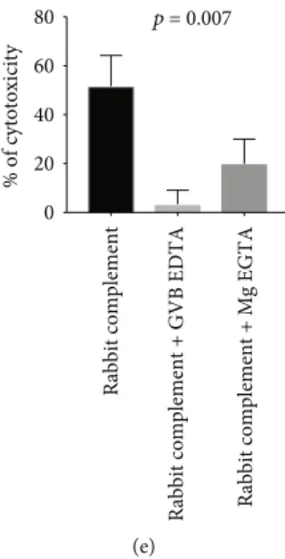
In conclusion, GVB-EDTA, which is known to inhibit the three pathways, strongly inhibited anti-PLA2R1-mediated cytotoxicity in all three patients, which confirm the role of the complement, while Mg-EGTA, which is known to inhibit only the classical and the lectin pathways, partially inhibited the anti-PLA2R1-mediated cytotoxicity, suggesting a potential limited activation of the alternative
pathway in some serum samples (p = 0:007) (Figure 2(e)).
Table 1: Clinical characteristics of patients in the study.
Clinical characteristics Cohort SOURIS
(48 patients)
Age (years) 59 ± 15
Gender ratio (M/F) 3.3
LOCF (months) 14 (3; 18)
Proteinuria at baseline (g/g) 5.1 (3.5; 7.2)
Serum albumin at baseline (g/l) 23.9 (21.2; 29.7) Serum creatinine at baseline (μmol/l) 103 (84.7; 135)
Proteinuria at LOCF (g/g) 1.9 (0.3; 3.4)
Patients treated with rituximab 37
Patients treated with other
immunosuppressors (Ponticelli) 4
Data are presented as mean ± SD or as median and interquartile range. LOCF: last observation carried forward.
Because Mg-EGTA induces inhibition of more than half of the complement-mediated cytotoxicity, the activation of the lectin or the classical pathway seems to be predominant. 3.4. Factors Associated with Cytotoxicity Induced by Anti-PLA2R1 Autoantibodies and Mediated by Complement. We analyzed the factors that may be associated with cytotoxicity (>10%) among the different serum samples from the 48 patients. Epitope spreading as well as high IgG4 anti-PLA2R1 titers was not associated with decreased cell viability (p = 0:72 and p = 0:50, respectively) (Figures 3(a) and 3(b)) while high level of total anti-PLA2R1 total IgG titer was
associated with cytotoxicity (p = 0:03, Figure 3(c)); moreover,
these two variables were correlated (rs = 0:45, p < 0:001) (Figure 3(d)). As suggested, the presence of anti-PLA2R1 IgG1, IgG2, or IgG3 antibodies was associated with
increased cell cytotoxicity (p = 0:03) (Figure 3(e)). Median
complement-mediated cytotoxicity was significantly higher for serum samples containing different IgG subclasses versus those containing only anti-PLA2R1 IgG4 antibodies (30%
(10; 40) versus 10% (0; 30), p = 0:02). There was an inverse
relationship for complement-mediated cytotoxicity between serum samples containing multiple IgG subclasses and those containing only IgG4, suggesting that IgG subclasses other
than IgG4 drive the observed cytotoxic effects (p = 0:015)
(Figure 3(f)). We failed to identify a correlation between titers of each subclass of IgG and cytotoxicity (Figure Sup 4) suggesting that it is the whole of subclasses that is respon-sible for the cytotoxic effect.
3.5. Prognostic Factors for Remission. During follow-up, 37 patients were treated with rituximab 1 g at 2-week interval, 25 entered into remission at month 6 (67.6%). Time to remis-sion after treatment was 6:1 ± 4:4 months.
0 20 40 60 80 Baseline Last observation n CysR CysRC1 CysRC1C7 (a) 0 5 10 15 20 25 30 IgG4 only 50% IgG4 +IgG3 36%
IgG4 + IgG1 + IgG3 8%
IgG4+ IgG2 4% IgG4 + IgG1 2%
(b)
IgG4 only IgG4 + IgG1-3 0 5 10 15 20 25 n Relapse First course p = 0.02 (c)
Figure 1: Description of samples with anti-PLA2R1 antibodies tested. (a) Epitope spreading profiles of patients at baseline (n = 48) and at last observation (n = 36). Follow-up was missing for 12 patients. C1: CTLD1; C7: CTLD7. CysR: immunized against CysR domain alone; CysRC1: immunized against CysR and CTLD1 domains; CysRC1C7: immunized against CysR, CTLD1, and CTLD7 domains. (b) IgG subclass profiles of patients at baseline (n = 48). (c) IgG subclass profiles of patients according to disease course: first course or relapse (n = 48). Note that patients with thefirst course of MN have a larger diversity of IgG subclasses (p = 0:02).
HEK T+ HEK T- HEK T+ HEK T-PLA2R1-positive serum Healthy donor serum
C+ C-(a) 0 20 40 60 80 100 % of cytotoxicity TET+ complement+ p < 0.0001 0 20 40 60 80 100 TET- complement+ p = 0.08 0 20 40 60 80 100 TET+ complement-p = 0.46 MN patients Healthy donors MN patients Healthy donors MN patients Healthy donors MN patients Healthy donors 0 20 40 60 80 100 TET- complement-p > 0.99 (b) TET- TET+ 130 100 70 55 (c)
Patient 1 (IgG3, IgG4) Patient 2 (IgG3, IgG4)
Patient 3 (IgG4) 0 20 40 60 80 % of cytotoxicity Rabbit complement
Rabbit complement+GVB EDTA Rabbit complement+Mg EGTA
(d)
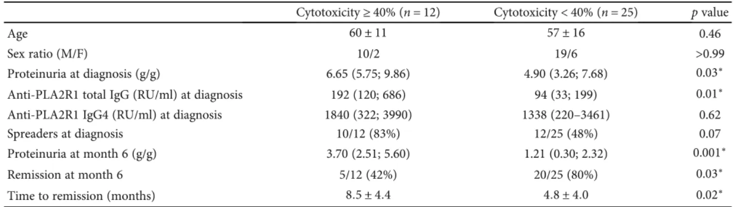
We compared clinical characteristics of patients with the
highest level of cytotoxicity (the third tertile defined by a
threshold of 40% or more of complement-mediated cytotox-icity) (Table 2). Patients with complement-mediated cytotoxicity ≥ 40% were similar at diagnosis for age and sex ratio but have the highest level of proteinuria (6.65 (5.75;
9.86) vs. 4.90 (3.23; 7.68), p = 0:03) and total IgG
anti-PLA2R1 titer at diagnosis (192 (120; 686) vs. 94 (33;
199), p = 0:01) while IgG4 anti-PLA2R1 titer and level of
epitope spreading were similar (1840 (322; 3990) vs. 1338
(220; 3461),p = 0:62, and 9/12 (75%) vs. 13/25 (52%), p =
0:08, respectively). After treatment with rituximab, fewer patients with high level of complement-mediated
cytotoxic-ity entered into remission (5/12 (42%) vs. 20/25 (80%),p =
0:03) in longer time (8:5 months ± 4:4 vs. 4:8 ± 4:0, p = 0:02) with higher proteinuria at month 6 (3.10 (2.51; 5.60) vs. 1.21
(0.30; 2.32),p = 0:001).
We then analyzed factors associated with remission at month 6 (Table 3) (using univariate analysis): high
anti-PLA2R1 total IgG titer, epitope spreading, and high level of
cytotoxicity (≥40%) were associated with active disease at
month 6 after 2 pulses of rituximab (185.0 (147.0; 893.0) vs.
73.0 (39.5; 209.0) (p = 0:01), 10/12 (83%) vs. 11/25 (44%)
(p = 0:04), and 7/12 (58%) vs. 5/25 (20%) (p = 0:03), respec-tively). As all these factors were correlated (Figure 1(c)and Figure Sup 5), multivariate analysis failed to identify an inde-pendent prognosis factor. Because each remission occurred at different time points, we performed a time-to-event analysis of renal survival. Renal event was defined by achieving
remis-sion within the year after thefirst course of rituximab. The rate
of remission was significantly lower for patients with high level
of cytotoxicity (≥40%) (p = 0:005, Figure 4(a)), with epitope
spreading (p = 0:002, Figure 4(b)), and high titer of total IgG
anti-PLA2R1 (p = 0:01, Figure 4(c)).
Patients with high level of cytotoxicity (≥40%) had six fold decreased chance to achieve remission (Odds ratio = 6:33 (1.39; 28.69)) with a mean time to achieve remission of 8:5 months ± 4:4 months vs. 4:8 months ± 4:0 months (p = 0:02).
p = 0.007
Rabbit complement
Rabbit complement + GVB EDTA Rabbit complement + Mg EGTA
0 20 40 60 80 % of cytotoxicity (e)
Figure 2: mediated cytotoxicity depends on complement in immunofluorescence cytotoxicity assay. (a) Anti-PLA2R1-mediated cytotoxicity depending on complement for a PLA2R1-positive serum and a healthy donor serum. HEK T+: HEK293T-REx cells transfected with PLA2R1 and induced with tetracycline to overexpress PLA2R1. HEK T-: HEK293T-REx cells transfected with PLA2R1 but not induced with tetracycline. C+: addition of rabbit complement. C-: no addition of rabbit complement. Dead cells appear in red; living cells appear in green. (b) Anti-PLA2R1 complement-mediated cytotoxicity in a cohort of 48 patients and 20 healthy donors in different conditions. Note a low level of cytotoxicity in noninduced HEK that tended to be significant (p = 0:08) caused by a minimal expression of PLA2R1 even in noninduced cells, as determined by western blot (c). All samples were tested using the same batch of HEK293 cells. A minimum of 50 cells per well was necessary for reading. (c) Expression of PLA2R1 in noninduced and induced HEK293 cells with tetracycline. Note a minimal expression of PLA2R1 in noninduced cells. (d) Analysis of complement activation pathways involved in anti-PLA2R1-mediated cytotoxicity for 3 PLA2R1-positive patients using two inhibitors of complement pathways. Patients 1 and 2 were positive for both IgG3 and IgG4 anti-PLA2R1 antibodies, while patient 3 was only positive for IgG4 anti-PLA2R1. An excess of GVB-EDTA (inhibitor of the three pathways of the complement) or Mg-EGTA (inhibitor of the classical and lectin pathways) was added in serum + complement and the complement-mediated cytotoxicity was measured. All samples were tested using the same batch of HEK293 cells. A minimum of 50 cells per well was necessary for reading. (e) Analysis of complement activation pathways involved in anti-PLA2R1-mediated cytotoxicity for 3 PLA2R1-positive patients. Note that GVB-EDTA strongly inhibits anti-PLA2R1-mediated cytotoxicity in all 3 patients, while Mg-EGTA, which is known to inhibit only the classical and lectin pathways, inhibits only partially the anti-PLA2R1-mediated cytotoxicity, suggesting a potential activation of the alternative pathway in some serum samples. A minimum of 50 cells per well was necessary for reading. HEK293 T-REx cells transfected with PLA2R1 and induced with tetracycline to express PLA2R1 were used. Negative controls (noninduced HEK T-REx cells) are not shown. All cytotoxicity assays were performed using the same batch of HEK293 T-Rex cells. A minimum of 50 cells per well was necessary for reading.
CysR CysRC1, CysRC1C7 0 5 10 15 20 25 n Cytotoxicity<10% Cytotoxicity>10% p = 0.72 (a) Cytotoxicity<10% Cytotoxicity>10% 0 2000 4000 6000
Anti-PLA2R1 IgG4 and
units (RU/ml) p = 0.50 (b) Cytotoxicity<10% Cytotoxicity>10% 0 500 1000 1500 2000
Anti-PLA2R1 total IgG titer
p = 0.03 (c) 0.0 0.2 0.4 0.6 0.8 0 500 1000 1500 2000 Cytotoxicity
Anti-PLA2R1 total IgG titer
rs = 0.45 p = 0.003
IgG4 only
IgG4 + IgG1 or IgG3
(d)
IgG4 only IgG4 + IgG1-3 0 5 10 15 20 25 n <10% cytotoxicity >10% cytotoxicity p = 0.03 (e) 0-10 10-20 20-40 >40 0-10 10-20 20-40 >40 0 5 10 15 % of cytotoxicity n IgG4 only IgG4 + IgG1-3 p = 0.015 (f)
Figure 3: Predictive factors of PLA2R1-mediated cytotoxicity in immunofluorescence cytotoxicity assay. (a) Relationship between anti-PLA2R1-induced cytotoxicity mediated by complement and epitope spreading profile at baseline (n = 48). CysR: immunized against CysR domain alone; CysRC1: immunized against CysR and CTLD1 domains; CysRC1C7: immunized against CysR, CTLD1, and CTLD7domains. (b) Relationship between anti-PLA2R1-induced cytotoxicity mediated by complement and Anti-PLA2R1 IgG4 (RU/ml) titers at baseline (n = 48). (c) Relationship between anti-PLA2R1-induced cytotoxicity mediated by complement and Anti-PLA2R1 total IgG titer at baseline (n = 48). (d) Correlation between Anti-PLA2R1 total IgG titer and complement-mediated cytotoxicity (n = 48). (e) Anti-PLA2R1-induced cytotoxicity mediated by complement according to anti-PLA2R1 IgG subclasses at baseline (n = 48). (f) Anti-PLA2R1-induced cytotoxicity mediated by complement regrouped by levels of cytotoxicity according to anti-PLA2R1 IgG subclasses at baseline (n = 48). Note that patients with high level of cytotoxicity (≥40%) are predominantly IgG4+IgG1-IgG3.
4. Discussion
Our results demonstrate that anti-PLA2R1 antibodies induce in vitro cytotoxicity mediated by complement activation as suggested by Kistler et al. who analyzed TRPC6 protective role from complement-mediated podocyte injury [33]. Sam-ples with high titer of anti-PLA2R1 total IgG have higher level of complement-mediated cytotoxicity, and these patients had exhibited a bad prognosis. In a previous work, we used human podocyte line in our standard conditions of
western blot (using 10-50μg/ml of total protein) to
deter-mine the approximate expression of PLA2R1 in human podocytes and observed a signal using positive control MN sample with PLA2R1 antibodies [13] as we observed in sim-ilar conditions using HEK293 cells overexpressing PLA2R1. This suggested that the level of PLA2R1 expression in HEK 293 cells overexpressing PLA2R1 is approximately compara-ble with the level of PLA2R1 expression in human podocyte. We demonstrate that the level of cytotoxicity correlates with total IgG PLA2R1 titer and not with IgG4 anti-PLA2R1. Patients with high level of cytotoxicity have more active disease at diagnosis, lower chance of remission after rituximab therapy, and longer time to enter into remission. Complement inhibitor (GVB-EDTA) strongly inhibited this
cytotoxicity. If our results are confirmed in other studies,
MN patients with high level of anti-PLA2R1 antibodies and
complement-mediated cytotoxicity could benefit from
adju-vant therapy using complement inhibitor associated with rituximab, which could probably induce less podocyte injury and earlier remissions.
There is a large body of evidence that anti-PLA2R1 IgG4 is predominant in MN [31, 34]. All patients were positive for IgG4 anti-PLA2R1. However, in addition to anti-PLA2R1 IgG4 antibodies, we observed in some patients the presence of IgG1, IgG2, or IgG3 anti-PLA2R1, as previously described [35, 36]. Moreover, IgG4 was the predominant subclass in relapsing MN. This result is in accordance with the IgG sub-class switch from IgG1, IgG2, and IgG3 to IgG4 observed in renal biopsies during disease progression by Huang et al. [25]. A similar IgG subclass switch was observed in other IgG4-mediated autoimmune diseases such as pemphigus vulgaris and idiopathic thrombotic thrombocytopenic pur-pura [25, 37, 38].
The exact contribution of each of the three complement pathways in MN remains unknown. The lectin pathway may play an important role, since the prevalence and staining intensity of mannose-binding lectin (MBL) deposits were much higher in PLA2R1-positive patients than in patients without MBL deposits [29]. The staining intensity of MBL in glomeruli also correlated with the IgG4 staining intensity and was an unfavorable predictor for remission of protein-uria and renal dysfunction. It is unclear how the lectin path-way (that is known to be an antibody-independent pathpath-way) is involved in MN (that is mediated by anti-PLA2R1 Table 2: Factors associated with cytotoxicity ≥ 40% (n = 37 patients treated with rituximab).
Cytotoxicity ≥ 40% (n = 12) Cytotoxicity < 40% (n = 25) p value
Age 60 ± 11 57 ± 16 0.46
Sex ratio (M/F) 10/2 19/6 >0.99
Proteinuria at diagnosis (g/g) 6.65 (5.75; 9.86) 4.90 (3.26; 7.68) 0.03∗
Anti-PLA2R1 total IgG (RU/ml) at diagnosis 192 (120; 686) 94 (33; 199) 0.01∗
Anti-PLA2R1 IgG4 (RU/ml) at diagnosis 1840 (322; 3990) 1338 (220–3461) 0.62
Spreaders at diagnosis 10/12 (83%) 12/25 (48%) 0.07
Proteinuria at month 6 (g/g) 3.70 (2.51; 5.60) 1.21 (0.30; 2.32) 0.001∗
Remission at month 6 5/12 (42%) 20/25 (80%) 0.03∗
Time to remission (months) 8:5 ± 4:4 4:8 ± 4:0 0.02∗
Data are presented asn (%) or mean ± SD or as median with interquartile range. Remission is defined as proteinuria < 3:5 g/g and serum albumin > 30 g/l. Spreaders are defined by samples with anti-CysR reactivity with additional anti-CTLD1 and/or anti-CTLD7 activities additional to anti-CysR reactivity.
Table 3: Factors associated with remission at month 6 in patients treated with rituximab (n = 37).
Remission at month 6 No remission at month 6 p value
Age 55 ± 15 64 ± 14 0.12
Sex ratio (M/F) 2.3 11 0.21
Proteinuria at diagnosis (g/g) 4.9 (3.3; 10.4) 7.2 (4.6; 11.4) 0.08
Anti-PLA2R1 total IgG (RU/ml) at diagnosis 73.0 (39.5; 209.0) 185.0 (147.0; 893.0) 0.01∗
Anti-PLA2R1 IgG4 (RU/ml) at diagnosis 885.0 (220.5; 2669.0) 2824.0 (420.5; 5538.0) 0.12
Spreaders at diagnosis 11/25 (44%) 10/12 (83%) 0.04∗
Complement-mediated cytotoxicity ≥ 40% 5/25 (20%) 7/12 (58%) 0.03∗
Data are presented asn (%) or mean ± SD or as median with interquartile range. Remission is defined as proteinuria < 3:5 g/g and serum albumin > 30 g/l. Spreaders are defined by samples with anti-CysR reactivity with additional anti-CTLD1 and/or anti-CTLD7 activities additional to anti-CysR reactivity.
antibodies). Some authors hypothesize that abnormalities of galactosylation of IgG anti-PLA2R1 could activate the lectin pathway [29]. We demonstrated that inhibition of the classi-cal and lectin pathways by Mg-EGTA inhibited the majority of complement-mediated cytotoxicity (completely for one patient and partially for two other patients). This partial inhi-bition would suggest a limited role of the alternative pathway in anti-PLA2R1-mediated cytotoxicity, which is still active in the presence of Mg-EGTA. Classical or lectin pathways are probably the predominant pathways implicated in MN. Bally et al. reported that PLA2R1-associated MN could develop in patients with IgG3 kappa anti-PLA2R1 antibodies having a complete MBL deficiency but capable of residual comple-ment activation mainly due to the alternative pathway [30]. Moreover, Seikrit et al. described a case of rapidly progressive renal failure in a patient with membranous nephropathy, related to the appearance of antibodies against the comple-ment regulatory protein, factor H. Inhibition of factor H led to hyperactivation of the alternative complement pathway [39]. This mechanism could explain the alternative pathway activation in some MN patients.
We described that during thefirst course of MN, several
serum samples contained anti-PLA2R1 IgGs not restricted
to the IgG4 subclass and these samples could induce more cytotoxicity than those containing only anti-PLA2R1 IgG4 autoantibodies. Thus, we suggest that in the presence of sev-eral anti-PLA2R1 IgG subclasses, sevsev-eral complement path-ways may be activated, and their contribution will depend on the respective antibody titers and ratios between the dif-ferent IgG subclasses. Segawa et al. also demonstrated that
the activation of the different complement pathways varied
according to the IgG subclasses presented in MN biopsy specimens [40]. It would be interesting to test which comple-ment pathway can be activated by each individual IgG sub-class of anti-PLA2R1 antibodies.
In different forms of human MN and animal models mediated by other antigens, each pathway of complement seems to be involved. Vivarelli et al. demonstrated a predom-inant role of the classical pathway for neutral endopeptidase protein-associated MN [41]. In a mouse model of MN, Luo et al. showed a role of the alternative pathway in the pathoge-nicity induced by glomerular subepithelial immune com-plexes [42]. However, no C3 deposition has been found in the renal tissue of mice following immunization with rabbit anti-THSD7A antibodies or purified human anti-THSD7A while these antibodies induced proteinuria and IgG deposits.
0 5 10 15 0 50 100 Months % of remission Cytotoxicity<40% Cytotoxicity>40% p = 0.005 (a) 0 5 10 15 0 50 100 Months % of remission Non spreaders Spreaders p = 0.002 (b) 0 5 10 15 0 50 100 Months % of remission
Anti-PLA2R1 titer<120 UI/ml Anti-PLA2R1 titer>120 UI/ml
p = 0.01
(c)
Figure 4: Factors associated with remission. Renal event is defined by remission (partial or complete) one year after diagnosis of MN. (a) Percent of patients achieving remission according to the level of complement-mediated cytotoxicity (n = 37). (b) Percent of patients achieving remission according to epitope spreading profile (n = 37). (c) Percent of patients achieving remission according to the level of total IgG anti-PLA2R1 (n = 37).
These results suggest that complement activation is not vital in the initiation of podocyte injury and proteinuria in this model [43, 44].
Based on our data, we can hypothesize a multistep mech-anism of anti-PLA2R1 cytotoxicity: at disease onset, sera containing multiple IgG subclasses (including IgG4) induce cytotoxicity mediated by various complement pathways, then anti-PLA2R1 IgG4 which becomes the predominant subclass leads to the inhibition of PLA2R1 interaction with collagen from the glomerular basement membrane [45–47] at this time, complement-mediated cytotoxicity is not the main pathogenic mechanism. A similar scenario has been described in other autoimmune diseases mediated by IgG4, like in idiopathic thrombotic thrombocytopenic purpura, where an IgG4 subclass switching is associated with increased inhibi-tion of ADAMTS13 enzymatic activity by anti-ADAMTS13
IgG4 antibodies [38]. These findings should now be
con-firmed in vitro with a podocyte model but will be more dif-ficult to confirm in in vivo animal models due to the lack of expression of PLA2R1 in the mouse kidney [48, 49].
Our results suggest a potential benefit of the use of eculi-zumab (a monoclonal anti-C5) in membranous nephropa-thy. As we demonstrated, all samples were not equivalent in complement mediated-cytotoxicity. This treatment could be beneficial in only a part of MN patients with a large diversity of IgG subclasses, high level anti-PLA2R1 total IgG, and high level of complement-mediated cytotoxicity.
This study has several limitations. First, we could not demonstrate the role of complement in all sera, probably because of the lack of sensitivity of our system but we choose a simple, validated, and routinely used test as per-formed in cross-match test before kidney transplantation. We cannot exclude other mechanisms of cytotoxicity not mediated by complement. Secondly, we indirectly demon-strated a limited role of the alternative pathway with inhi-bition assays. Third, we did not study the lectin pathway that would probably play a role in MN, which is an IgG4-related disease [50].
In conclusion, we have made several observations likely relevant to the mechanism of pathogenesis in MN. First, our in vitro data showed that anti-PLA2R1 antibodies induce complement-mediated cytotoxicity. Second, this complement-mediated cytotoxicity is associated with high level of anti-PLA2R1 total IgG and more severe disease. Third, classical and/or lectin pathways are predominant. However, the role of each individual anti-PLA2R1 IgG sub-class relative to each of the three complement pathways needs to be addressed in further studies, with a possible sequential scenario where the IgG subclass switch would also be associated with a switch of the complement activation pathways, from the classical and alternative pathways to the MBL pathway, as both the autoimmune response and the disease activity progress. Finally, if our results are con-firmed, our findings would suggest that MN patients could benefit not only from targeted therapy on B cells but also from a therapy using complement inhibitors and eventually combining the two therapeutic strategies towards a more
effective treatment for some MN patients with high level of
cytotoxicity to reduce podocyte injury.
Abbreviations
MN: Membranous nephropathy
PLA2R1: M-type phospholipase A2 receptor
THSD7A: Thrombospondin type 1 domain-containing 7A
KDIGO: Kidney Disease Improving Global Outcomes
MBL: Mannose-binding lectin.
Data Availability
The datasets used and/or analyzed during the current study are available from the corresponding author on reasonable request (after ethics approval).
Ethical Approval
The local ethics committee approved the study protocol (NCT02199145).
Consent
Information and written consent were obtained from all patients.
Disclosure
Gérard Lambeau and Barbara Seitz-Polski are co-last authors. An abstract of this study have been presented during
the 56thERA-EDTA congress.
Conflicts of Interest
The authors declare that they have no conflicts of interest.
Authors
’ Contributions
B.S.P. and M.L. participated in the research design, in the analysis of the data, and in the drafting and revising of the article. M.L. and C.P. performed the assays under the super-vision of G.L. and B.S.P. B.S.P., H.O., T.C., M.Z., S.B.S., T.C., C.C., N.J.C., B.K., and V.E. collected the clinical data. V.B. and G.D. produce HEK cells. All authors revised and gave final approval of the last version of this article. All the authors have validated the manuscript for publication.
Acknowledgments
We are grateful to Dr. Véronique Frémeaux-Bacchi (Paris, HEGP) for her precious advices. The sponsor of the NICE cohort was Centre Hospitalier Universitaire de Nice (NCT02199145). GL is a recipient of grants from CNRS, the French National Research Agency (ANR) through the Investments for the Future LABEX SIGNALIFE programme reference# ANR-11-LABX-0028-01 and MNaims# ANR-17-CE17-0012-01, the Fondation pour la Recherche Médicale (FRMING20140129210, SPF20150934219, FDT201805005509, and DEQ20180339193), and the Fondation du Rein (award and grant FdR 2018/FRM/G.L.). B.S.P. and V.L.M.E. are recipients of grants from the Centre Hospitalier Universitaire de Nice and the Direction Générale de l’offre de Soins of the
French Ministry of Health (PHRC2011-A01302-39 and NCT02199145). This work has been developed and sup-ported through the FHU Oncoage (Nice). BSP is the recipient
of a grant of “Amis de la Faculté de Médecine de Nice.”
M.L. received a grant from “La Société Francophone de
Néphrologie, Dialyse et Transplantation.”
Supplementary Materials
Figure Sup 1A: serum dilutions were associated to decreased (dilution 1 : 3) or negative (dilution 1 : 10) cytotoxicity.
Stan-dard rabbit complement (5μl/well, Cedarlane©, Ontario,
Canada) or OptiMEM was added and incubated for 1 hour at room temperature. Dead cells were revealed after adding 2.5μl/well of Fluoroquench AO/EB staining/quench (Ingen©Chilly-Mazarin, France) for 10 minutes in the dark.
The percentage of dead cells was estimated using a
fluores-cent microscope (Videomicroscope Zeiss LSM780). Figure Sup 1B and 1C: two investigators read plates blindly with a good correlation (rs = 0:81, p < 0:0001) (Figure 1(b)) and an excellent concordance (ICC = 0:89 (0.85; 0.92)) (Figure 1(c)). Figure Sup 2: various versions of gelatin veronal buffer (GVB) were used to determine the complement pathway involved in anti-PLA2R1 cytotoxicity. GVB supplemented with EDTA (GVB-EDTA) (ComplementTech©) was used to inhibit all complement activation pathways, whereas GVB supplemented with magnesium and EGTA (Mg-EGTA) (ComplementTech©) was used to inhibit the classical and lec-tin pathways, but not the alternative one. Three microliters of
three PLA2R1-positive sera were diluted in 5μl of GVB
sup-plemented with GVB-EDTA or Mg-EGTA and 5μl of
stan-dard rabbit complement. Cytotoxicity was assessed as described above. We confirmed in these conditions the inhibi-tion of the classical pathway activity by measuring CH50 (Total Haemolytic Complement Kits Binding Site©). All cyto-toxicity assays were performed using the same batch of HEK293 T-Rex cells. A minimum of 50 cells per well was nec-essary for reading. Figure Sup 3: ROC curve of cytotoxicity between MN patients and healthy donors. Using ROC curve
analysis, we identified a threshold > 10% of cytotoxicity
asso-ciated with a positive test with a sensitivity at 87.5% and spec-ificity at 84.6% (AUC = 0:90 (0.83 to 0.98), p < 0:0001). Figure Sup 4: correlation between anti-PLA2R11 titer and epitope spreading. (Supplementary Materials)
References
[1] P. Simon, M. P. Ramee, R. Boulahrouz et al.,“Epidemiologic data of primary glomerular diseases in western France,” Kid-ney International, vol. 66, no. 3, pp. 905–908, 2004.
[2] P. Ronco and H. Debiec, “Pathophysiological advances in membranous nephropathy: time for a shift in patient’s care,” The Lancet, vol. 385, no. 9981, pp. 1983–1992, 2015.
[3] L. H. Beck Jr., R. G. B. Bonegio, G. Lambeau et al., “M-type phospholipase A2 receptor as target antigen in idiopathic membranous nephropathy,” The New England Journal of Med-icine, vol. 361, no. 1, pp. 11–21, 2009.
[4] N. M. Tomas, L. H. Beck Jr., C. Meyer-Schwesinger et al., “Thrombospondin type-1 domain-containing 7A in idiopathic
membranous nephropathy,” The New England Journal of Med-icine, vol. 371, no. 24, pp. 2277–2287, 2014.
[5] C. Zaghrini, B. Seitz-Polski, J. Justino et al.,“Novel ELISA for thrombospondin type 1 domain-containing 7A autoantibodies in membranous nephropathy,” Kidney International, vol. 95, no. 3, pp. 666–679, 2019.
[6] A. Schieppati, L. Mosconi, A. Perna et al., “Prognosis of untreated patients with idiopathic membranous nephropa-thy,” The New England Journal of Medicine, vol. 329, no. 2, pp. 85–89, 1993.
[7] J. M. Hofstra, F. C. Fervenza, and J. F. M. Wetzels,“Treatment of idiopathic membranous nephropathy,” Nature Reviews Nephrology, vol. 9, no. 8, pp. 443–458, 2013.
[8] P. Ruggenenti, P. Cravedi, A. Chianca et al.,“Rituximab in idi-opathic membranous nephropathy,” Journal of the American Society of Nephrology, vol. 23, no. 8, pp. 1416–1425, 2012. [9] J. Radhakrishnan and D. C. Cattran, “The KDIGO practice
guideline on glomerulonephritis: reading between the (guide)-lines—application to the individual patient,” Kidney Interna-tional, vol. 82, no. 8, pp. 840–856, 2012.
[10] J. Floege, S. J. Barbour, D. C. Cattran et al.,“Management and treatment of glomerular diseases (part 1): conclusions from a Kidney Disease: Improving Global Outcomes (KDIGO) Con-troversies Conference,” Kidney International, vol. 95, no. 2, pp. 268–280, 2019.
[11] N. M. Tomas, E. Hoxha, A. T. Reinicke et al.,“Autoantibodies against thrombospondin type 1 domain-containing 7A induce membranous nephropathy,” The Journal of Clinical Investiga-tion, vol. 126, no. 7, pp. 2519–2532, 2016.
[12] J. M. Hofstra, L. H. Beck Jr., D. M. Beck, J. F. Wetzels, and D. J. Salant,“Anti-phospholipase A2receptor antibodies correlate with clinical status in idiopathic membranous nephropathy,” Clinical journal of the American Society of Nephrology, vol. 6, no. 6, pp. 1286–1291, 2011.
[13] B. Seitz-Polski, C. Payré, D. Ambrosetti et al., “Prediction of membranous nephropathy recurrence after transplantation by monitoring of anti-PLA2R1 (M-type phospholipase A2 receptor) autoantibodies: a case series of 15 patients,” Nephrol-ogy, Dialysis, Transplantation, vol. 29, no. 12, pp. 2334–2342, 2014.
[14] P. Ruggenenti, H. Debiec, B. Ruggiero et al., “Anti-phospho-lipase A2 receptor antibody titer predicts post-rituximab outcome of membranous nephropathy,” Journal of the American Society of Nephrology, vol. 26, no. 10, pp. 2545– 2558, 2015.
[15] B. Seitz-Polski, G. Dolla, C. Payré et al., “Epitope spreading of autoantibody response to PLA2R associates with poor prognosis in membranous nephropathy,” Journal of the American Society of Nephrology, vol. 27, no. 5, pp. 1517– 1533, 2016.
[16] B. Seitz-Polski, H. Debiec, A. Rousseau et al.,“Phospholipase A2 receptor 1 epitope spreading at baseline predicts reduced likelihood of remission of membranous nephropathy,” Journal of the American Society of Nephrology, vol. 29, no. 2, pp. 401– 408, 2018.
[17] B. Seitz-Polski, K. Dahan, H. Debiec et al.,“High-dose rituxi-mab and early remission in PLA2R1-related membranous nephropathy,” Clinical Journal of the American Society of Nephrology, vol. 14, no. 8, pp. 1173–1182, 2019.
[18] D. J. Salant,“Does epitope spreading influence responsiveness
nephropathy?,” Clinical Journal of the American Society of Nephrology, vol. 14, no. 8, pp. 1122–1124, 2019.
[19] M. J. Walport, “Complement. First of two parts,” The New England Journal of Medicine, vol. 344, no. 14, pp. 1058–1066, 2001.
[20] J. A. Schatz-Jakobsen, D. V. Pedersen, and G. R. Andersen, “Structural insight into proteolytic activation and regulation of the complement system,” Immunological Reviews, vol. 274, no. 1, pp. 59–73, 2016.
[21] D. J. Salant, C. Darby, and W. G. Couser,“Experimental mem-branous glomerulonephritis in rats. Quantitative studies of glomerular immune deposit formation in isolated glomeruli and whole animals,” The Journal of Clinical Investigation, vol. 66, no. 1, pp. 71–81, 1980.
[22] A. V. Cybulsky, H. G. Rennke, I. D. Feintzeig, and D. J. Salant, “Complement-induced glomerular epithelial cell injury. Role of the membrane attack complex in rat membranous nephrop-athy,” The Journal of Clinical Investigation, vol. 77, no. 4, pp. 1096–1107, 1986.
[23] A. Kuroki, T. Shibata, H. Honda, D. Totsuka, K. Kobayashi, and T. Sugisaki, “Glomerular and serum IgG subclasses in diffuse proliferative lupus nephritis, membranous lupus nephritis, and idiopathic membranous nephropathy,” Inter-nal Medicine, vol. 41, no. 11, pp. 936–942, 2002.
[24] H. Ohtani, H. Wakui, A. Komatsuda et al.,“Distribution of glomerular IgG subclass deposits in malignancy-associated membranous nephropathy,” Nephrology, Dialysis, Transplan-tation, vol. 19, no. 3, pp. 574–579, 2004.
[25] C. C. Huang, A. Lehman, A. Albawardi et al.,“IgG subclass staining in renal biopsies with membranous glomerulonephri-tis indicates subclass switch during disease progression,” Mod-ern Pathology, vol. 26, no. 6, pp. 799–805, 2013.
[26] J. S. van der Zee, P. van Swieten, and R. C. Aalberse,“Inhibition of complement activation by IgG4 antibodies,” Clinical and Experimental Immunology, vol. 64, no. 2, pp. 415–422, 1986. [27] M. H. Tao, S. M. Canfield, and S. L. Morrison, “The differential
ability of human IgG1 and IgG4 to activate complement is determined by the COOH-terminal sequence of the CH2 domain,” The Journal of Experimental Medicine, vol. 173, no. 4, pp. 1025–1028, 1991.
[28] N. Hayashi, S.’. Akiyama, H. Okuyama et al., “Clinicopatho-logical characteristics of M-type phospholipase A2 receptor (PLA2R)-related membranous nephropathy in Japanese,” Clinical and Experimental Nephrology, vol. 19, no. 5, pp. 797–803, 2015.
[29] N. Hayashi, K. Okada, Y. Matsui et al.,“Glomerular mannose-binding lectin deposition in intrinsic antigen-related membra-nous nephropathy,” Nephrology, Dialysis, Transplantation, vol. 33, no. 5, pp. 832–840, 2018.
[30] S. Bally, H. Debiec, D. Ponard et al., “Phospholipase A2 receptor-related membranous nephropathy and mannan-binding lectin deficiency,” Journal of the American Society of Nephrology, vol. 27, no. 12, pp. 3539–3544, 2016.
[31] C. Dähnrich, L. Komorowski, C. Probst et al.,“Development of a standardized ELISA for the determination of autoantibodies against human M-type phospholipase A2 receptor in primary membranous nephropathy,” Clinica Chimica Acta, vol. 421, pp. 213–218, 2013.
[32] P. I. Terasaki and J. D. McClelland,“Microdroplet assay of human serum cytotoxins,” Nature, vol. 204, no. 4962, pp. 998–1000, 1964.
[33] A. D. Kistler, G. Singh, M. M. Altintas et al.,“Transient recep-tor potential channel 6 (TRPC6) protects podocytes during complement-mediated glomerular disease,” Journal of Biologi-cal Chemistry, vol. 288, no. 51, pp. 36598–36609, 2013. [34] B. Seitz-Polski, G. Dolla, C. Payré et al.,“Cross-reactivity of
anti-PLA2R1 autoantibodies to rabbit and mouse PLA2R1 antigens and development of two novel ELISAs with different diagnostic performances in idiopathic membranous nephrop-athy,” Biochimie, vol. 118, pp. 104–115, 2015.
[35] J. M. Hofstra, H. Debiec, C. D. Short et al.,“Antiphospholipase A2 receptor antibody titer and subclass in idiopathic membra-nous nephropathy,” Journal of the American Society of Nephrology, vol. 23, no. 10, pp. 1735–1743, 2012.
[36] F. von Haxthausen, L. Reinhard, H. O. Pinnschmidt et al., “Antigen-specific IgG subclasses in primary and malignancy-associated membranous nephropathy,” Frontiers in Immunol-ogy, vol. 9, p. 3035, 2018.
[37] S. J. P. Warren, L. A. Arteaga, L. A. Diaz et al.,“The role of sub-class switching in the pathogenesis of endemic pemphigus foliaceus,” The Journal of Investigative Dermatology, vol. 120, no. 1, pp. 1–5, 2003.
[38] G. Sinkovits, Á. Szilágyi, P. Farkas et al.,“Concentration and subclass distribution of anti-ADAMTS13 IgG autoantibodies in different stages of acquired idiopathic thrombotic thrombo-cytopenic purpura,” Frontiers in Immunology, vol. 9, p. 1646, 2018.
[39] C. Seikrit, P. Ronco, and H. Debiec,“Factor H autoantibodies and membranous nephropathy,” The New England Journal of Medicine, vol. 379, no. 25, pp. 2479–2481, 2018.
[40] Y. Segawa, S. Hisano, M. Matsushita et al.,“IgG subclasses and complement pathway in segmental and global membranous nephropathy,” Pediatric Nephrology, vol. 25, no. 6, pp. 1091– 1099, 2010.
[41] M. Vivarelli, F. Emma, T. Pellé et al., “Genetic homogeneity but IgG subclass-dependent clinical variability of alloimmune membranous nephropathy with anti-neutral endopeptidase antibodies,” Kidney International, vol. 87, no. 3, pp. 602–609, 2015.
[42] W. Luo, F. Olaru, J. H. Miner et al.,“Alternative pathway is essential for glomerular complement activation and protein-uria in a mouse model of membranous nephropathy,” Fron-tiers in Immunology, vol. 9, p. 1433, 2018.
[43] Z. Wang, L. Wen, Y. Dou, and Z. Zhao, “Human anti-thrombospondin type 1 domain-containing 7A antibodies induce membranous nephropathy through activation of lectin complement pathway,” Bioscience Reports, vol. 38, no. 3, 2018. [44] N. M. Tomas, C. Meyer-Schwesinger, H. von Spiegel et al.,“A heterologous model of thrombospondin type 1 domain-containing 7A-associated membranous nephropathy,” Journal of the American Society of Nephrology, vol. 28, no. 11, pp. 3262–3277, 2017.
[45] P. Ancian, G. Lambeau, and M. Lazdunski,“Multifunctional activity of the extracellular domain of the M-type (180 kDa) membrane receptor for secretory phospholipases A2,” Bio-chemistry, vol. 34, no. 40, pp. 13146–13151, 1995.
[46] A.Škoberne, A. Behnert, B. Teng et al., “Serum with phospho-lipase A2 receptor autoantibodies interferes with podocyte adhesion to collagen,” European Journal of Clinical Investiga-tion, vol. 44, no. 8, pp. 753–765, 2014.
[47] S. Takahashi, K. Watanabe, Y. Watanabe et al.,“C-type lectin-like domain and fibronectin-like type II domain of
phospholipase A2receptor 1 modulate binding and migratory
responses to collagen,” FEBS Letters, vol. 589, no. 7, pp. 829– 835, 2015.
[48] M. Godel, F. Grahammer, and T. B. Huber,“Thrombospondin type-1 domain-containing 7A in idiopathic membranous nephropathy,” The New England Journal of Medicine, vol. 372, no. 11, pp. 1073–1075, 2015.
[49] C. Meyer-Schwesinger, G. Lambeau, and R. A. Stahl, “Throm-bospondin type-1 domain-containing 7A in idiopathic mem-branous nephropathy,” The New England Journal of Medicine, vol. 372, no. 11, pp. 1073–1075, 2015.
[50] I. Koneczny,“A new classification system for IgG4 autoanti-bodies,” Frontiers in Immunology, vol. 9, p. 97, 2018.
Stem Cells
International
Hindawi www.hindawi.com Volume 2018 Hindawi www.hindawi.com Volume 2018 INFLAMMATIONEndocrinology
International Journal ofHindawi www.hindawi.com Volume 2018 Hindawi www.hindawi.com Volume 2018
Disease Markers
Hindawi www.hindawi.com Volume 2018 BioMed Research InternationalOncology
Journal of Hindawi www.hindawi.com Volume 2013 Hindawi www.hindawi.com Volume 2018Oxidative Medicine and Cellular Longevity
Hindawi
www.hindawi.com Volume 2018
PPAR Research
Hindawi Publishing Corporation
http://www.hindawi.com Volume 2013 Hindawi www.hindawi.com
The Scientific
World Journal
Volume 2018 Immunology Research Hindawi www.hindawi.com Volume 2018 Journal ofObesity
Journal of Hindawi www.hindawi.com Volume 2018 Hindawi www.hindawi.com Volume 2018 Computational and Mathematical Methods in Medicine Hindawi www.hindawi.com Volume 2018Behavioural
Neurology
Ophthalmology
Journal of Hindawi www.hindawi.com Volume 2018Diabetes Research
Journal ofHindawi
www.hindawi.com Volume 2018
Hindawi
www.hindawi.com Volume 2018
Research and Treatment
AIDS
Hindawi
www.hindawi.com Volume 2018
Gastroenterology Research and Practice
Hindawi www.hindawi.com Volume 2018