HAL Id: inserm-00108759
https://www.hal.inserm.fr/inserm-00108759
Submitted on 13 Jun 2008HAL is a multi-disciplinary open access
archive for the deposit and dissemination of sci-entific research documents, whether they are pub-lished or not. The documents may come from teaching and research institutions in France or abroad, or from public or private research centers.
L’archive ouverte pluridisciplinaire HAL, est destinée au dépôt et à la diffusion de documents scientifiques de niveau recherche, publiés ou non, émanant des établissements d’enseignement et de recherche français ou étrangers, des laboratoires publics ou privés.
the Elderly.
Tasmine Akbaraly, Isabelle Hininger-Favier, Isabelle Carriere, Josiane Arnaud,
Véronique Gourlet, Anne-Marie Roussel, Claudine Berr
To cite this version:
Tasmine Akbaraly, Isabelle Hininger-Favier, Isabelle Carriere, Josiane Arnaud, Véronique Gourlet, et al.. Plasma Selenium Over Time and Cognitive Decline in the Elderly.. Epidemiology, Lippincott, Williams & Wilkins, 2007, 18 (1), pp.52-58. �10.1097/01.ede.0000248202.83695.4e�. �inserm-00108759�
EDE 05-547 Original Article
Title: Plasma selenium over time and cognitive decline in the elderly: Results of the EVA Study
Abbreviated running title: Plasma selenium over time and cognitive decline
Word count, main text: 3277 Word count, abstract: 199
Authors:
N.Tasnime Akbaraly 1, Isabelle Hininger-Favier 2, Isabelle Carrière1, Josiane Arnaud 2,4, Veronique Gourlet 3, Anne-Marie Roussel2, Claudine Berr1.
Authors’ Institutions:
(1)
Inserm, E361, Montpellier, 34000 France ; Université Montpellier1, Montpellier, F-34000 France;
(2)
NVMC, UFR de Pharmacie, Université J. Fourrier, 38000 Grenoble, France
(3)
INSERM, U708, Paris, F- 75000 France; Université Paris 6, Paris, F-75000, France
(4)
Département de Biologie intégrée, CHU de Grenoble, 38000 Grenoble, France
Address for correspondence: N.Tasnime AKBARALY
INSERM E0361 Hôpital La Colombière, 39 Avenue Charles Flahault, BP 34493. 34093 Montpellier Cedex 5, France
Phone: 33 (0)4 99 614 569 Fax : 33 (0)4 99 614 579
E-mail : [email protected]
N. Tasnime Akbaraly was supported by a grant from the French Alzheimer’s disease Association
The EVA study was carried out under an agreement between INSERM and the Merck, Sharp and Dohme-Chibret Laboratories (WestPoint, PA) and was supported by EISAI laboratory (France).
Total no. of pages:22 Text pages: 16 Table pages: 6 Figure pages:0
Date submitted: 6 December 2005 Date accepted: 31 July 2006
ABSTRACT
Background: Because brain oxidative stress is a cause of cognitive impairment, selenium,
which is an antioxidant, may protect against cognitive decline. The aim of the study was to
examine whether declining selenium levels over time are associated with cognitive decline in
a cohort of community-dwelling French elderly.
Methods: During 1991-1993, 1389 subjects (age 60-71 years) were recruited into a nine-year
longitudinal study with 6 waves of follow-up. Cognitive functions were evaluated by
neuropsychological tests. To take into account the entire set of cognitive measurements and
the within-subject correlations between measures, we analyzed mixed linear and logistic
models to study associations between selenium change and cognitive decline.
Results: After controlling for potential confounders, cognitive decline was associated with
decreases of plasma selenium over time. Among subjects who had a decrease of their plasma
selenium levels, the greater the decrease in plasma selenium, the higher the probability of
cognitive decline. Among subjects who had an increase of their plasma selenium levels,
cognitive decline was greater in subjects with the smallest selenium increase. There was no
association between short-term (2-year) selenium change and cognitive changes.
Conclusion: Selenium status decreases with age, and may contribute to declines in
The increase in life expectancy and the exponential increase in the number of elderly people
give new importance to the development of preventive strategies to reduce or delay the onset of
cognitive impairment, a major component of age-related diseases. One leading cause of cognitive
impairment, is an increase in brain oxidative stress.1,2 Selenium, an antioxidant and a main
constituent of brain selenoproteins, may be particularly important for the maintenance of brain
functions.3 Seleno-glutathione peroxidase (GSH-Px) constitutes an important line of defense against
free radicals acting against hydrogen peroxide and lipid peroxidation.4 However, the most important
selenoprotein for cerebral functions is probably selenoprotein P.5 This protein is synthesized at the
cerebral level and protects the brain against oxidative damage, particularly peroxinitrite. 6
Furthermore, selenium status decreases with old age. 7-10 Therefore, marginal or deficient selenium
status may be a risk factor for a decline of cognitive functions. Selenium and cognition changes could
also both reflect the ageing process.
Only one study has investigated the relationship between longitudinal cognitive decline and baseline
selenium level; 11 the highest declines in cognitive functions were associated with the lowest plasma
selenium concentrations at baseline. Limited data are available from selenium supplementation
studies in the elderly. 12,13 In the EPESE cohort (n=2082), Gray et al.13 showed that subjects who
currently used antioxidant supplements (vitamins C, E, A and selenium or zinc) had a lower risk of
cognitive decline than non-users. A randomized, double blind, placebo controlled trial on 86 subjects
showed that subjects supplemented in trace elements (including selenium) and vitamins for 1 year
had a an improvement in the 6 different cognitive test assessments.12 However, it is impossible to
isolate the specific effect of selenium in these two studies with multi-antioxidant supplementation
The present study considered selenium change during follow-up, rather than just considering
first 2-year) and long-term selenium changes (9-year) with cognitive changes during the 9-year
follow-up of the EVA study (“Etude du Vieillissement Artériel”).
METHODS
Subjects cohorts
The EVA study is a nine-year longitudinal study with 6 waves of follow-up. 14 During the
first two-years (1991-1993; EVA0), 1389 volunteers (574 men and 815 women) born between 1922
and 1932 and residing in the town of Nantes (Western France) were recruited from electoral rolls,
and to a lesser extent, via information campaigns. The subsequent follow-up waves were EVA2
(1993-1995), EVA3 (1995-1997), EVA4 (1997-1999), and EVA5 (1999-2000); the sixth and last
follow-up of the EVA study (EVA6) was conducted between June 2000 and December 2001. The
numbers of subjects who completed a general questionnaire and a cognitive evaluation at each wave
were 1272 at EVA2, 1188 at EVA3, 1042 at EVA4, 792 at EVA 5 and 702 at EVA6. The study
protocol was approved by the Ethical Committee of the University Center Hospital of
Kremlin-Bicêtre, Paris. Signed informed consent was obtained from all participants at enrollment.
Data collection
Questionnaire and general medical examination
The general questionnaire at baseline allowed us to obtain information on socio-demographic
factors such as sex, age, educational achievement (no school or primary school/ high school or
university), and consumption habits such as smoking (current/ former/ non-smokers) and alcohol
consumption ( 20 mL/ <20 mL per day). Alcohol intake was determined from the subject’s
estimated average amount of alcoholic beverages ingested weekly and expressed in ml alcohol /day.
Two independent measures of systolic and diastolic blood pressure were made with a digital
electronic tensiometer after a 10-minute rest.
Cognitive evaluation
At each wave, trained neuropsychologists evaluated cognition with a neuropsychological
battery of tests, including assessment of a range of cognitive domains and a global test, the Mini
Mental Status Examination (MMSE).15 Visual attention was assessed with the Trail Making Test part
B (TMTB).16 The Digit Symbol Substitution (DSS) from the Wechsler Adult Intelligence
Scale-Revised measured sustained attention and logical reasoning.17 Psychomotor speed was evaluated with
the Finger Tapping Test (FTT).
Biological parameters
At EVA0, EVA2 and EVA6, selenium was determined in serum, using electrothermal atomic
absorption spectrometry (Perkin Elmer 5100 ZT, Norwalk, CT, USA) as previously described 18 A
selenium electrode-less discharge lamp and a Zeeman longitudinal background correction were
used. Serum was diluted in a solution containing 0.1 M nitric acid and 0.2 % (w/v) Triton X 100. 10
µl of this dilution and 5 µl of matrix modifier were introduced onto the platform of a pyrolytic
graphite furnace. Concentration was obtained using an addition calibration. Seronorm trace element
serum was used as internal quality control (Sero, Billingstad, Norway). We considered the long-term change of plasma selenium by the difference between the plasma selenium measured at the
9-year follow up and at baseline (n=751). We also assessed the short-term plasma selenium variability
by the difference between the 2-year measurement and baseline (n=1064). Total plasma cholesterol
and plasma glucose level were also measured using routine methods.
At each wave of follow-up, a new general questionnaire was completed by participants;
clinical examination, neuropsychological tests and blood sampling were also repeated. We thus
updated all variables during the study except for alcohol consumption, height and weight, which were
Statistical Methods
The characteristics of subjects at inclusion were described in 2 groups: those who did and did
not complete the follow-up. To test the differences between these two groups, Chi square test and
the Student T test were used. Percentile distribution and means with SD were described for both
cognitive and selenium change variables. Classical linear regressions were used to assess the
association between 9-year selenium change and cognitive change, and the association between
2-year selenium change and cognitive changes during the 2, 4, 6 and 9 2-years of follow-up. Analyses
were controlling for age, sex, education and selenium level at baseline.
To simultaneously take into account cognitive changes at each wave of the study and the
within-subject correlation of measurements, we used mixed models (MIXED procedure in SAS; SAS
Institute, Cary, NC) to analyze associations between cognitive and selenium changes. We also
dichotomized cognitive decline by using two cut-offs. For each subject and at each wave, we
calculated the cognitive score difference between that wave and baseline. Cut-off points
corresponded to the 25th and 10th percentile of the distributions of the mean of these differences. For
MMSE, the cognitive decline variable was first defined by a loss of 2 points and then by a loss of 3
points (25th and 10th percentiles, respectively). The same cut-offs were used for all cognitive
continuous variables and corresponded to differences of -3 and -6 points for DSS, +2 and -7 taps for
FTT and a time difference of 7.8 and 32.4 seconds for TMTB. To analyze these dichotomous
cognitive variables, we performed a mixed logistic model with Gaussian random effect (NLMIXED
procedure in SAS).19
Selenium changes were analyzed as continuous variables. Analyses were, first, adjusted for
time and selenium level at baseline. Time was an explanatory variable and considered to be a
the effect of time is more pronounced in older subjects, by introducing an interaction term between
these two co-variables. Secondly, analyses were adjusted for other potential confounding factors
(which could be fixed or updated at each wave of the study) associated with cognition or selenium
level, such as socio-demographic factors, consumption habits, but also health factors or indicators
such as: BMI (in kg/m²), hypertension (systolic or diastolic blood pressure 140 or 90 mm Hg respectively, or use of hypertensive drugs or report of hypertension medical history), diabetes
(plasma glucose level 7.80 mmol/L or use of anti-diabetic drugs or report of diabetes medical history), dyslipidemia (total cholesterol 6.2 mmol/L or use of lipid-lowering drugs or report dyslipidemia medical history), and history of cardiovascular diseases (self-reported history of
myocardial infarction, angina pectoris, stroke or use of vascular drugs).
Results of mixed linear models are expressed by linear regression coefficient (β) with their
95% confidence interval (CI). Results of mixed logistic models are expressed by odds ratio (OR)
with their 95% (CI). All statistical analyses were performed using SAS software version 9.1 (SAS
Institute, Inc. Cary, North Carolina).
RESULTS
Mean (+SD) age was 65.0 years (+3.0 years) for both genders. The other main characteristics
of the study population at baseline have been previously described.14 Characteristics of the 702
subjects who completed the 9-year follow-up, were compared to the 687 who did not (including 101
deaths) and are reported in Table 1. Subjects who did not complete the whole study were more likely
to be men, current or former smokers, and persons with hypertension, a history of cardiovascular
disease or a higher BMI (Table1). We also showed that cognitive performances at baseline were
lower in subjects who did not complete the whole follow-up. While plasma selenium level was
associated with mortality,20 there was no association between loss-to-follow-up and plasma selenium
Classical linear regression analyses
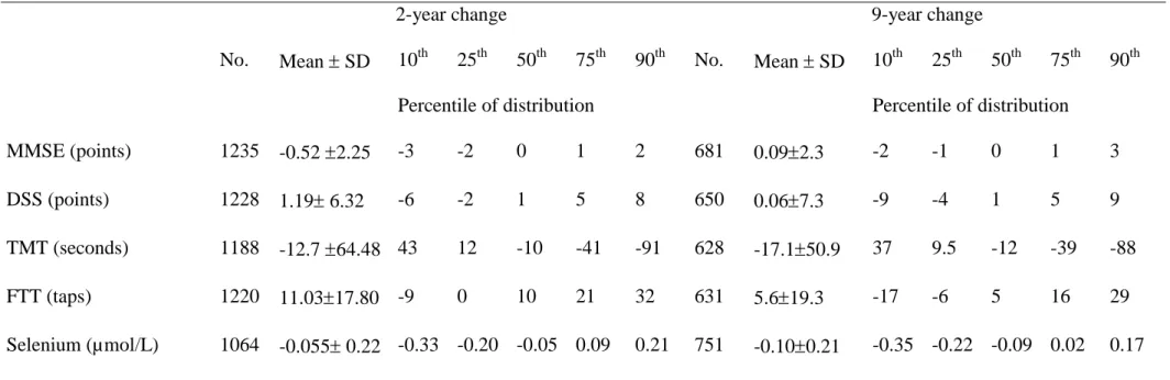
In the EVA study population, baseline mean plasma selenium level ( standard deviation) was 1.09 μmol/L ( 0.20 μmol/L). We observed a decrease of plasma selenium over the entire follow-up period. Means of selenium declined –0.055 (SD0.20) μmol/L at 2 years and –0.096 (SD+0.21) µmol/L at the 9-year follow-up (Table 2). We noted an increase in cognitive performances means
(Table 2). To assess the short- and long-term association between selenium and cognitive changes,
we performed linear regression models between 2-year and 9-year selenium change and cognitive
changes during 2,4, 6 and 9-year follow-up (Table 3). Associations were adjusted for age, sex,
education and selenium level at baseline. We showed that 2-year selenium change was not associated
with cognitive change during 2, 4, 6 or 9-year follow-up, for any of the cognitive tests. Nine-year
selenium change was associated with 9-year cognitive change for MMSE, but not for the other
cognitive tests.
Mixed Models
Factors associated with cognitive change and selenium change
To take into account all cognitive changes during the follow-up and the within-subject
variability, we performed mixed linear models. We used these models mainly to determine which
factors were associated with cognitive change by restricting analyses to MMSE test. Cognitive
change was associated with time of follow-up (=0.09; 95% CI =0.07 to 0.11), but not with age at inclusion. The interaction term between time of follow-up and age at inclusion was not significant,
suggesting that the effect of time on cognitive decline was no more pronounced in older subjects than
in younger ones for the narrow age range studied (60-70 years at baseline). Diabetes and
hypertension were modestly associated with a higher decrease of cognitive performances (=0.31 [0.004 to 0.62] and =0.14 [-0.02 to 0.30], respectively). Sex, education, tobacco status, alcohol
consumption, BMI, history of cardiovascular diseases, dyslipidemia and baseline plasma selenium
level were not associated with cognitive performances change during the follow-up (data not shown).
During the follow-up, occurrence of cardiovascular events, as well as obesity, were associated with
greater declines in plasma selenium (data not shown). No association was found between plasma
selenium and other factors.
Association between 2-year selenium change and 9-year cognitive change
Using crude mixed linear models, modelling cognitive change (2, 4, 6, 8 and 9-year) by the
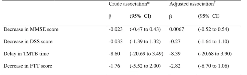
first 2-year selenium change, there was no association with any of the four cognitive tests (Table 4). These results were confirmed in the multivariate models; (for the MMSE, β=0.01 (-0.52 to 0.50); for DSS, β= -0.27 ( -1.6 to 1.1); for TMTB, β= -8.4 (-20.7 to 3.9); for FTT, β= -2.8 (-6.7 to 1.1).
To understand the relationship between cognitive decline and 2-year selenium change, we
used a mixed logistic model with Gaussian random effects (Table 6). In these models, cognitive
decline was considered as a dichotomous variable. After controlling for time and plasma selenium
level at baseline, the short-term plasma selenium decrease was only weakly associated with cognitive
decline. There results were not changed after controlling for potential confounding factors (Table 6).
Association between selenium change and cognitive change during the 9 years of follow-up
For these analyses, we considered cognitive and selenium variables simultaneously measured
at inclusion, 2 years and 9 years. In these mixed linear models, cognitive performances (as
continuous variables) were related to selenium levels and time of follow-up. Interaction terms
between selenium and time of follow-up expressed the change of selenium according to cognitive
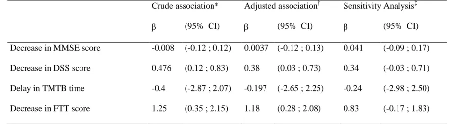
change during follow-up (Table 5). In the crude analyses, selenium changes were associated with
DSS and FTT but not with TMTB or MMSE. Analyses adjusted for time, sex, education, diabetes,
However, for these models, to be applicable distributions of cognitive variables should be Gaussian;
this condition was met for DSS and FTT but not for MMSE and TMTB. In sensitivity analysis, we
applied the same models limited to subjects who had all three selenium measurements and cognitive
evaluation (inclusion, 2 and 9-year). Results were quite similar, although with lower power because
analyses were carried out on 570 subjects instead of the initial 1371.
Associations between selenium change and cognitive decline were studied by using a mixed
logistic model with Gaussian random effect (Table 6). After controlling for time, sex, education,
plasma selenium level at baseline, diabetes, hypertension, dyslipidemia and history of cardiovascular
diseases, we observed that probability of cognitive decline increased with the decrease of plasma
selenium change over time. Among subjects who had a decrease of their plasma selenium levels, the
higher the decrease of plasma selenium, the higher probability of cognitive decline. Among subjects
who had an increase of their plasma selenium levels, the probability of cognitive decline was higher
in subjects with the smallest selenium increase. Plasma selenium change was associated with MMSE
decline at the 2-points cut-off (OR=2.31; CI =1.12 to 4.77)) but not at the 3-points cut-off (1.41; 0.52
to 3.83)). We observed an association for the DSS at the 25th cut-off (2.60; 1.22 to 5.57)), and for
TMTB at the 10th percentile (3.18 (1.14 to 8.88)). For FTT, decline increased when selenium
decreased for the two cut-offs we considered (for the 10th percentile, 2.40 [1.21 to 4.77] and for the
25th percentile, 4.33 [1.60 to 11.72]).
DISCUSSION
The EVA study is a longitudinal study of cognitive and vascular aging in an elderly general
population. The study provided plasma selenium measurements at 3 points in time, and assessments
of cognitive functioning at 6 points in time. Declines in selenium were associated with cognitive
controlling for potential confounders. There were weaker associations between short-term selenium
change and cognitive changes during the follow-up.
The EVA cohort is a free-living community dwelling population with normal
neuropsychological test results at inclusion and a selenium status similar to that recently reported in
French adults.21 All co-variables for every model were updated at each waves of the study, except
BMI and alcohol consumption for which we only took into account the baseline data. Alcohol
consumption use may be associated with both cognitive decline and changes in selenium. However,
correlations between alcohol consumption of the first three waves were very strong and we made the
assumption regarding these correlations and the change in tobacco status (which is linked to alcohol
behavior) that, in the EVA cohort, alcohol consumption remained constant during the follow-up.22
In epidemiologic studies of cognitive decline, biological status is generally considered as a
static exposure associated with health changes. In this study, we extended our approach to dynamic
exposure, considering selenium changes during a time period rather than as a single measurement.
However, this approach requires appropriate statistical analyses. Classical linear regressions are well
suited to study selenium change and cognitive change between two given moments. For our study,
however, this method is too restrictive when considering our initial objective, the wealth and
complexity of our data and sample selection. Mixed models allow us to simultaneously take into
account measurements of the whole follow-up, and results are thus more powerful because sample
selection is not as strong. We performed mixed linear or logistic models considering cognitive
change as continuous or categorical . As cognitive variables are not strictly normally distributed,
conditions for computing mixed linear models are not optimal. The associations between selenium
The various statistical models applied to our data offer both advantages and limits. However
we think the mixed logistic model is best suited to study long-term cognitive decline assessed by
neuropsychological tests in a healthy elderly population.
The study’s obvious main limit is sample selection throughout follow-up. Subjects who did
not complete the whole follow-up had the lowest cognitive performances at inclusion, and we could
hypothesize that these subjects also had the highest cognitive decline during the follow-up. However,
they did not differ with regard to selenium level at baseline or plasma selenium decrease for the first
two years. Moreover, with analyses limited to subjects who had the three plasma selenium
measurements and complete cognitive assessment (n=570), results were quite similar even though
power was reduced. Finally, a relationship between plasma selenium and cognition change is found
in subjects with weak cognitive decline. Sample selection may limit interpretation of our results, but
should not affect the relationship itself.
In previous works conducted on the same cohort at baseline 14 and after the four-year
follow-up, 11 we showed that, at baseline, plasma selenium and cognitive functions were positively
associated. Baseline selenium plasma was lower in the subjects who develop cognitive decline as
measured by 4-year decline of 3 points in MMSE scores. We therefore hypothesize that plasma
selenium may be one of the factors associated with cognitive decline. This hypothesis is supported by
the role of selenium in redox reactions and in thyroid hormone synthesis, both being involved in
cognitive impairment.23,24 In addition, the brain contains large amounts of selenium and represents a
target organ with respect to selenium supply and retention.5 So far, genes for at least 25
selenoproteins 25 have been identified in the human genome and most of them are expressed in the
brain (although their specific roles for normal brain functions and neurological diseases have not
We found an association between plasma selenium change and 9-year cognitive decline only
with 9-year selenium change, not 2-year change. These results are in agreement with the effect of
antioxidant supplementation observed in some long-term studies 13,26 and the lack of effect after a
6-month period in mild Alzheimer disease’s patients.27 Nevertheless, these intervention studies were
conducted with multi-vitamin and mineral supplements, thus making it impossible to identify a
specific selenium effect. It is also possible that health changes, including poor cognitive function,
may lead to dietary or behavioral changes that affect selenium levels.
Taken as a whole, our results suggest that plasma selenium decrease is associated with
cognitive decline. The real importance of selenium in brain and the capacity of brain to manage
selenium depletion is just beginning to be explored.5 Molecular biology has recently contributed to
the recognition of selenium and selenium -dependent enzymes as modulators of brain function. Our
results are in agreement with this approach even if a recent experimental work on knock-out mice
suggest that plasma selenium cannot reflect brain selenium status due to the maintenance of brain
selenoprotein P synthesis.28
Our results, together with information on involvement of selenoproteins in brain functions,
support possible relationships between selenium status and neuropsychological functions in aging
people. In this context, the preventive effect of selenium supplementation at a nutritional level needs
to be evaluated with large-scale studies. This dynamic approach could shed new lights on the
ACKNOWLEDGMENTS
N. Tasnime Akbaraly was supported by a grant from the French Alzheimer’s disease Association The EVA study was carried out under an agreement between INSERM and the Merck, Sharp and
REFERENCES
1. Finkel T, Holbrook NJ. Oxidants, oxidative stress and the biology of ageing. Nature 2000;408(6809):239-247.
2. Harman D. Aging; a theory based on free radical and radiation chemistry. J.Gerontol. 1956;2:298-300. 3. Chen J, Berry MJ. Selenium and selenoproteins in the brain and brain diseases. J Neurochem
2003;86(1):1-12.
4. Rayman MP. The importance of selenium to human health. Lancet 2000;356:233-241.
5. Schweizer U, Schomburg L, Savaskan NE. The neurobiology of selenium: lessons from transgenic mice. J Nutr 2004;134(4):707-10.
6. Mostert V. Selenoprotein P: properties, functions, and regulation. Arch Biochem Biophys 2000;376(2):433-8.
7. Berr C, Nicole A, Godin J, Ceballos-Picot I, Thevenin M, Dartigues JF, Alperovitch A. Selenium and oxygen-metabolizing enzymes in elderly community residents: a pilot epidemiological study. J Am Geriatr Soc 1993;41(2):143-8.
8. Ducros V, Faure P, Ferry M, Couzy F, Biajoux I, Favier A. The sizes of the exchangeable pools of selenium in elderly women and their relation to institutionalization. Br J Nutr 1997;78(3):379-96. 9. Olivieri O, Stanzial AM, Girelli D, Trevisan MT, Guarini P, Terzi M, Caffi S, Fontana F, Casaril M,
Ferrari S, et al. Selenium status, fatty acids, vitamins A and E, and aging: the Nove Study. Am J Clin Nutr 1994;60(4):510-7.
10. Savarino L, Granchi D, Ciapetti G, Cenni E, Ravaglia G, Forti P, Maioli F, Mattioli R. Serum concentrations of zinc and selenium in elderly people: results in healthy nonagenarians/centenarians. Exp Gerontol 2001;36(2):327-39.
11. Berr C, Balansard B, Arnaud J, Roussel AM, Alperovitch A. Cognitive decline is associated with systemic oxidative stress: the EVA study. Etude du Vieillissement Arteriel. J Am Geriatr Soc 2000;48(10):1285-91.
12. Chandra RK. Effect of vitamin and trace-element supplementation on cognitive function in elderly subjects. Nutrition 2001;17(9):709-12.
13. Gray SL, Hanlon JT, Landerman LR, Artz M, Schmader KE, Fillenbaum GG. Is antioxidant use protective of cognitive function in the community-dwelling elderly? Am J Geriatr Pharmacother 2003;1(1):3-10.
14. Berr C, Coudray C, Bonithon-kopp C, Roussel AM, Mainard F, Alperovitch A, group atEs. Demographic and cardiovascular risk factors in relation to antioxidant status; the EVA study. Int.J.Vitam.Nutr.R 1998;68:26-35.
15. Folstein M, Anthony JC, Parhad I, Duffy B, Gruenberg EM. The meaning of cognitive impairment in the elderly. J.Am.Geriatr.Soc. 1985;33:228-235:228-235.
16. Robins Wahlin TB, Backman L, Wahlin A, Winblad B. Trail Making Test performance in a community-based sample of healthy very old adults: effects of age on completion time, but not on accuracy. Arch Gerontol Geriatr 1996;22(1):87-102.
17. Wechsler D. The Wechsler Adult Intelligence Scale-Revised. New-York, 1981.
18. Arnaud J, Prual A, Preziosi P, Favier A, Hercberg S. Selenium determination in human milk in Niger: influence of maternal status. J Trace Elem Electrolytes Health Dis 1993;7(4):199-204.
19. Diggle P, Heagerty P, Liang K-Y, Zeger S. Analysis of Longitudinal Data. Oxford stastistical science series. 2nd edition ed. United States: Oxford University Press, 2002.
20. Akbaraly NT, Arnaud J, Hininger-Favier I, Gourlet V, Roussel AM, Berr C. Selenium and mortality in the elderly: results from the EVA study. Clin Chem 2005;51(11):2117-23.
21. Galan P, Viteri FE, Bertrais S, Czernichow S, Faure H, Arnaud J, Ruffieux D, Chenal S, Arnault N, Favier A, Roussel AM, Hercberg S. Serum concentrations of beta-carotene, vitamins C and E, zinc and selenium are influenced by sex, age, diet, smoking status, alcohol consumption and corpulence in a general French adult population. Eur J Clin Nutr 2005;59(10):1181-90.
22. Dufouil C, Tzourio C, Brayne C, Berr C, Amouyel P, Alperovitch A. Influence of apolipoprotein E genotype on the risk of cognitive deterioration in moderate drinkers and smokers. Epidemiology 2000;11(3):280-4.
23. Basun H, Forssell LG, Wetterberg L, Winblad B. Metals and trace elements in plasma and
cerebrospinal fluid in normal aging and Alzheimer's disease. J Neural Transm Park Dis Dement Sect 1991;3(4):231-58.
24. Smorgon C, Mari E, Atti AR, Dalla Nora E, Zamboni PF, Calzoni F, Passaro A, Fellin R. Trace elements and cognitive impairment: an elderly cohort study. Arch Gerontol Geriatr Suppl 2004(9):393-402.
25. Kryukov GV, Castellano S, Novoselov SV, Lobanov AV, Zehtab O, Guigo R, Gladyshev VN. Characterization of mammalian selenoproteomes. Science 2003;300(5624):1439-43.
26. Mendelsohn AB, Belle SH, Stoehr GP, Ganguli M. Use of antioxidant supplements and its association with cognitive function in a rural elderly cohort: the MoVIES Project. Monongahela Valley
Independent Elders Survey. Am J Epidemiol 1998;148(1):38-44.
27. Planas M, Conde M, Audivert S, Perez-Portabella C, Burgos R, Chacon P, Rossello J, Boada M, Tarraga LL. Micronutrient supplementation in mild Alzheimer disease patients. Clin Nutr 2004;23(2):265-72.
28. Schweizer U, Streckfuss F, Pelt P, Carlson BA, Hatfield DL, Kohrle J, Schomburg L. Hepatically derived selenoprotein P is a key factor for kidney but not for brain selenium supply. Biochem J 2005;386(Pt 2):221-6.
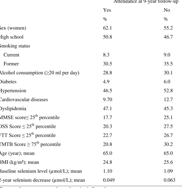
Table 1: Baseline characteristics* of subjects according to 9-year follow-up status†
Attendance at 9-year follow-up
Yes No % % Sex (women) 62.1 55.2 High school 50.8 46.7 Smoking status Current 8.3 9.0 Former 30.5 35.5
Alcohol consumption (≥20 ml per day) 28.8 30.1
Diabetes 4.9 6.0 Hypertension 46.5 52.8 Cardiovascular diseases 9.70 12.7 Dyslipidemia 47.1 45.3 MMSE score≤ 25th percentile 17.7 25.1 DSS Score ≤ 25th percentile 20.3 27.5 FTT Score ≤ 25th percentile 22.7 26.7 TMTB Score ≥ 75th percentile 20.8 30.2
Age (year); mean 65.0 65.0
BMI (kg/m²); mean 24.8 25.6
Baseline selenium level (µmol/L); mean 1.10 1.09 2-year selenium decrease (µmol/L); mean 0.049 0.063 * Expressed as percentage unless otherwise indicated
† Number of subjects for whom information was available on specific baseline variables: among those followed up at 9 years, n=677-702 (except the 2 selenium variables, n-607); among those not followed up, 653-687 (except baseline selenium, n= 684 and 2-year decrease, n= 457)
Table 2: Means and percentile distributions of selenium and cognitive change from baseline
2-year change 9-year change
No. Mean SD 10th 25th 50th 75th 90th No. Mean SD 10th 25th 50th 75th 90th Percentile of distribution Percentile of distribution
MMSE (points) 1235 -0.52 2.25 -3 -2 0 1 2 681 0.092.3 -2 -1 0 1 3 DSS (points) 1228 1.19 6.32 -6 -2 1 5 8 650 0.067.3 -9 -4 1 5 9 TMT (seconds) 1188 -12.7 64.48 43 12 -10 -41 -91 628 -17.150.9 37 9.5 -12 -39 -88 FTT (taps) 1220 11.0317.80 -9 0 10 21 32 631 5.619.3 -17 -6 5 16 29 Selenium (µmol/L) 1064 -0.055 0.22 -0.33 -0.20 -0.05 0.09 0.21 751 -0.100.21 -0.35 -0.22 -0.09 0.02 0.17
Table 3: Linear regressions models for the association between 2, 4, 6 and 9-year cognitive change and 2 and 9-year selenium change. Results are
expressed as linear regression coefficient with 95% confidence intervals. Cognitive change from Baseline
2 years 4 years 6 years 9 years
No. (95% CI) No. (95% CI) No. (95% CI) No. (95% CI) Selenium Change 2-year MMSE 1034 -0.12 (-0.28 to 0.04) 975 -0.04 (-0.18 to 0.10) 867 -0.028 (-0.18 to 0.12) 583 0.2 (-0.02 to 0.42) DSS 1027 -0.02 (-0.45 to 0.41) 968 -0.004 (-0.44 to 0.43) 859 -0.061 (-0.55 to 0.43) 555 -0.05 (-0.70 to 0.60) TMTB 995 -2.41 (-7.04 to 2.22) 937 -1.9 (-5.58 to 1.78) 832 -1.92 (-5.51 to 1.67) 541 -2.18 (-6.71 to 2.35) FTT 1021 -0.49 (-1.69 to 0.71) 963 -0.91 (-2.13 to 0.31) 853 -0.7 (-1.95 to 0.55) 546 0.74 (-1.02 to 2.50) 9-year MMSE 570 0.38 (0.14 to 0.62) DSS 542 0.53 (-0.20 to 1.26) TMTB 524 -3.2 (-8.61 to 2.21) FTT 533 1.6 (-0.40 to 3.60)
Table 4: Association between selenium change during the first 2 years and cognitive change up to 9 years of follow-up: results of mixed linear
models
For these analyses, we used the plasma selenium measurements at inclusion and at 2-year and the cognitive evaluations assessed at
inclusion, 2, 4, 6 and 9 years.
* Associations adjusted for time and baseline plasma selenium level
†Association adjusted for time, sex, education, baseline plasma selenium level, diabetes, dyslipidemia, hypertension and history of cardiovascular diseases.
Crude association* Adjusted association†
(95% CI) (95% CI) Decrease in MMSE score -0.023 (-0.47 to 0.43) 0.0067 (-0.52 to 0.54)
Decrease in DSS score -0.033 (-1.39 to 1.32) -0.27 (-1.64 to 1.10)
Delay in TMTB time -8.60 (-20.69 to 3.49) -8.39 (-20.68 to 3.90)
Table 5: Associations between plasma selenium changes and cognitive changes during the 9 years of follow-up: Results of mixed linear models.
For these analyses, we used the plasma selenium measurements and cognitive evaluation assessments at inclusion, 2 years and 9 years
* Associations adjusted for time and baseline plasma selenium level
†Association adjusted for time, sex, education, baseline plasma selenium level, diabetes, dyslipidemia, hypertension and history of cardiovascular diseases.
‡Sensitivity Analysis carried on the 570 subjects who had all three plasma selenium measurements and cognitive evaluations. Crude association* Adjusted association† Sensitivity Analysis‡
(95% CI) (95% CI) (95% CI) Decrease in MMSE score -0.008 (-0.12 ; 0.12) 0.0037 (-0.12 ; 0.13) 0.041 (-0.09 ; 0.17)
Decrease in DSS score 0.476 (0.12 ; 0.83) 0.38 (0.03 ; 0.73) 0.34 (-0.03 ; 0.71)
Delay in TMTB time -0.4 (-2.87 ; 2.07) -0.197 (-2.65 ; 2.25) -0.24 (-2.98 ; 2.50)
Table 6: Associations between 2-year or 9-year plasma selenium change decrease and 9 year
cognitive decline. Results of mixed logistic models expressed by Odds Ratios and 95%
confidence intervals of cognitive decline according to the decrease of selenium change*
*For each subject and at each wave, the cognitive score differences between the wave i and
baseline were calculated. Cut-off points < 25th of the distributions of the mean of these
differences corresponded to a difference of -2 points for MMSE, -3 points for DSS, +2 taps
for FTT and 7.8 seconds for TMTB. Cut-off points < 25th of the distributions of the mean of
these differences corresponded to a difference of -3 points for MMSE, -6 points for DSS, -7
taps for FTT and 32.4 seconds for TMTB
Cut-off level < 25th < 10th OR† (95% CI) OR† (95% CI) Selenium decrease 2-year MMSE 1.07 0.59 ; 1.95 1.04 0.49 ; 2.21 DSS 1.56 0.74 ; 3.28 1.51 0.59 ; 3.87 TMTB 1.32 0.64 ; 2.73 1.35 0.51 ; 3.54 FTT 0.91 0.47 ; 1.74 0.69 0.30 ; 1.59 9-year MMSE 2.31 1.12; 4.77 1.41 0.52 ; 3.83 DSS 2.60 1.22 ; 5.57 2.22 0.78 ; 6.33 TMTB 1.97 0.95 ; 4.11 3.18 1.14 ; 8.88 FTT 2.40 1.21 ; 4.77 4.33 1.60 ; 11.72
†Adjusted on time, sex, education, plasma selenium level at baseline, diabetes, hypertension, dyslipidemia and history of cardiovascular diseases