HAL Id: inserm-00913076
https://www.hal.inserm.fr/inserm-00913076
Submitted on 22 May 2014HAL is a multi-disciplinary open access archive for the deposit and dissemination of sci-entific research documents, whether they are pub-lished or not. The documents may come from teaching and research institutions in France or abroad, or from public or private research centers.
L’archive ouverte pluridisciplinaire HAL, est destinée au dépôt et à la diffusion de documents scientifiques de niveau recherche, publiés ou non, émanant des établissements d’enseignement et de recherche français ou étrangers, des laboratoires publics ou privés.
Leptin and regulatory T-lymphocytes in idiopathic
pulmonary arterial hypertension.
Alice Huertas, Ly Tu, Natalia Gambaryan, Barbara Girerd, Frédéric Perros,
David Montani, Dominique Fabre, Elie Fadel, Saadia Eddahibi, Sylvia
Cohen-Kaminsky, et al.
To cite this version:
Alice Huertas, Ly Tu, Natalia Gambaryan, Barbara Girerd, Frédéric Perros, et al.. Leptin and reg-ulatory T-lymphocytes in idiopathic pulmonary arterial hypertension.. European Respiratory Jour-nal, European Respiratory Society, 2012, 40 (4), pp.895-904. �10.1183/09031936.00159911�. �inserm-00913076�
Leptin and regulatory T lymphocytes in idiopathic pulmonary arterial hypertension
Alice Huertas1,2,3, Ly Tu1,2,3, Natalia Gambaryan1,2,3, Barbara Girerd1,2,3, Frédéric
Perros1,2,3, David Montani1,2,3, Dominique Fabre1,2,3, Elie Fadel1,2,3, Saadia Eddahibi1,2,3,
Sylvia Cohen-Kaminsky1,2,3, Christophe Guignabert1,2,3, Marc Humbert1,2,3
1 Université Paris-Sud, Faculté de Médecine, Kremlin-Bicêtre, F-94276;
2 Centre National de Référence de l’Hypertension Pulmonaire Sévère, Service de
Pneumologie et Réanimation Respiratoire, Hôpital Antoine Béclère, AP-HP, Clamart, F-92140;
3 INSERM U999 Hypertension Artérielle Pulmonaire: Physiopathologie et Innovation
Thérapeutique, LabEX LERMIT Laboratory of Excellence in Research on Medication and Innovative Therapeutics (LERMIT), Centre Chirurgical Marie Lannelongue, Le Plessis-Robinson, F-92350
Corresponding author: Prof. Marc Humbert
Service de Pneumologie et Réanimation Respiratoire, Centre National de Référence de l'Hypertension Pulmonaire Sévère, Hôpital Antoine Béclère, Assistance Publique Hôpitaux de Paris, Université Paris-Sud 11
157, rue de la Porte de Trivaux, 92140 Clamart, France Tel: +33 1 45 37 47 72, Fax: +33 1 46 30 38 24
Abstract
Immune mechanisms and autoimmunity seem to play a significant role in idiopathic pulmonary arterial hypertension (IPAH) genesis and/or progression but the pathophysiology is still unclear. Recent evidence has demonstrated a detrimental involvement of leptin in promoting various autoimmune diseases by controlling regulatory T lymphocytes. Despite this knowledge, the role of leptin in IPAH is currently unknown. We hypothesized that leptin, synthesized by dysfunctional pulmonary endothelium, might play a role in the immunopathogenesis of IPAH by regulating circulating regulatory T lymphocytes function. First, we collected serum and regulatory T lymphocytes from controls, IPAH and scleroderma-associated (SSc-PAH) patients; secondly, we recovered tissue samples and cultured endothelial cells after surgery or transplantation in controls and IPAH patients, respectively. Our findings indicate that serum leptin was higher in IPAH and SSc-PAH patients compared to controls. Circulating regulatory T lymphocytes number was comparable in all groups, the percentage of those expressing leptin receptor was higher in IPAH and SSc-PAH compared to controls, whereas their function was reduced in IPAH and SSc-PAH patients compared to controls in a leptin-dependent manner. Furthermore, endothelial cells from IPAH patients synthesized more leptin than controls. Our data suggest that endothelial-derived leptin may play a role in the immunopathogenesis of IPAH.
Key words: dysimmunity, endothelial dysfunction, leptin, pulmonary arterial hypertension, regulatory T lymphocytes
Introduction
Idiopathic pulmonary arterial hypertension (IPAH) corresponds to precapillary pulmonary hypertension in which there is neither a family history of the disease, nor an identified risk factor and is defined as a resting mean pulmonary arterial pressure mPAP 5 mm(g with a normal pulmonary artery wedge pressure 5 mm(g 1. Although the pathophysiology of IPAH has been extensively studied in the past decades and several new pathways have been identified, the etiology of this disease is still not clearly understood. It is now well established that inflammation plays an important role in IPAH 2-6 and increasing data are also supporting the hypothesis that immunologic disorders could be present in IPAH patients: circulating autoantibodies have been detected 7-8 and recent evidence are indicating that regulatory T cells (Treg) could play a role in PAH 9. Despite these findings, little is known about the exact role of inflammation and dysimmunity in the development of IPAH and it remains unclear how immune mechanisms contribute to the pathogenesis of IPAH.
Recent evidence has demonstrated a detrimental involvement of leptin, a cytokine-like hormone mainly secreted by adipocytes, in promoting the pathogenesis of various autoimmune diseases 10-11. Although leptin, product of the obese (ob) gene, has been discovered as the appetite regulator, it regulates a wide range of physiological functions 10-13. Interestingly, it has also been shown that leptin can promote chronic autoimmune disorders by regulating Treg onset and/or function 14. Intriguingly, little is known about the pulmonary effects of leptin, and in particular its inflammatory and/or immunologic role in IPAH.
Treg are T lymphocytes known to dampen autoreactive responses and are able to delay the onset and progression of autoimmune disorders: reduced frequency of Treg
and/or defective suppressor function have been observed in these diseases 14-15. So far, little is known about the role of leptin on the lung and in particular about its effects on the pulmonary vessels.
IPAH is characterized by small-sized pulmonary arteries/arterioles in which there is intimal hyperplasia with medial and adventitial hypertrophy, hyperplasia and fibrosis. Cells from the vessel wall are known to play an important role in the pathogenesis of IPAH. In particular, pulmonary endothelial cells (P-EC) represent a critical cell type. Endothelial dysfunction is characterized by an altered vasoconstriction/vasodilatation balance and disorganized pro-proliferative and apoptotic-resistant phenotype that can lead to the formation of plexiform lesions mostly described in IPAH patients 16.
We hypothesized that leptin, synthesized by dysfunctional P-EC, might play a role in the immunopathology of IPAH by regulating circulating Treg function. We addressed this issue using freshly recovered circulating Treg from IPAH patients. Our findings indicate that Treg function is reduced in IPAH patients compared to controls in a leptin-dependent manner. Furthermore, using cultured cells recovered after lung transplantation in IPAH patients, we demonstrated that there is a major increase in leptin synthesis from IPAH P-EC compared to controls. Taken together, our data suggest that leptin could derive from endothelial cells and may play a role in the immunopathogenesis and/or progression of IPAH.
Methods
Blood samples were collected in patients with IPAH and in scleroderma-associated PAH (SSc-PAH) during usual follow-up and in control subjects (Table 1). All patients were treated with PAH specific treatments. Inclusion criteria were age above 18 years and PAH diagnosis confirmed by right-heart catheterization with a stable clinical and heamodynamic status for the last 3 months. Exclusion criteria were an heritable form of PAH, anaemia, thyroid dysfunction, diabetes, metabolic syndrome, and immunosuppressive or corticosteroid therapies in the last 6 months for SSc-PAH.. Characteristics at diagnosis and follow-up were stored in the Registry of the French Network of Pulmonary Hypertension set up in agreement with French bioethics laws (French Commission Nationale de l'Informatique et des Libertés) and all patients gave their written informed consent.
Leptin, leptin receptor and pro-inflammatory measurements
Serum samples were collected and stored at -80°C. For the measurement of leptin, C-reactive protein (CRP), tumor necrosis factor (TNF)-, monocyte chemoattractant protein (MCP)-1, interleukin-6 and 1, human AlphaLISA or ELISAs kits were used accordingly to manufacturer’s instructions PerkinElmer and R&D Systems.). Circulating soluble leptin receptor (sObR) levels were measured using ELISAs (as recommended by the manufacturer; R&D Systems).
Flow cytometry analysis
After blood samples withdrawal, peripheral blood mononuclear cells (PBMCs) from IPAH, SSc-PAH patients and controls were obtained by standard Ficoll gradient centrifugation. The cells were carefully washed with PBS and resuspended in a staining buffer containing 10% of human serum. The cells were then fluorescently labeled with
the following monoclonal antibodies: anti-CD4 conjugated with either Alexa 488 (Becton Dickinson) or Pacific Blue (BioLegend); anti-CD25 conjugated with Alexa 647 (BioLegend); CD127 conjugated with PerCp-Cy5.5 (Becton Dickinson); and anti-FoxP3 conjugated with Pacific Blue (BioLegend), under non-permeabilized or permeabilized conditions (IntraPrep, following manufacturer’s instructions, Beckman Coulter), to enable surface or intracellular staining, respectively. Flow cytometry gating conditions and the mean fluorescence intensity (MFI) were set and normalized, respectively, against isotype- and fluorophore-matched non-immune IgGs.
It is now well known that CD127 expression inversely correlates with FoxP3 activation 17. To test whether Treg analysis and cell count were similar using these different markers, we randomly selected 5 subjects from each group and compared the two Treg phenotypes , ie CD4+CD25+CD127lowFoxP3+, and CD4+CD25+CD127low (the subgroup characteristics are presented as online supplemental data). The cell count was similar by using these 2 different surface markers (Figure 4C and Table 2). Based on these preliminary tests and to preserve the intracellular space, we avoided PBMCs and defined Treg as CD4+CD25+CD127low (Figure 3A). Treg cell count was expressed as the
percentage of total CD4+ cells.
In order to evaluate the ObR expression, PBMCs were also stained under non-permeabilized conditions with a monoclonal antibody against ObR fluorescently conjugated with PE (R&D Systems). Treg expressing ObR were defined as CD4+CD25+CD127lowObR+ and were quantified as the percentage of CD4+CD25+CD127low.
To determine the functional status of the Treg expressing ObR, we stained them under permeabilized conditions with a monoclonal antibody against phosphorylated STAT3 (pSTAT3) conjugated with Alexa 488 (Cell Signaling Technology).
To assess the effect of leptin on Treg functional status, we stimulated freshly isolated PBMCs from healthy controls with recombinant human leptin at a concentration of 250ng/ml for 5 minutes (R&D Systems, 18). Then, we stained and analyzed the Treg and pSTAT3 as described above.
Flow cytometry data were acquired with a flow cytometer (MACSQuant) and analyzed by FlowJo software program (Tree Star, Inc).
Human pulmonary tissues and immunohistostaining
Lung specimens were obtained at the time of lung transplantation from patients with IPAH (n=10), at the Marie Lannelongue Hospital, Le Plessis-Robinson, France. Control-lung specimens were obtained from patients without any evidence of pulmonary vascular disease who underwent lobectomy or pneumonectomy for localized lung cancer (n=15). Lung specimens were fixed in 4% paraformaldehyde and embedded in Paraffin. 5-μm sections were dewaxed and rehydrated progressively. Citrate buffer pH6 was used for antigen retrieval, and endogenous peroxidase was quenched with hydrogen peroxide. Sections were blocked with 5% of bovine serum albumin and incubated overnight with the following antibodies: polyclonal antibody against leptin (dilution: 1/100; Santa Cruz Biotechnologies, Santa Cruz, CA, USA) and monoclonal antibody against leptin receptor (dilution: 1/50; R&D systems Europe, Lille, France). Biotin/Streptavidin-peroxidase systems, DAB substrate was used for relevation (Universal LSAB HRP Kit, Dako, Trappes, France). Nuclei were counterstained with hematoxylin and mounted. Controls used for these antibodies included omission of the primary antibody and substitution of the primary antibody by isotype control.
Human microvascular P-EC isolated from lung tissue fragments using immunomagnetic purification were cultured as previously described 19. To quantify leptin in conditioned media, the isolated cells were seeded on 6-well plates at a density of 1×10⁵ cells/well. After 24 hours, cells were washed twice with PBS then incubated for
24h in serum-free MCDB131 (without the addition of growth factors). Leptin was measured in conditioned media using ELISAs (as recommended by the manufacturer; R&D Systems).
Statistical analysis
Results are expressed as mean ± sem. A p<0.05 level of statistical significance was used for all analyses. The Shapiro-Wilk test was used to ensure that data had a normal distribution. All between-groups comparisons were assessed using One Way ANOVA; post-hoc analysis of significant variables was performed using Tukey Test with all pairwise multiple comparisons. Differences between two selected groups (controls and IPAH) were compared using unpaired t-test. Pearson correlations were used to establish associations between the dependent variables (i.e., leptin, ObR expression on Treg) and relevant independent variables. All statistical procedures were carried out using GraphPad Prism version 5.0 (GraphPad Software Inc.)
Results
Leptin and pro-inflammatory cytokines serum concentration
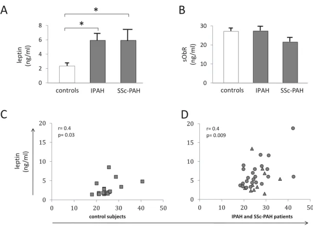
To investigate the role of leptin in PAH pathogenesis, we first quantified serum leptin concentration and found that both IPAH and SSc-PAH patients had a significantly
higher leptinemia than controls. No difference was found between IPAH and SSc-PAH (Figure 1A and Table 2).
In order to compare the relative free leptin index and the leptin bioavailability in these 3 groups of subjects, we measured sObR levels. We found a similar level in the 3 groups without any statistically significant difference (Figure 1B and Table 2). These data indicate that circulating active leptin levels are increased in both IPAH and SSc-PAH. Correlations between serum leptin levels and body mass index (BMI) in controls and PAH patients are shown in Figure 1C-D. Furthermore, no correlation was found between circulating leptin levels and disease severity expressed as haemodymic and/or functional parameters. Since leptin has been shown to act as a key player in inflammatory conditions, we tested whether serum leptin concentration in PAH correlated with serum pro-inflammatory cytokines. CRP and TNF- concentrations were normal in all groups, without any statistically significant difference among the groups (Figure 2A and Table 2). MCP-1, IL-6 and IL-1 were significantly increased in IPAH and SSc-PAH patients compared to controls (Table 1). Furthermore, there was no correlation between the pro-inflammatory cytokines and leptin levels (Figure 2B-C).
These data indicate that in PAH patients, in IPAH and in SSc-PAH, serum leptin levels are increased independently of pro-inflammatory cytokines.
ObR expression on Treg cell membrane
Since our findings indicated a possible role of leptin in PAH pathogenesis, we analyzed the expression of its receptor ObR on Treg. After withdrawing peripheral venous blood samples from controls and PAH patients, we first selected Treg among the fluorescently tagged PBMCs by flow cytometry analysis.
We selected Treg and analyzed the expression of ObR on their membrane (Figure 3A). Flow cytometry analysis revealed that ObR expression was markedly increased in PAH patients, as compared to controls (Figure 3B-C and Table 2), either when expressed as percentage of Treg cells (Figure 3C and Table 2), or as absolute numbers (Figure 3D and Table 2). ObR expression was similar in IPAH and SSc-PAH. Interestingly, the expression of ObR on Treg cell membrane did not correlate with BMI or serum leptin levels (Figure 3E-H).
Taken altogether, these findings demonstrate that ObR expression is markedly increased in IPAH patients compared to controls and to a similar extent as in SSc-PAH. Interestingly, the level of ObR expression is independent of the patients’ BM) or serum leptin levels.
Circulating Treg levels
To further explore the role of leptin on Treg in PAH, we first examined the Treg cell count. Flow cytometry analysis revealed a normal Treg cell count in all groups, controls and PAH patients. We expressed the Treg as percentage of CD4+ cells as well as
in absolute numbers and also found a normal Treg cell count (Figure 4A-B and Table 2). These data indicate that leptin does not seem to influence Treg numbers in PAH patients.
Functional status of Treg
Our next objective was to determine the extent to which leptin could play a role on Treg function. As readout of ObR activity, we measured pSTAT3 amounts, because STAT3 is known to participate in the intracellular signaling pathways of ObR 11. After Treg permeabilization, we quantified pSTAT3 by flow cytometry (Figure 5A).
Interestingly, the number of Treg-pSTAT3+ was markedly decreased in PAH patients
compared to controls, and similarly in IPAH and SSc-PAH (Figure 5B-C and Table 2). In order to further investigate the relationship between leptin and Treg function, we stimulated freshly isolated PBMCs from healthy controls with recombinant leptin, we stained them as described above and measured pSTAT3 amounts by flow cytometry. Treg-pSTAT3+ were significantly decreased when stimulated by leptin, clearly showing a leptin-dependent effect on Treg functional status (Figure 5D). Taking these results together, we hypothesize that leptin regulates Treg in PAH by inhibiting them through the ObR binding.
Leptin secretion by endothelial cells
Leptin has recently been detected in lung tissue 20. Since our findings suggested a mechanistic role for leptin in Treg functional regulation and P-EC represent a critical cell type in IPAH patients 16, our next aim was to investigate whether P-EC could represent one of the sources of leptin in PAH. To test this hypothesis, after exclusion of inflammatory mechanisms as a source of leptin (Figure 2B-C), we performed immunohistochemistry on lung specimens from controls and IPAH patients and we stained them for leptin and ObR. A more intense immunoreactivity was noted for leptin in the endothelium of distal pulmonary arterial walls in IPAH patients versus controls. In contrast, no significant changes in ObR were found between IPAH and control patients. Therefore, we assessed that P-EC are an important source of leptin in the lung. To further assess whether the synthesis and the release of leptin by P-EC were increased in PAH, we isolated microvascular P-EC from lung specimens from IPAH patients and from controls. We measured leptin protein amount in the cell supernatant and found a significant increase in leptin in IPAH patients compared to controls (Figure
6). These findings indicate that P-EC contribute to the increased secretion of leptin measured in IPAH patients.
Discussion
One of the central unanswered questions in PAH pathogenesis relates to the role of inflammation and dysimmunity in the development and/or progression of IPAH. It remains unclear how immune mechanisms contribute to the pathogenesis of IPAH. We approached this question by determining the role of leptin and endothelial dysfunction in circulating Treg regulation. Our data suggest, for the first time to our knowledge, that leptin may play a role in the immunopathogenesis of IPAH by inhibiting Treg function, through its receptor upregulation on circulating Treg surface. Furthermore, we are also showing, using P-EC isolated from PAH patients and lung tissues, that P-EC could represent one of the sources of leptin.
Leptin, Treg function and ObR expression upregulation
Despite strong evidence for the role of leptin in autoimmunity, the precise mechanism of its activity is still controversial. Much of the difficulty relies on the highly pleiotropic activities of leptin on both the neuroendocrine and immune systems. In the immune system, leptin can directly affect the activity of numerous immune cell types of both the innate and the adaptive systems. A critical protective role for Treg in several autoimmune diseases is now well established 14-15, 17 but it is still controversial whether leptin induces Treg anergy and/or hyporesponsiveness in autoimmune diseases. In order to identify Treg by flow cytometry, we have chosen to use surface
markers because recent studies have indicated that, in humans, expression of endogenous FoxP3 is not sufficient to induce Treg activity or to identify Treg cells 21, 22
In this study, we establish a unique link between leptin and Treg cells in PAH, by showing that leptin can modulate the hyporesponsiveness of Treg in vivo. Freshly isolated Treg from PAH patients display higher expression of ObR on their surface compared to healthy controls. At the molecular level, circulating PAH Treg cells express low level of pSTAT3, which represents the major signalling pathway in Treg. In contrast, the number of circulating Treg is in the normal range and it is similar in PAH patients as well as in controls. Interestingly, these results were similar in the two major PAH subgroups tested, IPAH and SSc-PAH, in which there is an established associated immunological disorder. Furthermore, when stimulated by recombinant leptin, Treg cells express lower levels of pSTAT3, clearly indicating a direct effect of leptin on Treg functional status. It appears that, in PAH, leptin controls Treg function rather than Treg numbers that could participate to the immunopathogenesis of PAH.
Leptin and inflammation
Leptin production is not only regulated by food intake, but also by various hormones, as well as by several inflammatory mediators, both in humans and in experimental models 10-11. The pro-inflammatory properties of leptin are similar to those of other acute-phase reactants. Generally, leptin increases in the course of acute infection, sepsis and inflammation 10-11. It is now clear that inflammation plays an important role in PAH pathogenesis. On the other hand, some studies suggested an association between leptin levels and inflammatory markers such as soluble TNF receptors or CRP 23 that was not confirmed by others 24. We approached this
controversial question by measuring serum pro-inflammatory cytokine levels in PAH and controls. Conversely to recent data published by Quark and colleagues 25, we found normal levels of CRP without any difference within the groups. We confirmed our previous data showing increase in IL-6 and 1and normal levels of TNF- in PAH patients 2. We showed no correlation between leptin levels and inflammatory markers. Of note, patients did not receive therapies susceptible to decrease inflammatory markers, such as statins, and there was no active systemic disease in the patients with SSc-PAH we studied, as evidenced by the lack of steroid and/or immunosuppressant therapy at the time of SSc-PAH management, and the normal CRP levels measured in these subjects. This suggests that in the PAH patients we studied, increased levels of leptin were not linked to markers of systemic inflammation.
Pulmonary endothelial cells as a source of leptin?
Leptin expression and secretion is constitutively produced by adipocytes and has been first restricted to this tissue only, but recent reports have demonstrated that it is also produced at low levels by other tissues 26. Interestingly, it has been shown that another important source of leptin are the Treg cells themselves, which both secrete leptin and express leptin receptor 10. Thus, leptin can mediate a negative autocrine loop in Treg, which can promote the onset and/or the progression of autoimmune diseases 22. Theoretically, the blockade of leptin by antagonists, antibodies or soluble receptors should inhibit leptin bioavailability and reduces its effects. So far, this has been shown in animal models with experimental conditions 10. In humans, leptin administration does not efficiently improve immune function in the normal or obese individual but only in individuals with congenital leptin deficiency and lipodystrophy 27. The question of the existence of another source of leptin involved in the immune
regulation in humans remains unanswered. Endothelial dysfunction represents one the major cellular PAH characteristics that could contribute to increased leptin secretion 3. By using P-EC isolated from PAH and lung specimens, we are showing here for the first time, that PAH pulmonary endothelial cells produce more leptin than controls. This can explain, at least in part, the increased serum leptin levels in PAH patients compared to controls, despite the normal number of Treg cells. Further studies are needed to confirm that endothelial cells represent the source of leptin and the role of leptin on Treg in PAH. In conclusion, our findings address a central unanswered question in PAH pathogenesis, namely that relating to the role of dysimmunity in the development and/or progression of IPAH. Our evidence that leptin and endothelial cells play a determining role in the regulation of Treg in IPAH reveals a previously unknown mechanism in the IPAH immunopathogenesis, which can contribute to the onset and/or progression of the disease. Our findings indicate for the first time that leptin must be considered as an actor of immunological disorders in IPAH.
Acknowledgements
Dr. Huertas is supported by the Josso Award 2010 from the French Medical Research Foundation.
References
1. Simonneau G, Robbin IM, Beghetti M, Channick RN, Delcroix M, Denton CP, Elliott CG, Gaine SP, Gladwin MT, Jing ZC, Krowka MJ, Langleben D, Nakanishi N, Souza R. Updated clinical classification of pulmonary hypertension. J Am Coll Cardiol 2009; 54: Suppl S.
2. Humbert M, Monti G, Brenot F, Sitbon O, Portier A, Grangeot-Keros L, Duroux P, Galanaud P, Simonneau G, Emilie D. Increased interleukin-1 and interleukin-6 serum concentrations in severe primary pulmonary hypertension. Am J Respir
Crit Care Med 1995; 151: 1628-1631.
3. Humbert M, Morrell NW, Archer SL, Stenmark KR, MacLean MR, Lang IM, Christman BW, Weir EK, Eickelberg O, Voelkel NF, Rabinovitch M. Cellular and molecular pathobiology of pulmonary arterial hypertension. J Am Coll Cardiol 2004; 43: Suppl. 12,13S–24S.
4. Perros F, Dorfmüller P, Souza R, Durand-Gasselin I, Mussot S, Mazmanian M, Hervé P, Emilie D, Simonneau G, Humbert M. Dendritic cell recruitment in lesions of human and experimental pulmonary hypertension. Eur Respir J 2007; 29: 462-468.
5. Dorfmuller P, Perros F, Balabanian K, Humbert M. Inflammation in pulmonary arterial hypertension. Eur Respir J 2003; 22: 358–363.
6. Tuder RM, Groves B, Badesch DB, Voelkel NF. Exuberant endothelial cell growth and elements of inflammation are present in plexiform lesions of pulmonary hypertension. Am J Pathol 1994; 144: 275–285.
7. Tamby MC, Chanseaud Y, Humbert M, Fermanian J, Guilpain P, Garcia-de-la-Peña-Lefebvre P, Brunet S, Servettaz A, Weill B, Simonneau G, Guillevin L, Boissier MC,
Mouthon L. Anti-endothelial cell antibodies in idiopathic and systemic sclerosis associated pulmonary arterial hypertension. Thorax 2005; 60: 765-772.
8. Terrier B, Tamby MC, Camoin L, Guilpain P, Broussard C, Bussone G, Yaïci A, Hotellier F, Simonneau G, Guillevin L, Humbert M, Mouthon L. Identification of target antigens of antifibroblast antibodies in pulmonary arterial hypertension.
Am J Respir Crit Care Med 2008; 177: 1128-1133.
9. Tamosiuniene R, Tian W, Dhillon G, Wang L, Sung YK, Gera L, Patterson AJ, Agrawal R, Rabinovitch M, Ambler K, Long CS, Voelkel NF, Nicolls MR. Regulatory T Cells Limit Vascular Endothelial Injury and Prevent Pulmonary Hypertension.
Circ Res 2011 Aug 25 [Epub ahead of print].
10. Lam QL, Lu L. Role of leptin in immunity. Cell Mol Immunol 2007; 4: 1-13.
11. La Cava A, Matarese G. The weight of leptin in immunity. Nat Rev Immunol 2004; 4: 371-379.
12. Lord GM, Matarese G, Howard JK, Baker RJ, Bloom SR, Lechler RI. Leptin modulates the T-cell immune response and reverses starvation-induced immunosuppression. Nature 1998; 394: 897-901.
13. Lam QL, Liu S, Cao X, Lu L. Involvement of leptin signaling in the survival and maturation of bone marrow-derived dendritic cells. Eur J Immunol 2006; 36: 3118-3130.
14. Matarese G, Carrieri PB, La Cava A, Perna F, Sanna V, De Rosa V, Aufiero D, Fontana S, Zappacosta S. Leptin increase in multiple sclerosis associates with reduced number of CD4(1)CD251 regulatory T cells. Proc Natl Acad Sci USA 2005; 102: 5150–5155.
15. Wing K, Sakaguchi S. Regulatory T cells exert checks and balances on self tolerance and autoimmunity. Nat Immunol 2010; 11: 7-13.
16. Cool CD, Stewart JS, Wehareha P, Miller GJ, Williams RL, Voelkel NF, Tuder RM. Three-dimensional reconstruction of pulmonary arteries in plexiform pulmonary hypertension using cell-specific markers. Evidence for a dynamic and heterogeneous process of pulmonary endothelial cell growth. Am J Pathol 1999; 155: 411–419.
17. Liu W, Putnam AL, Xu-Yu Z, Szot GL, Lee MR, Zhu S, Gottlieb PA, Kapranov P, Gingeras TR, Fazekas de St Groth B, Clayberger C, Soper DM, Ziegler SF, Bluestone JA. CD127 expression inversely correlates with FoxP3 and suppressive function of human CD4+ T reg cells. J Exp Med 2006; 203: 1701-1711.
18. De Rosa V, Procaccini C, Calì G, Pirozzi G, Fontana S, Zappacosta S La Cava A, Matarese G. A key role of leptin in the control of regulatory T cell proliferation.
Immunity 2007; 26: 241-255.
19. Tu L, Dewachter L, Gore B, Fadel E, Dartevelle P, Simonneau G, Humbert M, Eddahibi S, Guignabert C. Autocrine FGF2 Signaling Contributes to Altered Endothelial Phenotype in Pulmonary Hypertension. Am J Respir Cell Mol Biol 2011; 45: 311-322.
20. Bellmeyer A, Martino JM, Chandel NS, Scott Budinger GR, Dean DA, Mutlu GM. Leptin resistance protects mice from hyperoxia-induced acute lung injury. Am J
Respir Crit Care Med 2007; 175: 587-594.
21. Gavin MA, Torgerson TR, Houston E, DeRoos P, Ho WY, Stray-Pedersen A, Ocheltree EL, Greenberg PD, Ochs HD, Rudensky AY. Single-cell analysis of normal and FOXP3-mutant human T cells: FOXP3 expression without regulatory T cell development. Proc Natl Acad Sci USA 2006; 103: 6659–6664.
22. Wang J, Ioan-Facsinay A, van der Voort EI, Huizinga TW, Toes RE. Transient expression of FOXP3 in human activated nonregulatory CD4+ T cells. Eur J
Immunol 2007; 37: 129-138.
23. Shamsuzzaman AS, Winnicki M, Wolk R, Svatikova A, Phillips BG, Davison DE, Berger PB, Somers VK. Independent association between plasma leptin and C-reactive protein in healthy humans. Circulation 2004; 109: 2181-2185.
24. Gomez-Ambrosi J, Salvador J, Silva C, Rotellar F, Gil MJ, Cienfuegos JA, Frühbeck G. Leptin therapy does not affect inflammatory markers. J Clin Endocrinol Metab 2005; 90: 3803.
25. Quarck R, Nawrot T, Meyns B, Delcroix M. C-reactive protein: a new predictor of adverse outcome in pulmonary arterial hypertension. J Am Coll Cardiol 2009; 53: 1211-1218.
26. Flier JS. Obesity wars: molecular progress confronts an expanding epidemic. Cell 2004; 116: 337-350.
27. Farooqi IS, Matarese G, Lord GM, Keogh JM, Lawrence E, Agwu C, Sanna V, Jebb SA, Perna F, Fontana S, Lechler RI, DePaoli AM, O'Rahilly S. Beneficial effects of leptin on obesity, T cell hyporesponsiveness, and neuroendocrine/metabolic dysfunction of human congenital leptin deficiency. J Clin Invest 2002; 110: 1093-1103.
Tables
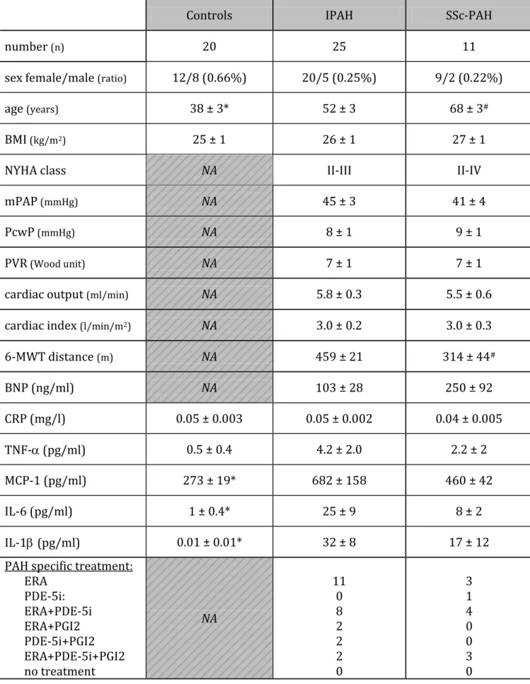
Table 1: Subjects anthropometric and hemodynamic characteristics
Controls IPAH SSc-PAH
number (n) 20 25 11
sex female/male (ratio) 12/8 (0.66%) 20/5 (0.25%) 9/2 (0.22%)
age (years) 38 ± 3* 52 ± 3 68 ± 3#
BMI(kg/m2) 25 ± 1 26 ± 1 27 ± 1
NYHA class NA II-III II-IV
mPAP(mmHg) NA 45 ± 3 41 ± 4
PcwP(mmHg) NA 8 ± 1 9 ± 1
PVR(Wood unit) NA 7 ± 1 7 ± 1
cardiac output (ml/min) NA 5.8 ± 0.3 5.5 ± 0.6
cardiac index (l/min/m2) NA 3.0 ± 0.2 3.0 ± 0.3
6-MWT distance (m) NA 459 ± 21 314 ± 44# BNP (ng/ml) NA 103 ± 28 250 ± 92 CRP (mg/l) 0.05 ± 0.003 0.05 ± 0.002 0.04 ± 0.005 TNF- (pg/ml) 0.5 ± 0.4 4.2 ± 2.0 2.2 ± 2 MCP-1 (pg/ml) 273 ± 19* 682 ± 158 460 ± 42 IL-6 (pg/ml) 1 ± 0.4* 25 ± 9 8 ± 2 IL-1(pg/ml) 0.01 ± 0.01* 32 ± 8 17 ± 12
PAH specific treatment: ERA PDE-5i: ERA+PDE-5i ERA+PGI2 PDE-5i+PGI2 ERA+PDE-5i+PGI2 no treatment NA 11 0 8 2 2 2 0 3 1 4 0 0 3 0
6-MWT: 6 minute walk test; BMI: body mass index; BNP: brain natriuretic peptide; CRP: C-reactive protein; ERA: endothelin receptor antagonist; IL-6: interleukin-6; IL-1: interleukin-1beta; IPAH: idiopathic pulmonary arterial hypertension; MCP-1: monocyte chemoattractant protein-1; mPAP: mean pulmonary arterial pressure; NA: not applicable; NYHA: New York Heart Association; PcwP: pulmonary capillary wedge pressure; PDE-5i: phosphodiesterase type 5 inhibitor; PGI2: prostacyclin; PVR: pulmonary vascular resistance; SSc-PAH: scleroderma-associated PAH; TNF-: tumor necrosis factor alpha. *:p<0.05 between controls versus IPAH and versus SSc-PAH patients; #:p<0.05 between IPAH and SSc-PAH patients.
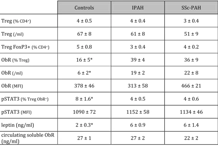
Table 2: Regulatory T lymphocytes, leptin receptor and serum leptin data
Controls IPAH SSc-PAH
Treg (% CD4+) 4 ± 0.5 4 ± 0.4 3 ± 0.4 Treg (/ml) 67 ± 8 61 ± 8 51 ± 9 Treg FoxP3+ (% CD4+) 5 ± 0.8 3 ± 0.4 4 ± 0.2 ObR (% Treg) 16 ± 5* 39 ± 4 36 ± 9 ObR(/ml) 6 ± 2* 19 ± 2 22 ± 8 ObR(MFI) 378 ± 46 313 ± 58 466 ± 21
pSTAT3(% Treg ObR+) 8 ± 1.6* 4 ± 0.5 4 ± 0.6
pSTAT3 (MFI) 1090 ± 72 1152 ± 58 1134 ± 46
leptin (ng/ml) 2 ± 0.3* 6 ± 0.9 6 ± 1.4
circulating soluble ObR
(ng/ml) 27 ± 1 27 ± 2 22 ± 2
FoxP3: forkhead box P3; IPAH: idiopathic pulmonary arterial hypertension; MFI: mean fluorescence intensity; ObR: leptin receptor; pSTAT3: phosphorylated Signal Transducer
and Activator of Transcription 3; SSc-PAH: scleroderma-associated PAH; Treg: regulatory T lymphocytes. *:p<0.05 between controls versus IPAH and versus SSc-PAH patients.
Figure Legends
Figure 1: Leptin measurements
A: Group data of leptin measurement in serum from controls (n=20), idiopathic pulmonary arterial hypertension (IPAH, n=25) and scleroderma-associated PAH (SSc-PAH, n=11) patients. Values are expressed as mean ± sem. *: p<0.05 between controls
versus IPAH and versus SSc-PAH patients. B: Group data of circulating soluble leptin
receptor (sObR) measurement in serum from controls (n=20), IPAH (n=25) and SSc-PAH (n=11) patients. Values are expressed as mean ± sem. C: Correlation between serum leptin and body mass index in controls (square dots). D: Correlation between serum leptin and body mass index in patients considered as a unique group (IPAH: round dots and SSc-PAH patients: triangular dots).
Figure 2: C-reactive protein measurements
A: Group data of C-reactive protein (CRP) measurement in serum from controls (n=20), idiopathic pulmonary arterial hypertension (IPAH, n=25) and scleroderma-associated PAH (SSc-PAH, n=11) patients. Values are expressed as mean ± sem. B: Correlation between CRP and serum leptin in controls (square dots). C: Correlation between CRP and serum leptin in patients considered as a unique group (IPAH: round dots and SSc-PAH patients: triangular dots).
Figure 3: Leptin receptor (ObR) expression
A: Single FACS dot plots representing an example of phenotype strategy by using surface stainings analysed by flow cytometry in a control subject. B: Single FACS dot plot representing regulatory T lymphocytes (Treg) ObR+ in control subject, idiopathic
patients. C: Group data of ObR expression on circulating Treg, expressed as percentage of the total Treg population, in control subjects (n=20), IPAH (n=25) and SSc-PAH (n=11) patients. Values are expressed as mean ± sem. *:p<0.05 between controls versus IPAH and versus SSc-PAH patients. D: Group data of ObR expression on circulating Treg, expressed as absolute numbers, in control subjects (n=20), IPAH (n=25) and SSc-PAH (n=11) patients. Values are expressed as mean ± sem. *:p<0.05 between controls versus IPAH and versus SSc-PAH patients. E: Correlation between Treg ObR+ and body mass
index (BMI) in controls (square dots). F: Correlation between Treg ObR+ and BMI in
patients considered as a unique group (IPAH: round dots and SSc-PAH patients: triangular dots). G: Correlation between Treg ObR+ and serum leptin in controls (square
dots). H: Correlation between Treg ObR+ and serum leptin in patients considered as a
unique group (IPAH: round dots and SSc-PAH patients: triangular dots).
Figure 4: Regulatory T lymphocytes (Treg)
A: Group data of circulating Treg cell count, expressed as percentage of the total CD4+
population, in control subjects (n=20), idiopathic pulmonary arterial hypertension (IPAH, n=25) and scleroderma-associated PAH (SSc-PAH, n=11) patients. Values are expressed as mean ± sem. B: Group data of circulating Treg cell count, expressed as absolute numbers, in control subjects (n=20), IPAH (n=25) and SSc-PAH (n=11) patients. Values are expressed as mean ± sem. C: Group data of circulating Treg cell count, defined as CD4+CD25+CD127+FoxP3+ and expressed as percentage of the total
CD4+ population in control subjects (n=5), IPAH (n=5) and SSc-PAH (n=5) patients.
Figure 5: Functional status of regulatory T lymphocytes (Treg) expressing leptin receptor (ObR)
A: Single FACS dot plots representing an example of staining strategy by using surface markers to phenotype Treg and intracellular staining with anti-phosphorylated Signal Transducer and Activator of Transcription 3 (pSTAT3) antibody, analysed by flow cytometry in a control subject. B: Single FACS dot plot representing pSTAT3 in Treg ObR+ in control subjects, idiopathic pulmonary arterial hypertension (IPAH) and
scleroderma-associated PAH (SSc-PAH) patients. C: Group data of Treg ObR+ pSTAT3+,
expressed as percentage of the total Treg ObR+ population, in control subjects (n=20),
IPAH (n=25) and SSc-PAH (n=11) patients. Values are expressed as mean ± sem. *:p<0.05 between controls versus IPAH and versus SSc-PAH patients.
D: Group data of Treg ObR+ pSTAT3+, expressed as percentage of the total Treg ObR+
population in control subjects peripheral blood mononuclear cells (n=3) after leptin stimulation (leptin +) or without any stimulation (leptin -). Values are expressed as mean ± sem. *:p<0.05.
Figure 6: Leptin production by human pulmonary endothelial cells
A: Staining for leptin in lung specimens in a representative pulmonary artery from controls (left panel) and IPAH patients (right panel).
B: Staining for leptin receptor in lung specimens in a representative pulmonary artery from controls (left panel) and IPAH patients (right panel).
C: Group data of leptin production by human pulmonary endothelial cells, in control subjects (n=15) and in idiopathic pulmonary arterial hypertension (IPAH, n=10). Values are expressed as mean ± sem. *: p<0.05 between controls versus IPAH patients.
Figure 1 BMI (kg/m2)
A
0 2 4 6 8 le pt in (ng /m l)controls IPAH SSc-PAH
*
*
0 10 20 30controls IPAH SSc-PAH
sO bR (ng /m l)
B
0 5 10 15 20 0 10 20 30 40 50 0 5 10 15 20 0 10 20 30 40 50 r= 0.4 p= 0.03 r= 0.4 p= 0.009C
le pti n (ng /m l)D
IPAH and SSc-PAH patients control subjects
0 5 10 15 20 0,00 0,05 0,10 0,00 0,02 0,04 0,06 0,08 0,10 C -r e acti ve pr o te in (m g /l )
controls IPAH SSc-PAH
A
Figure 2 0 5 10 15 20 0,00 0,05 0,10 le pti n (ng /m l) C-Reactive Protein (mg/l)B
C
r= 0.43 p= 0.14 r= -0.3 p= 0.06IPAH and SSc-PAH patients control subjects
Figure 3
CD4+cells
4% Treg
CD4+CD127lowCD25+ Treg ObR
+ SSC SSC CD 1 2 7 CD 1 2 7 ObR CD25 CD4 FSC
A
0 102 103 104 105 0 102 103 104 105 14-03-11 sample9.011.fcsÉCD127low FL3-A: ObR-PE-A F L 4 -A : C D 1 2 7 P e rC P .C y 5 .5 -A 22.9 0 102 103 104 105 0 102 103 104 10528mai10 sample ctrl 7.011.fcsÉCD127low
FL3-A: ObR PE-A
F L 4 -A : C D 1 2 7 P e rC P C y 5 .5 -A 2.53 0 102 103 104 105 0 102 103 104 105
18nov2010 sample 13.008.fcsÉCD127low
FL3-A: ObR PE-A
F L 4 -A : C D 1 2 7 P e rC P -C y 5 .5 -A 10.7
B
CD 1 2 7 ObRcontrols IPAH SSc-PAH
Figure 3 0 10 20 30 40 50 O bR +ce lls (% T re g )
controls IPAH SSc-PAH
*
*
0 5 10 15 20 25 30 ObR +ce lls /m lcontrols IPAH SSc-PAH
D
*
*
0 10 20 30 40 50 60 70 0 20 40 60 0 10 20 30 40 50 60 70 0 5 10 15 20 O bR +ce lls (% T re g ) BMI (kg/m2) 0 10 20 30 40 50 60 70 0 20 40 60
E
Figure 3 0 10 20 30 40 50 60 70 0 5 10 15 20 leptin (ng/ml) O bR +ce lls (% T re g )F
G
H
r= 0.1 p= 0.9 r= -0.1 p= 0.7 r= -0.2 p= 0.2 r= -0.2 p= 0.3IPAH and SSc-PAH patients control subjects
IPAH and SSc-PAH patients control subjects
0 1 2 3 4 5 T re g (% C D 4 + ce lls )
controls IPAH SSc-PAH
A
Figure 4 0 20 40 60 80 100 T re g /mlcontrols IPAH SSc-PAH
B
0 1 2 3 4 5 6 Tr e g Fo xP 3 + (% C D 4 + ce lls )controls IPAH SSc-PAH
C
NS NS
Figure 5
CD4+
Treg
CD4+CD127lowCD25+ Treg ObR
+ SSC SSC CD 1 2 7 CD 1 2 7 ObR CD25 CD4 FSC pSTAT3
A
0 102 103 104 105 0 50K 100K 150K 200K 250K 25janv11 sample 1.009.fcsÉObR+FL2-A: pSTAT3 alexa488-A
S S C -A : S S C -A 20.7 0 102 103 104 105 0 50K 100K 150K 200K 250K 22-03-11 sample 14.017.fcsÉObR+
FL2-A: pSTAT3 A488-A
S S C -A : S S C -A 1.95 0 102 103 104 105 0 50K 100K 150K 200K 250K 22-03-11 sample 24.027.fcsÉObR+
FL2-A: pSTAT3 A488-A
S S C -A : S S C -A 2.93 CD 1 2 7 pSTAT3
B
Figure 5 0 2 4 6 8 10 Tr e g ObR +pST A T 3 + (% Tr e g O bR +)
controls IPAH SSc-PAH
C
*
*
D
0 2 4 6 8 leptin - + Tr e g O bR +pST A T 3 + (% Tr e g O bR +)*
Figure 6 0 20 40 60 80 100 120 controls IPAH le pti n (pg /m l)