HAL Id: inserm-02169494
https://www.hal.inserm.fr/inserm-02169494
Submitted on 1 Jul 2019
HAL is a multi-disciplinary open access archive for the deposit and dissemination of sci-entific research documents, whether they are pub-lished or not. The documents may come from teaching and research institutions in France or abroad, or from public or private research centers.
L’archive ouverte pluridisciplinaire HAL, est destinée au dépôt et à la diffusion de documents scientifiques de niveau recherche, publiés ou non, émanant des établissements d’enseignement et de recherche français ou étrangers, des laboratoires publics ou privés.
stimulate cytolytic T cells and offer increased protection
against EBV infection ex vivo and in mice
Dwain van Zyl, Ming-Han Tsai, Anatoliy Shumilov, Viktor Schneidt, Rémy
Poirey, Bettina Schlehe, Herbert Fluhr, Josef Mautner, Henri-Jacques
Delecluse
To cite this version:
Dwain van Zyl, Ming-Han Tsai, Anatoliy Shumilov, Viktor Schneidt, Rémy Poirey, et al.. Immunogenic particles with a broad antigenic spectrum stimulate cytolytic T cells and offer increased protection against EBV infection ex vivo and in mice. PLoS Pathogens, Public Library of Science, 2018, 14 (12), pp.e1007464. �10.1371/journal.ppat.1007464�. �inserm-02169494�
RESEARCH ARTICLE
Immunogenic particles with a broad antigenic
spectrum stimulate cytolytic T cells and offer
increased protection against EBV infection ex
vivo and in mice
Dwain G. van ZylID1,2,3,4, Ming-Han TsaiID1,3,4¤, Anatoliy ShumilovID1,3,4, Viktor Schneidt1,2,3,4, Re´my Poirey
ID1,3,4, Bettina Schlehe5, Herbert Fluhr5, Josef MautnerID4,6, Henri-Jacques DelecluseID1,2,3,4*
1 German Cancer Research Center (DKFZ) Unit F100, Heidelberg, Germany, 2 Faculty of Biosciences,
Heidelberg University, Heidelberg, Germany, 3 Institut National de la Sante´ et de la Recherche Me´dicale (INSERM) Unit U1074, Heidelberg, Germany, 4 German Center for Infection Research (DZIF),
Braunschweig, Germany, 5 Frauenklinik, University Hospital Heidelberg, Heidelberg, Germany, 6 Children’s Hospital, Technische Universita¨ t Mu¨nchen, & Helmholtz Zentrum Mu¨nchen, Munich, Germany
¤ Current address: Institute of Microbiology and Immunology, National Yang-Ming University, Taipei, Taiwan
*h.delecluse@dkfz.de
Abstract
The ubiquitous Epstein-Barr virus (EBV) is the primary cause of infectious mononucleosis and is etiologically linked to the development of several malignancies and autoimmune dis-eases. EBV has a multifaceted life cycle that comprises virus lytic replication and latency programs. Considering EBV infection holistically, we rationalized that prophylactic EBV vac-cines should ideally prime the immune system against lytic and latent proteins. To this end, we generated highly immunogenic particles that contain antigens from both these cycles. In addition to stimulating EBV-specific T cells that recognize lytic or latent proteins, we show that the immunogenic particles enable the ex vivo expansion of cytolytic EBV-specific T cells that efficiently control EBV-infected B cells, preventing their outgrowth. Lastly, we show that immunogenic particles containing the latent protein EBNA1 afford significant pro-tection against wild-type EBV in a humanized mouse model. Vaccines that include antigens which predominate throughout the EBV life cycle are likely to enhance their ability to protect against EBV infection.
Author summary
Human herpesviruses are tremendously successful pathogens that establish lifelong infec-tion in a substantial proporinfec-tion of the populainfec-tion. The oncogenicγ-herpesvirus EBV, like other herpesviruses, expresses a plethora of open-reading frames throughout its multiface-ted life cycle. We have developed a prophylactic vaccine candidate in the form of immu-nogenic particles that contain several EBV antigens. This is in stark contrast to the vast majority of EBV vaccines candidates that contain only one or two EBV antigens. Our
a1111111111 a1111111111 a1111111111 a1111111111 a1111111111 OPEN ACCESS
Citation: van Zyl DG, Tsai M-H, Shumilov A,
Schneidt V, Poirey R, Schlehe B, et al. (2018) Immunogenic particles with a broad antigenic spectrum stimulate cytolytic T cells and offer increased protection against EBV infection ex vivo and in mice. PLoS Pathog 14(12): e1007464.
https://doi.org/10.1371/journal.ppat.1007464
Editor: Rajiv Khanna, QIMR Berghofer Medical
Research Institute, AUSTRALIA
Received: October 23, 2018 Accepted: November 8, 2018 Published: December 6, 2018
Copyright:© 2018 van Zyl et al. This is an open access article distributed under the terms of the
Creative Commons Attribution License, which permits unrestricted use, distribution, and reproduction in any medium, provided the original author and source are credited.
Data Availability Statement: All relevant data are
within the manuscript and its Supporting Information files.
Funding: Funding has been received from the
German Cancer Research Center (Project number F100), Inserm (Project number U1074) and Deutsche Krebshilfe (70110621).The funders had no role in study design, data collection and analysis, decision to publish, or preparation of the manuscript.
immunogenic particles were shown capable of stimulating several EBV-specific T-cell clonesin vitro. The immunogenic particles were also capable of expanding cytolytic EBV-specific T cellsex vivo and provided a protective benefit in vivo when used as a prophylac-tic vaccine.
Introduction
The Epstein-Barr virus (EBV) is aγ-herpesvirus that establishes asymptomatic infection in the majority of the human population. EBV infects both B cells and epithelial cells, but it is in the former in which EBV establishes latency and persists lifelong [1]. Despite being carried asymp-tomatically by most individuals, the global disease burden of EBV is substantial. EBV is the pri-mary cause of infectious mononucleosis (IM), accounts for 200,000 new cancer cases annually [2] and is linked to the development of autoimmune diseases (e.g. multiple sclerosis) [3].
Shortly after the discovery of EBV, vaccination was touted as a possible means of control-ling or eliminating EBV-associated diseases [4]. Despite EBV being the first human oncogenic virus to be discovered, and in spite of several decades of EBV vaccine research, no prophylactic EBV vaccine has made it onto the market. So far, the majority of prophylactic vaccine proto-types have focused on the major viral envelope glycoprotein gp350 [5]. One study, in which soluble gp350 was used for vaccination purposes, reported a decrease in the frequency of IM in vaccinated individuals over a study period of 18 months, but vaccination did not reduce the frequency of infection with the wild type virus [6]. Long-term information on the vaccinated cohort is not available.
Herpesviruses have complex life cycles and primary infection, latency and reactivation are achieved through the expression of a large number of open-reading frames [7–9]. Considering the number of antigens that are expressed during the EBV life cycle, it is not surprising that EBV infection is controlled in healthy individuals through humoral and cellular immune responses that target a variety of lytic and latent proteins [10]. Considering the breadth of EBV-specific immune responses in healthy individuals, it is surprising that EBV vaccine proto-types have only targeted a limited set of lytic [6,11,12] or latent proteins [13].
We previously generated a vaccine candidate in the form of EBV virus-like particles (VLPs) and light particles (LPs) [14]. Deletion of EBV proteins involved in DNA packaging produced particles that were DNA-free, non-infectious and highly immunogenic. Whilst EBV VLPs/LPs are likely to contain several dozen lytic proteins (viz. envelope, tegument and capsid proteins) [15], they are devoid of latent proteins. To address this shortcoming, we enlarged the antigenic spectrum of VLPs/LPs to include immunodominant latent proteins. We interrogated the anti-genicity of VLPs/LPs containing latent antigensin vitro, ex vivo and in vivo.
Results
Enlarging the antigenic spectrum of EBV virions to include latent proteins
To generate immunogenic particles with an enlarged antigenic spectrum, we aimed to intro-duce latent protein fragments into EBV VLPs/LPs. Since BNRF1 is abundant within virions [15], we rationalized that latent antigens could be introduced into VLPs/LPs by fusing them to BNRF1 (Fig 1A). However, since wild-type EBV (wtEBV) can be accurately and sensitively quantified through qPCR [14], we first modified BNRF1 of wtEBV to test this assumption. The BNRF1 of wtEBV was modified to contain a fragment from the highly antigenic latent protein EBNA3C [16] (S1 Fig). Bacterial artificial chromosome (BAC) DNA from wtEBV was
Competing interests: The authors have declared
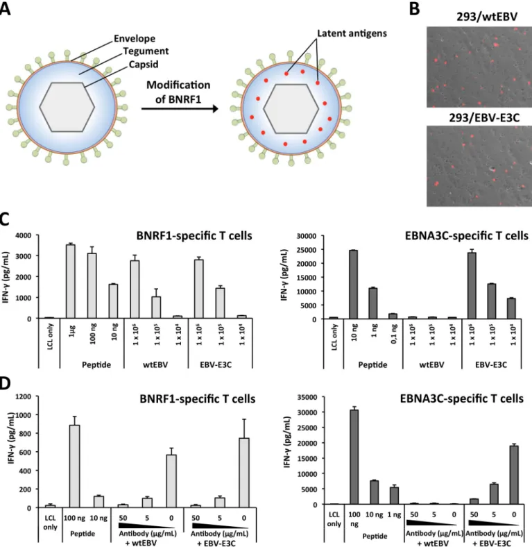
Fig 1. The antigenic spectrum of EBV virions is enlarged through the construction of BNRF1-latent protein gene fusions. (A) The insertion of latent protein
epitopes into the major tegument protein BNRF1 enables them to be incorporated into the tegument of VLPs/LPs. (B) Modification of BNRF1 to include an antigenic fragment from EBNA3C (E3C) does not influence lytic replication in producer cells. The late lytic gene product gp350 was detected at the surface of induced 293/wtEBV and 293/EBV-E3C producer cells by staining withα-gp350 and α-mouse IgG-Cy3 antibodies. (C) EBV virions that encode a BNRF1-EBNA3C fusion protein stimulate BNRF1- and EBNA3C-specific CD4+T cells. Autologous LCLs were pulsed with various amounts (1 x 10⁴to 1 x 10⁶genome equivalents
(geq)) of wtEBV or EBV-E3C and then cocultured with CD4+T cells that were specific for BNRF1 VSD (1006–1017 aa) or EBNA3C 5H11 (325–339 aa) epitopes. In
parallel, LCLs were pulsed with control peptides (μg to ng quantities) prior to coculture with CD4+T cells. T-cell activation was determined by measuring secreted IFN-γ by ELISA. (D) A neutralizing antibody that recognizes gp350 impairs the antigenicity of wtEBV and EBV-E3C. The neutralizing antibody 72A1 was titrated (50, 5 and 0μg/mL) and incubated with 1 x 10⁶geq of wtEBV and EBV-E3C. Thereafter, supernatants were used in T-cell activation assays. The data displayed in
each chart represents triplicate values and error bars represent standard deviation. Furthermore, each graph is a representative experiment of at least three.
https://doi.org/10.1371/journal.ppat.1007464.g001
modified to contain EBNA3C and then stably introduced into 293 cells to generate a virus pro-ducer cell line (293/EBV-E3C). The integrity of the EBV-E3C BAC DNA within propro-ducer cells was confirmed with restriction analysis (S1 Fig). Transfection of the lytic transactivator BZLF1 gene into 293/EBV-E3C and 293/wtEBV yielded a similar percentage of cells that expressed the late lytic protein gp350 (Fig 1B). This indicates that modification of BNRF1 to include a latent antigen fragment does not influence lytic replication. Next, we compared the antigenic-ity of EBV-E3C and wtEBV viruses in T-cell activation assays (Fig 1C). Autologous lympho-blastoid cell lines (LCLs) were pulsed with the two viruses or peptide controls and then cocultured with BNRF1- [17] or EBNA3C- [18] specific CD4+T cells. This confirmed that modified virions, containing a BNRF1-EBNA3C fusion protein, were able to simulate EBNA3C- and BNRF1-specific CD4+T cells. Conversely, wtEBV that contained unmodified BNRF1 was only able to stimulate the BNRF1-specific CD4+T cells (Fig 1C). In all cases, the dose of the virus applied correlated to the response generated by the T cells, with as little as 1 x 10⁴virions (genome equivalents (geq)) being able to generate responses from the BNRF1- and EBNA3C-specific T cells. Importantly, BNRF1-specific CD4+T cells recognized modified and unmodified EBV to the same extent. This indicates that BNRF1-latent antigen fusion proteins enlarged the antigenic spectrum of EBV without influencing the antigenicity of BNRF1. Next, we tested whether the enlarged antigenic spectrum of EBV-E3C was exclusively due to BNRF1-latent antigen fusions contained within virions. To this end, virus supernatants were pre-incubated with anti-gp350 neutralizing antibody [19] prior to being used in T-cell recogni-tion assays (Fig 1D). This showed that the neutralizing antibody was able to abolish the antige-nicity of EBV-E3C. Altogether, these results confirm that BNRF1-latent antigen fusion proteins are successfully packaged into virions and enlarge their antigenic spectrum.
Antigenic diversification of VLPs/LPs lacking gp110
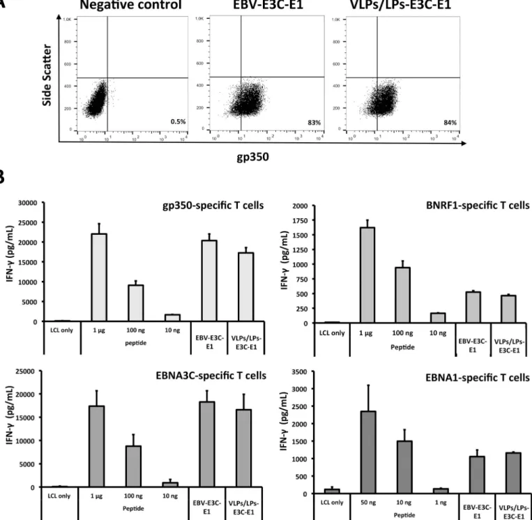
Next, we confirmed the antigenicity of BNRF1-latent antigen fusion proteins in gp110-nega-tive VLPs/LPs. Since gp110 has been shown to preclude viral and host membrane fusion [20], and abrogate toxicity [21], we exclusively used gp110-negative VLPs/LPs in the present study. We concurrently modified VLPs/LPs and wtEBV to encode EBNA3C and EBNA1 fragments, respectively generating 293/VLPs/LPs-E3C-E1 and 293/EBV-E3C-E1 producer cells (S2 Fig). EBNA1, like EBNA3C, is a highly immunogenic latent protein that is frequently recognized by the population [16,22]. The DNA-free VLPs/LPs-E3C-E1 were quantified using flow cytome-try (S3 Fig) and then compared to an equivalent amount of EBV-E3C-E1 that was quantified with qPCR (Fig 2A). This confirmed that flow cytometry enabled the reliable quantification DNA-free VLPs/LPs. Next, VLPs/LPs-E3C-E1 were analysed in T-cell activation assays along-side EBV-E3C-E1 (Fig 2B). This showed that the VLPs/LPs, like EBV virions, were able to stimulate BNRF1- [17], gp350- [23], EBNA3C- [18] and EBNA1- [24] specific CD4+T cells when they were modified to contain EBNA3C and EBNA1 fragments (Fig 2B). Furthermore, the modified VLPs/LPs stimulated the various lytic protein- and latent protein-specific T cells to the same extent as modified EBV. This confirmed that VLPs/LPs could be used as a platform to generate immunogenic particles that comprise lytic and latent antigens. Furthermore, the lack of gp110 does not negatively influence the antigenicity of the VLPs/LPs, indicating that their safety can be increased without compromising their antigenicity.
Modified VLPs/LPs expand T cells that efficiently control recently infected
B cells
Since EBV-specific T cells play a crucial role in controlling EBV-infection [25,26], we tested whether modified VLPs/LPs could expand EBV-specific T cells with protective value. To this
Fig 2. Modified VLPs/LPs that lack gp110 are antigenic and stimulate multiple EBV-specific T cells. (A) VLPs/LPs and EBV that contain EBNA3C (E3C) and
EBNA1 (E1) bind to B cells to a similar degree. Equal quantities of EBV-E3-E1 (1 x 10⁶geq) and VLPs/LPs-E3C-E1 (1 x 10⁶particles) were incubated with Elijah B
cells, stained withα-gp350 (72A1) and α-mouse IgG-Cy3 antibodies and then analysed with flow cytometry. The values displayed on plots indicate the percentage of B cells that were bound by virus or VLPs/LPs. (B) VLPs/LPs-E3C-E1 retain their antigenic character in the absence of gp110. Autologous LCLs were pulsed with control peptides, VLPs/LPs-E3C-E1 (1 x 10⁶particles) or EBV-E3C-E1 (1 x 10⁶geq) and cultured with T cells that were specific for gp350 1D6 (65–69 aa), BNRF1
VSD (1006–1017 aa), EBNA3C 5H11 (325–339 aa) or EBNA1 3E10 (475–489 aa) epitopes. T-cell activity was determined by quantifying IFN-γ release with ELISA. The data illustrated in the graphs are average of triplicate values and error bars represent standard deviation. Furthermore, each graph is a representative experiment of at least three.
https://doi.org/10.1371/journal.ppat.1007464.g002
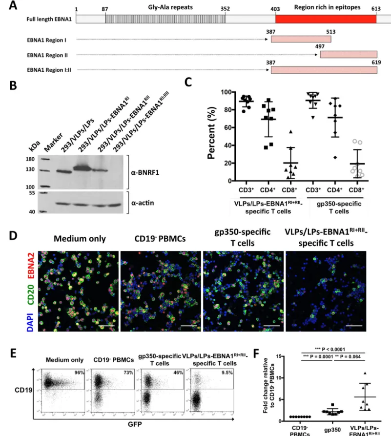
end, epitope-rich regions from EBNA1, arbitrarily named region I, region II and region I:II (Fig 3A), were used to generate VLPs/LPs producer cells that encode EBNA1 (S4 Fig). Analysis of the producer cells with western blot showed that the 293/VLP/LP-EBNA1RI:IIproducer cell line was unable to express the large BNRF1-EBNA1 fusion, whilst the 293/VLP/LP-EBNA1RI and 293/VLP/LP-EBNA1RIIproducer cells successfully expressed their BNRF1-EBNA1 fusions (Fig 3B). Hence VLPs/LPs-EBNA1RI:IIwere excluded from analysis. VLPs/LPs-EBNA1RIand VLPs/LPs-EBNA1RIIwere combined (VLPs/LPs-EBNA1RI+RII) and used to stimulate bulk PBMCs from unhaplotyped EBV-positive donors (Fig 3C). As a control, PBMCs from the same donors were also expanded with an antigen-armed antibody (AgAb) that contained the major EBV glycoprotein gp350. AgAbs were originally developed as a targeted therapy for B cell malignancies [18,27], but were repurposed in the present study to expand EBV-specific T cells of interest (S5 Fig). Stimulation of PBMCs from EBV-positive donors with VLPs/LPs-EBNA1RI+RIIor gp350-AgAb expanded similar numbers of CD4+, CD8+and total T cells from the PBMCs of EBV-positive donors (Fig 3C). Next, primary B cells from four donors were infected with highly infectious gp110highB95-8 [28] and then cocultured with VLPs/LPs-EBNA1RI+RII- or gp350-AgAb-expanded T cells. After 5 days,ex vivo cultures were analyzed by immunofluorescence for the presence of EBV-infected B cells (Fig 3D). This showed that nearly all the B cells inex vivo cultures were EBNA2-positive, confirming that virtually all the B cells were successfully infected with EBV. However, there were noticeably fever EBV-infected B cells (CD20+EBNA2+) in the presence of VLPs/LPs-EBNA1RI+RII-specific T cells than gp350-specific T cells. This suggested that VLPs/LPs-EBNA1RI+RII-specific T cells were more efficient at controlling the EBV-infected B cells during the first five days of infection compared to gp350-specific T cells. Next, VLPs/LPs-EBNA1RI+RII- and gp350-AgAb-expanded T cells from eight donors were cocultured with infected B cells as before and quantitatively analysed with flow cytometry (Fig 3E and 3F). This confirmed thatex vivo cultures containing VLPs/LPs-EBNA1RI+RII-specific T cells had the lowest percentage of CD19+cells. This indi-cates that VLPs/LPs-EBNA1RI+RII-specific T cells are more proficient than gp350-specific T cells at controlling EBV-infected B cells during the first five days of infection.
Modified VLPs/LPs expand T cells that restrict the outgrowth of
B95-8-and M81-infected B cells
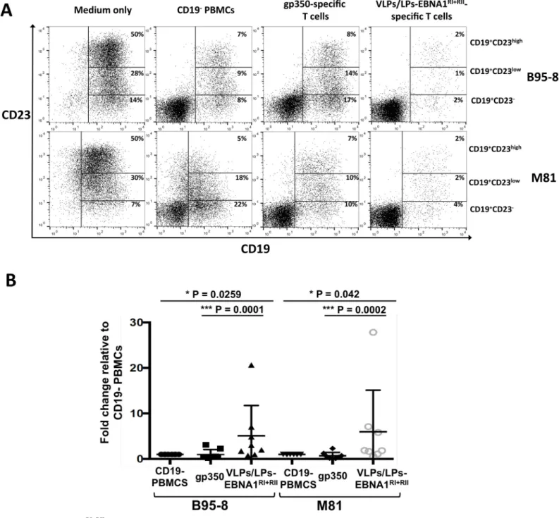
Having shown that VLP/LPs-EBNA1RI+RII-specific T cells efficiently control B95-8-infected B cells, we tested whether they could restrict the outgrowth of infected B cells over a longer period. Additionally, we tested whether VLP/LPs-EBNA1RI+RII-specific T cells could prevent the outgrowth of B cells infected with the prototypic B95-8 strain or the distantly related M81 strain [29] from Hong Kong. We stimulated PBMCs from eight EBV-positive donors as before (seeFig 3) and then cocultured them with autologous B cells that were infected with gp110high B95-8 and gp110highM81. As a positive and negative control, infected B cells were respectively cultured with CD19-PBMCs or in medium only. After 15 days,ex vivo cultures were analysed with flow cytometry to detect outgrowing B cells (Fig 4). Since proliferating B cells express CD23, outgrowing B cells were identified by detecting CD19+CD23+double-positive cells [30,31]. EBV-infected B cells were found to consist of CD19+CD23-, CD19+CD23lowand CD19+CD23highpopulations when they were cultured in medium only, with the majority of B cells being of the CD19+CD23highvariety (Fig 4A). Comparatively, in the presence of CD19 -PBMCs, gp350-specific T cells and VLPs/LPs-EBNA1RI+RII-specific T cells, the number of CD19+CD23+cells were considerably reduced. This indicated that proliferating B cells were restricted in these cultures. Interestingly, whilst gp350-specific T cells were shown to be more efficient than CD19-PBMCs at controlling infected B cells during the early phase of infection
Fig 3. VLPs/LPs containing EBNA1 fragments expand T cells that efficiently target EBV-infected B cells. (A) Epitope-rich regions from EBNA1 were utilized to
construct modified VLPs/LPs. EBNA1 has a single stretch of ~200 amino acids that contains more than 25 described CD4 and CD8 epitopes. Regions encompassing the majority of EBNA1 T-cell epitopes, arbitrarily named region I, II and I:II, were used to generate VLP/LPs-EBNA1RI, VLP/LPs-EBNA1RIIand VLP/LPs-EBNA1RI:II.
(seeFig 3), the CD19-PBMCs of several donors were considerably more adept at restricting B-cell outgrowth than gp350-specific T B-cells over the longer 15 day period (Fig 4B). This suggest that the PBMCs from some donors contained EBV-specific T cells, other than gp350-specific T cells, that were able to restrict B-cell outgrowth. However, it is evident that proliferating B cells were restricted to a greater degree inex vivo cultures that contained VLPs/LPs-EBNA1RI
+RII
-specific T cells. Moreover, this was observed for B95-8- and M81-infected B cells and for all donors (Fig 4B). This confirms that VLPs/LPs equipped with EBNA1 expand EBV-specific T cells that efficiently restrict B cells infected with B95-8 and M81 EBV.
Modified VLPs/LPs stimulate cytolytic CD4
+T cells that recognize lytic
and latent cycle antigens
Having shown that VLPs/LPs-EBNA1RI+RII-specific T cells control EBV-infected cells, it indicated that they were cytolytic in character. However, since VLPs/LPs-EBNA1 RI+RII-stimulated PBMCs contained primarily CD4+T cells (Fig 3C), it was unclear whether VLPs/ LPs-EBNA1RI+RIIare capable of stimulating EBV-specific CD8+T cells. To this end, we tested the ability of LCLs pulsed with VLPs/LPs-EBNA1RI, containing the EBNA1 HPV CD8+ epi-tope, to stimulate an EBNA1 HPV-specific T-cell clone [18]. This showed that the EBNA1 HPV-specific CD8+T-cell clone was unable to recognize LCLs pulsed with VLPs/LPs-EBNA1RI(S6A Fig). In contrast, LCLs pulsed with the VLPs/LPs-EBNA1RIwere well recog-nized by the BNRF1 VSD-specific CD4+T-cell clone. This suggests that the autologous LCLs were unable to cross-present the EBNA1 HPV epitope from VLPs/LPs-EBNA1RIto CD8+T cells. We also assessed IFN-gamma secretion by CD4+and CD8+T cells after the stimulation of bulk PBMCs with VLPs/LPs-EBNA1RI+RII(S6B Fig). This revealed that both CD4+and CD8+IFN-gamma producing cells were detected upon stimulation with VLPs/LPs-EBNA1RI+RII (S6C Fig). However, it was evident that IFN-γ+
CD4+T cells were more numerous than IFN-γ+
CD8+T cells and suggests that the stimulation of PBMCs with VLPs/LPs-EBNA1RI+RII pref-erentially expands EBV-specific CD4+T cells. Next, we expanded EBNA1- and gp350-specific CD4+T cells from VLPs/LPs-EBNA1RI+RII-stimulated PBMCs and analyzed them for their cytotoxic potential. Bulk PBMCs from an unhaplotyped EBV-positive donor was stimulated with VLPs/LPs-EBNA1RI+RIIfor two rounds, after which gp350-AgAb (S5 Fig) or EBNA1-A-gAb (S7 Fig) were used to expand gp350- and EBNA1-specific CD4+T cells (Fig 5A). The expanded CD4+T cells were confirmed to be specific for either EBNA1 or gp350 (Fig 5B). The ex vivo expanded CD4+T cells specifically responded to EBNA1-AgAb or gp350-AgAb and to EBNA1 3G2 or gp350 1D6 epitope peptides. Next, we determined whether the EBNA1- and gp350-specific CD4+T cells were capable of expressing CD107a, a surrogate marker for the release of cytolytic granules [32]. Autologous LCLs were pulsed withα-CD20, EBNA1-AgAb
The gly-ala repeats that impede HLA class I-restricted presentation is also shown. (B) Expression of BNRF1-EBNA1 fusion proteins by induced 293/VLPs/LPs-EBNA1RI, 293/VLPs/LPs-EBNA1RIIand 293/VLPs/LPs-EBNA1RI:IIproducer cells. Western blot analysis was performed withα-BNRF1 and α-actin antibodies. (C) VLPs/LPs containing EBNA1 predominantly expand CD4+T cells. VLPs/LPs-EBNA1RIand VLPs/LPs-EBNA1RIIwere combined in a 1:1 ratio (VLPs/LPs-EBNA1RI
+RII) and used to stimulate PBMCs from eight unhaplotyped EBV-positive donors. The PBMCs from the same donors were stimulated in parallel with gp350-AgAb.
Ex vivo cultures were stained for CD3, CD4 and CD8 after two stimulation cycles and analysed with flow cytometry. The percentage of CD4+, CD8+and total T cells
(CD3+) inex vivo cultures are shown. (D-F) VLPs/LPs-EBNA1RI+RII
-specific T cells efficiently control EBV-infected B cells during the first 5 days of infection. PBMCs from EBV-positive donors were stimulated as described in (C) and then cocultured with B cells that were infected overnight with recombinant B95-8. Additionally, infected B cells were cultured in medium only or with CD19-depleted (CD19-) PBMCs.
Ex vivo cultures were analysed five days post-infection with
immunofluorescence (D) and flow cytometry (E and F). (D) Representative immunofluorescence results from four donors are shown. Cells were stained withα-CD20, α-EBNA2 and DAPI prior to analysis. Scale bars represent 50 μm. (E) Representative flow cytometry data from eight donors are shown. Ex vivo cultures were assessed for the frequency of CD19+cells. Percentages shown are of total cells. Since the recombinant B95-8 strain encodes GFP, CD19+GFP+double-positive cells could be observed. (F) A summary of flow cytometry results from eight donors. The percentage of CD19+cells in all cultures are expressed relative to the percentage of CD19+
cells in the presence of CD19-PBMCs. Statistical analysis was performed using a two-tailed student t-test.
or gp350-AgAb then cocultured with the EBNA1- and gp350-specific CD4+T cells. This showed that both CD4+T-cell lines upregulated CD107a in response to the relevant antigen (Fig 5C). However, approximately 50% of gp350-specific CD4+T cells expressed CD107a, whilst only 10% of EBNA1-specific CD4+T cells expressed CD107a. Next, we tested the ability
Fig 4. VLPs/LPs-EBNA1RI+RII-specific T cells prevent the outgrowth of B95-8- and M81-infected B cells. PBMCs from eight unhaplotyped EBV-positive donors were stimulated with gp350 or VLPs/LPs-EBNA1RI+RIIas described inFig 3, and then cocultured with primary B cells that were infected with B95-8 or M81. In parallel,
the infected B cells were cultured in medium only or with CD19-PBMCs. After 15 days,ex vivo cultures were stained for CD19 and CD23 and then analysed by flow
cytometry. (A) Representative flow cytometry data from eight donors. The percentage of CD19+CD23-, CD19+CD23lowand CD19+CD23highB cells inex vivo cultures
are shown. (B) A summary of data obtained from eight donors. The percentage of CD19+CD23+B cells in all cultures are expressed relative to the percentage of
CD19+CD23+B cells in the presence of CD19-PBMCs. Statistical analysis was performed using a two-tailed student t-test. Only P values lower than 0.05 are shown.
https://doi.org/10.1371/journal.ppat.1007464.g004
of EBNA1- and gp350-specific CD4+T cells to release the mediator of cytolysis granzyme B (Fig 5D). Both the EBNA1- and gp350-specific CD4+T cells released granzyme B in response to the relevant AgAb and epitope peptide. Lastly, we tested whether the EBNA1- and gp350-specific CD4+T cells were capable of directly lysing autologous LCLs pulsed with antigen (Fig 5E). This showed that the both the EBNA1- and gp350-specific CD4+T cells specifically lysed LCLs pulsed with epitope peptides and VLPs/LPs that contained EBNA1. Taken together, these results confirm that VLPs/LPs-EBNA1RI+RIIhave the ability to stimulate cytolytic CD4+ T cells specific for lytic and latent antigens. These results are consistent with previous studies that showed EBNA1- [30,33,34] and gp350- [35] specific T cells to be cytolytic.
Vaccination with EBV VLPs/LPs containing EBNA1 protects mice from
wtEBV infection
Having shown that modified VLPs/LPs were antigenicin vitro and ex vivo, we assessed whether VLPs/LPs- EBNA1RI+RIIhad protective abilitiesin vivo. To this end, mice reconsti-tuted with human immune system components, susceptible to EBV infection and capable of exerting EBV-specific immune control [36], were used to interrogate VLPs/LPs- EBNA1RI+RII. Humanized NSG-A2 (huNSG-A2) mice were randomly grouped and injected intraperitone-ally with PBS, unmodified VLPs/LPs (1 x 10⁶particles) or VLPs/LPs-EBNA1RI+RII(1 x106 par-ticles), using poly (I:C) as an adjuvant (Fig 6A). Four weeks later, mice were boosted using the same dose. Animals were challenged with gp110highB95-8 (1 x 10⁵GRUs) six weeks after the last boost and euthanized eight weeks later. From the literature we knew that this titer would enable infection without gross development of tumors [36]. The spleens of challenged animals were analysed by histology (Fig 6B). This showed that all animals contained human CD20-and CD3-positive cells in their spleens. However, there was no correlation between the abun-dance of CD20- and CD3-positive cells and the different treatments.In situ hybridization revealed the presence of interspersed cells that expressed EBV-encoded RNAs (EBERs) in the spleens of mice from the PBS and unmodified VLPs/LPs groups. In total, 60% of mice from the PBS group were found to contain EBER+cells, while 37.5% of the mice from the VLPs/LPs group contained EBER+cells (Fig 6C). Statistical analysis showed that this observation was
Fig 5. VLPs/LPs containing EBNA1 enable the expansion of cytolytic gp350- and EBNA1-specific CD4+T cells. (A) The
expansion of gp350- and EBNA1-specific T cells using VLPs/LPs-EBNA1RI+RIIand AgAb. PBMCs from EBV-positive individuals contain multiple EBV-specific T cells. This includes T cells that recognize EBNA1 (red cells), gp350 (green cells) or other structural antigens (orange cells). Stimulation of PBMCs with VLPs/LPs-EBNA1RI+RIIexpanded these various T
cells. Thereafter, EBNA1- and gp350-specific T cells were selectively expanded by respectively using EBNA1-AgAb and gp350-AgAb.Ex vivo expanded T cells were stained for CD3 and CD4 and analysed with flow cytometry. EBNA1- (red) and
gp350- (green) specific T cells are shown. Unstained cells are shown in grey. (B) Theex vivo expanded CD4+T cells are
specific for EBNA1 or gp350. Autologous LCLs were pulsed with EBNA1-AgAb, gp350-AgAb, EBNA1 3G2 (514–528 aa) epitope or gp350 1D6 (65–69 aa) epitope and then cocultured with the CD4+T cells. The release IFN-γ was quantified by ELISA. Each data point is the average of three values and error bars represents standard deviation. Each experiment is a representative of at least three. (C) EBNA1- and gp350-specific CD4+T cells express CD107a. Autologous LCLs were pulsed
withα-CD20, EBNA1-AgAb or gp350-AgAb and then cocultured with the CD4+T cells. Thereafter, cells were stained for
CD4 and CD107a and then analysed with flow cytometry. The percentage of CD4+T cells that express CD107a are indicated. The data displayed are representative of three experiments. (D) EBNA1- and gp350-specific CD4+T cells release granzyme B. Autologous LCLs were pulsed with EBNA1-AgAb, gp350-AgAb or relevant peptides (EBNA1 3G2 and gp350 1D6) and then cocultured with the CD4+T cells. The release of granzyme B was quantified with an ELISA. The data displayed in each chart
represent triplicate values and error bars represent standard deviation. Each graph is a representative experiment of at least three. (E) EBNA1- and gp350-specific CD4+T cells lyse target cells pulsed with VLPs/LPs-EBNA1RI+RII. Autologous LCLs were pulsed overnight with VLPs/LPs- EBNA1RI+RII(gray line), EBNA1 3G2 (red line), gp350 1D6 (green line) or EBNA3C
5H11 (black line) peptides. Thereafter, the pulsed LCLs were incubated with calcein AM and then cocultured with increasing amounts of the EBNA1- or gp350-specific CD4+T cells. Effector to target (E:T) ratios of 1 to 30 were used. The release of calcein from targeted cells was measured at 535 nm after excitation with 485 nm light. Each data point is the average of three values and error bars represents error bars. Furthermore, each experiment is a representative of two experiments.
https://doi.org/10.1371/journal.ppat.1007464.g005
statistically insignificant (P > 0.05). None of the spleen samples from the VLPs/LPs-EBNA1RI
+RII
group were found to contain EBER+cells. This result was confirmed to be statistically sig-nificant from the PBS (P = 0,009) and VLPs/LPs (P = 0.035) groups. Next, qPCR was used to detect the presence of EBV in the peripheral blood of challenged animals (Fig 6D). This showed that 100% of mice from the PBS group contained EBV DNA in their peripheral blood, compared to 62% of the VLPs/LPs group and 14% of the VLPs/LPs-EBNA1RI+RIIgroup. Once more, statistical analysis revealed that the observed difference between the PBS and VLPs/LPs group was not significant (P > 0.05). However, statistical analysis showed that the difference between the VLPs/LPs-EBNA1RI+RIIgroup and the PBS (P = 0.0017) and VLPs/LPs (P = 0.0286) groups was significant. In summary, this indicates that vaccination with VLPs/LPs-EBNA1RI+RIIafforded significantly better protection during the eight-week challenge period than vaccination with unmodified VLPs/LPs. However, whether vaccination with VLPs/LPs-EBNA1RI+RIIwould offer long-term protection against EBV infection in humans remains unknown. Nonetheless, our results bode well for the development of second generation VLPs/ LPs that contain multiple latent antigens.
Discussion
DNA-free EBV VLPs/LPs are structurally complex, composed of multiple lytic gene products, incapable of infection and highly immunogenic [14]. Considering the breadth of EBV-specific immune responses in healthy individuals [10], it is likely that prophylactic vaccination would benefit from an immunogen that contains EBV-antigens that are expressed during lytic repli-cation and latency. To this end, we used EBV VLPs/LPs as a platform to generate immuno-genic particles that comprise lytic and latent antigens. Considering the importance of T-cell responses in controlling of EBV infection [37], we extensively assessed the ability of modified VLPs/LPs to stimulate EBV-specific T cells.
Modification of EBV VLPs/LPs to contain latent antigens successfully enlarged their anti-genic spectrum, enabling the stimulation of several lytic protein- and latent protein-specific CD4+T-cell clones. The antigenicity of EBV VLPs/LPs containing EBNA1 were also analyzed in anex vivo setting by stimulating bulk PBMCs from EBV-positive donors to yield EBV-spe-cific T-cell lines.Ex vivo generated T-cell lines contained EBV-specific CD4+and CD8+T cells, but it was evident that CD4+T cells were more numerous. The preferential expansion of CD4+T cells from bulk PBMCs is supported by earlier studies which have shown that EBV structural proteins are weakly immunogenic towards CD8+T cells [38,39] and are instead immunodominant targets of CD4+T cells [26]. The ability of B cells to efficiently present sev-eral structural proteins from incoming virions to CD4+T cells [35,40] suggests that structural protein-specific CD4+T cells are likely to play a more dominant role than CD8+T cells during the very early stages of infection. However, the recent identification of BcLF1-specific T-cell clones that can recognize LCLs pulsed with UV-inactivated EBV [41] suggests that structural protein-specific CD8+T cells may also play an important role during the early stage of infec-tion. In contrast, we found that LCLs pulsed with modified EBV VLPs were unable to stimulate
Fig 6. Vaccination of humanized mice with VLPs/LPs-EBNA1RI+RIIconfers protective immunity. (A) Immunization
schedule of humanized NSG-A2 mice and challenge with wtEBV. Three groups of mice were vaccinated and boosted with PBS (n = 5), VLPs/LPs (n = 8) or VLPs/LPs-EBNA1RI+RII(n = 7), with poly (I:C) serving as adjuvant. Mice were challenged with
B95-8 (GRU = 1 x10⁵) six weeks after the boost. After eight weeks, all animals were euthanized and their tissues analysed for evidence
of EBV infection. (B) Histological analysis of mice spleens after challenge with wtEBV. Spleen sections of mice were stained with H&E,α-human CD20 antibody, α-human CD3 antibody and a probe specific for EBER non-coding RNAs. Scale bars represent 50μm. (C) Incidence of EBV infection based on EBER staining of spleens. (D) Incidence of EBV-infection based on real-time qPCR analysis of peripheral blood. Statistical analysis was performed on the results shown in (C) and (D) using a one-tailed Chi-square test. Only P values lower than 0.05 are shown.
https://doi.org/10.1371/journal.ppat.1007464.g006
an EBNA1-specific CD8+T-cell clone, implying that cross-presentation might be limited to particular structural antigens. Future studies will certainly shed light on this newly discovered phenomenon.
We have shown that T cells expanded with modified VLPs are considerably more efficient at controlling recently infected B cells compared to those expanded with gp350. Since VLPs/ LPs contain multiple EBV antigens, they likely expanded a polyclonal pool of EBV-specific T cells that simultaneously recognize different antigenic epitopes displayed by infected B cells. Indeed, recently infected B cells have been shown to display multiple structural proteins [35,40]. The inability of gp350-specific T cells to competently target recently infected B cells has profound implications for prophylactic vaccine design, since T-cell responses would be of paramount importance if a small number of EBV virions are not neutralized and manage to infect a subset of cells. The major envelope glycoprotein gp350, arguably the most intensively studied EBV antigen, has been investigated as a prophylactic vaccine for several decades and continues to garner attention [6,11,12,42,43]. The continued focus on gp350 is sensible consid-ering gp350 is chiefly responsible for attachment to B cells during the infection process and is a frequent target of neutralizing antibodies [44]. Indeed, emerging gp350 vaccines have improved in their ability to generate neutralizing antibodies [12,43]. However, the ability of gp350-specific T cells to provide optimal protection against recently EBV-infected cells has previously not been seriously questioned. Our analyses suggest that gp350-specific T cells alone are suboptimal for targeting EBV-infected B cells during the early phase of infection. This implies that prophylactic vaccines are more likely to generate protective T-cell responses during the early phase of EBV-infection if they include multiple lytic antigens. Additionally, prophylactic vaccines composed exclusively of gp350 would not elicit antibody responses that protect against epithelial cell infection. EBV infects epithelial cells independently of gp350 [45], a process recently shown to rely on gH/gL [46,47]. From this perspective, a vaccine com-posed of gH/gL has an advantage over gp350 since it may prevent epithelial and B cell infection [48]. Since VLPs/LPs contain both gp350 and gH/gL, along with several other EBV glycopro-teins, they are likely to generate antibody responses that protects against B-cell and epithelial cell infection. Whilst we did not focus on antibody responses in the present study, the struc-tural similarity of VLPs/LPs to wtEBV suggests they would enable the generation of neutraliz-ing antibodies to several EBV glycoproteins in their natural context. Indeed, UV-irradiated EBV has previously been shown to generate potent neutralizing antibodies [43]. From this per-spective, prophylactic vaccines that contain numerous EBV antigens, including glycoproteins that mediate B-cell and epithelial cell infection, are likely to stimulate superior protective anti-body and T-cell responses.
Humoral and cellular immune responses that recognize multiple structural proteins are likely to provide a protective benefit by targeting virions, recently infected cells and cells undergoing reactivation [35,49,50]. However, they are unlikely to offer protect against latently infected cells. The establishment of type I, II or III latency in just a handful of cells would enable the number of EBV-infected cells to increase through simple cell division [51]. The inability of vaccination to protect against latently infected cells is especially important to con-sider since it is latency transcription programs that predominate during IM, EBV-associated lymphoproliferative disease and EBV-positive malignancies [52]. The ability of EBV-specific T cells to control EBV-infected cells and prevent post-transplant lymphoproliferative disease (PTLD) strongly suggests that prophylactic vaccination should elicit latent protein-specific T cells in addition to lytic protein-specific T cells [53–55]. Since the mouse challenge model in the present study extended for eight weeks, the antigenic load of structural proteins are likely to have been markedly reduced relative to that of latent proteins. Vaccination of humanized mice with VLPs containing EBNA1 afforded more protection against EBV infection than
vaccination with unmodified VLPs/LPs. Since humanized mice produce little or no EBV-spe-cific antibodies [36,56], it likely that protection against EBV infection did not involve humoral immunity. Rather, it is more probable that vaccination with VLPs/LPs-EBNA1RI+RIIelicited protective T cell responses. The recognition of structural proteins and EBNA1 possibly enabled the humanized mice to target EBV-infected cells during the whole EBV life cycle (except latency 0). Indeed, EBNA1 is the only latent protein that is expressed during all forms of EBV latency (except type 0) and in all EBV-positive malignancies [30]. Whether vaccination with lytic and latent EBV antigens can afford protection against EBV in humans remains to be determined.
Prototype EBV vaccine candidates have thus far not directly addressed the daunting task of affording protection against the plethora of EBV strains that exist worldwide. In recent years, the advent of high throughput sequencing has enabled the detailed analysis of various EBV strains [57]. This has revealed that EBV-encoded proteins can be highly polymorphic.
Recently, T-cell epitopes from several EBV strains have been compared [58]. This revealed that epitopes differ considerably between different EBV strains, with almost 50% of CD4+epitopes and almost 30% of CD8+epitopes, including their flanking region, varying between the B95-8 and M81 strains. Whilst EBV strain heterogeneity presents a hurdle for T-cell immunity, our results show that polyclonal memory T cells stimulated with modified B95-8 VLPs/LPs can restrict B cells transformed with B95-8 and M81 at a relatively early stage of infection. How-ever, it is unknown whether modified B95-8 VLPs/LPs would afford protection against M81in vivo. Since latent genes are more polymorphic than lytic genes, it is possible that B95-8 latent antigens would not afford optimal protection against those encoded by other EBV strains. The identification of T-cell epitopes that are conserved amongst different EBV strains would cer-tainly aid vaccine design efforts, enabling this hurdle to be overcome. Nonetheless, a vaccine consisting of multiple EBV proteins, possibly from different strains, is also likely to combat the problem of EBV strain heterogeneity.
EBV VLPs/LPs are likely to be further improved as a prophylactic vaccine through the inclusion of antigens in addition to EBNA1. Several interesting candidates, latent and lytic proteins, have recently been highlighted [40]. Modification of multiple tegument proteins will enable the incorporation of several antigens into EBV VLP/LPs. By incorporating the most immunodominant EBV antigens into VLPs/LPs it will enable them to prime the immune sys-tem against viral antigens that are expressed at all stages of infection and in all types of EBV-associated tumors, whilst enabling the generation of neutralizing antibodies against surface viral proteins. Such a multipronged approach is likely to increase the protection afforded by the EBV VLPs/LPs.
Materials and methods
Ethics statement
Peripheral blood mononuclear cells (PBMCs) were isolated from healthy donors that provided written informed consent (ethical approval granted by the Ethikkommission of the Medizi-nische Fakulta¨t Heidelberg (S-603/2015)) or from anonymous buffy coats purchased from the Institut für Klinische Transfusionsmedizin und Zelltherapie (IKTZ) in Heidelberg and did not require ethical approval. Animal experiments were approved (approval number G156-12) by the federal veterinary office at the Regierungspra¨sidium Karlsruhe (Germany) and were per-formed in strict accordance with German animal protection law (TierSchG). Mice were han-dled in accordance with good animal practice, as defined by the Federation of European Laboratory Animal Science Associations (FELASA) and the Society for Laboratory Animal Science (GV-SOLAS), and were housed in the class II containment laboratory of the German Cancer Research Center.
Cell lines and primary cells
Cell lines include EBV-positive Raji cells (ATCC CCL-86) [59], EBV-negative Elijah B cells (kindly provided by Prof. A.B. Rickinson), 293 cells (ATCC CRL-1573) [60], T cells specific for EBNA1 3E10, EBNA3C 5H11, gp350 1D6 and BNRF1 VSD epitopes (kindly provided by Prof. J. Mautner) and autologous LCLs (kindly provided by Prof. J. Mautner). Peripheral blood mononuclear cells (PBMCs) were isolated using Ficoll-Paque Plus and primary B cells were isolated using Dynabeads CD19 Pan B (Invitrogen) and DETACHaBEAD CD19 kit (Invitrogen). RPMI containing 10% fetal calf serum (F7524, Sigma) was used to culture 293, Raji and Elijah cells. T-cell clones and lines were cultured as previously described [17,35].
Construction and production of AgAbs
AgAbs were constructed using sequences coding for EBNA1 (390–622 aa) and gp350 (1–470 aa). Latent protein-coding sequences were PCR amplified and introduced downstream of an α-CD20 HC gene contained within the pRK5 expression vector [18]. The primers used to con-struct AgAbs are listed inS1 Table. Theα-CD20 antibody and AgAbs were produced by trans-fecting the appropriate heavy chains and theα-CD20 light chain into 293 cells using
polyethylenimine (PEI). The following day the PEI-containing medium was removed and replaced with serum-free FreeStyle 293 expression medium and cells were incubated for three days. Supernatants were centrifuged at 400 x g for 10 minutes and filtered through a 0,22μm filter.
Recombinant BAC DNA and stable producer cell lines
Recombinant BAC DNA was constructed using galK recombination [61]. In the present study,
wtEBV (B95-8) [62] or VLPs/LPs (B95-8ΔBFLF1/BFRF1a/BALF4) [21] BAC DNA were
modi-fied to encode latent protein fragments. Only VLPs/LPs lacking BALF4, encoding the glyco-protein gp110, were utilized in the present study due to their enhanced safety [20,21]. The primers used for the construction of BAC mutants, as well as a description of all BAC mutants, are shown inS1 Table. The first step in galK recombination was the insertion of the galK cas-sette into the BNRF1 ORF of wtEBV or VLPs/LPs BAC DNA. Subsequently, the galK cascas-sette was replaced with DNA fragments encoding latent protein moieties. Outgrowing colonies were analysed with restriction digestion and sequencing to confirm the integrity of BNRF1-la-tent protein fusions. Stable producer cells were generated with the recombinant BAC DNA as previously described [63].
Production of virus and VLPs/LPs
An expression plasmid encoding BZLF1 (p509) was transfected into producer cells to induce virus or VLPs/LPs production [14]. For the production of EBV (B95-8 or M81) forex vivo and in vitro studies, the pRA plasmid encoding gp110 was cotransfected with p509 for increased infectivity [28]. The liposome-based transfectant Metafectene (Biontex) was used to carry out transfections overnight. Subsequently, Metafectene-containing medium was removed and replaced with fresh medium. Transfected cells were incubated for three days before superna-tants were harvested. Supernasuperna-tants were centrifuged at 400 x g for 10 minutes and filtered through a 0.44μm filter. VLPs/LPs used for ex vivo T-cell expansions and animal experiments were produced in serum-free FreeStyle 293 expression medium (Gibco). In all other cases, virus and VLPs/LPs were produced on RPMI supplemented with 10% FCS. Lastly, virus and VLPs/LPs used in animal experiments were concentrated at 18 000 x g for 3 hours and resus-pended in PBS.
Real-time qPCR
Virus titers were determined by real-time qPCR as previously described [14]. In brief, virus-containing supernatants were treated with DNase I (5 units) and proteinase K (1mg/mL). Next, real-time qPCR analysis was carried out using primers and probe specific for the EBV BALF5 gene. To determine the presence of EBV in peripheral blood, genomic DNA from vac-cinated and challenged animals was compared to unchallenged animals.
Quantification of VLPs/LPs with flow cytometry
wtEBV, previously quantified with real-time qPCR, was titrated (1, 0.75, 0.5, and 0.25 x 10⁷ geq) and bound to Elijah B cells at 4˚C. Cells were washed, stained withα-gp350 (clone 72A1) andα-mouse IgG-Cy3 antibodies and analysed with flow cytometry. MFI values were deter-mined for different amounts of virus. A standard curve was generated for EBV genomes vs MFI. Concurrently, supernatants containing VLPs/LPs were incubated with Elijah B cells and stained as above. MFI values obtained for VLPs/LPs were extrapolated off the standard curve to quantify VLPs/LPs.
T-cell activation assays
IFN-γ in cell culture supernatants was determined as previously described [18]. Autologous LCLs were pulsed overnight with antigen and cocultured for a minimum of 18 hours with T-cells at an E:T ratio of 1:1. Supernatants were analysed by ELISA (Mabtech). In blocking stud-ies with neutralizing antibody (72A1 clone), virus containing supernatants were preincubated with antibody for 1 hour at 37˚C before being used in T-cell activation assays.
Short-term
ex vivo stimulation of PBMCs with VLPs/LPs-EBNA1
RI+RIIor
gp350-AgAb
Bulk PBMCs from EBV-positive donors were pulsed with VLPs/LPs-EBNA1 (1 x 10⁶particles) or gp350-AgAb (20 ng). After two days, cultures were supplemented with IL-2 (10 U/mL) and thereafter maintained in medium containing IL-2. Cells were restimulated 10 days later using IL-2 (10 U/mL) and the same amount of VLPs/LPs-EBNA1 or gp350-AgAb. One week later, cells were analysed for the presence of CD4, CD8 and CD3 expressing cells or where cocul-tured with primary B cells that were infected overnight with EBV.
Targeting of recently infected B cells by VLPs/LPs-EBNA1-stimulated
PBMCs
Bulk PBMCs from four EBV-positive donors were stimulated for two rounds with VLPs/LPs-EBNA1 (1 x 10⁶particles) or gp350-AgAb (20 ng) in the presence of IL-2 (10 U/mL). Autolo-gous primary B cells were infected overnight with B95.8 (MOI = 3) and then cocultured with the stimulated PBMCs, CD19-depleted PBMCs or medium only.Ex vivo cultures were ana-lysed with flow cytometry and immunofluorescence 5 days post-infection to observe EBV-pos-itive cells. Cells were stained withCD19-APC (HIB19 clone) prior to flow cytometry and α-CD20 (L26 clone),α-EBNA2 (PE2 clone) and DAPI prior to immunofluorescence.
Restriction of B cell outgrowth by VLPs/LPs-EBNA1-stimulated PBMCs
Bulk PBMCs from eight EBV-positive donors were stimulated for two rounds with VLPs/LPs-EBNA1 (1 x 10⁶particles) or gp350-AgAb (20 ng) in the presence of IL-2 (10 U/mL). B cells were infected with B95-8 or M81, respectively using an MOI of 3 or 30 to account for their
different transforming abilities [29].Ex vivo cultures stained with α-CD19-APC (HIB19 clone) andα-CD23-PE-Cy7 (EBVCS2 clone) antibodies and analysed by flow cytometry.
Expansion of EBNA1 and gp350-specific CD4
+T cells from
VLPs/LPs-EBNA1-stimulated PBMCs
PBMCs from an EBV-positive donor were stimulated for one round with VLPs/LPs-EBNA1RI+RII (1 x 10⁶particles) in the absence of IL-2. After two weeks, cells were restimulated using irradi-ated (40 Gy) autologous PBMCs, pulsed with the same dose of VLPs/LPs-EBNA1RI+RII, in the presence of IL-2. After another two weeks, EBNA1- or gp350-specific T cells were expanded by stimulating cells biweekly with AgAbs (10–50 ng) that contained EBNA1 or gp350. Autolo-gous LCLs, generated using B95-8ΔZR, were used as antigen presenting cells after the fifth round of stimulation. T cells were maintained in AIM V medium supplemented with 10%
pooled human serum, IL-2 (10 U/mL), 10 mM HEPES, 2 mM L-glutamine, 50μg/mL
genta-micin and 0.4 mg/ mL ciprofloxacin.
Intracellular IFN-gamma staining
Bulk PBMCs (5 x 10⁶cells) from EBV-positive donors were pulsed with VLPs/LPs-EBNA1RI+RII (1 x 10⁶particles) and supplemented with IL-2 (10U/mL) two days later. After another six days, cultures were restimulated with medium, EBNA1 peptide (PepTivator, Miltenyi), gp350-AgAb (20 ng) or VLPs/LPs-EBNA1RI+RII(1 x 106particles) in the presence of bredfeldin A (Biolegend) for 4,5 hours. Cells were stained withα-CD4-APC (RPA-T4 clone) and α-CD8-PE-Cy7 (RPA-T8), fixed/permeabilized (Fixation/Permeabilization solution kit, BD Biosciences), stained withα-IFN-γ-PE (B27 clone) and analysed by flow cytometry.
Generation, vaccination and challenge of humanized NSG-A2 mice
NSG-A2 mice (NOD.Cg-PrkdcscidIl2rgtm1WjlTg (HLA-A2.1) 1Enge/SzJ) were humanized with CD34+hematopoietic progenitor cells (HPCs) that were isolated from human fetal liver tissue (Advanced Bioscience Resources, USA) [64]. Newborn mice were irradiated (1 Gy) and injected intrahepatically with CD34+HPCs. After twelve weeks, the presence of human CD45+ cells in the peripheral blood of mice was determined to confirm successful humanization. In total, 20 humanized NSG-A2 (huNSG-A2) mice were randomly grouped according to similar-ity of humanization ratios and injected intraperitoneally in a single blind fashion with PBS, VLPs/LPs (1 x 10⁶particles) or VLPs/LPs-EBNA1RI+RII(1 x 106particles). In all cases, 50μg poly (I:C) was used as adjuvant. Animals were boosted one month later with the same treat-ments. One and a half months after the boost, animals were injected intraperitoneally with 1 x 10⁵GRUs of B95-8. Mice were sacrificed eight weeks post-infection and their blood and tissues analysed for evidence of EBV infection [64]. All the VLPs/LPs and virus used in animal experi-ments were obtained by centrifuging supernatants at 18 000 x g for 3 hours and resuspending in PBS.
Supporting information
S1 Fig. Construction of EBV BAC DNA encoding a BNRF1-latent protein fusion. (A) Galk
recombination was carried out with a 150 bp fragment corresponding to 320–344 aa and 633– 656 aa of EBNA3C.EcoRI and BamHI restriction sites before and after recombination are shown, as are the size of fragments generated by these enzymes. (B) Restriction digestion with EcoRI and BamHI confirmed that EBV-E3C BAC DNA from 293 producer cells generated the same restriction fragments as EBV-E3C BAC DNA constructed inE.coli. White arrows
emphasize DNA fragments that are different between wtEBV DNA (B95-8) and DNA modi-fied with galK recombination.
(TIF)
S2 Fig. Constructing EBV and VLP/LP BAC DNA encoding a BNRF1-EBNA3C-EBNA1 fusion protein. A 300 bp fragment encoding the EBNA3C (E3C) (320–344 aa and 633–656 aa)
and EBNA1 (E1) (476–505 aa and 522–546 aa) was introduced into EBV (A) and VLP/LP (B) BAC DNA using galK recombination.EcoRI and BamHI restriction sites before and after recombination are shown, as well as the size of fragments generated by these enzymes.
Restric-tion digesRestric-tion withEcoRI and BamHI confirmed that EBV-E3C-E1 (C) and VLP/LP-E3C-E1
(D) BAC DNA from 293 producer cells generated the same restriction fragments as BAC DNA constructed inE.coli. White arrows emphasize DNA fragments that are different between unmodified EBV and VLP/LP DNA modified with galK recombination.
(TIF)
S3 Fig. Quantification of VLPs/LPs using flow cytometry. (A) The ability of VLPs/LPs and
wtEBV to bind B cells was exploited for quantification purposes. B cells exposed to wtEBV or VLPs/LPs were stained withα-gp350 (72A1) and α-mouse IgG-Cy3 antibodies and analysed with flow cytometry. (B) wtEBV, previously quantified with qPCR, was added in increasing amounts (geq) to B cells and analysed with flow cytometry. This revealed a linear relationship between virus titer (geq) and median fluorescence intensity (MFI). Similarly, the MFI was determined for VLPs/LPs-E3C-E1 and the linear relationship between MFI and virus titer used for their quantification.
(TIF)
S4 Fig. Construction of VLP/LP BAC DNA encoding BNRF1-EBNA1 fusion proteins.
VLP/LP BAC DNA was modified with galK recombination using EBNA1-coding sequences corresponding to region I (A), II (B), and I:II (C).EcoRI restriction sites and restriction frag-ments are shown. Restriction digestion of BAC DNA withEcoRI confirmed that VLP/LP-EB-NA1RI(D), VLP/LP-EBNA1RII(E) and VLP/LP-EBNA1RI:II(F) from producer cells was the same as BAC DNA constructed inE.coli. White arrows highlight DNA fragments that are dif-ferent between unmodified VLP/LP BAC DNA and galK modified VLP/LP BAC DNA. (TIF)
S5 Fig. Validation of gp350-AgAb as a tool for expanding gp350-specific T cellsex vivo.
The ligand-binding domain (1–470 aa) of gp350 was fused to the CH3 domain ofα-CD20, expressed in 293 cells and used to stimulate (dotted lines) the PBMCs from an unhaplotyped EBV-positive donor in the presence of IL-2. After a minimum of six stimulation cycles,ex vivo cultures were stained for CD3 and CD4 and analysed by flow cytometry. The percentage of CD3+CD4+double-positive cells inex vivo cultures is shown. Unstained cells are shown in grey. A T-cell activation assay was performed to confirm that the expanded T cells were spe-cific for gp350-AgAb. Autologous LCLs were pulsed with medium, unmodifiedα-CD20 or gp350-AgAb and then cocultured with the CD4+T cells. The release of IFN-γ was measured by ELISA.
(TIF)
S6 Fig. Modified VLPs/LPs predominately stimulate CD4+T cells. (A) Autologous LCLs
were pulsed with unmodified VLPs/LPs (1 x 10⁶particles) or VLPs/LPs-EBNA1RI(1 x 106 par-ticles) and then cocultured with T cells specific for the CD4-restricted BNRF1 VSD epitope (1006–1017 aa) or the CD8-restricted EBNA1 HPV epitope (407–417 aa). T-cell activity was determined by quantifying IFN-γ release with ELISA. The assay was performed in triplicate
and standard deviations are illustrated. (B) PBMCs from EBV-positive donors were stimulated with VLPs/LPs-EBNA1RI+RIIfor a single round and the frequencies of IFN-γ+CD8+(top row) and IFN-γ+
CD4+(bottom row) T cells were determined after restimulation with medium, EBNA1 peptide, gp350-AgAb and VLPs/LPs-EBNA1RI+RII. Representative data from six experiments are shown and displayed percentages are of total cells. (C) A summary of IFN-γ secretion from six donors. Statistical analysis was performed using a two-tailed student t-test. Only P values lower than 0.05 are shown.
(TIF)
S7 Fig. Validation of EBNA1-AgAb as a tool for expanding EBNA1-specific T cellsex vivo.
An epitope-rich region of EBNA1 (390–622 aa) was fused to the CH3 domain ofα-CD20, expressed in 293 cells and used to stimulate (dotted lines) the PBMCs from an unhaplotyped EBV-positive donor in the presence of IL-2. After a minimum of six stimulation cycles,ex vivo cultures were stained for CD3 and CD4 and analysed with flow cytometry. The percentage of CD3+CD4+double-positive cells are shown. Unstained cells are shown in grey. A T-cell activa-tion assay was performed to confirm the specificity of the expanded T cells towards the
EBNA1-AgAb. Autologous LCLs were pulsed with medium, unmodifiedα-CD20 or
EBNA1-AgAb and then cocultured with the CD4+T cells. The release of IFN-γ was measured by ELISA.
(TIF)
S1 Table. List of oligonucleotides.
(PDF)
Acknowledgments
We would like to thank the German Cancer Aid (Deutsche Krebshilfe) for supporting this study (D.v.Z and V.S) and the staff of the animal facility of DKFZ for their excellent assistance.
Author Contributions
Conceptualization: Dwain G. van Zyl, Re´my Poirey, Henri-Jacques Delecluse. Formal analysis: Dwain G. van Zyl, Ming-Han Tsai, Anatoliy Shumilov. Investigation: Dwain G. van Zyl, Ming-Han Tsai, Anatoliy Shumilov. Methodology: Ming-Han Tsai, Viktor Schneidt, Josef Mautner. Project administration: Henri-Jacques Delecluse.
Resources: Viktor Schneidt, Re´my Poirey, Bettina Schlehe, Herbert Fluhr. Supervision: Josef Mautner, Henri-Jacques Delecluse.
Visualization: Dwain G. van Zyl.
Writing – original draft: Dwain G. van Zyl.
Writing – review & editing: Dwain G. van Zyl, Josef Mautner, Henri-Jacques Delecluse.
References
1. Hutt-Fletcher LM (2014) Epstein-Barr virus replicating in epithelial cells. Proc Natl Acad Sci U S A 111: 16242–16243.https://doi.org/10.1073/pnas.1418974111PMID:25385596
2. Young LS, Rickinson AB (2004) Epstein-Barr virus: 40 years on. Nat Rev Cancer 4: 757–768.https:// doi.org/10.1038/nrc1452PMID:15510157
3. Draborg AH, Duus K, Houen G (2013) Epstein-Barr virus in systemic autoimmune diseases. Clin Dev Immunol 2013: 535738.https://doi.org/10.1155/2013/535738PMID:24062777
4. Epstein MA, Achong BG (1973) The EB virus. Annu Rev Microbiol 27: 413–436.https://doi.org/10. 1146/annurev.mi.27.100173.002213PMID:4356531
5. Cohen JI (2015) Epstein-barr virus vaccines. Clin Transl Immunology 4: e32.https://doi.org/10.1038/ cti.2014.27PMID:25671130
6. Sokal EM, Hoppenbrouwers K, Vandermeulen C, Moutschen M, Leonard P, et al. (2007) Recombinant gp350 vaccine for infectious mononucleosis: a phase 2, randomized, double-blind, placebo-controlled trial to evaluate the safety, immunogenicity, and efficacy of an Epstein-Barr virus vaccine in healthy young adults. J Infect Dis 196: 1749–1753.https://doi.org/10.1086/523813PMID:18190254
7. Bencun M, Klinke O, Hotz-Wagenblatt A, Klaus S, Tsai MH, et al. (2018) Translational profiling of B cells infected with the Epstein-Barr virus reveals 5’ leader ribosome recruitment through upstream open read-ing frames. Nucleic Acids Res 46: 2802–2819.https://doi.org/10.1093/nar/gky129PMID:29529302
8. Murphy E, Rigoutsos I, Shibuya T, Shenk TE (2003) Reevaluation of human cytomegalovirus coding potential. Proc Natl Acad Sci U S A 100: 13585–13590.https://doi.org/10.1073/pnas.1735466100
PMID:14593199
9. Arias C, Weisburd B, Stern-Ginossar N, Mercier A, Madrid AS, et al. (2014) KSHV 2.0: a comprehen-sive annotation of the Kaposi’s sarcoma-associated herpesvirus genome using next-generation sequencing reveals novel genomic and functional features. PLoS Pathog 10: e1003847.https://doi.org/ 10.1371/journal.ppat.1003847PMID:24453964
10. Taylor GS, Long HM, Brooks JM, Rickinson AB, Hislop AD (2015) The immunology of Epstein-Barr virus-induced disease. Annu Rev Immunol 33: 787–821. https://doi.org/10.1146/annurev-immunol-032414-112326PMID:25706097
11. Gu SY, Huang TM, Ruan L, Miao YH, Lu H, et al. (1995) First EBV vaccine trial in humans using recom-binant vaccinia virus expressing the major membrane antigen. Dev Biol Stand 84: 171–177. PMID:
7796951
12. Kanekiyo M, Bu W, Joyce MG, Meng G, Whittle JR, et al. (2015) Rational Design of an Epstein-Barr Virus Vaccine Targeting the Receptor-Binding Site. Cell 162: 1090–1100.https://doi.org/10.1016/j.cell. 2015.07.043PMID:26279189
13. Elliott SL, Suhrbier A, Miles JJ, Lawrence G, Pye SJ, et al. (2008) Phase I trial of a CD8+ T-cell peptide epitope-based vaccine for infectious mononucleosis. J Virol 82: 1448–1457.https://doi.org/10.1128/ JVI.01409-07PMID:18032491
14. Pavlova S, Feederle R, Gartner K, Fuchs W, Granzow H, et al. (2013) An Epstein-Barr virus mutant pro-duces immunogenic defective particles devoid of viral DNA. J Virol 87: 2011–2022.https://doi.org/10. 1128/JVI.02533-12PMID:23236073
15. Johannsen E, Luftig M, Chase MR, Weicksel S, Cahir-McFarland E, et al. (2004) Proteins of purified Epstein-Barr virus. Proc Natl Acad Sci U S A 101: 16286–16291.https://doi.org/10.1073/pnas. 0407320101PMID:15534216
16. Long HM, Haigh TA, Gudgeon NH, Leen AM, Tsang CW, et al. (2005) CD4+ T-cell responses to Epstein-Barr virus (EBV) latent-cycle antigens and the recognition of EBV-transformed lymphoblastoid cell lines. J Virol 79: 4896–4907.https://doi.org/10.1128/JVI.79.8.4896-4907.2005PMID:15795275
17. Adhikary D, Behrends U, Boerschmann H, Pfunder A, Burdach S, et al. (2007) Immunodominance of lytic cycle antigens in Epstein-Barr virus-specific CD4+ T cell preparations for therapy. PLoS One 2: e583.https://doi.org/10.1371/journal.pone.0000583PMID:17611619
18. Yu X, Ilecka M, Bartlett EJ, Schneidt V, Bhat R, et al. (2015) Antigen-armed antibodies targeting B lym-phoma cells effectively activate antigen-specific CD4+ T cells. Blood 125: 1601–1610.https://doi.org/ 10.1182/blood-2014-07-591412PMID:25568348
19. Hoffman GJ, Lazarowitz SG, Hayward SD (1980) Monoclonal antibody against a 250,000-dalton glyco-protein of Epstein-Barr virus identifies a membrane antigen and a neutralizing antigen. Proc Natl Acad Sci U S A 77: 2979–2983. PMID:6248876
20. Neuhierl B, Feederle R, Adhikary D, Hub B, Geletneky K, et al. (2009) Primary B-cell infection with a del-taBALF4 Epstein-Barr virus comes to a halt in the endosomal compartment yet still elicits a potent CD4-positive cytotoxic T-cell response. J Virol 83: 4616–4623.https://doi.org/10.1128/JVI.01613-08PMID:
19244320
21. Shumilov A, Tsai MH, Schlosser YT, Kratz AS, Bernhardt K, et al. (2017) Epstein-Barr virus particles induce centrosome amplification and chromosomal instability. Nat Commun 8: 14257.https://doi.org/ 10.1038/ncomms14257PMID:28186092
22. Calarota SA, Chiesa A, Zelini P, Comolli G, Minoli L, et al. (2013) Detection of Epstein-Barr virus-spe-cific memory CD4+ T cells using a peptide-based cultured enzyme-linked immunospot assay. Immunol-ogy 139: 533–544.https://doi.org/10.1111/imm.12106PMID:23560877
23. Adhikary D, Behrends U, Feederle R, Delecluse HJ, Mautner J (2008) Standardized and highly efficient expansion of Epstein-Barr virus-specific CD4+ T cells by using virus-like particles. J Virol 82: 3903– 3911.https://doi.org/10.1128/JVI.02227-07PMID:18272580
24. Linnerbauer S, Behrends U, Adhikary D, Witter K, Bornkamm GW, et al. (2014) Virus and autoantigen-specific CD4+ T cells are key effectors in a SCID mouse model of EBV-associated post-transplant lym-phoproliferative disorders. PLoS Pathog 10: e1004068.https://doi.org/10.1371/journal.ppat.1004068
PMID:24853673
25. Heller KN, Gurer C, Munz C (2006) Virus-specific CD4+ T cells: ready for direct attack. J Exp Med 203: 805–808.https://doi.org/10.1084/jem.20060215PMID:16549599
26. Mautner J, Bornkamm GW (2012) The role of virus-specific CD4+ T cells in the control of Epstein-Barr virus infection. Eur J Cell Biol 91: 31–35.https://doi.org/10.1016/j.ejcb.2011.01.007PMID:21458882
27. Schneidt V, Ilecka M, Dreger P, van Zyl DG, Fink S, et al. (2018) Antibodies conjugated with viral anti-gens elicit a cytotoxic T cell response against primary CLL ex vivo. Leukemia.
28. Neuhierl B, Feederle R, Hammerschmidt W, Delecluse HJ (2002) Glycoprotein gp110 of Epstein-Barr virus determines viral tropism and efficiency of infection. Proc Natl Acad Sci U S A 99: 15036–15041.
https://doi.org/10.1073/pnas.232381299PMID:12409611
29. Tsai MH, Raykova A, Klinke O, Bernhardt K, Gartner K, et al. (2013) Spontaneous lytic replication and epitheliotropism define an Epstein-Barr virus strain found in carcinomas. Cell Rep 5: 458–470.https:// doi.org/10.1016/j.celrep.2013.09.012PMID:24120866
30. Gurer C, Strowig T, Brilot F, Pack M, Trumpfheller C, et al. (2008) Targeting the nuclear antigen 1 of Epstein-Barr virus to the human endocytic receptor DEC-205 stimulates protective T-cell responses. Blood 112: 1231–1239.https://doi.org/10.1182/blood-2008-03-148072PMID:18519810
31. Nikiforow S, Bottomly K, Miller G (2001) CD4+ T-cell effectors inhibit Epstein-Barr virus-induced B-cell proliferation. J Virol 75: 3740–3752.https://doi.org/10.1128/JVI.75.8.3740-3752.2001PMID:11264363
32. Zaritskaya L, Shurin MR, Sayers TJ, Malyguine AM (2010) New flow cytometric assays for monitoring cell-mediated cytotoxicity. Expert Rev Vaccines 9: 601–616.https://doi.org/10.1586/erv.10.49PMID:
20518716
33. Munz C, Bickham KL, Subklewe M, Tsang ML, Chahroudi A, et al. (2000) Human CD4(+) T lympho-cytes consistently respond to the latent Epstein-Barr virus nuclear antigen EBNA1. J Exp Med 191: 1649–1660. PMID:10811859
34. Bickham K, Munz C, Tsang ML, Larsson M, Fonteneau JF, et al. (2001) EBNA1-specific CD4+ T cells in healthy carriers of Epstein-Barr virus are primarily Th1 in function. J Clin Invest 107: 121–130.https:// doi.org/10.1172/JCI10209PMID:11134187
35. Adhikary D, Behrends U, Moosmann A, Witter K, Bornkamm GW, et al. (2006) Control of Epstein-Barr virus infection in vitro by T helper cells specific for virion glycoproteins. J Exp Med 203: 995–1006.
https://doi.org/10.1084/jem.20051287PMID:16549597
36. Strowig T, Gurer C, Ploss A, Liu YF, Arrey F, et al. (2009) Priming of protective T cell responses against virus-induced tumors in mice with human immune system components. J Exp Med 206: 1423–1434.
https://doi.org/10.1084/jem.20081720PMID:19487422
37. Tangye SG, Palendira U (2017) Human immunity against EBV-lessons from the clinic. 214: 269–283.
https://doi.org/10.1084/jem.20161846PMID:28108590
38. Pudney VA, Leese AM, Rickinson AB, Hislop AD (2005) CD8+ immunodominance among Epstein-Barr virus lytic cycle antigens directly reflects the efficiency of antigen presentation in lytically infected cells. J Exp Med 201: 349–360.https://doi.org/10.1084/jem.20041542PMID:15684323
39. Abbott RJ, Quinn LL, Leese AM, Scholes HM, Pachnio A, et al. (2013) CD8+ T cell responses to lytic EBV infection: late antigen specificities as subdominant components of the total response. J Immunol 191: 5398–5409.https://doi.org/10.4049/jimmunol.1301629PMID:24146041
40. Brooks JM, Long HM, Tierney RJ, Shannon-Lowe C, Leese AM, et al. (2016) Early T Cell Recognition of B Cells following Epstein-Barr Virus Infection: Identifying Potential Targets for Prophylactic Vaccina-tion. PLoS Pathog 12: e1005549.https://doi.org/10.1371/journal.ppat.1005549PMID:27096949
41. Forrest C, Hislop AD, Rickinson AB, Zuo J (2018) Proteome-wide analysis of CD8+ T cell responses to EBV reveals differences between primary and persistent infection. PLOS Pathogens 14: e1007110.
https://doi.org/10.1371/journal.ppat.1007110PMID:30248160
42. Epstein MA, Morgan AJ, Finerty S, Randle BJ, Kirkwood JK (1985) Protection of cottontop tamarins against Epstein-Barr virus-induced malignant lymphoma by a prototype subunit vaccine. Nature 318: 287–289. PMID:2999604
43. Ogembo JG, Muraswki MR, McGinnes LW, Parcharidou A, Sutiwisesak R, et al. (2015) A chimeric EBV gp350/220-based VLP replicates the virion B-cell attachment mechanism and elicits long-lasting neu-tralizing antibodies in mice. J Transl Med 13: 50.https://doi.org/10.1186/s12967-015-0415-2PMID: