7'
The Design Process Applied to the Development of Orthopedic Transcutaneous Electrical Nerve Stimulation (TENS) Devices
for the Treatment of Acute Pain
by
Kenneth Jerome Francis Michlitsch S.B., Mechanical Engineering
Massachusetts Institute of Technology, 1998
SUBMITTED TO THE DEPARTMENT OF MECHANICAL ENGINEERING IN
PARTIAL FULFILLMENT OF THE REQUIREMENTS FOR THE DEGREE OF MASTER OF SCIENCE IN MECHANICAL ENGINEERING
AT THE
MASSACHUSETTS INSTITUTE OF TECHNOLOGY JUNE 1999
© 1999 Kenneth Jerome Francis Michlitsch. All rights reserved.
The author hereby grants MIT permission to reproduce and to distribute publicly paper and electronic copies of this thesis document in whole or in part.
Signature of Author:
Department of Mechanical Engineering April 30, 1999
C e rtifie d b y : 1z;;a - n
Ern-e-so E.Bac
Professor of Mechanical Engineering
osis Supervisor
Accepted by:
Ain A. Sonin
TTS INSTITUTE Professor of Mechanical Engineering
Chainnan, Committee for Graduate Students
RON 1999.
The Design Process Applied to the Development of Orthopedic Transcutaneous Electrical Nerve Stimulation (TENS) Devices
for the Treatment of Acute Pain
by
Kenneth Jerome Francis Michlitsch
Submitted to the Department of Mechanical Engineering April 30, 1999, in Partial Fulfillment of the Requirements for the Degree of Master of Science in
Mechanical Engineering
ABSTRACT
The principal objective of this study was to apply a structured design process to the development of novel orthopedic devices that incorporate transcutaneous electrical nerve stimulation (TENS) for the treatment of acute pain. A modular design was adopted which allows a standardized controls and battery package to be used in a variety of splints, braces, and immobilizers. Prototype knee and wrist splints were created as platforms to test modularity and to serve as proofs of concept. This paper serves as a case study in the application of a structured design process.
Thesis Supervisor: Ernesto E. Blanco Title: Professor of Mechanical Engineering
3
Muchas Gracias
With any large accomplishment in life, credit will go to a few or even a single person, but very little can be accomplished without the support of a great many people so that one person can shine. If I can take a little liberty and create a Dickensianly long sentence, I'd like to thank Dr. Bill Colston of the Medical Technologies Program (MTP) at Lawrence Livermore National Laboratory (LLNL) for his total commitment to seeing that I had both a gratifying master's project and the resources to accomplish that project (I enjoyed bankrupting you, Bill); thanks are also due to Dr. Luiz DaSilva of MTP for taking a chance on an engineer in a group of physicists; to Bob Langland of LLNL, for putting up with Kirk and I over the last three years, for many free dinners, for working to finagle finances, for giving me the freedom to identify projects I found intellectually stimulating, and for other things too numerous to mention here: thank you; to Steve Michelson, CEO of Cyclotec Medical Industries, for allowing a student you knew very little about to come in and have significant influence over the direction of your company - I learned a great
deal in large part due to the autonomy I was given to develop my own ideas and follow pathways whose commercial viability wasn't always clear; to Ernesto E. Blanco, Professor of Mechanical Engineering at MIT, for agreeing to supervise a thesis project from 3000 miles away - his support and direction provided the guidance I needed to complete a large project in a very limited amount of time; to Dr. Sergey Pevney, Director of Biofil Ltd. in Russia, for wonderful background information and for rapid fabrication of TENS control device parts vital to the success of the project; to Dan Schumann and Kelye Allen of the LLNL plastic shop for their use of innovative techniques in the prototype production of my knee brace that allowed me to produce production-quality parts at reasonable costs: to Woodie Manchester for flex circuitry; to Ginny Morgan of Ginny Inc. for keeping me on track about what was really possible and for amazingly fast turnaround in the development of the first wrist splints; to my coworkers at LLNL for always keeping life lighthearted; and, finally, to my family and friends whose love and support are as deep and endless as the sea and the stars in the sky: there is absolutely no way I'd be at this point without them.
In the course of my studies, several people very dear to me have passed away.
I dedicate this paper to their memories. I am a better person because they were.
Grandma Jane Fortino Grandpa Math Michlitsch
Uncle Nino Pontarelli
And so tonight, I, along with one of my great heroes, can take pride in the words of that black spiritual, "Free at last. Free at last. Thank God, Almighty, I am free at last!!"
TABLE OF CONTENTS
Section Page Number
1.0 INTRODUCTION... 6
2.0 BACKGROUND...9
2.1 The Nervous System...9
2.1.1 The Cellular Scale...9
2.1.2 The Organ Scale...11
2.1.3 The Patient Scale...13
2.2 Pain ... 16
2.2.1 Acute vs. Chronic Pain & the Pain Cycle...16
2.2.2 Historical Development of Pain Theory & Descending Pain Inhibition Pathways...17
2.2.3 Methods of Treating the Symptomatic Aspects of Pain...20
2.3 TENS, a 'New' Modality for Pain Modulation... 22
2.3.1 A Brief History of Medical Electricity...22
2.3.2 What is TENS...2.4 2.3.3 TENS: Possible Mechanisms of Action...27
2.3.4 Clinical Efficacy...28
3.0 DESIGN PROCEDURE...31
3.1 A Generic Product Development Process...31
3.1.1 Concept Development...31
3.1.2 System-level Design...33
3.1.3 D etail D esign...33
3.1.4 Testing & Refinement...33
3.1.5 Production Ramp-up... 34
3.2 A Technology-push, Platform Product Process...36
4.0 PROTOTYPE DISCUSSION...38
4.1 Technology-push, Platform Strategy...39
4.1.1 Modular TENS Control Device...39
4.1.2 'Band-AidTM' TENS Devices & Electrodes Designed for Specific Etiologies...50
5
4.1.3 Remotely Operated TENS Devices...52
4.1.4 Orthopedic TENS Devices...53
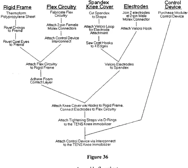
4.2 TENS Knee Immobilizer...54
4.2.1 Concept Development...54
4.2.2 System-level Design...73
4.2.3 Detail Design...85
4.2.4 Testing & Refinement...85
4.2.5 Production Ramp-up (Future Work)...92
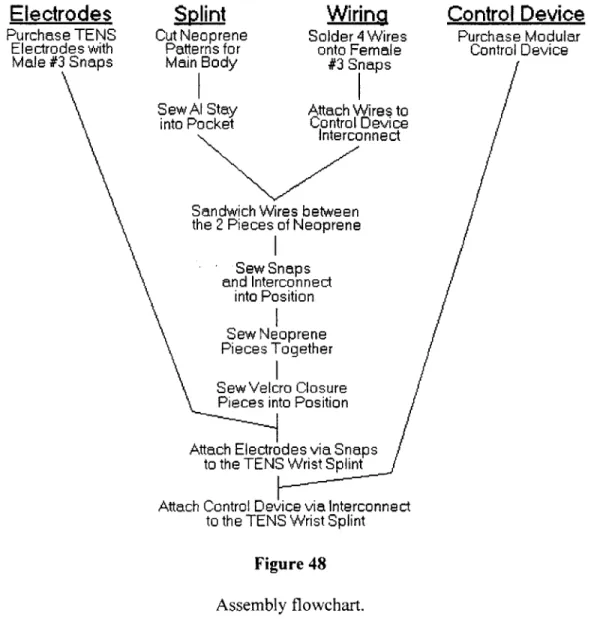
4.3 TENS Wrist Splint...92
4.3.1 Concept Development...92
4.3.2 System-level Design...102
4.3.3 Detail Design...106
4.3.4 Testing & Refinement...106
4.3.5 Production Ramp-up (Future Work)...IlI 5.0 CONCLUSIONS & FUTURE WORK...112
6.0 ACKNOWLEDGMENTS...114
7.0 REFERENCES...115
1.0 INTRODUCTION
A structured design process was applied to the development of novel orthopedic devices
that incorporate transcutaneous electrical nerve stimulation (TENS) for the treatment of acute pain. A modular design was adopted that allows a standardized controls and battery package to be used in a variety of splints, braces, and immobilizers. Electrode arrays were developed for many different areas where TENS therapy has been shown effective including the neck, cervical spine, wrist, shoulder, elbow, hand, thumb, scapula, abdomen, knee, hip, and ankle. A prototype knee immobilizer and a wrist splint with integrated TENS technology were created as platforms to test modularity and to serve as proofs of concept.
Therapeutic TENS devices developed as a new modality for the treatment of pain in the late 1960s due in large part to a greater understanding of the nervous system and the ways in which. pain signals are transmitted and muted. Gate control- theory, the seminal work on pain developed by psychologist Ronald Melzack and physiologist Patrick D. Wall whom met while faculty at MIT [1], most directly led to TENS. It holds that the painful stimulation applied at afferent nerve endings is not the same as the painful stimulation transmitted to the brain. Instead, stimulation is pooled at the dorsal horn of the spinal cord. There, the substantia gelatinosa modulates the signal actually passed to consciousness based on input from both nerve endings modulating pain and those modulating touch. Melzack and Wall theorized that an increase in the stimulation of nerve endings modulating touch would decrease the sensation of pain due to 'gating' at the dorsal horn. TENS seeks to stimulate touch nerve fibers at a pain site with low current electrical waveforms below the threshold of nerve endings modulating pain.
TENS is an attractive alternative to conventional methods of relief for the symptomatic
aspects of pain. Drug therapy can be highly addictive with severe side effects. Surgical procedures are even more drastic than medication, can lead to debilitation, and are often ineffective. TENS has been proven safe with very minimal side effects. Its contraindications are few and clinical trials have shown that it can help a significant percentage of people that needlessly suffer from pain.
Pain is often classified into two categories, acute and chronic, based on its duration. Acute pain is short lived with an identifiable cause and purpose. Chronic is much longer lasting, and its cause and purpose is not always clear. TENS has traditionally been used as a pain modulation technique in the treatment of chronic pain. However, research suggests that TENS is most effective in early treatments and when applied close to the onset of pain. It is therefore perhaps better suited to the modulation of acute pain. Acute pain sufferers represent a much larger patient population than chronic sufferers. This study sought to seamlessly integrate TENS into products aimed at the acute pain market. Acute pain sufferers have different needs than those with chronic pain. They generally live more active lifestyles and require therapies sympathetic to their needs. Currently available TENS devices consist of a TENS stimulator connected by long, entangling wires to TENS electrodes placed at the site of pain. The user alters stimulation parameters to find optimum settings. The devices are often unwieldy and their use is not
7
always intuitive. Advances in battery technology and microprocessing capability have made it possible to create more intuitive, essentially wireless TENS devices that limit impact and interference with the lifestyle of the patient.
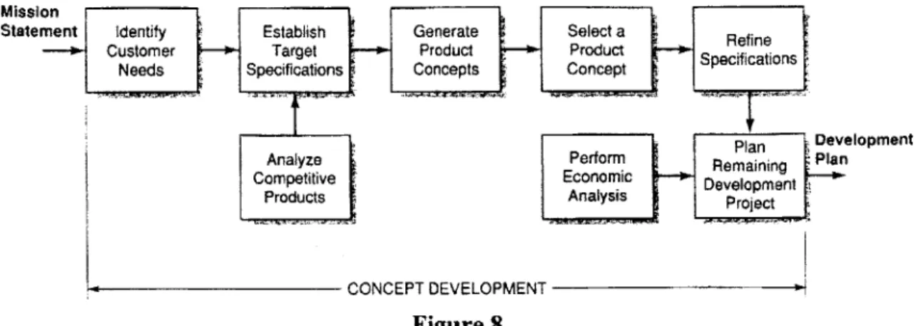
This papers presents an engineering study aimed at applying a structured design process to the development of orthopedic TENS devices for the treatment of acute pain. The major steps in a generic design process are concept development, system-level design, detail design, testing and refinement, and production ramp-up. Concept development can be further delineated into identification of customer needs, establishment of target specifications, analysis of competitive products, concept generation, concept selection, refinement of specifications, economic analysis, and project planning.
A new and innovative modular TENS control device was adopted as the platform from
which an entire product line is being developed. The product line focuses on three areas of concurrent development: 'Band-Aid"' TENS devices; remotely operated TENS devices; and orthopedic braces, splints, and immobilizers with integrated TENS technology. This study focused on the final area. Specifically, c prototype knee immobilizers and wrist splints were developed. The devices are presented herein from problem statement through prototype in relation to the structured design process. Methodologies for each of the constitutive steps in the process are provided in the context of application to these orthopedic TENS devices.
The goals of this paper are twofold:
1. To provide the practitioner of product development with a case study for examination
and discussion of the major steps of development, design methodologies, pitfalls, and lessons learned
2. To facilitate and expand the use of TENS for the treatment of acute pain by creating devices focused on the needs of the designated patient population
Within these two goals, it is my hope that both product developers and members of the medical community can learn about the issues of importance to one another so that communication between our disciplines can proceed in a slightly more informed manner, thereby facilitating advances of importance to us all.
Before the TENS knee immobilizer and wrist splint are presented, a background chapter develops an understanding of the problem. As this paper is at least in part written for the practicing engineer, it expects very little prior knowledge about the structure or function of the human nervous system. The salient features at the cellular, organ, and patient scales are discussed. Types of pain, pain transmission and modulation theory, and methods of treating its symptomatic aspects are then presented. The background section concludes with a discussion of TENS. In turn, it addresses historical development, possible mechanisms of action, and clinical efficacy.
Once the reader has attained a solid understanding of the background and impetus for the study, a chapter on the design procedure presents a structured design process. Next, prototype discussion gets to the heart of the study and systematically examines the
application of the design process to a real-world problem: namely, the development of prototype TENS knee immobilizes and wrist splints for the treatment of acute pain. Finally, conclusions sum up the work and discuss the role of a structured methodology in successful development efforts. By remaining focused on the needs of the patient
9
2.0 BACKGROUND
Before application of the structured design process continues, an understanding of the nervous system, pain theory, and TENS is necessary. Section 2.1 lays out a simplified model of the nervous system at each of three scales relevant to any medical device: the cellular (10 pim), organ (1 cm) and patient scales (1 m). Section 2.2 then establishes a solid foundation for a discussion of pain and its treatment by addressing its physical, physiological, and psychological aspects. Pain transmission and modulation theory is developed in its historical context from Specificity and Pattern through Gate Control and opiate receptors. Gate Control theory, first proposed in 1965, provided scientific justification for the use of electrical stimulation as a method of pain modulation. The discovery of spinal opiate and nonopiate receptors shortly thereafter provided further evidence that noxious stimuli could, in fact, be modulated by selective stimulation of afferent nerve fibers.
Section 2.3 begins with a brief history of electrotherapy. It then discusses TENS, its possible mechanisms of action, its clinical efficacy, and its use as part of comprehensive therapy aimed at breaking the Pain Cycle. As a modality, TENS has been shown safe and effective in relieving the symptomatic aspects of pain.
2.1 The Nervous System
The human nervous system allows us to interact with our environment in ways that are both elegant in their simplicity and fantastically incomprehensible in their subtlety. It is responsible for the rapid transfer of information in the form of electrical signals. Section
2.1 develops an understanding of its function from the microscopic to the macroscopic
level.
2.1.1 The Cellular Scale
The nervous system at this fundamental scale is relatively well characterized in the literature. Basic information on its structure and function is available from a variety of sources. The discussion here is principally developed from Review of Medical Physiology by William F. Ganong [2] and Electrical Nerve Stimulation: Theory, Experiments, and Applications by Frank Rattay [3].
The cells of the nervous system are called neurons. The human nervous system contains about 1 trillion neurons, which exhibit significant differentiation appropriate to their specialized functions. The great majority of these cells are concentrated in the central nervous system (CNS). The remainder comprise the peripheral nerve system, which is made up of sensory, or afferent, neurons that send information from the body to the CNS and motor, or efferent, neurons (also known as motoneurons) that send information from the CNS to the body.
Despite their specialization, most neurons are comprised of the same basic parts. The spinal motor neuron of Figure 1 illustrates the salient features.
Cll body (som'a) Initial sagmantNoeoRave hwnclL Axon hillock Terminal buttons D endrites
Input
Conductile
Output
Region
Region
Region
Generation
Region
Figure 1
Spinal motor neuron with myelinated axon.
(Adapted from Ganong, William F. Review of Medical Physiology, 18th ed. Copyright 1997 by Appleton & Lange; Stamford, Connecticut [2; p. 48].)
The soma is the cell body of the neuron, which encloses the nucleus. One or more
processes extend out from this body. A unipolar neuron has only one process, a bipolar has two, and a multipolar neuron has three or more processes. The spinal motor neuron has 5-7. One process is usually much longer that the others and is known as the nerve fiber, or axon. The shorter processes are called dendrites. The axon carries outgoing information to areas up to a meter away from the cell body. The first section of the axon is called the initial segment. The initial segment generates propagated electrical impulses. At the end region, the axon divides into several branches, which conclude at
terminal buttons. At this widening, known as the synapse, the neuron is in close contact
with other nerve cells. The terminal buttons store synaptic transmitters that the neuron secretes to communicate with other cells. Up to 200,000 synapses, which bring in information from other nerve cells, cover the dendrites and cell body. Some promote, while others inhibit, excitation. Heat, electricity, chemicals, pressure, or other stimuli at the Input Region excite sensory neurons. These inputs sum together and propagate into the axon. If they are small, no information will be transmitted by the neuron. However, excitation above a certain threshold generates an action potential, or electrical impulse. This is commonly known as the all-or-nothing principle.
To summarize, four key regions make up the neuron. The Input Region, comprised of the dendrites and soma, integrates local potential changes from multiple synaptic connections. Next, a Generation Region (the initial segment or the initial node of
ll
Ranvier) actually creates propagated action potentials when local potential changes exceed the threshold value. Then, a Conductile Region (the axon) transmits the propagated impulses to the nerve endings. Finally, an Output Region releases synaptic transmitters, and the information is conveyed to other neurons.
A long cylinder of material known as axoplasm, surrounded by an electrically excitable
membrane, makes up the axon. Axon diameter and insulation by myelin, a protein-lipid complex composed of the cell membrane of Schwann cells wrapped around the axon, determines conduction velocity. Neurons surrounded by this complex are deemed myelinated. The myelin sheath covers the axon except at its ending and at the nodes of
Ranvier. Nodes of Ranvier are about 1 pm in size and about 1mm apart. Myelin
significantly increases a neuron's electrical insulation and, in turn, its conduction velocity.
Some nerve cells are merely surrounded by Schwann cells without the myelin wrapping; these are termed unmyelinated. Conduction velocity in unmyelinated neurons is much slower than in myelinated and is proportional to the square root of fiber diameter.
2.1.2 The Organ Scale
The sensory neurons of the peripheral nervous system embedded in the dermis of the skin are of pertinent interest to TENS (and gate theory). Peripheral neurons specialize to convey different types of information and are generally classified by either letter or number according to their specialization. Table I classifies the neurons by letter and lists their function, axonal diameter, and electrical characteristics. Table 2 then lists their less common numerical classification.
Table 1
Nerve fiber types in mammalian nerve.*
(From Ganong, William F. Review of Medical Physiology, 18th ed. Copyright 1997 by Appleton & Lange; Stamford, Connecticut [2; p. 57].)
Absolute
Fiber Conduction Spike Refractory
Diameter Velocity Duration Period
Fiber Type Function (pM) (m/s) (ms) (ms)
A a Proprioception; 12-20 70-120 Somatic motor
p
Touch, pressure 5-12 30-70 y Motor to Muscle 0.4-0.5 0.4-1 spindles 3-6 15-306 Pain, cold, touch 2-5 12-30
B Preganglionic autonomic <3 3-15 1.2 1.2
C
Dorsal Root Pain, temperature, 2 2
some mechanoreception, 0.4-1.2 0.5-2 reflex responses
Sympathetic Postganglionic 0.3-1.3 0.7-2.3 2 2
sympathetics I I I
*A and B fibers are myelinated; C fibers are unmyelinated.
Table 2
Numerical classification of sensory neurons.
(From Ganong, William F. Review of Medical Physiology, 18th ed. Copyright 1997 by Appleton & Lange; Stamford, Connecticut [2; p. 57].)
Number Origin Fiber Type
Ia Muscle spindle, annulo-spinal ending. A-a
Ib Golgi tendon organ. A-a
II Muscle spindle, flower-spray ending; A-P
touch, pressure.
III Pain and cold receptors; some touch A-6
receptors.
IV Pain, temperature, and other receptors. Dorsal root C
Note that small diameter A-6 and dorsal root C fibers are the nociceptors - nerves that convey painful excitation. The A-8 fibers are myelinated and conduct information much more rapidly than the C fibers. Pain provoked by A-8 fibers is usually of short duration but is sharp, distinct, and well localized. Pain carried by C fibers is more of the dull, aching variety that is diffuse and long lasting [4; pp. 2-3]. Also notice that the much
13
larger diameter and faster conducting A-p fibers transmit touch and pressure stimuli.
This will be of particular interest when discussing Gate Control theory in Section 2.2.
2.1.3 The Patient Scale
The patient scale encompasses the whole of the human nervous system, which influences reflexes, hearing, vision, smell, taste, posture, autonomic actions, emotions, memory, behavior, temperature, touch, pain, and a host of other life-sustaining activities. The nervous system is far too broad a topic to discuss here in any detail, but of pertinent interest to TENS are the transmission pathways governing touch and pain. How does stimulation of sensory neurons at a location significantly remote from the brain enter consciousness; affect emotion, memory, conditioning, etc.; and provoke a coordinated response from the body? Is it possible to modulate the information passed to the brain and thereby diminish or relieve the symptomatic aspects of pain? This section attempts to explain the sensory pathways in somewhat general terms. Although some details are still unclear, especially in the cognitive functions of the brain, the pathways are well characterized in the literature, and have been for some time. The discussion here is primarily based on Essentials of Neurology by Lord Walton [5] and An Evaluation of Transcutaneous Electrical Nerve Stimulation for the Treatment of Pain Related to Spinal Cord Injury by Jason N. Doctor [6]. Secondary information is also derived from Relief of Pain by TENS by Bengt Sjoelund and Margareta Eriksson [7], and Pain Mechanisms:
A New Theor by Ronald Melzack and Patrick Wall [1].
Figure 2
The sensory pathways. The central fibers of the sensory nerves enter the cord through the posterior roots. The ascending branches of those mediating fine touch, vibration and kinaesthetic sensation run in the posterior white columns to the gracile and cuneate nuclei. Here they relay and cross to form the medial lemniscus, which traverses the medulla and pons and midbrain. The fibers mediating pain and temperature and gross touch enter the dorsal grey column and relay in the substantia gelatinosa. The second order neurons cross the cord and form two tracts, the anterior and lateral spinothalamic tracts. These come together in the medulla and then join the medial lemniscus. This large sensory bundle of fibers (the lemniscus) which carries all forms of sensation from the opposite side of the body terminates in the ventral nuclei of the thalamus. From the thalamus, tertiary neurons pass to the sensory cortex. (The small drawing to the right shows the positions of the medial lemniscus and the spinothalamic tract in the medulla.)
(Conybeare's Textbook of Medicine, 16th ed. W.N. Mann, Editor. Copyright 1975 by Churchill Livingstone; Edinburgh, UK [8].)
15
Action potentials regulating touch, pain, temperature, etc. are synaptically transmitted to the cells of the first sensory neuron in the posterior root ganglia. These neurons are bipolar, having peripheral dendrites that convey afferent impulses from the sensory receptors, and central axons that enter the spinal cord in the posterior nerve roots or dorsal horn [5; p. 164]. Here, all impulses are pooled and the analgesic effects of TENS
are thought to take place. Neurons arising from the substantia gelatinosa modify or 'gate' the sensory stimuli (especially pain) sent on to transmission cells (T cells) based on the pool of incoming impulses. T cells then convey the information on to higher levels. Section 2.2 discusses gating in more detail.
After gating in the posterior horn, pain and temperature impulses cross the spinal cord and travel up the lateral portion of the spinothalamic tract on secondary neurons. Most touch stimuli travel up the posterior column for a short distance, then cross over the midline and join the anterior portion of the spinothalamic tract. Meanwhile, other impulses travel up the posterior columns. The sensory fibers of the spinothalamic tracts enter the brain at the medulla oblongata. Figure 3 presents the major structures of the brain.
Precentral Central Postcentral
sulcus sulcus suicus
Cingulate sulcus Parieto-occipital Paracentral fissure lobule yrus Calcarine 9 7 7 111 fissure Corpus callosum
-Olfactory tract -C Cerebellarrbla
Interventricular hemisphere
Vermis of cerebellum
Hypothalamus
Fourth ventricle
Optic chiasma Medulla oblongata
Mammillary body
Thalamus (lateral wall of third ventricle) Pons Pineal body Midbrain Cerebral aqueduct
Figure 3
A diagram of the medial aspect of the right cerebral hemisphere.
(From Gardner, E. Fundamentals of Neurology, 4th ed. Copyright 1963 by Saunders; Philadelphia and London [9; p. 128].)
Upon reaching the medulla oblongata. the fibers travel upwards through the pons and midbrain and terminate in the thalamus. This represents the end of the sensory portion of the nervous system. Tertiary fibers communicate with the sensory cortex and coordinate a response.
The thalamus regulates emotion while the cerebral cortex deals with higher order functions such as memory and learning. Although great progress has been made in determining where different forms of information are processed or stored in the brain,
how such information is stored and processed into a response is still unclear. For TENS,
perception is of much greater importance than response. Although studies have shown that pain can be modulated directly in the brain [6], TENS acts at lower levels in the spinal cord. For our purposes, it is therefore unnecessary to discuss processes of the brain in more detail at this time.
This section gave a brief description of the nervous system at the cellular, organ, and patient scales. It provided the necessary background for an informed discussion on pain: what it is, how it's caused, and methods of controlling it. These, and other issues, are developed in Section 2.2.
2.2 Pain
Yes - Pain! Only you cause humans to become really human. -Alphonse de Lamartine: Harmonies
Pain. The word conjures complex and highly personal images of hardship. However, pain can act as a diagnostic aid and lead to a comprehensive treatment program aimed at correcting pathology, controlling symptomatic manifestation, and preventing recurrence. Its symptomatic aspects can broadly be grouped into three categories: physical (trauma, heat, cold), physiological (spasm, inflammation, sensory deprivation, ANS dysfunction), and psychological (emotional). Alleviation of its symptomatic aspects must occur in conjunction with an attempt to rehabilitate the structure that has given rise to the symptoms [10; p. 7]. This section begins with a description of acute and chronic pain and discusses the pain cycle. It then continues Section 2.1's analysis of the pain transmission pathways by developing our understanding in a historical context and specifically focusing on the mechanisms of pain transmission and inhibition. Finally, it lists current techniques available for the relief of pain.
2.2.1 Acute vs. Chronic Pain & the Pain Cycle
Pain has an element of blank; It cannot recollect
When it began or if there were
A time when it was not.
-Emily Dickinson
Pain is often classified into two types, acute and chronic, in accordance with its duration. Acute pain is short lived, normally lasting no more than a few weeks. It usually leaves
17
no permanent psychological distress, and its cause and purpose are readily identifiable.
By effectively treating the cause while modulating the pain (with heat, massage,
medication, TENS, etc.), relief and rehabilitation can rapidly be obtained [11].
Chronic pain is a different and much more complex animal. Pain that has lasted longer than 6 months is generally termed chronic. Chronic pain is both a physical and psychological problem. Its cause is often unclear, its biological purpose is suspect, and it can persist indefinitely. Chronic pain patients frequently abuse medications, undergo surgical procedures without benefit, and become depressed, hypochondriacal, and hysterical [10]. By some estimates, chronic pain is the most costly health problem in the United States. When direct medical expenses, lost income, lost productivity, compensation payments, and legal fees are all added together, the annual bill approaches $50 billion [11].
In their book Clinical Transcutaneous Electrical Nerve Stimulation, Mannheimer and Lampe describe the pain cycle [10; pp. 7-9]. When trauma occurs, the corresponding pain causes the body to protect itself by guarding. Guarding produces muscle tension, which reduces the blood supply and increases metabolites in the affected area. Prolonged guarding leads to fatigue of the nerve fibers and more pain. This, in turn, leads to more guarding, more psychological stress; and the pain is progressively amplified. Clearly, it is important to manage the pain response in order to prevent the pain cycle in those with acute pain, and to break the cycle in those with chronic pain.
If acute pain persists without improvement for more than a few weeks, it may very well
become a chronic disorder. Therefore, it is imperative that the cause of such pain be treated quickly and aggressively in conjunction with a pain modulating technique such as
TENS to relieve the symptomatic aspects. It must be noted that strictly treating the
symptomatic aspects without addressing the cause can precipitate false expectations of healing and lead to chronic behavior. Although most clinicians agree that TENS could be very useful in the treatment of acute pain, research has surprisingly focused on its application to chronic pain. The reasons are many and are described in more detail in Section 2.3. This study sought to integrate TENS into user-friendly, over-the-counter orthopedic devices for the symptomatic treatment of acute pain. Chapter 4 details these efforts.
2.2.2 Historical Development of Pain Theory & Descending Pain Inhibition Pathways Until the publication of Melzack and Wall's seminal work on pain transmission in 1965, thoughts on pain were nearly equally divided into two opposing schools of thought -and had been since before the turn of the century. In 1894, M. von Frey and A. Goldscheider independently proposed pain transmission theories: specificity and pattern, respectively. Specificity held that "pain is a specific modality like vision and hearing, with its own central and peripheral apparatus," while pattern theory held that pain is produced by "intense stimulation of nonspecific receptors."[1] In pattern theory, there was no such thing as specific fibers or specific endings.
In light of Section 2.1. it is clear that neither theory is strictly correct. Pattern theory recognized the significance of both stimulation intensity and summation in the central nervous system, but it did not, and could not, account for the physiological fact of neuronal specialization. Specificity theory accounted for specialization, but it also assumed a direct link between the point of stimulation and the brain and did not account for central summation.
Melzack and Wall recognized of the shortcomings of both theories and focused on the role of the substantia gelatinosa in pain transmission and modulation. Their work is generally referred to as the Gate Control Theory of Pain. They conceived pain transmission as the control loop seen in Figure 4.
CENTRAL
CONTROL
GATE CONTROL SYSTEM
L
INPUT S G T --- AC TION
SYSTE M
S
Figure 4
Schematic diagram of the gate control theory of pain mechanisms: L, the large-diameter fibers; S, the small-diameter fibers. The fibers project to the substantia gelatinosa (SG) and first transmission (T) cells. The inhibitory effect exerted by the SG on the afferent fiber terminals is increased by activity in L fibers and decreased by activity in S fibers. The central control trigger is represented by a line running from the large-fiber system to the central control mechanisms; these mechanisms, in turn, project back to the gate control system. The T cells project to the entry cells of the action system. +, Excitation;
-, inhibition.
(From Melzack, R. & Wall, P. D. Pain Mechanisms: A New Theory. Science, Vol. 150, Number 3699. 11/19/65 [1].)
The theory holds that the painful stimulation actually transmitted to the brain (and other higher levels) is not the same as the painful stimulation applied at afferent nerve endings. Instead, the substantia gelatinosa modulates the signal based on incoming input from small and large diameter nerve fibers. Signals from small diameter axons (including the
19
A-6 and dorsal root C fibers that transmit pain) act to increase the intensity of pain sent to
higher levels while signals from large diameter fibers (including A-p fibers that transmit touch) act to decrease the intensity of pain. Thus, the substantia gelatinosa acts as a 'gate' to regulate pain.
Meanwhile, the central trigger accounts for the ability of attention, emotion, and memory to influence afferent conduction. Efferent fibers from the brain can act by descending inhibitory pathways and activate the control cells in the dorsal horns. Thereby, the brain can directly close the gate [4; p. 13]. Examples include war victims and athletes who often do not experience pain until after the battle or athletic contest is complete.
Although the new theory was not perfect in explaining the highly complex mechanisms of pain, it focused further research on the mechanisms of both transmission and the descending inhibitory pathways. .This research directly led to several 'new' treatment modalities for control of the symptomatic aspects of pain. TENS is one such modality.
TENS seeks to stimulate large diameter A-p fibers in the painful area with non-painful,
low current electrical waveforms - thereby closing the gate. Wall and Sweet [12] conducted the first clinical trials with electrical stimulation based on gate control in 1967. Section 2.3 discusses TENS in much greater detail.
A great deal of work has been done since 1965 in an attempt to prove or disprove the gate
control theory. The research has led to a much fuller understanding of the physiological mechanisms and descending inhibitory pathways of pain. Significantly, the method in which the body regulates, modulates, and transmits information between neurons is much clearer at the chemical level. In 1973, Terenius discovered that endogenous opiates and opiate-like drugs bind to specific receptors in the brain to produce their pain relieving effect [4; p. 14]. Serotonin was the first inhibitory neurotransmitter identified. The discovery of opiate peptides, which inhibit pain transmission in the dorsal horn of the spinal cord and in the brain, soon followed. The first peptide was termed enkephalin, and the others are known as endorphins [11].
The descending pathways of pain inhibition have also been characterized much more fully. They are mentioned here briefly. Pain can be inhibited at several different physiological levels. The integrated activity of neurons at these levels is collectively known as the descending pain system. Inhibition appears to occur in the CNS at four levels. The first three levels are in the brain: first, within the cortex and diencephalon, then in the periaqueductal gray (PAG) of the midbrain, and finally in the rostroventral medulla, which includes the nucleus raphe magnus (NRM). The fourth level is the aforementioned dorsal horn of the spinal cord. Opiate and nonopiate neurotransmitters act at these levels to inhibit pain [6; pp. 9-10].
A new generation of drugs developed after the gate theory is also expected to gain FDA
approval in the very near future. These analgesics attack specific pain mechanisms and promise to be more effective with fewer side effects.
2.2.3 Methods of Treating the Symptomatic Aspects of Pain
Now that we know what pain is, how it's transmitted, and how it's modulated, the focus must shift to its treatment. Many treatment modalities exist, but the gold standard by which all other treatments are assessed is analgesic medications. Drug therapy acts at all four levels of the descending pain system, as well as peripherally by relieving hyperalgesia (increased sensitivity to noxious stimuli) which occurs at the site of injury [2].
Available drugs are broadly grouped into narcotics and non-narcotics. The best and most widely used non-narcotic drug is aspirin. Aspirin reduces inflammation and desensitizes nociceptors. However, the threat of serious side effects in children requires that care be taken in its administration. Of the narcotics, morphine is the standard. 20-25 other narcotic drugs are also in use; these are either wholly synthetic or semi-synthetic opiate derivatives and include heroin, codeine, Darvon, and Demerol. Narcotic side effects include sedation, confusion, nausea, constipation, and depressed respiration. Unfortunately, all of the more powerful analgesics are highly addictive. There is no efficacy ceiling, so the dosage can always be increased as a tolerance is developed. However, increasing the dosage increases the incident of side effects and physical addiction. Other available drug therapies include the antidepressants, tranquilizers,
antihistamines, anticonvulsants, and corticosteroids [5, 11].
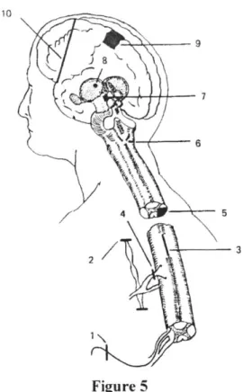
Long-term, intractable chronic pain, at times leads to more drastic surgical methods of pain relief. Figure 5 illustrates several of these procedures.
21 10 9 f8\ 6 Figure 5
Diagram of various surgical procedures designed to alleviate pain: 1, nerve section; 2, sympathectomy (for visceral pain); 3, myelotomy to section spinothalamic fibers in anterior white commissure; 4, posterior rhizotomy; 5, anterolateral cordotomy; 6, medullary tractotomy; 7, mesencephalic tractotomy; 8, thalamotomy; 9, cingulate gyrectomy; 10, prefrontal lobotomy.
(From Ganong, William F. Review of Medical Physiology, 18th ed. Copyright 1997 by Appleton & Lange; Stamford, Connecticut [2; p. 138].)
Other, more traditional methods of pain relief should be attempted before drug therapy and surgery. These include rest, temporary immobilization, stretching, application of heat and cold, exercise, and massage. Psychological treatment may also be appropriate when the cause of pain cannot be identified or when lifestyle adjustments are necessary to deal with the pain. Some people find relief in alternative therapies, such as acupuncture and chiropractic. The placebo effect should also be attempted: a consistent 1/3 of patients responds to placebos for pain relief.
Finally, there is electrotherapy; this includes direct stimulation of the brain and spinal cord via surgically implanted electrodes at the four levels of the descending pathways and stimulation of afferent neurons via electrodes placed on the skin surface in an attempt to close the gate in the dorsal horn (TENS).
This section has developed an understanding of acute and chronic pain, as well as the pain cycle. It's also explained the pain transmission pathways in a historical context while focusing on the mechanisms of inhibition. Finally, current techniques available for the relief of pain were discussed. Section 2.3 concludes the background chapter by focusing more closely on TENS: its history, an explanation of the technology, its mechanisms of action, and its clinical efficacy.
2.3 TENS, a 'New' Modality for Pain Modulation
This section begins with a history of electrotherapy. It then discusses TENS and its possible mechanisms of action. Finally, clinical efficacy is addressed.
2.3.1 A Brief History of Medical Electricity Is there a thing of which it is said,
"See, this is new"? It has already been,
in the ages before us.
The people of long ago are not remembered, nor will there be any remembrance
ofpeople yet to come by those who come after them. -Ecclesiastes 1:10-11
Although the modem use of electricity for pain modulation only dates to the mid- 1 960s, its origins can be traced at least to ancient Rome, and probably to Egypt circa 2500 BC. Stone carvings in tombs of that era indicate that an electric fish native to the Nile was used to treat pain [7; p. 3]. The book Compositiones Medicae by the Roman physician Scribonius Largus contains the first written record of such treatments. Written in the year 46 AD, the text recommends use of a torpedo fish for the treatment of headache and gout
[3; p. 3]. Figure 6, excerpted from an 18th century text, reproduces a section of Largus's
23
s Capitis dolor
quemuis
ueterem
&
intolerabilem protinustollit,& imp era
petuum remediat torpedo uiua nigr,
impofira coloco
g
indolore eft, donec
definat dolor,
&
obfrupefcat ea pars:
quo-d
camn
primi
fenferitremouelut
remedium. ne fenfus auferatur eius par
cis. Plures
agt-parldatfCunt
eius
generfs
torpedines
*,-quia
nonung
aixad
duas,
trestreref
Pn-dercuatioid-eft, torpor
1
quod
fig
efemediationis,
Srneokrus LARCUS DESIGNATUS
"Scripta mea Latina medicalis, codex instar" editio prima, cap. XI. De compositione medicamentum liber.
Ed. J.M.Berthold, Argentor, 1736. Figure 6
An excerpt from the first written account of the use of electrical stimulation for the treatment of pain, written in 46 AD by the Roman physician Scribonius Largus.
(From Jenker, F. L. Electric Pain Control. Copyright 1995
by Springer-Verlag; New York [2; frontispiece].)
The translation of Figure 6 reads:
Chapter XI: Unbearable headaches, as enduring as they may be, are relieved immediately and cured forever by the living black electric ray if it is held in contact with the aching area until pain disappears and this part is numbed; as soon as this is felt, the remedy should be removed such as not to interfere with sensation. There are several species of these fishes; since the effect does not occur at once, but after 2 or 3 applications, healing, that is numbness which is the sign of healing...
The text goes on to say:
Chapter XIII: For every type of podagra place an electric ray under the feet, just when the pain comes on. But do not stand on a dry part of the beach, but where the sea is wetting it. Keep the contact to the ray long enough to make foot and skin areas numb up to the knee. The pain will disappear immediately and will hold cured for the future. In this fashion Antheros, a public employee, who had been liberated by Tiberius, was cured [13; p. 5].
Of course, electrical eels aren't really suited to clinical use. With an increased
Richard Lovett published the first book written in English on medical electricity in 1756 under the title Subtil Medium Proved. John Wesley, founder of the Methodist Church, was an early proponent of electrotherapy and published his own text on the subject in
1759. Wesley saw electricity as the soul of the universe [10; p. 1]. W. J. Oliver, an
American Physician working in the mid-19th century, reported that he could effectively modulate pain during surgery by applying well-moistened dressings attached to copper wires above and below the area of surgery. Although this and other early work was promising, anesthetics were introduced a few decades later, and the scientific community quickly abandoned electrical stimulation as a method of pain modulation. Interest wasn't rekindled until research in the 1950s and '60s led to the gate theory [7; pp. 6-7].
The underpinnings for modem scientific thought about the efficacy of electrical stimulation can be traced to Melzack and Wall's now famous gate control theory, first published in 1965. It gave credibility and directly led to the development of TENS. Shortly after the gate theory was published, researchers attempted to modulate pain by direct electrical stimulation of the dorsal column of the spinal cord. In 1967 Wall and Sweet conducted the first trials on the clinical use of TENS for the treatment of pain [12].
TENS seeks to stimulate afferent A-p fibers with low current electrical stimulation
applied at a pain site via conducting electrodes. The gate theory predicts that this non-painful stimulation can diminish or block the non-painful stimulation transmitted to higher centers. TENS was originally used to evaluate which patients would respond favorably to direct stimulation of the dorsal column. It quickly became apparent that afferent stimulation was nearly as effective as direct stimulation in relieving pain for many patients [10; p. 3]. Thus, TENS was born as a new and freestanding modality for the treatment of pain. It has since evolved into an industry valued in excess of $200 million annually.
2.3.2 What is TENS?
The gate control theory of pain suggested that nociceptively evoked activity in ascending pathways could be modulated by mechanoreceptive (touch) stimuli. It followed that if an extra supply of mechanoreceptive stimuli could be generated at a pain site, the intensity and duration of the pain should be reduced or eliminated. Large, mechanoreceptive
(A-P)
nerve fibers can be selectively activated with electrical current due to their low inner longitudinal resistance [7; p. 21]. Low resistance allows stimulation at intensities below threshold for other nerve endings. The easiest way to selectively activate these fibers isby attaching electrodes to the skin at the site of pain. A non-painful, alternating current
electrical waveform is then applied to the electrodes. This technique is known as Transcutaneous Electrical Nerve Stimulation, or TENS. Most TENS devices produce asymmetric biphasic waveforms with a narrow pulse followed by a negative spike with exponential delay. This produces a zero net DC current. A few devices produce a monopolar spike [14; p. 2]. Figure 7 shows a few common waveforms.
25
0
K
-0
0
Figure 7
Commonly used TENS waveforms.
(From Mykleburst, Joel B. Neural Stimulation. Vol. I. Copyright 1985 by CRC Press; Boca Raton, FL [14; p. 3].)
The stimulation parameters - frequency, pulse width, and amplitude - can be altered to activate different pain modulation mechanisms. Frequency refers to the number of electrical waveforms delivered per second, pulse width is the duration of each waveform, and amplitude refers to the height of each pulse above baseline. Skin resistance, body weight, and inter-electrode distance all influence amplitude. Thus, amplitude is often adjusted on a patient-to-patient basis in the milliampere range in order to find an optimum setting [6; p. 29]. Frequencies used range from 1-200Hz, while pulse widths vary between 0 and about 500ps. Much progress has been made in the clinical use of
TENS since its inception. Patients who do not respond or are uncomfortable with one set
of parameters, or mode, are often more receptive to a different mode. A discussion of the basic modes currently in clinical use follows.
Conventional (high
frequency/low
intensity) TENSConventional TENS is based directly on the gate theory and was the mode used by Wall and Sweet in the very first TENS study [12]. Conventional TENS consists of high frequency (50-100Hz), small pulse width (40-100pts) and moderate intensity waveforms.
Several researchers report that this is the most clinically successful mode [15, 16]. It produces a tingling sensation and immediately increases the pain threshold. The onset of relief is very rapid, but the after-effect is limited, usually lasting no longer than the period of stimulation [17].
Acupuncture-like (low
frequency/high
intensity) TENSAcupuncture-like TENS employs low frequencies (1-4Hz), large pulse widths (150-250us), and high intensities near the pain threshold. Motor nerve fibers must be activated, and visual muscle twitching should be produced.
In contrast to conventional TENS, this mode produces a gradual increase in the pain threshold. After-effect is longer as the threshold gradually decreases back to baseline
[17].
Burst Stimulation
TENS efficacy has a tendency to temporarily decrease over the period of one therapy
session. The body seems to adjust to the stimulation parameters and decrease their effectiveness in a process called accommodation. Burst TENS modulates the frequency
by sending waveforms in packets, or bursts, thereby not allowing the body to adjust.
Waveforms are delivered for a period of time, stopped, then restarted. The bursts are conducted at frequencies ranging from 0.25-2Hz. The burst mode can be conducted with either conventional or acupuncture-like TENS parameters [17].
Modulation TENS
Much like the burst mode, modulation TENS seeks to limit the body's accommodation, or ability to adjust to one set of parameters. Beginning with either conventional or acupuncture-like settings, frequency., pulse width, and amplitude are dynamically altered during stimulation in a range of up to 50% around the baseline.
TENS has been used successfully for relief of the symptomatic aspects of many different
types of acute and chronic pain including dental procedures, labor and delivery, musculoskeletal problems, traumatic and post-surgical orthopedic conditions, postoperative recovery, spinal trauma, chronic back pain, and a host of other conditions where avoidance of narcotic analgesics is desirable [18]. TENS has no major side effects or drawbacks. It is safe and is effective in a statistically significant portion of the patient population as discussed in Section 2.3.4.
Although no deaths or serious injuries have been reported, TENS is contraindicated during pregnancy, for patients with pacemakers, and for use across mucous membranes. It is also to be avoided in patients with dementia or psychological disease. The most commonly reported problems are skin allergies or irritation due to energy absorption from the TENS device to the skin [17]. Skin problems can usually be avoided by the use of non-irritating electrogels and by correct placement of TENS electrodes on the surface.
TENS does not treat the cause of nociception, but merely attacks its symptomatic aspects.
It must therefore be stressed that TENS should be used as part of comprehensive therapy aimed at breaking the pain cycle. If used properly TENS has the ability to help a great number of people that needlessly suffer from pain.
27
2.3.3 TENS: Possible Mechanisms of Action
Although TENS was originally justified on the basis of the gate theory, newer research has led to a fuller understanding of the mechanisms activated by TENS stimulation. It
now appears that conventional and acupuncture-like modes activate different mechanisms in the descending pain pathways. Several of the prevailing theories are touched upon here.
Gate Control
Gate control has already been discussed extensively, and is only touched upon here. Stimulation of large diameter afferent fibers inhibits input from small diameter fibers, thereby blocking the pain signal. Gating is thought to take place in the dorsal horn of the spinal cord. The theory predicts analgesia at frequencies between about 75 and 150Hz, but suggests that relief should stop immediately after stimulation is removed. Conventional TENS can be justified on the basis of this theory.
Endogenous Opioid Pathways
Acupuncture-like TENS is thought to act by the release of endogenous opiates [19, 20]. To test this theory, researchers used the opiate antagonist naloxone to determine whether the analgesic effects of TENS could be reversed upon its administration. The effects of acupuncture-like TENS were reversed while conventional TENS analgesia was unaffected, suggesting that the acupuncture-like mode acts by the release of endogenous opiates while the conventional mode acts by other mechanisms [21]. There is also speculation that TENS analgesia is modulated by non-opiate substances, such as GABA, adenosine, or glycine, but little research has been conducted to substantiate these claims [15].
Peripheral Nerve Block
This theory suggests that TENS acts in a manner consistent with local anesthetics that block nerve conduction [13; p. 32]. Impairment of both painful and non-painful sensation during and after TENS has been clinically observed. It appears that C-fibers may be more susceptible to inhibition that myelinated fibers. Thus, conventional TENS may inhibit small afferent input while stimulating large afferent fibers. According to the gate theory, both actions should reduce pain [15].
Others suggest that analgesia occurs as a consequence of sensory distraction with a continuous peripheral stimulus, and not by central neuronal modulation [22]. Some even feel that at least certain TENS modes do nothing more than provide a placebo effect that allows patients to heal themselves [23]. Alternatively, one study concludes that the effects of long-term TENS usage cumulate over time, thus producing plastic changes in the neural pathway [24]. Still others suggest that nerve fatigue leads to a decrease in sensation. From a clinical standpoint, perhaps the mechanisms of action are unimportant:
TENS has and will continue to bring relief to many patients that have not been able or do
not wish (due to side effects) to seek relief by conventional means. Section 2.3.4 focuses on clinical efficacy.
2.3.4 Clinical Efficacy
Double-blind clinical trials to determine what percentage of patients truly benefit from TENS have been very difficult because a placebo cannot be found that is both believable to the patient and doesn't provide some physiological effect [15]. Also, stimulation parameters, pain syndromes, social and psychological considerations, duration of therapy sessions, length of observation, other concurrent pain modalities in use (drugs, massage, exercise, heat, cold, etc.), and a host of other factors have varied from study to study. With that said, Doctor compiled a review of the most reliable studies conducted on TENS efficacy [6]. Table 3 reproduces the review.
Table 3
Notable controlled TENS trials.
(From Doctor, Jason N. An Evaluation of Transcutaneous Electrical Nerve Stimulation (TENS) for the Treatment of Pain Related to a Spinal Cord Injury, Appendices 1-2.
Doctoral Thesis: Univ. of CA, San Diego & San Diego State Univ., 1996 [6].)
Study Subjects Treatment Results
[Design Features*1
Melzack N = 53 Negative Response Active TENS sig. decreased
(1975) Chronic Pain Sight Control (n = 15); no pain relative to control. output (n = 7). Crossover.
[e]
Cooperman et al. N =50 Active TENS (n = 26) and Active TENS sig. less med.
(1975) Post Surgery Sham (n = 24). usage. No sig. difference in
[b] duration of ICU stay.
Thorsteinsson et al. N = 93 Active TENS and Placebo Active TENS sig. less pain
(1978) Chronic Pain TENS (no current). than control. Crossover.
[a]
Long et al. N = 150 Supra-threshold TENS, Supra-threshold TENS sig.
(1979) Chronic Pain subliminal TENS, no less pain than control. battery TENS. Crossover.
Ali et al. N = 40 Active TENS (n = 15), no Active TENS sig. less
(1981) Elective stimulation TENS (n = medicine usage, sig. higher Cholecystectomy 10), no TENS (n = 15). pO2.
[b]
Hansson & Ekbolm N = 62 High Freq. TENS (n No sig. difference between
(1983) Acute Orofacial Pain 22), Low Freq. TENS (n high and low freq. TENS.
= 20), Placebo TENS (n Active TENS sig. less pain
= 20). than placebo.
[b]
Abelson et al. N = 32 Active TENS vs. Placebo Active TENS sig. less pain
(1983) Rheum. Arthritis with (no stimulation). and greater grip strength than
Wrist Involvement Crossover. control.
[a, c]
Lewis et al. N = 28 Active TENS vs. Placebo Sig. improvement in pain (1984) Osteoarthritis with (no stimulation). index & decreased med.
Knee Pain Crossover. usage in both groups. No sig. [a, c] difference between groups.
46% response rate active vs. 43% response rate placebo.
29
Langley et al. N = 33 High Freq. TENS, Low All groups showed sig.
(1984) Rheum. Arthritis and Freq. TENS, Placebo decreases in resting & grip
Chronic Hand TENS. pain. No sig. diff. btwn
Involvement [a, b, c, d, e] groups on power or work scores, overall pain, joint tenderness, grip or resting pain.
Lehman et al. N = 53 Acupuncture TENS No sig. diff. btwn groups on
(1986) Chronic Low Back (n = 17) physician ratings of pain, Pain Active TENS Low Freq. self-rated pain, trunk
(n= 18), Placebo TENS strength, back flex/ext, med (n = 18). use, return to work, post-tx or
[a, b, c] f/u. All groups showed sig. improv. over time.
Smith et al. N = 18 Active TENS (n = 9), No sig. diff. btwn groups post
(1986) Caesarian Section Placebo TENS (n = 9). treatment on pain or med.
Surgery [b, c] usage.
Ordog N = 100 Active TENS, Placebo Active TENS sig. diff. than
(1987) Acute Trauma TENS, Active TENS + 3 placebo at day 2 but not at Outpatients (Sprains, Tylenol, Placebo TENS + day 30. No sig. diff. in med. Lacerations, Fractures, 3 Tylenol. usage for active or placebo.
etc...) [a, b, c]
Finsen N = 51 Active TENS (n = 17), No sig. diff. btwn groups on
(1987) Lower Leg Sham TENS (n = 19), level of pain or med. intake at Amputations Sham TENS + 4 weeks or 1 year follow-up.
Chlorpromazine.
[b, c]
Deyo et al N = 145 Active TENS (n 36), No sig. diff. in any outcome
(1990) Chronic Low Back Sham TENS (n = 36), for subjects receiving active Pain Active TENS + Exercise or sham TENS. Sig.
(n = 37), Sham TENS + improvement was obs. in Exercise (n = 36). every group pre to post, but [a, b, c. d, e] rtn to baseline at 3 months. Marchand et al. N =42 Active TENS (n = 14), Active TENS group showed
(1993) Chronic Low Back Placebo TENS (n = 12), sig. greater reductions in pain Pain Patients control (n = 16). intensity post tx than other
[b, c] two groups. But, no diff. between active TENS and placebo on unpleasantness ratings. Both showed sig.
diff. in comparison to control. *[a]= double-blind trial.
[b]= noncrossover randomized design.
[c] = credible placebo with functional "on" light and/or hum. Or all units encased to preserve blind.
[d] = attention focus control across conditions.
[e] = verbal suggestion to placebo group that active treatment may or may not produce a sensation.
Doctor feels that studies made after 1984 are somewhat more reliable as they used better experimental design and more plausible control conditions. Taken collectively, the results on efficacy are somewhat inconclusive. However, even if TENS can produce little more lasting analgesia than placebos, this still represents a very significant portion of the patient population (30-50%) and justifies its use as a method of pain intervention.
Also note that studies dealing with acute pain generally give favorable results. If the effects of TENS do, in fact, cumulate, this is not surprising. For the purposes of this study, the focus is on acute pain of short duration. As can be seen from Table 3, most work has focused on chronic pain, but the evidence available suggests that TENS is most effective during the first therapy sessions. If this is true, it may be much better suited to acute pain relief.
Section 2.1 developed a broad-based understanding of the human nervous system. It examined the system's function from the micro- to macroscopic levels. Section 2.2 then discussed acute and chronic pain, the pain cycle, the transmission pathways, mechanisms of modulation, and available treatment options. Finally, this section gave a history of electrotherapy, explained TENS, discussed its possible mechanisms of action, and reported on its efficacy. We can now proceed to the design process and its application to the development of stand-alone orthopedic TENS devices for the treatment of acute pain.