M A J O R A R T I C L E
Serum Immunoglobulin A Cross-Strain Blockade
of Human Noroviruses
Lisa C. Lindesmith,1Martina Beltramello,2Jesica Swanstrom,1Taylor A. Jones,1Davide Corti,2,3Antonio Lanzavecchia,2,4 and Ralph S. Baric1
1
Department of Epidemiology, University of North Carolina, Chapel Hill;2Institute for Research in Biomedicine, Bellinzona, Switzerland;3Humabs BioMed SA, Bellinzona, Switzerland; and4Institute of Microbiology, ETH Zurich, Switzerland
Background. Human noroviruses are the leading cause of acute viral gastroenteritis, justifying vaccine develop-ment despite a limited understanding of strain immunity. After genogroup I (GI).1 norovirus infection and immu-nization, blockade antibody titers to multiple virus-like particles (VLPs) increase, suggesting that GI cross-protection may occur.
Methods. Immunoglobulin (Ig)A was purified from sera collected from GI.1-infected participants, and potential neutralization activity was measured using a surrogate neutralization assay based on antibody blockade of ligand binding. Human and mouse monoclonal antibodies (mAbs) were produced to multiple GI VLPs to characterize GI epitopes.
Results. Immunoglobulin A purified from day 14 post-GI.1 challenge sera blocked binding of GI.1, GI.3, and GI.4 to carbohydrate ligands. In some subjects, purified IgA preferentially blocked binding of other GI VLPs compared with GI.1, supporting observations that the immune response to GI.1 infection may be influenced by pre-exposure history. For other subjects, IgA equivalently blocked multiple GI VLPs. Only strain-specific mAbs rec-ognized blockade epitopes, whereas strain cross-reactive mAbs recrec-ognized nonblockade epitopes.
Conclusions. These studies are the first to describe a functional role for serum IgA in norovirus immunity and thefirst to characterize human monoclonal antibodies to GI strains, expanding our understanding of norovirus immunobiology.
Keywords. blockade antibody; BOB assay; IgA; monoclonal antibody; neutralization; Norwalk virus; norovirus.
Noroviruses (NoVs) are a leading cause of gastroenteritis outbreaks in all age groups, contributing to an estimated 21 million illnesses per year in the United States [1]. Although genogroup II (GII).4 NoV strains predominate worldwide, a large number of diverse NoV strains cocir-culate at endemic levels. Genogroup I (GI) strain infec-tions occur most frequently in children and the elderly
[2,3]. Norovirus disease is usually self-limiting in healthy individuals, but it can be severe in the very young, elder-ly, and immunocompromised [3–5]. A NoV vaccine would benefit these vulnerable groups as well as the mil-itary, food-handlers, and support-care workers.
An understanding of the complex antigenic relation-ship between NoV strains will aid in vaccine develop-ment. Currently, a multivalent (GI.1/GII.4C) virus-like particle (VLP) vaccine is in phase I clinical trials [6]. Cross-strain reactive blockade Ab responses, a potential measure of strain cross-neutralization and a correlate to protective immunity in GI.1-challenged participants [7], were identified in serum samples collected during an initial reactogenicity trial of the vaccine [8]. These data support the hypothesis that some similar neutral-izing antibody (Ab) epitopes might exist within both GI and GII NoV strains that could potentially provide targets for a broadly protective vaccine.
Likewise, we have previously characterized Ab respons-es in humans after GI.1 NoV experimental infection [9],
Received 27 April 2015; accepted 1 June 2015.
Presented in part: Annual Meeting for American Society for Virology, June 2014, Fort Collins, CO.
Correspondence: Ralph S. Baric, PhD, 3304 Hooker Research Center, 135 Dauer DR, CB7435, School of Public Health, University of North Carolina-Chapel Hill, Chapel Hill, NC 27599 (rbaric@email.unc.edu).
Open Forum Infectious Diseases
© The Author 2015. Published by Oxford University Press on behalf of the Infectious Diseases Society of America. This is an Open Access article distributed under the terms of the Creative Commons Attribution-NonCommercial-NoDerivs licence (http:// creativecommons.org/licenses/by-nc-nd/4.0/), which permits non-commercial reproduction and distribution of the work, in any medium, provided the original work is not altered or transformed in any way, and that the work is properly cited. For commercial re-use, please contact journals.permissions@oup.com.
DOI: 10.1093/ofid/ofv084
and we found that GI.1 infection induced blockade Ab cross-reactive within a panel of GI VLPs. In this follow-up study using the same serum samples, we extend these observations to identify serum immunoglobulin (Ig)A as a component of the cross-GI blockade Ab response, and we characterize the antigenic relationship between GI VLPs using a panel of monoclonal Abs (mAbs). These mAbs include thefirst reported human mAbs to GI NoV strains.
MATERIALS AND METHODS
Ethics Statement
Serum samples were collected from participants infected with GI.1–1968 in an unpublished pilot study. Participants provided written informed consent, the original study was approved by the Institutional Review Board, and the guidelines for human ex-perimentation were followed in the conduct of clinical research [9].
Human Serum Samples and Genogroup I Virus-Like Particles
The archived, deidentified serum samples collected from GI.1-infected participants and GI VLPs used in these studies are
described in refs. [9,10]. Not all archival samples were available for analyses. Any missing samples are noted in the respective figure legends.
Enzyme Immunoassay, Blockade Antibody, and Blocking of Binding Assays
Enzyme immunoassay (EIA) and blockade Ab assays were per-formed as described for GI VLPs [8] with the following exceptions for the blockade assay: (1) 0.5 µg/mL VLP was used, and (2) the VLP-serum and VLP-porcine gastric mucin binding steps were incubated at 37°C. Blocking of binding (BOB) assay procedures [11] are the same as EIA procedures except that serial dilutions of mouse mAbs were added to GI.1-coated plates before the ad-dition of anti-GI.1 human mAbs at a concentration equal to the EC50titer (effective concentration at 50%) [8,11]. All sera and
mAbs were assayed in duplicate in a minimum of at least 2 in-dependent assays for each VLP.
Avidity Index Calculation
Avidity indexes were determined as described for IgG and IgA EIA as described in ref. [8], with the inclusion of a 10-minute, 7
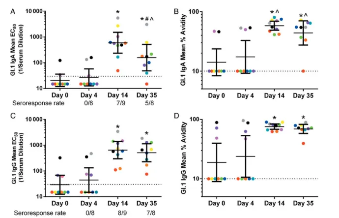
Figure 1. Genogroup I (GI).1 infection induces high-titer, high-avidity homotypic serum immunoglobulin (Ig)A and IgG. Serum samples were assayed for IgA and IgG reactivity to GI.1 by enzyme immunoassay (EIA) and avidity assays. Each color of circle represents the mean EC50for a participant. The group
geometric mean titer (line) and 95% confidence intervals (error bars) for each timepoint are also reported. Samples with titers or avidities below the limit of detection (dashed line) were assigned values of 15 for the EIA assay or 10 for the avidity assay for statistical analysis. *Significantly different from day O (Wilcoxon). ^IgA significantly different from IgG at the same timepoint (Wilcoxon). #Day 14 significantly different from day 35 (Wilcoxon). Three samples were not available for analysis, 1 each on day 0 (gray), day 4 (yellow), and day 35 ( pink). The seroresponse rate is equal to (# Samples Tested/# Matched with day 0).
M urea incubation step after thefirst wash postprimary Ab in-cubation [12]. The index was defined as the average ratio of op-tical density (OD) of Ab bound in the presence of 7 M urea compared with the average OD of Ab bound in the absence of urea at the dilution closest to the Ab EC50. Samples with
Ab levels below the limit of detection were assigned an index of 10 for statistical purposes. All sera and mAbs were assayed in duplicate in a minimum of at least 2 independent assays for each VLP.
Immunoglobulin A Purification
Immunoglobulin A was purified from serum samples using Peptide M/Agarose (InvivoGen) following the manufacturer’s directions, except samples were diluted 6-fold in phosphate-buffered saline beforefiltration instead of being dialyzed. Puri-fied IgA samples were confirmed as IgG negative by EIA.
Monoclonal Antibody Production and Purification
Mouse mAbs to GI.1 and GI.4 and human mAbs reactive to GI VLPs were isolated, produced, and purified as reported previ-ously [13,14].
Statistical Analysis
Statistical analyses were done using GraphPad Prism 6.02. EC50
values or geometric mean titers (GMTs) between days postchal-lenge, or VLPs were compared using the one-way analysis of variance (ANOVA) with Dunnett posttest (ANOVA), when at least 3 values were compared or a Wilcoxon test (Wilcoxon) when only 2 values were compared. A difference was considered significant if the P value was < .05.
RESULTS
Genogroup I .1 Infection Induces Homotypic Immunoglobulin A and Immunoglobulin G With High Titer and Increased Avidity
Genogroup I.1 infection results in significant rises in serum IgG titer to GI.1 at day 14 postchallenge [9]. To further characterize this serum response, we compared GI.1-reactive IgA and IgG titers and avidities at days 0, 4, 14, and 35. Avidity was measured as an indicator of the degree of strain-specific somatic hypermu-tation. Immunoglobulin A GMTs at day 0 and day 4 were below the assay limit of detection. Immunoglobulin A titers peaked at day 14 and were significantly lower at day 35 (Figure1A) (Wil-coxon,P = .004). Immunoglobulin A avidity GMTs were corre-sponding low (<30%) at day 0 and day 4, peaked at day 14 (58%), and remained elevated at day 35 (44%) (Figure1B). Im-munoglobulin G GMTs were low at day 0 and 4, peaked at day 14, and remained elevated at day 35 (Figure1C). Immunoglob-ulin G avidity GMTs were low at days 0 and 4 (<30%) and high at days 14 (76%) and 35 (69%) (Figure 1D). Compared with IgG, IgA titers were 3.2-fold lower at day 35 and avidities were significantly lower at days 14 (Wilcoxon, P = .01) and 35 (Wilcoxon,P = .004), indicating that the GI.1-induced IgA Ab
titers wane more quickly and are less affinity matured compared with IgG Abs.
Serum Immunoglobulin A Has Cross-Genogroup I Virus-Like Particle Blockade Activity
The ability of serum to block binding of NoV VLPs to carbohy-drate ligands is used as a surrogate neutralization assay in the absence of a validated NoV cell culture system [7,8]. We have previously shown that the day 14 sera from G1.1-infected sub-jects block binding of GI.1, GI.2, GI.3, and GI.4 VLPs to ligand [9]. To evaluate the role IgA plays in this broadly blocking ac-tivity, we purified IgA from day 14 sera and measured the IgA blockade Ab titer against GI VLPs. Eight participants had IgA titers high enough to facilitate IgA purification. As reported for unfractionated sera, purified IgA blocked GI.1, GI.3, and GI.4 (Figure2). All IgA samples blocked binding of more than 1 GI VLP (Table1). In 4 of these 8 (50%), IgA blockade potency was highest for GI.1. Of note, IgA from subject 3 blocked binding of all 3 GI VLPs similarly. Like unfractionated sera, sera depleted of IgA retained broad-GI blockade Ab function, supporting a role for other Ab isotypes, in addition to IgA, in sera blockade activity (Table1).
Genogroup I Virus-Like Particles Share Common Antibody Epitopes
Immunoglobulin A and sera assays suggest that GI strains may share cross-reactive Ab epitopes, possibly as a result of multiple infections. Currently, little is known about GI Ab epitopes and
Figure 2. Day 14 immunoglobulin (Ig)A blocks multiple genogroup I (GI) virus-like particles (VLPS) from binding ligand. Immunoglobulin A was af-finity purified from day 14 serum samples of subjects infected with GI.1 and tested for blockade potential against GI.1, GI.3, and GI.4 VLPs and mean EC50titers calculated. Each color of circle represents the mean
EC50for a participant. The group geometric mean titer (line) and 95%
con-fidence intervals (error bars) for each VLP are also reported. Samples with titers above the limit of detection (dashed line) were assigned the value of 32 for statistical analysis. Purified IgA was not recovered from serum from participant 1 and 7, each of which had GI.1-reactive IgA titers <200 µg/mL. *Mean EC50blockade titer is significantly different from GI.1 (analysis of
variance).
no GI blockade epitopes have been reported. Therefore, we iso-lated a panel of mouse and human mAbs to directly determine whether GI strains share common epitopes or whether GI.1 in-fection activates multiple strain-specific Ab responses as has been reported for GII.4 strains postvaccination (Table2) [8]. Two of 5 mouse mAbs to GI.1 were strain-specific, whereas 3 recognize multiple GI VLPs by EIA (Figure3A). Contrary to polyclonal Ab responses post-GI.1 infection, all 5 mAbs prefer-entially recognize GI.1. None of the mouse GI.1 mAbs block binding of GI.1 VLP to carbohydrate ligand (Figure3B). In comparison, all 4 mouse mAbs to GI.4 were strain-specific to GI.4 (Figure3C) and effectively blocked binding of GI.4 to car-bohydrate ligand (Figure3D). These Abs are thefirst reported mAbs with GI.4 blockade activity.
Norovirus seropositivity approaches 100% in adults, and pre-exposure history may shape the Ab response to NoV. Therefore, we prepared GI-reactive mAbs from blood-banked human pe-ripheral blood mononuclear cells [14] and tested for cross-GI reactive epitopes. Of the 4 human mAbs tested, 2 reacted with
more than 1 GI VLP, 1 reacted with GI.1, and another reacted with GI.3 (Figure 4A). Neither of the cross-reactive mAbs blocked binding of any tested GI VLP to ligand (Figure4B). However, the mAbs that only recognized GI.1 or GI.3 both blocked binding of the VLPs to ligand. These mAbs are the first reported human mAbs to GI strains and demonstrate the existence of both cross-GI reactive Ab epitopes and strain-specific GI blockade epitopes.
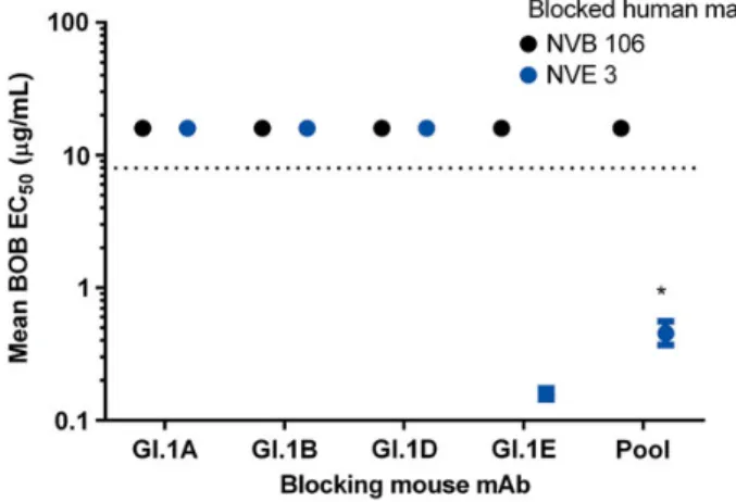
Previous studies with GII.4 NoVs have identified strain-specific epitopes on the surface of the viral particle. Using the paired human and mouse mAbs to GI.1 and an Ab binding competition assay (BOB assay) [11] allowed us to gage the rel-ative spatial arrangement of the GI.1 blockade epitope recog-nized by human mAb NVB 106 to the nonblockade epitopes recognized by the mouse GI.1 mAbs. None of the mouse GI.1 mAbs were able to block binding of NVB 106 in the BOB assay, suggesting that the GI.1 blockade epitope is spatially distinct from the nonblockade epitopes (Figure5). The GI.1E (mouse GI cross-reactive mAb) blocked binding of the human GI
Table 1. Genogroup I Virus-Like Particle Blockade by Day 14 Serum Unfractionated, Serum Depleted of IgA and Purified IgA Subject VLP Serum EC50a,b Serum-IgA EC50a,b Purified IgA EC50b,c,d 2 GI.1 308.50 (224.20–424.40) 103.40 (74.79–142.90) 7.31 (5.91–9.04) GI.3 94.72 (68.63–130.700) 40.56 (35.45–46.40) 32.00 (32.00–32.00) GI.4 2417.00 (2067.00–2827.00) 410.40 (467.90–359.90) 2.53 (2.31–2.78) 3 GI.1 1271.00 (955.00–1691.00) 257.20 (192.20–344.20) 4.46 (3.76–5.30) GI.3 1404.00 (1202.00–1639.00) 340.10 (302.30–382.60) 4.54 (4.11–5.03) GI.4 1148.00 (644.60–2043.00) 515.00 (613.60–432.10) 4.40 (4.06–4.77) 4 GI.1 430.50 (318.80–581.50) 71.20 (53.88–94.09) 5.05 (4.12–6.19) GI.3 193.20 (171.80–217.30) 83.62 (71.08–98.38) 10.01 (8.92–11.31) GI.4 575.10 (530.80–623.10) 75.49 (62.97–90.51) 8.40 (7.69–9.20) 6 GI.1 845.80 (770.60–928.50) 174.50 (147.10–207.10) 1.86 (1.69–2.05) GI.3 109.80 (92.68–130.20) 21.60 (16.65–28.03) 1.00 (0.90–1.11) GI.4 847.60 (688.60–1043.00) 381.90 (334.70–435.70) 6.20 (5.16–7.46) 7 GI.1 662.80 (502.30–874.60) 32.05 (16.01–64.15) 3.20 (2.90–3.54) GI.3 141.40 (122.50–163.10) 55.43 (47.71–64.40) 4.75 (4.46–5.10) GI.4 2313.00 (2068.00–2587.00) 315.60 (245.50–405.70) 1.16 (1.05–1.28) 8 GI.1 278.80 (220.40–352.60) 149.90 (111.70–201.20) 3.95 (3.51–4.44) GI.3 276.20 (225.20–338.70) 79.41 (64.31–98.06) 9.61 (7.90–11.68) GI.4 302.80 (190.80–480.30) 98.00 (57.19–167.90) 6.81 (5.41–8.57) 9 GI.1 342.50 (263.80–444.90) 134.00 (178.70–100.50) 5.45 (4.79–6.19) GI.3 270.10 (234.90–310.50) 100.10 (82.32–121.80) 12.03 (10.72–13.50) GI.4 593.10 (490.70–716.90) 65.85 (34.43–125.90) 8.17 (7.10–9.42) 10 GI.1 3802.00 (3097.00–4668.00) 631.40 (462.80–861.30) 0.48 (0.42–0.54) GI.3 5334.00 (4428.00–6424.00) 1095.00 (994.3–1206) 0.96 (0.90–1.02) GI.4 12095.00 (10066.00–14533.00) 5331.00 (4554.00–6241.00) 0.93 (0.79–1.09)
Abbreviations: Ab, antibody; ANOVA, analysis of variance; GI, genogroup I; Ig, immunoglobulin; VLP, virus-like particle.
a
Mean blockade Ab EC501/serum dilution (95% confidence interval). b
VLP mean EC50values significantly different from GI.1 mean EC50values are bolded (ANOVA). c
Mean blockade IgA EC50µg/mL (95% confidence interval). d
Samples with titers above the limit of detection were assigned the value of 32 for statistical analysis.
Table 2. Characteristics of Anti-Genogroup I Norovirus Monoclonal Antibodies
mAb Clone Name Species Isotype Immunogen %Aviditya(VLP) Blockade
GI.1A 68.21.17 Mouse IgG2ak GI.1 50 (GI.1) –
GI.1B 140.6.12 Mouse IgG2ak GI.1 66 (GI.1) –
10 (GI.3) –
GI.1C 140.42.8 Mouse IgM GI.1 62 (GI.1) –
10 (GI.3) –
GI.1D 300.1.28 Mouse IgG2ak GI.1 66 (GI.1) –
41 (GI.3) –
10 (G1.4) –
GI.1E 300.4.2 Mouse IgG2ak GI.1 66 (GI.1) –
GI.4A 4A2.6.4 Mouse IgG2bk GI.4 41 (GI.4) GI.4
GI.4B 4A2.14.13 Mouse IgG2bk GI.4 84 (GI.4) GI.4
GI.4C 4B4.2.7 Mouse IgG2ak GI.4 78 (GI.4) GI.4
GI.4D 8A6.16.11 Mouse IgG2bk GI.4 84 (GI.4) GI.4
NVF 144 144 Human IgG1 Natural Infection 86 (GI.3) GI.3
NVB 106 106 Human IgG1 Natural Infection 98 (GI.1) GI.1
NVB 84 84 Human IgG1 Natural Infection 86 (GI.3) –
72 (GI.4) –
NVE 3 E3 Human IgG1 Natural Infection 100 (GI.1) –
33 (GI.3) –
10 (GI.4) –
Abbreviations: EIA, enzyme immunoassays; GI, genogroup I; Ig, immunoglobulin; mAb, monoclonal antibody; VLP, virus-like particle.
a
Avidity was only determined for VLPs with positive EIA results (see Figures3and4).
Figure 3. Reactivity of mouse anti-genogroup I (GI) monoclonal antibodies. Mouse monoclonal antibodies against GI.1 (A and B) or GI.4 virus-like par-ticles (VLPs) (C and D) were assayed for enzyme immunoassay (EIA) reactivity (A and C) and blockade activity (B and D) for GI.1, GI.3, and GI.4 VLPs, and mean EC50titers were determined. Antibodies with EIA or blockade titer above the limit of detection (dashed line) were assigned values of 4 µg/mL or 16
µg/mL, respectively, for statistical analysis. *Mean EC50significantly different from the immunizing strain VLP (analysis of variance).
cross-reactive mAb (NVE 3), indicating that the mouse mAbs generated to VLPs recognize relevant epitopes that are targeted during human infection. Pooled mouse mAbs did not block binding of NVB 106 and blocked NVE 3 less well than GI.1E alone, indicating that BOB activity may be related to epitope distance and frequency and not to steric hindrance from epitope saturation.
DISCUSSION
Noroviruses are characterized by extensive antigenic diversity among the GI and GII genotypes. Identification of type-specific and cross-genotype epitopes will provide epitope-defined diag-nostic assays that will facilitate development of a broadly protec-tive NoV vaccine and an immune therapeutic. To begin to understand the origins of cross-reactive Ab responses, we ana-lyzed IgA and IgG serum titers and avidities in samples that present cross-reactive GI Abs at day 14 post-GI.1 infection [9]. The GI.1-specific IgG and IgA titers and avidities remained consistent at day 4 postchallenge and peaked at day 14. Com-pared with IgG, IgA titer and avidity waned more quickly, possibly reflecting the homing of IgA Ab-secreting cells from the peripheral blood to mucosal sites [15]. Prechallenge IgG and IgA titers with avidities above 40% were detected in 2 of the 10 (20%) participants who became infected with GI.1.
Furthermore, high avidity was not predictive of blockade func-tion among mAbs. Although the design of this study does not allow prediction of correlates of protective immunity, together, these data suggest that the location of Ab binding may be more important in determining protection from infection than Ab titer or avidity.
A functional role for serum IgG in NoV immunity has been established by demonstrating that human IgG mAbs have blockade activity [14,16] and that IgG memory B-cell titers cor-relate with protection from GI.1 infection [17]. Mucosal IgA ti-ters, as measured in saliva, have also been shown to correlate with protection from GI.1 infection [17,18]. These studies are thefirst to assess the blockade activity of IgA. To distinguish IgA blockade potency from IgG, IgA was purified from the day 14 serum samples with total GI.1-reactive IgA titer greater than 200 µg/mL. All 8 IgA preparations had cross-GI blockade activity, identifying IgA as a component of the serological blockade Ab response to GI NoVs. Supporting studies with polymeric monoclonal IgA indicating that IgA is strain cross-reactive [19], purified IgA did not preferentially block ligand
binding of GI.1 compared with other GI VLPs. The IgA re-sponse may have been generated to cross-reactive epitopes in response to either the current GI.1 infection or an earlier GI ex-posure. Supporting this hypothesis, serum and serum depleted of IgA from multiple participants, and IgA from one participant blocked the 3 GI VLPs equivalently, indicating that conserved GI blockade epitopes exist or that infection with 1 strain elicits
Figure 5. Genogroup (GI).1 blockade epitope is structurally distinct from GI.1 nonblockade epitopes. Mouse monoclonal antibodies (mAbs) to GI.1 nonblockade epitopes (mAbs GI.1A, B, D, E) were measured for ability to block GI.1 virus-like particle binding of human mAbs to a strain-specific blockade epitope (black circle, NVB106) or a conserved nonblockade epi-tope (gray circle, NVE 3) in an epiepi-tope-specific blocking-of-binding (BOB) assay. Mouse antibodies unable to block human antibody binding at titers within the limit of detection (dashed line) were assigned values of 16 µg/ mL for statistical analysis. *Mean BOB EC50titer significantly different
from GI.1E for the same human antibody (Wilcoxon). Pool: 2 µg/mL each GI.1A, B, D, and E.
Figure 4. Reactivity of human anti-genogroup I (GI) monoclonal antibod-ies (Abs). Human monoclonal Abs reactive with GI virus-like particles (VLPs) were assayed for enzyme immunoassay (EIA) reactivity (A) and blockade activity (B) for GI.1, GI.3, and GI.4 VLPs, and mean EC50titers
were determined. Antibodies with EIA or blockade titer above the limit of detection (dashed line) were assigned values of 4 µg/mL or 16 µg/mL, respectively, for statistical analysis. *Mean EC50titer significantly different
from GI.3 titer (analysis of variance).
pre-exposure memory responses against other closely related GI strains. More importantly, thisfinding was observed using an-tigen-limiting conditions, designed to emphasize strain anti-genic differences, as opposed to our previous analysis of these samples in which assays were optimized for sensitivity resulting in no group difference in serum IgG or blockade Ab titers between GI VLPs [8,9]. To clearly define Ab epitopes, large
panels of human mAb are needed. The data presented here sug-gest that clone selection processes should include IgA as well as IgG isotypes.
Mouse mAbs to GI VLPs have been described by others [20,
21], but none have identified GI blockade Ab potency or epi-topes, contrary to GII.4 NoVs [14,22]. Therefore, we developed a panel of GI mAbs to evaluate the antigenic relationship be-tween GI VLPs. Unlike GI.1 infection in humans, GI.1 or GI.4 immunization of mice resulted in Abs that preferentially recognize the immunizing strain, again suggesting that pre-ex-posure history may shape the GI Ab response in humans. Six of the GI Abs appear to have type-specific blockade activity against the panel of GI VLPs tested because all recognized only 1 GI VLP. However, we cannot definitively infer from these data that blockade epitopes are strain-specific, because representa-tives of each of the GI genotypes were not tested. The remaining 7 mAbs recognized nonblockade epitopes. In particular, none of the 5 mouse GI.1 mAbs had blockade activity. Although atypical in our experience with NoV mAbs, it is unlikely that the lack of blockade potency is related to immunization or hy-bridoma production protocols because the mouse GI.1 mAbs were developed simultaneously with the mouse GI.4 mAbs and all of the GI.4 mAbs recognize a blockade epitope. Anti-bodies can confer protection from infection by additional means besides blocking of ligand interaction, including comple-ment lysis, opsonization, and Ab-dependent cell cytolysis; therefore, a nonblocking phenotype does not decrease the bio-logical significance of these Abs.
CONCLUSIONS
Most cross-reactive mouse and human GI mAbs had a higher avidity to a single GI VLP and less avidity to other GI VLPs, supporting the hypothesis that strain cross-reactive Abs are gen-erated to sequences in a single strain that are conserved within other GI VLPs, although to varying degrees of homology. Struc-tural studies of Ab-bound VLPs and bioinformatics and genetic tools to create VLPs with altered predicted Ab epitopes com-bined with a large panel of mAbs may define GI strain-specific and strain cross-reactive epitopes that can be exploited as diag-nostic tools to measure epitope-specific responses and to refine the design of a broadly protective NoV vaccine. Additional human challenge studies are critical for creating mAb panels, mapping relevant epitopes, and defining correlates of protective immunity.
Acknowledgments
We thank the SERCEB Mouse Monoclonal Core for assistance with hy-bridoma development; Victoria Madden and C. Robert Bagnell Jr. of Mi-croscopy Services Laboratory; Department of Pathology and Laboratory Medicine, University of North Carolina-Chapel Hill for expert technical support; and Christine Moe and Juan Leon (Emory University) for provid-ing the deidentified sera from GI.1-infected participants.
Disclaimer. The funders had no role in study design, data collection, analysis, decision to publish, or preparation of the manuscript.
Financial support. This work was funded by grants from the National Institutes of Health, Allergy and Infectious Diseases (R01 AI056351, R56 A15-0756 and U19 AI109761 CETR) and Southeastern Regional Center for Excellence in Biodefense (U54 AI057157).
Potential conflicts of interest. A. L. is the scientific founder and share-holder of Humabs BioMed SA. D. C. and M. B. are employees of Humabs Bi-omed, a company that develops anti-invectives human monoclonal antibodies. All authors have submitted the ICMJE Form for Disclosure of Potential Conflicts of Interest. Conflicts that the editors consider relevant to the con-tent of the manuscript have been disclosed.
References
1. The Centers for Disease Control and Prevention (CDC). Updated noro-virus outbreak management and disease prevention guidelines. MMWR Recomm Rep2011; 60:1–18.
2. Bruggink LD, Dunbar NL, Marshall JA. Norovirus genotype diversity as-sociated with gastroenteritis outbreaks in aged-care facilities [Epub ahead of print]. Epidemiol Infect2015; doi:10.1017/S095026881500031X. 3. Payne DC, Vinje J, Szilagyi PG, et al. Norovirus and medically attended
gastroenteritis in U.S. children. N Engl J Med2013; 368:1121–30. 4. Bok K, Green KY. Norovirus gastroenteritis in immunocompromised
patients. N Engl J Med2012; 367:2126–32.
5. Trivedi TK, DeSalvo T, Lee L, et al. Hospitalizations and mortality as-sociated with norovirus outbreaks in nursing homes, 2009–2010. JAMA 2012; 308:1668–75.
6. Treanor JJ, Atmar RL, Frey SE, et al. A novel intramuscular bivalent norovirus virus-like particle vaccine candidate-reactogenicity, safety, and immunogenicity in a phase 1 trial in healthy adults. J Infect Dis 2014; 210:1763–71.
7. Atmar RL, Bernstein DI, Harro CD, et al. Norovirus vaccine against ex-perimental human Norwalk virus illness. N Engl J Med2011; 365: 2178–87.
8. Lindesmith LC, Ferris MT, Mullan CW, et al. Broad blockade antibody responses in human volunteers post immunization with a multivalent norovirus VLP candidate vaccine: immunological analyses from a phase I clinical trial. PloS Med2015; 12:e1001807.
9. Lindesmith LC, Donaldson E, Leon J, et al. Heterotypic humoral and cellular immune responses following Norwalk virus infection. J Virol 2010; 84:1800–15.
10. Tian P, Yang D, Jiang X, et al. Specificity and kinetics of norovirus bind-ing to magnetic bead-conjugated histo-blood group antigens. J Appl Microbiol2010; 109:1753–62.
11. Lindesmith LC, Donaldson EF, Beltramello M, et al. Particle conforma-tion regulates antibody access to a conserved GII.4 norovirus blockade epitope. J Virol2014; 88:8826–42.
12. Khurana S, Verma N, Yewdell JW, et al. MF59 adjuvant enhances diver-sity and affinity of antibody-mediated immune response to pandemic influenza vaccines. Sci Transl Med 2011; 3:85ra48.
13. Swanstrom J, Lindesmith LC, Donaldson EF, et al. Characterization of blockade antibody responses in GII.2.1976 snow mountain virus-infect-ed subjects. J Virol2014; 88:829–37.
14. Lindesmith LC, Beltramello M, Donaldson EF, et al. Immunogenetic mechanisms driving norovirus GII.4 antigenic variation. PLoS Pathog 2012; 8:e1002705.
15. Sundararajan A, Sangster MY, Frey S, et al. Robust mucosal-homing antibody-secreting B cell responses induced by intramuscular
administration of adjuvanted bivalent human norovirus-like particle vaccine. Vaccine2015; 33:568–76.
16. Debbink K, Lindesmith LC, Donaldson EF, et al. Emergence of new pandemic GII.4 Sydney norovirus strain correlates with escape from herd immunity. J Infect Dis2013; 208:1877–87.
17. Ramani S, Neill FH, Opekun AR, et al. Mucosal and cellular immune responses to Norwalk virus [Epub ahead of print]. J Infect Dis2015; doi:10.1093/infdis/jiv053.
18. Lindesmith L, Moe C, Marionneau S, et al. Human susceptibility and resistance to Norwalk virus infection. Nat Med2003; 9:548–53. 19. Muramatsu M, Yoshida R, Yokoyama A, et al. Comparison of antiviral
activity between IgA and IgG specific to influenza virus hemagglutinin:
increased potential of IgA for heterosubtypic immunity. PLoS One 2014; 9:e85582.
20. Crawford SE, Ajami N, Parker TD, et al. Mapping broadly reactive Nor-ovirus genogroup I and II monoclonal antibodies. Clin Vaccine Immu-nol2015; 22:168–77.
21. Hale AD, Tanaka TN, Kitamoto N, et al. Identification of an epitope common to genogroup 1 "norwalk-like viruses". J Clin Microbiol 2000; 38:1656–60.
22. Debbink K, Donaldson EF, Lindesmith LC, Baric RS. Genetic mapping of a highly variable norovirus GII.4 blockade epitope: poten-tial role in escape from human herd immunity. J Virol2012; 86: 1214–26.