HAL Id: hal-01993625
https://hal.univ-grenoble-alpes.fr/hal-01993625
Submitted on 26 Nov 2020HAL is a multi-disciplinary open access archive for the deposit and dissemination of sci-entific research documents, whether they are pub-lished or not. The documents may come from teaching and research institutions in France or abroad, or from public or private research centers.
L’archive ouverte pluridisciplinaire HAL, est destinée au dépôt et à la diffusion de documents scientifiques de niveau recherche, publiés ou non, émanant des établissements d’enseignement et de recherche français ou étrangers, des laboratoires publics ou privés.
Time-Resolved Serial Femtosecond Crystallography,
Towards Molecular Movies of Biomolecules in Action
Jacques-Philippe Colletier, Giorgio Schirò, Martin Weik
To cite this version:
Jacques-Philippe Colletier, Giorgio Schirò, Martin Weik. Time-Resolved Serial Femtosecond Crystal-lography, Towards Molecular Movies of Biomolecules in Action. Petra Fromme; Sebastien Boutet; Mark Hunter. X-ray Free Electron Lasers, Springer International Publishing, pp.331-356, 2018, �10.1007/978-3-030-00551-1_11�. �hal-01993625�
Time-resolved serial femtosecond crystallography,
towards molecular movies of biomolecules in action
Authors: Jacques-Philippe Colletier1, Giorgio Schirò1, Martin Weik1
Affiliations:
1Institut de Biologie Structurale, Univ. Grenoble Alpes, CEA, CNRS, F-38044 Grenoble,
France.
1. Introduction
Biological macromolecules, such as proteins, nucleic acids and complexes thereof, are characterized by specific structural and dynamic features that are the basis of their respective biological activity, and define their dynamic personalities (Henzler-Wildman and Kern 2007). Understanding macromolecular activity thus requires studying structural changes over time and on various time-scales, such as equilibrium fluctuations and conformational changes orchestrating enzyme catalysis or enabling signal transduction. The first step in human vision, for instance, is the sub-picosecond time-scale photoisomerization of the retinal pigment in rhodopsin (Schoenlein et al. 1991), which within microseconds leads to the conformational changes required for activation of transducin, the regulatory protein that initiates the signaling cascade beyond the macromolecular level.
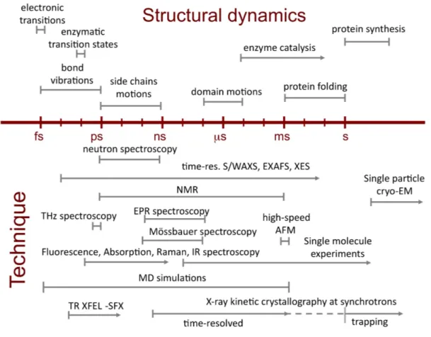
Complementary biophysical techniques each open a window in time on macromolecular dynamics occurring from femtoseconds to minutes (Figure 1). Among those, kinetic X-ray crystallography permits trapping of macromolecular conformational intermediates along a reaction pathway, and their characterization at the atomic level of spatial resolution (Bourgeois and Weik 2009). The basic concept underlying this ensemble of techniques is that macromolecular activity can be triggered within a crystal, and the structure of intermediate states characterized, provided that (i) the macromolecule is active in the crystalline state; (ii) an efficient and synchronous triggering of activity can be attained (at room-temperature) for all (or most) probed molecules; and (iii) the structural information can be recorded on a time scale shorter than the lifetime of the intermediate state of interest. As within a macromolecular crystal neighboring molecules are bathed in solvent (on average around 50% of the crystal volume) and generally make limited direct contacts, they most often preserve their functionally relevant
structural dynamics and therefore remain active in the crystalline state (Mozzarelli and Rossi 1996). After triggering of the biological reaction within macromolecular crystals, functionally relevant conformational changes are either arrested by flash-cooling the crystal, allowing characterization of the structure by conventional cryo-crystallography, or followed in real time by time-resolved crystallography. The temporal resolution of the latter is limited to 100 ps if carried out in the form of Laue crystallography at synchrotrons. The advent of X-ray free electron lasers (X-ray FEL) has pushed the resolution to the sub-ps regime, allowing ultrafast changes to be studied by time-resolved serial femtosecond crystallography. Below, we introduce the principles of time-resolved crystallography and summarize its implementation at synchrotron sources, before offering a review of time-resolved structural studies carried out so far using serial femtosecond crystallography (SFX (Chapman et al. 2011)) at X-ray FELs. Challenges, limitations and perspectives of time-resolved SFX round out this chapter.
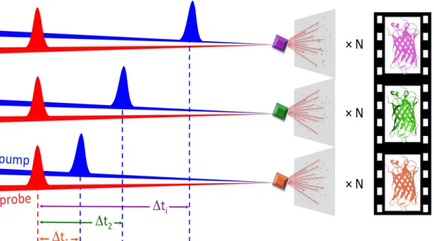
2. Principles of time-resolved crystallography In time-resolved crystallography, a reaction in the crystalline macromolecule is triggered (pump) at t=0 and a diffraction pattern (probe) collected after a well-defined lapse of time ∆t1 (pump-probe delay). The pump-probe sequence is repeated sufficiently enough for a complete diffraction data set to be produced from which a macromolecular structure can be determined that features conformational changes characteristic of the time delay ∆t1 after reaction initiation. The pump-probe delay is then changed to ∆t2 and the procedure repeated, etc. The structure determined at each ∆ti represents a frame in a molecular movie featuring conformational changes along the reaction pathway (Figure 2). A complete and intelligible time-resolved study of the mode of action of the rhodopsin protein mentioned above, for instance, would require structural data to be collected over ten orders of magnitude, from the fs time scale where the actinic photon is absorbed, up to the µs timescale where transduction is activated.
Efficient and synchronous reaction triggering is key to a successful pump-probe experiment. The pump needs to trigger the reaction faster than the process of interest (synchrony) and in a large fraction of the crystalline molecules (efficiency) so that structural changes can be observed by ensemble-averaged crystallography. The pump method to be used depends on the reaction to be triggered: UV-visible light activates
inherently light-sensitive proteins or so-called caged compounds complexed with light-insensitive proteins; diffusion of substrates initiates enzyme catalysis (see Chapter 13 in this book); a temperature jump shifts conformational equilibria (Kubelka 2009); X-ray irradiation can provide electrons to trigger redox processes (Schlichting et al. 2000) or the breakdown of strained intermediate states (Colletier et al. 2008); electric field pulses allow studying protein mechanics (Hekstra et al. 2016). By far the most widespread method is optical triggering because of its straightforward technical implementation, the availability of crystals for a large number of light-sensitive proteins with cyclic reactions to be studied, and the accessibility of ultrafast (fs-ps) time scales.
The X-ray probe produces diffraction patterns that allow structure determination at ∆ti after reaction initiation. The probe needs to be pulsed so that structural changes can be captured, analogous to shutter opening and closing in photography. The probe-pulse length must be shorter than the process of interest and sets, together with the pump-pulse length, the best time resolution that can be attained. At synchrotron sources, the pulse length cannot be shorter than about 100 ps, making it impossible to study processes faster than that limit. Also, time-resolved crystallography at synchrotrons is in practice limited to cyclic reactions because the pump-probe sequence is repeated multiple times on one or several macro-sized crystals. Using time-resolved (TR) SFX at X-ray FELs, however, cyclic as well as non-cyclic reactions can be studied since each microcrystal is probed only once by a single X-ray shot. Furthermore, with their femtosecond X-ray pulses, X-ray FELs have extended the time resolution to the sub-ps regime, permitting the structural study of ultrafast processes such as those immediately following photon absorption in light-sensitive processes. Last but not least, the risk of X-ray radiation damage to biological macromolecules, known to compromise structural data collected at synchrotrons (Garman and Weik 2017), is abolished in most SFX studies because diffraction data are collected before chemical and structural damage has had the time to develop (Neutze et al. 2000). Hence, the advent of X-ray FELs has revived and extended the reach of time-resolved crystallography.
3. Time-resolved crystallography at synchrotron sources
The advent of third-generation synchrotron sources and undulator beamlines, poised with 1018 and 106 higher peak brilliance than X-ray tubes and second-generation bending
magnet sources, respectively, permitted the introduction of time-resolved crystallography by means of the so-called Laue technique (Moffat 1989). In this approach, crystals of inherently photosensitive proteins or of non-photosensitive proteins complexed with photolabile precursors of their substrates or products – so called caged compounds – are activated by exposure to a femtosecond or nanosecond laser (pump) at room temperature. A highly intense polychromatic X-ray beam is then used to probe the structure, at various ∆ti. The advantage of using a white (~1-5 % bandwidth) or pink (~0.05 to 0.1 % bandwidth) polychromatic beam is that enough photons are elastically scattered, even from a single X-ray bunch, to enable collection of a diffraction pattern on which enough information is present to derive a partial dataset. Combining multiple such diffraction images, typically 10 to 30 exposures to the polychromatic X-ray beam of one or more crystals in different orientations, experimentalists can then produce a full dataset for each ∆ti. The time resolution of Laue crystallography at synchrotrons is inherently limited to 100 ps, corresponding to the pulse length of a single electron bunch generating the X-rays. Furthermore, the methodology requires large (0.5 mm), highly ordered, radiation-resistant protein crystals (to enable multiple exposures to the polychromatic beam without loss of resolution due to X-ray damage) with small unit cells (to minimize Bragg-peak overlaps), and works best with proteins undergoing cyclic photoreactions (see above). In the case of highly radiation sensitive samples, non-reversible photoreactions, or in studies where non-inherently photosensitive proteins are light-functionalized by complexation with caged compounds (Schlichting et al. 1990), more crystals are indeed needed. In the case of large proteins with correspondingly large unit cell parameters (e.g. photosystems I and II, or cytochrome c oxidase), data processing is complicated, due to overlaps between diffracted spots even when using a pink beam. Hence the methodology has failed to enable time-resolved crystallography on a large variety of proteins, and has mostly remained limited to a handful of proteins including the small GTPase Ras (Schlichting et al. 1990), myglobin (Šrajer et al. 1996; Bourgeois et al. 2003; Schotte et al. 2003; Bourgeois et al. 2006), the photoactive yellow protein (Genick et al. 1997; Ihee et al. 2005; Schotte et al. 2012; Jung et al. 2013), and the photosynthetic reaction center (Baxter et al. 2004; Wohri et al. 2010). The largest protein to have been successfully studied by time-resolved Laue crystallography is hemoglobin (Knapp et al. 2006). The advent of X-ray FELs and monochromatic SFX has allowed the resolution of
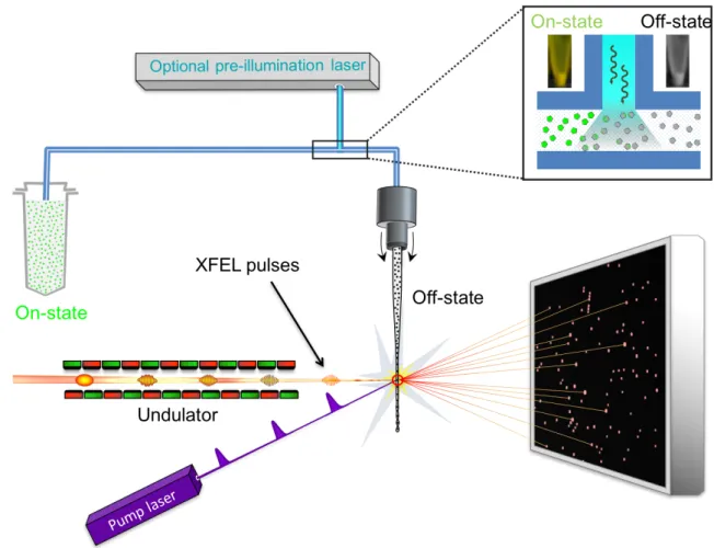
time-resolved crystallography to be improved to the fs timescale, and to extend the feasibility of time-resolved studies to larger and radiation sensitive proteins. 4. Time-resolved serial femtosecond crystallography at X-ray FELs – an inventory In time-resolved serial femtosecond crystallography (TR-SFX) following a pump-probe data collection scheme, macromolecular crystals are streamed across the pulsed X-ray FEL beam typically by means of a liquid jet (Weierstall et al. 2012; Sierra et al. 2012), a high-viscosity injector (Weierstall et al. 2014; Botha et al. 2015; Sugahara et al. 2015) or by acoustic droplet injection coupled with a conveyer-belt drive (Fuller et al. 2017). When the protein is inherently photosensitive or has been photosensitized by complexation with a caged-compound, crystals can be activated by an optical laser pulse (pump) prior to interaction with the X-ray pulse (probe), generating a diffraction pattern (Figure 3). Repetition of this process on many crystals provides a full data set at a well-defined ∆ti from which the corresponding structural snapshot can be determined. Varying ∆ti then allows generating frames of a molecular movie. If macromolecular activity is triggered by diffusion of a small molecule into the crystal, two solutions containing the crystals and the small molecule are mixed prior to interaction with an X-ray pulse. This so-called mix-and-inject strategy (Schmidt 2013) is extensively discussed in Chapter 13 of this book and has been successfully applied to study ligand-binding in adenine riboswitches (Stagno et al. 2017) and ß-lactamase (Kupitz et al. 2017). Time-resolved crystallography experiments at X-ray FELs remedy several shortcomings that limit those carried out at synchrotron sources. Firstly, the achievable time resolution of maximal 100 ps at a synchrotron is improved to 10 fs, owing to the short length of FEL pulses. Secondly, the high peak brilliance of X-ray FEL pulses generates exploitable diffraction from micron-sized crystals that can be fully activated by the optical pump laser. Indeed, absorption by optically dense protein crystals (Figure 4) limits light penetration to outer layers in the case of large crystals (at least several tens of microns in size) such as those required by time-resolved synchrotron studies. And third, the very nature of (TR)-SFX, wherein the sample is replenished after each shot in a serial manner,
enables time-resolved structural studies on non-reversible reactions, provided that enough sample can be produced for the envisaged delivery system.
The first TR-SFX study was carried out on crystals of a photoactive complex of photosystem I (PSI) with ferredoxin (Aquila et al. 2012) with pump-probe delays of 5 µs and 10 µs. Only virtual powder patterns were used in the analysis, since the low amount of indexed patterns per time-delay precluded extraction of structure factors. At 5 µs, a slight increase in diffracted intensities was observed, which was interpreted as a correlated structural change induced by electron transfer in the PSI – ferredoxin complex. At 10 µs, diffraction intensities dropped, rationalized as originating from the disordering of crystals upon dissociation of ferredoxin from the binding pocket, in line with time-resolved optical spectroscopy. Since this proof-of-principle study, several pump-probe TR-SFX studies have been carried out on various proteins (myoglobin, photoactive yellow protein, bacteriorhodopsin, photosystem II, a reversibly photoswitchable green fluorescent protein, cytochrome c oxydase, nitrite oxide reductase), and on timescales ranging from seconds to sub-picoseconds (Figure 6). These results are briefly presented below. The first high-resolution (1.6 Å) TR-SFX study was carried out on the photoactive yellow protein (PYP) with pump-probe delays of 1 µs and 10 ns using optical pump laser pulses on the ns regime (Tenboer et al. 2014). The difference Fourier maps, computed between data sets of illuminated (light) and non-illuminated (dark) crystals, showed structural changes qualitatively similar to those observed in TR synchrotron Laue studies at comparable time delays, demonstrating validity of the TR-SFX method. Yet, peaks in the TR-SFX difference maps were higher than in the corresponding Laue difference maps, owing to the more extensive pump-laser activation of the micron-sized crystals used in the TR-SFX studies, as compared to those used in Laue experiments. As mentioned previously, usability of µm- and sub-µm-sized crystals is one of the advantages of X-ray FEL time-resolved studies. Further steps into the uncharted regime of ps resolution were subsequently taken, as discussed in detail in the next section. It is of important note that with much of the biological dynamics of interest being in the ns or slower time scales, many TR-SFX studies utilize the unique advantages of X-ray FEL
based methods to study relatively slow dynamics compared to the X-ray FEL pulse duration, such as those involved in photosystem II (PS II) activity. The high scientific interest in the water-splitting cycle of PS II has made it so far the macromolecular system most studied by TR-SFX (Figure 6). PSII is a large membrane-bound protein complex (700 kDa) involved in photosynthesis that produces dioxygen by catalyzing light-driven water splitting at a so-called oxygen evolving complex (OEC) containing a Mn4CaO5 cluster. The photocycle of PSII is a reversible reaction that involves sequential absorption of four photons generating four consecutive redox states of the cluster that cycles through five S-states (S0 to S4; so called “Kok cycle”) on the µs to ms time scale (Kok et al. 1970). Each of the four steps is characterized by a non-unitary quantum yield, so that a mixture of states has accumulated at the end of one photocycle; therefore, the reaction is in practice irreversible and each crystal may be used only once. A high-resolution (1.9 Å) structure of PSII has been solved by synchrotron macromolecular crystallography (Umena et al. 2011), but determination of the accurate OEC geometry had been hampered by the high X-ray sensitivity of this metalloenzyme (Yano et al. 2005). The high-radiation sensitivity, combined with the experimentally non-reversible reaction, makes TR-SFX the method of choice to study the Kok cycle. Using femtosecond FEL pulses, it was indeed possible to determine the radiation-damage free structure of PS II and its OEC at 1.95 Å resolution by rotating and translating a large single crystal at 100 K in a stepwise fashion (Suga et al. 2015). Comparison of the synchrotron and X-ray FEL structures revealed a more compact OEC with bond lengths 0.1 – 0.2 Å shorter in the X-ray FEL structure. Characterizing the radiation-damage free high-resolution structure of PS II (Suga et al. 2015) is yet only the first step in understanding the atomic details of water splitting, and TR-SFX studies that aimed at resolving time-dependent structural changes following light absorption were then carried out. In the first published TR-SFX study on PS II, simultaneous SFX and X-ray emission spectroscopy (XES) data were collected on crystals in the dark-adapted S1 state, and in the S2 state generated by light illumination with a pump-probe delay of 0.4 – 0.5 s (Kern et al. 2013). Electron density maps of the S2 structure at 5.7 Å resolution did not show differences with respect to the S1 structure and it was concluded that structural changes, if present, are too small to be seen at that resolution. The simultaneous collection of SFX and XES data not only allowed assigning the dominant S-state to the structural data collected, but also provided evidence that the fs pulses probed the intact electronic structure of metal centers in the OEC. Subsequent studies focused on structural changes
that accompany the transition to the S3 state. While Kupitz and coworkers reported changes in the structure of an S3 enriched state at 5.5 Å resolution compared to S1 (Kupitz et al. 2014), a similar study by Kern and coworkers came to the conclusion that any structural changes related to the transitions between S1 and S3 must be smaller than what can be detected at the resolution achieved (between 4.5 and 5.2 Å)(Kern et al. 2014; Sauter et al. 2016). The resolution of S3 enriched structures was then improved to 2.25 Å (Young et al. 2016) and 2.35 Å (Suga et al. 2017), yet without reaching a consensus on the nature of associated conformational changes. In one case (Young et al. 2016), ~0.1 Å changes in metal distances in the OEC were reported, i.e. changes small enough to be at the limit of uncertainty. In the other (Suga et al. 2017), peaks were observed on the OEC in Fourier difference maps that were interpreted as the appearance of a new oxygen atom and the displacement of a water molecule. Awaiting further improvements in diffraction resolution, the jury is thus still out as to time-resolved changes in the OEC during water splitting. Another molecular mechanism actively studied by TR-SFX is the structural sequence of photo-intermediates in the light-driven proton pump bacteriorhodopsin (bR). Despite being a well-studied model for membrane proteins, certain structural details of the bR photocycle remain controversially discussed because of a possible convolution with radiation-induced changes in cryo-trapped structural intermediates solved at synchrotron sources (Wickstrand et al. 2015). A proof-of-principle that TR-SFX on bR is possible has been provided by Nogly and co-workers (Nogly et al. 2016), using a thick high-viscosity jet to present lipidic-cubic phase (LCP) grown bR crystals to the X-ray beam. Using femtosecond excitation and a pump-probe delay of 1 ms, the structure of the M photointermediate could be solved at 2.3 Å resolution. As multiple consecutive excitations are not possible during the fs pump-pulse duration (see detailed discussion in section 5), the intermediate-state occupancy was limited to 13%. The first extensive TR-SFX study on bR presented diffraction data collected following nanosecond excitation and spanning five orders of magnitude in time, from ns to ms (Nango et al. 2016). Transient structures solved at 2.1 Å resolution covered photointermediates K to M, and provided a three-dimensional view of the structural changes underlying unidirectional proton pumping. In the earliest time-point studied (16 ns), the chromophore (retinal) had already photoisomerized, so that ultrafast changes right after photon absorption are still to be uncovered.
Two heme proteins other than myoglobin (see below) have been studied by time-resolved crystallography at an X-ray FEL, namely cytochrome c oxidase (Shimada et al. 2017) and NO reductase (Tosha et al. 2017). In the former, CO release has been followed 20 ns and 100 µs after photolysis with a ns pump-laser, and the characterized structural changes allowed establishing a mechanism for functionally relevant closing of a water channel (Shimada et al. 2017). Similar to the characterization of the radiation-damage free high-resolution structure of PS II (Suga et al. 2015), and at variance with most TR-SFX studies published so far, cytochrome c oxidase crystals were not presented as micron-sized samples in random orientations within a jet or by a conveyer-belt drive, but rather large (up to 500 µm) macrocrystals were rotated and translated through the FEL beam (serial femtosecond rotational crystallography (SF-ROX (Hirata et al. 2014)). The achievement standing out in the study on NO reductase is the first and successful use of caged compounds in TR-SFX. The enzyme was indeed rendered photosensitive by complexation with a photolabile precursor of NO (caged NO), which was cleaved on the µs time scale by means of ns laser irradiation at 308 nm. X-ray data were collected at a pump-probe delay of 20 ms, allowing determination of the structure of the ferric NO complex at 2.1 Å resolution. Unlike corresponding synchrotron structures, the TR-SFX structure was devoid of X-ray radiation damage, as corroborated by accompanying QM/MM studies (Tosha et al. 2017).
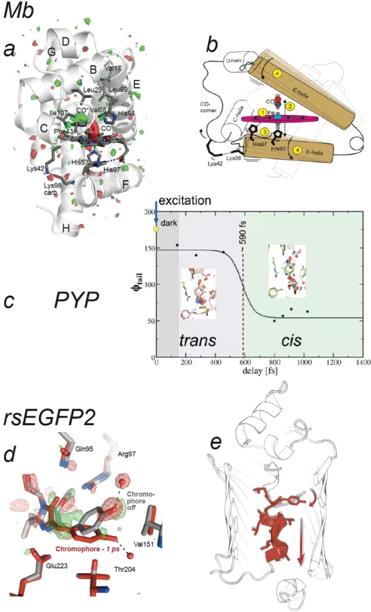
Ultrafast TR-SFX on the fs – ps time scale. Three TR-SFX studies have so far been
published beyond the 100 ps resolution-limit of synchrotron-based Laue crystallography. Pump-probe delays of a picosecond and shorter allowed entering the time scale of photochemical reactions – a regime inaccessible by X-ray sources other than X-ray FELs – enabling visualization of ultra-fast light-induced motions in carbonmonoxy myoglobin (Barends et al. 2015) and characterization of excited-state photoisomerization intermediates in PYP (Pande et al. 2016) and in a reversibly photoswitchable variant of the green fluorescent protein (GFP, (Coquelle et al. 2018)). In carbonmonoxy (CO) myoglobin (Mb), structural changes following light-activated cleavage of the Fe-CO bond were captured at nominal delays of 0.5 ps, 1 ps, 3 ps, 10 ps, 50 ps and 150 ps (Barends et al. 2015). In line with results from other methods, the crystallographic maps showed that within one ps after photodissociation, CO has moved to its primary docking site, the heme has domed, and side-chains of neighboring residues have moved (Figure 5a). Significant
main-chain conformational changes were observed on the ps time scale with helices moving essentially as rigid bodies, in line with time-resolved solution scattering experiments (Levantino et al. 2015). The jitter between pump laser and X-ray FEL pulses caused the nominally 500 fs time delay data to contain images with various true time delays, so that they could be sorted into datasets with average time delays of -100 to +600 fs, with bin widths of ~100 fs. Refinement against these ultrafast datasets revealed that residues in the heme-binding pocket showed an oscillatory behaviour with a 500 fs period upon cleavage of the CO. These oscillations were noted to fit very well inside the general normal mode model for protein dynamics (Miller 1994), in which fast coherent motions ultimately couple with larger-scale displacements of entire helices (Figure 5b). In PYP, structural changes of the p-coumaric acid chromophore after femtosecond illumination at 450 nm have been followed at nominal pump-probe delays of 0.3 ps, 0.6 ps and 3 ps (Pande et al. 2016). A structural transition has been proposed to occur at around 0.59 ps, associated with a trans-to-cis isomerization of the chromophore that involves some atoms moving by as much as 1.3 Å, on the sub-ps time scale (Figure 5c). At delays shorter than 0.59 ps, the chromophore displays a trans, yet distorted, conformation, suggesting that it is still in the electronic excited state. At longer pump-probe delays, a cis chromophore is observed, indicating relaxation to the ground state. In the reversibly photoswitchable fluorescent protein rsEGFP2, used for nanoscale imaging of living cells by means of REversible Saturable Optical Linear Fluorescence Transitions (RESOLFT) microscopy (Grotjohann et al. 2012), photoisomerization intermediates have been characterized 1 and 3 ps after femtosecond illumination of the crystalline protein in its non-fluorescent off-state with the hydroxybenzylidene imidazolinone chromophore in the planar trans configuration (Coquelle et al. 2018). One ps after illumination, the chromophore is fully twisted, with its two rings oriented perpendicular to each other (Figure 5d). Quantum-mechanics/molecular-mechanics (QM/MM) and excited-state molecular dynamics simulations and time-resolved absorption spectroscopy in solution indicated that the twisted chromophore conformation after 1 ps corresponds to that of the electronic excited S1 state, close to the conical intersection where the system relaxes back to the ground state. The entire helix carrying the chromophore shifts along its axis, so as to permit chromophore twisting (Figure 5e). Three ps after illumination, the twisted chromophore conformation is less occupied than at 1 ps, and features indicative of the presence of the cis chromophore are present in the difference Fourier map. The twisted
chromophore (Figure 5d,e) thus represents a photoisomerization intermediate, half-way between trans and cis isomers. The maximum displacement of a chromophore atom (phenol OH group) is 4.5 Å in rsEGFP2 – that is, much larger than in the case of PYP (1.3 Å) – possibly explaining why photoisomerization takes longer in rsEGFP2 than in the PYP (at 1 ps, rsEGFP2 is still in the excited state, while PYP is already in the ground state again). The excited-state structure of rsEGFP2 (Figure 5d) was the basis for rationally improving this protein used as a molecular label in super-resolution fluorescence microscopy. The twisted chromophore conformation indicated that mutating Val151 (Figure 5d) into an alanine residue should facilitate photoswitching by enlarging the pocket at the tip of the chromophore. Indeed, the V151A mutant turned out to have a two-fold increased photoswitching quantum yield (Coquelle et al. 2018).
Since TR-SFX on the picosecond time scale requires pumping with a femtosecond pulse, reaction initiation is limited by the primary quantum yield. As a consequence, reaction initiation in the PYP study was low (structurally estimated to be 13% (Pande et al. 2016)) and the intermediate-states in rsEGFP2 were present at a total occupancy of maximum 7% (Coquelle et al. 2018). An additional caveat of femtosecond excitation is the requirement of using high pump laser power densities (380 GW/cm2 were used for Mb (Barends et al. 2015), 570 GW/cm2 for PYP (Pande et al. 2016) and 400 GW/cm2 for rsEGFP2 (Coquelle et al. 2018), which carries the risk of creating artifacts (see more extensive discussion below). Consequently, all three picosecond TR-SFX studies relied on complementary QM/MM and optical spectroscopy experiments to interpret and validate the intermediate-state structures obtained. 5. Challenges and limitations of TR-SFX One might face several challenges and limitations when attempting to carry out TR-SFX. The first and most obvious is the necessity to obtain well-diffracting crystals of the protein of interest, or a functional fraction thereof. When crystals are not available, or when crystal packing hinders or even prevents some of the macromolecular motions required for biological activity, time-resolved solution scattering (Cammarata et al. 2008) can be used to follow conformational changes at low resolution after triggering macromolecular activity (see Chapter 16 of this book for further discussions on this matter). Below, we
discuss those challenges and limitations that arise when well-diffracting crystals have already been obtained.
Spectroscopic characterization of protein microcrystals. A prerequisite for TR-SFX on
photosensitive proteins is to characterize the kinetics of photoproduct formation by spectroscopic methods. The major limiting factor in spectroscopic studies of optically dense protein crystals is the difficulty to grow them in a suitable shape for spectroscopy. Special approaches to produce large, flat crystals for spectroscopic studies were developed in some cases (Kort et al. 2003; Gerwert et al. 1988) but they cannot be easily generalized to every crystallization condition. Furthermore, it may well be that the crystalline form most suited for spectroscopic studies is not that which diffracts to the highest resolution or is best suited for the TR structural studies, in terms of solvent content, nature of crystal contacts, presence of precipitating agents, unit cell dimensions and space group, radiation sensitivity, etc. An alternative strategy is to characterize the kinetics of photoproduct formation by the protein in solution, and assume the results to be valid for the crystalline state. However, there are several examples showing that not only the time constants but also the kinetic schemes can be altered in protein crystals (Ng et al. 1995; Efremov et al. 2006; Yeremenko et al. 2006; Tosha et al. 2017). A recent work showed the feasibility of single-wavelength spectroscopic characterization of PYP microcrystals after optical femtosecond excitation (Hutchison et al. 2016).
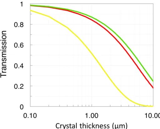
Pump light penetration in protein crystals. The protein concentration in crystals can be
as high as 30-60 mM, resulting in a very high optical density in the wavelength range where the chromophore moiety absorbs. As shown in Figure 4 for photosensitive proteins recently studied by TR-SFX, the penetration depth for pump-laser light at the wavelengths used in TR structural studies is limited to only a few microns or less. In case of optical activity, the light penetration can also be dependent on the crystal orientation that is generally randomly distributed in the case of TR-SFX. It is recommended to tune experimental parameters like crystal size, pump-light wavelength and pump-light polarization in order to ensure sufficient light penetration into the crystal – notably when using an experimental setup where the pump and probe beams are not collinear, i.e. when the probability increases of probing a poorly illuminated part of the crystal.
Excitation efficiency and pump power density issues. An important parameter to be
taken into account when designing a TR-SFX experiment is the expected excitation efficiency by the pump light. The excitation efficiency is defined as the ratio between the number of protein molecules wherein the photoreaction under study has effectively been triggered and the total number of photons impinging on the crystal. This efficiency is the product of the absorption cross-section at the given pump-light wavelength and polarization, and the primary quantum yield of the photoreaction. The primary quantum yield is the probability that an absorbed photon triggers the photoreaction. When the pump-pulse length is shorter than the excited-state lifetime, the extent of reaction initiation is limited by the primary quantum yield. For pump-pulses being longer than the lifetime of the excited state, a protein molecule that after excitation has decayed back to the ground state (instead of progressing further along the photoreaction pathway) can be excited more than once, resulting in an extent of reaction initiation that exceeds the primary quantum yield. For example, in recent TR-SFX experiments on PYP, whose excited state lifetime is about 500 fs (Lincoln et al. 2012), the extent of reaction initiation was 40% with nanosecond excitation (Tenboer et al. 2014). With femtosecond excitation, the extent of reaction initiation was only 13%, comparable to the primary quantum yield (Pande et al. 2016). To study excited-state structural dynamics on the femtosecond/picosecond time scale, only femtosecond excitation can be used. The usual practice is to excite the sample with a photon density corresponding to an average of at least one absorbed photon per chromophore. Considering typical values of protein concentrations in a crystal, of microcrystal sizes and of extinction coefficients, one absorbed photon per chromophore corresponds to a peak power density of the order of tens of GW/cm2 for femtosecond excitation. Such a high value is not only far beyond illumination power densities in any biologically relevant condition, but can also produce parasitic processes, like e. g. photobleaching, ionization and radical formation, which will compete with the photophysical process under study and may produce structural artefacts. Moreover, it is common practice to exceed the regime of one absorbed photon per chromophore, in order to compensate for possible pump-laser intensity fluctuations and position drifts affecting the pump-probe spatial overlap. Illustratively, the three recent femtosecond experiments discussed above used power densities of the order of 380 GW/cm2 (Barends et al. 2015),
access to X-ray FEL beam time has so far precluded the possibility of routinely performing pump power titrations, as required to define the linear regime within which excitation efficiency is maximized. Therefore, it is recommended to perform at least a preliminary thorough spectroscopic characterization of the system upon femtosecond excitation at various pump-power densities, prior to engaging in TR-SFX studies.
Time resolution and pump-probe synchronization diagnostics in ultra-fast experiments. The time resolution of an ultrafast TR-SFX experiment is given by the
convolution of the femtosecond pump pulse temporal profile with the femtosecond probe pulse temporal profile, and should in principle also be in the femtosecond range. However, the effective time resolution also depends on the precision of the pump-probe synchronization. Optical laser pulses are synchronized to the X-ray FEL pulses by radiofrequency phase-locking technology. Currently, the precision of temporal synchronization with this technology is limited by the shot-to-shot jitters of optical and X-ray FEL pulses, which is on the order of more than 100 fs (Glownia et al. 2010), and by thermal drifts of the order of a few picoseconds after several hours, which is the typical time scale of a TR-SFX data collection. Although there is an effort to improve the pump-probe synchronization precision and stability (Löhl et al. 2010; Danailov et al. 2014), the approach currently used is to directly measure, on a shot-to-shot basis, the relative arrival time delay of optical and X-ray pulses close to the experimental interaction region. This real-time diagnostic approach allows re-sorting data recorded for a given pump-probe delay, by substituting the nominal time delay assigned to each image with the time delay corrected after post-processing. This method was shown to allow sub-10 fs precision in time delay determination (Harmand et al. 2013) and was successfully applied to process TR-SFX data with time resolution of ~100 fs (Barends et al. 2015; Pande et al. 2016), limited by crystallographic data redundancy rather than by the timing diagnostic itself.
Structure refinement of low-occupancy states. In pump-probe TR-SFX experiments on
photosensitive proteins, limited pump-light penetration into protein crystals (Figure 4) and non-unity excitation efficiency together result in only a fraction of the probed molecules being excited and effectively undergoing conformational changes. The probed diffraction signal is thus a mixture of that arising from molecules within which the reaction has been triggered, and from molecules that are still in the resting state. Most
often, the 2mFo-DFc experimental electron density maps for the datasets without (dark) and with (light) pump excitation hardly differ and straightforward building of a model for the transient structure is not possible. Consequently, insights into conformational changes are obtained by examination of structure-factor amplitude Fourier difference maps, calculated by subtracting observed structure factor amplitudes for the dark data set (Fodark) from those of the light data set (Folight), and then phasing this difference with phases calculated from the dark model. Experimental Folight – Fodark differences can be weighted prior to map calculation based on the Bayesian likelihood that each Folight - Fodark structure factor amplitude difference is observed at a given resolution within measurement errors (sigmas) – an approach termed Q-weighting (Ursby and Bourgeois 1997). Q-weighting was introduced as a means to improve the estimates of difference amplitudes and to reduce the noise in difference maps calculated from time-resolved polychromatic Laue data, but it also increases the signal-to-noise ratio of amplitude differences from monochromatic datasets. In Folight - Fodark maps, negative and positive peaks indicate positions from which electron density has vanished and appeared upon pumping, respectively. Using this information, and prior knowledge from the resting-state model, the crystallographer may build a model for the transient structure, include it as an alternate conformer in the light structure, and then refine this new model in terms of coordinates and occupancy in the reciprocal space. An initial guess for the validity and occupancy of the transient conformer in the light structure can be obtained by computing Fclight –Fcdark maps for various relative occupancies of the resting-state and transient states in the light structure, and comparing peak heights (on residues of interest) in these and in the experimental Folight - Fodark map. Another approach is to compute extrapolated structure factors for the transient structure at 100% occupancy (Fextlight), and then to perform difference refinement against these (Terwilliger and Berendzen 1995). By this method, 2mFextlight-DFcdark and mFextlight-DFcdark electron density maps can be obtained that will guide model-building for the transient structure. The transient structure is then refined by minimizing the residual between the extrapolated and calculated structure factor amplitudes (Fermi et al. 1982; Terwilliger and Berendzen 1995; Šrajer et al. 1996). Again, an initial estimate of the occupancy of the transient state in the light dataset is needed to generate extrapolated structure factors, using the following equation: Fextlight = a * Q * (Folight - Fodark) + Fodark, where Q is the optional Bayesian-based weighting factor and 1/a is the occupancy of the time-evolved state in the light structure. This estimate
may again be obtained by computing Fclight –Fcdark maps and comparing peak heights on residues of interest in these and in the experimental Folight –Fodark map (Duan et al. 2013). Alternatively, occupancy-scaling can rely on a similar comparison between the experimental Folight –Fodark map, and the mFextlight-DFcdark maps computed using extrapolated Fextlight for various occupancies of the transient state in the light dataset (Coquelle et al. 2018).
Kinetic analysis of time-resolved diffraction patterns. A TR-SFX experiment produces a
time-dependent set of electron density maps spanning a given time range and possibly containing time-dependent changes in density as the reaction proceeds. The strategy to interpret such a time-resolved dataset depends on the time scale explored, which we divide into dynamic (ultrafast, i.e. fs-ps) and kinetic (several ps and longer) regimes. In the dynamic regime, structural changes reflect the coherent dynamics of atomic motions, allowing a direct comparison in terms of structure and time scale with results from molecular dynamics simulations. Time-dependent maps each provide a snapshot of the protein in motion and allow tracking non-exponential structural evolutions such as side chain oscillations in Mb (Barends et al. 2015). In the kinetic regime, the time evolution of the crystalline protein ensemble is governed by the energy barriers separating structural intermediates. The time-dependence of density maps then arises from the variation in population of the underlying time-independent intermediate structures, each associated with a reaction intermediate. The objective of a kinetic analysis of time-dependent maps is to extract a set of time-independent maps, each corresponding to a different reaction intermediate that can be used to determine the reaction pathway along with rate coefficients connecting the different reaction intermediates. The two main strategies described in the literature for a kinetic analysis are singular value decomposition (SVD) (Schmidt et al. 2003) and cluster analysis (Kostov and Moffat 2011). An exhaustive description of the two methods goes beyond the scope of this chapter and we refer to the dedicated literature (Kostov and Moffat 2011; Schmidt et al. 2003; Rajagopal et al. 2004). Briefly, the SVD method, extensively used in the analysis of any kind of time-resolved data, is a mathematical procedure that represents each data set by two sets of vectors, weighted by corresponding so-called singular values. The first set of singular vectors forms a time-independent orthonormal basis on which all time-dependent data can be decomposed; the second set of singular vectors describes the time-dependence of the first set. Since the basis vectors are weighted by their singular values, the data matrix can be approximated by a subset of vectors which contains signal above the noise level. The obtained reduced representation of
the data sets allows interpreting the time evolution of singular vectors in terms of a kinetic model. The cluster analysis is a general method used in statistics to group objects contained in an ensemble, based on a similarity defined by a given mathematical measure. This method has been proposed to deconvolute time-independent reaction intermediates from an intrinsically noisy and time-dependent electron density datasets produced by time-resolved crystallography, via a so-called “analytical trapping procedure”. A complete SVD or cluster analysis has not yet been carried in any of the published TR-SFX studies.
Sample presentation and consumption. The ultra-short and highly-intense nature of X-ray FEL pulses is what allows time-resolved studies down to the sub-ps timescale, at the atomic level of resolution. These characteristics are also at the origin of the high sample consumption rate of SFX and TR-SFX experiments. Indeed, because crystals virtually stand still during the fs exposure, all measured reflection intensities are partial, requiring collection from myriads of crystals to obtain a meaningful estimate of structure factor amplitudes. Furthermore, the intense X-ray beam eventually destroys the crystals, requiring the sample to be constantly replenished. Different means to present crystalline samples to the X-ray FEL beam exist, and they are extensively discussed in Chapter 6 of this book. All of them share the prospect of enabling TR-SFX studies, although on different systems and timescales. The first – and to date most successful – approach has been injection of crystalline samples across the X-ray FEL beam by means of gas-focused liquid jets (Weierstall et al. 2012). Sample consumption is the highest when working with liquid jets, with one out of several hundred thousand crystals interacting with an X-ray pulse at a repetition rate of 120 Hz (Schlichting and Miao 2012), due to the high injection speed (typically 10 m/s) required for the stability and sub-10 µm thickness of the jet (important for minimizing background scattering noise from the jet itself). Liquid jets can be highly stable and enable collection of data at pump-probe delays from sub-ps to a few µs, with pump excitation outside the injector nozzle. Because of the high jet speed, longer pump-probe delays are not easily accessible. Liquid jets remain the first choice in cases where crystalline sample production quantity is not an issue. Illustratively, all three ps timescale pump-probe TR-SFX studies published thus far were conducted using such an injection system, requiring about 1 g of protein crystals for a 5-shift experiment at the LCLS (Barends et al. 2015; Pande et al. 2016; Coquelle et al. 2018). To reduce sample consumption, crystals can be grown or embedded in a viscous carrier matrix prior to
(Weierstall et al. 2014; Botha et al. 2015; Sugahara et al. 2015). The first matrix of the kind was a lipidic cubic phase, in which crystals of the human serotonin receptor where grown before injection across the X-ray FEL beam (Liu et al. 2013), allowing structure determination from as little as 0.5 mg of protein. Rapidly after, other inert media were proposed that also form easily-extrudable gel-like matrices from crystals that do not grow in a lipidic cubic phase (Sugahara et al. 2015; Botha et al. 2015; Conrad et al. 2015; Sugahara et al. 2016; Sugahara et al. 2017; Kovacsova et al. 2017). High-viscosity jets have been used in pump-probe TR-SFX experiments on bR (Nogly et al. 2016; Nango et al. 2016), where pump-probe delays in the ms range were accessed due to the slow jet speed. Weaknesses of high-viscosity jets for TR-SFX include reduced transparency that limits the excitation efficiency (Tosha et al. 2017), the possibility of fluctuating jet speeds that reduce precision of longer pump-probe delays and the inability to address diffusion-based processes that are prohibited by the high viscosity of the matrix. Furthermore, the slow flow leads to concerns about pre-illumination of the sample from the previous laser pulses, a problem that is worse for irreversible reactions. Recently, the design was reported of a conveyer belt on which crystals may ride following acoustic droplet ejection, allowing for multiple illuminations, and/or exposure to solute or vapors prior to interaction with the X-ray beam (Fuller et al. 2017). Hence, the conveyer belt system is in theory adapted both for pump-probe and diffusion based TR-SFX experiments. Published work to date used only the pump-probe mode, to provide insights into the photocycle of photosystem II (Young et al. 2016), but it is expected that other experiments will soon take advantage of the versatility and completeness of the setup. Weaknesses are that the size of ejected drops remains high and that the device does not run in vacuum, both generating high background. TR-SFX crystallography data can also be collected from a much smaller number of crystals presented sequentially to the X-ray beam in a cryo-loop, as recently shown for the cytochrome c oxidase (Shimada et al. 2017). About 40 large crystals (0.5 mm) per time point were used, each of which was exposed at multiple angles and positions while maintained in a humid air flow at 4°C. Chip-based micro-compartmented systems allowing sequestration of microcrystals and their presentation to the X-ray beam at a speed matching the repetition rate of most X-ray FEL sources have also been proposed and demonstrated, which could enable TR-SFX on microcrystals with minimal background and high hit rates (Oghbaey et al. 2016; Owen et al. 2017; Roedig et al. 2017). Solid-support based-approaches thus hold the promise of allowing a more
efficient use of crystalline material and beamtime, during TR-SFX experiments on photosensitive proteins or proteins complexed with caged compounds. In combination with microfluidic devices, solid-support based-approaches could also provide insight into diffusion-based mechanisms, by means of a variation on the mix-and-inject approach (Schmidt et al., 2013).
6. Perspective on future applications of TR-SFX
Since the first study in 2012 (Aquila et al. 2012), an increasing number of different light-sensitive proteins have been studied by TR-SFX (Figure 6). Even if most of them are model systems that have been extensively studied by various biophysical techniques, TR-SFX experiments, in particular those carried out on the ultrafast time scale, provided new insights. For some model systems (e.g. bR and other rhodopsins), ultra-fast TR-SFX is yet to come. For others (e.g. PS II), further insight from TR-SFX awaits increased diffraction resolution. Time-resolved changes along the reaction pathway are still to be uncovered by TR-SFX in a large number of inherently photosensitive proteins (e.g. reaction center, phytochromes, cryptochromes, …), as well as in photoenzymes. Also, it is expected that more and more inherently light insensitive proteins will be studied by TR-SFX using caged compounds or synthetic photoswitches (Broichhagen et al. 2015). In these cases, the functional relevance of light-induced motions will have to be assessed carefully. With the proven possibility to be carried out on the ultrafast time scale, TR-SFX provides the unique opportunity to link photochemistry (fs-ps motions) and photobiology (µs-ms motions) for the first time on a structural basis. The prospect of understanding the entire chain of reactions, from photon absorption to signal transduction in the case of rhodopsin, or to proton pumping in the case of bR, is now finally within reach. The complementarity of X-ray FELs and synchrotrons, in particular the highly brilliant 4th generation sources to come up in the next years, is likely to gain shape in the near future. Indeed, serial synchrotron crystallography has gained momentum in the past three years (Gati et al. 2014; Stellato et al. 2014; Heymann et al. 2014; Nogly et al. 2015; Botha et al. 2015; Coquelle et al. 2015; Huang et al. 2015; Schubert et al. 2016; Jaeger et al. 2016; Oghbaey et al. 2016; Owen et al. 2017; Martin-Garcia et al. 2017; Beyerlein et al. 2017; Meents et al. 2017; Weinert et al. 2017) and will be able to cover time-resolved serial
limited storage rings produce the required photon flux. TR-SFX might then mostly focus on ultrafast studies and on cases where X-ray induced changes need to be avoided.
Thus far, all TR-SFX studies but two ((Kupitz et al. 2017) (Stagno et al. 2017)) have been carried out on light sensitive proteins with an optical laser as the pump method (Figure 6). Other triggering methods mentioned in section 2 (T-jump, THz irradiation, X-ray irradiation, …) are likely to be incorporated into TR-SFX protocols to study light-insensitive proteins, as will be the mix-and-inject approach discussed in Chapter 13 of this book.
TR-SFX studies published to date ‘only’ provided one or up to a dozen (Nango et al. 2016) snapshots of a protein structure after reaction initiation, limited by the extremely scarce X-ray FEL beam time and the requirement to collect a large number of indexable diffraction patterns per data set. With three more X-ray FELs having started operation in 2017, on the one hand, and optimization of SFX data collection and data processing schemes, on the other hand, both bottlenecks are expected to be mitigated, so that TR-SFX may soon provide real-time molecular movies of biomolecules in action.
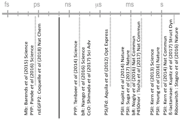
Figures Figure 1: Time-scale of protein motions and biophysical techniques to study them.
Figure 2 : Principle of pump-probe time-resolved crystallography. A crystalline macromolecule is probed by X-ray pulses (‘probe’, red beam) generated by a synchrotron or an X-ray FEL source. The time domain in the diffraction experiment is defined by the synchronization of X-ray pulses with optical laser pulses (‘pump’, blue beam). The interaction of optical laser pulses with the sample triggers a reaction and defines the time t=0. The evolution of the protein structure at a generic time ∆ti is monitored by variation of the ∆t delay between pump and probe. Every pump-probe sequence is repeated N times to produce a complete dataset from which a structural model can be determined.
Figure 3: General setup for TR-SFX experiments at X-ray FELs. There are a variety of
ways to present crystals to the (probe) X-ray beam in SFX experiments. In the present example, crystals are jetted across the pulsed X-ray beam, and thousands of diffraction patterns are collected for each dataset. In TR-SFX, a pump laser is used to trigger conformational changes in the crystalline protein, and data are collected by the X-ray probe beam at various pump-probe delays. The experiment may require photoconversion of crystals within a pre-illumination device (Schiro et al. 2017), as in the case of the photoswitchable rsEGFP2 that needed to be switched by 488 nm light from the fluorescent on-state (resting state) to the non-fluorescent off-state that was subsequently activated by a pump laser at 400 nm (Coquelle et al. 2018).
Figure 4 : Low pump-light penetration depth in chromophore-containing protein crystals results in only a fraction of the probed molecules being excited and effectively undergoing conformational changes. The figure shows the calculated transmission of pump light across crystals of the three proteins that have so far been studied by TR-SFX on the ps or sub-ps time scale, i.e. myoglobin (Mb; (Barends et al. 2015)), the photoactive yellow protein (PYP; (Pande et al. 2016)) and the reversibly photoswitchable fluorescent protein rsEGFP2 (Coquelle et al. 2018) as a function of crystal thickness. In these studies, concentrations of the crystalline Mb, PYP and rsEGFP2 were 53, 63 and 27 mM, respectively. The molar extinction coefficients of Mb, PYP and rsEGFP2 in its off state are 13950, 45500 and 22000 M-1.cm-1 at 532, 450 and 400 nm, respectively. Of note, the molar extinction coefficients used for these calculations were those of the ground states and assumed to be constant across the crystal over the pulse length. A more sophisticated calculation that takes into account the change of penetration depth as time progresses and molecules bleach has been presented for PYP (Tenboer et al. 2014).
Figure 5 : Three TR-SFX studies entered the ultra-fast time scale of photochemical reactions with pump-probe delays of a picosecond and shorter. (a) Structural changes in myoglobin (Mb) occurring 0.5 ps after photoexcitation (Barends et al. 2015). The Folight - Fodark difference electron density map is contoured at +3s (green) and -3s (red). (b) Oscillatory side-chain dynamics, excited by photodissociation of the heme,
reflect fast coherent motions that ultimately couple with larger-scale displacement of entire helices. (c) A structural transition at around 590 fs in the photoactive yellow protein (PYP) has been associated with trans-to-cis isomerization of the chromophore (Pande et al. 2016). (d,e) Structural changes in the reversibly photoswitchable fluorescent protein rsEGFP2 occurring 1 ps (red model) after photoexcitation of the non-fluorescent off-state (grey model) feature a fully twisted chromophore in the electronically excited state. The Folight - Fodark difference electron density map is overlaid in (d) and contoured at +3.5s (green) and -3.5s (red). (e) Chromophore twisting at 1 ps is accompanied by a shift in the entire helix that carries the chromophore. Panels (a,b) and (e) were extracted from published work ((Barends et al. 2015) and (Coquelle et al. 2018), respectively). Panels (c) and (d) were provided by Marius Schmidt and Nicolas Coquelle, respectively.
Figure 6 : All TR-SFX experiments published by early 2018 and their pump-probe time scales. Except for the riboswitch (Stagno et al. 2017) and ß-lactamase (Kupitz et al. 2017) studies, all TR-SFX experiments have been conducted on light-sensitive proteins with optical pump-pulses so far.
References Aquila A, Hunter MS, Doak RB, Kirian RA, Fromme P, White TA, Andreasson J, Arnlund D, Bajt S, Barends TR, Barthelmess M, Bogan MJ, Bostedt C, Bottin H, Bozek JD, Caleman C, Coppola N, Davidsson J, DePonte DP, Elser V, Epp SW, Erk B, Fleckenstein H, Foucar L, Frank M, Fromme R, Graafsma H, Grotjohann I, Gumprecht L, Hajdu J, Hampton CY, Hartmann A, Hartmann R, Hau-Riege S, Hauser G, Hirsemann H, Holl P, Holton JM, Homke A, Johansson L, Kimmel N, Kassemeyer S, Krasniqi F, Kuhnel KU, Liang M, Lomb L, Malmerberg E, Marchesini S, Martin AV, Maia FR, Messerschmidt M, Nass K, Reich C, Neutze R, Rolles D, Rudek B, Rudenko A, Schlichting I, Schmidt C, Schmidt KE, Schulz J, Seibert MM, Shoeman RL, Sierra R, Soltau H, Starodub D, Stellato F, Stern S, Struder L, Timneanu N, Ullrich J, Wang X, Williams GJ, Weidenspointner G, Weierstall U, Wunderer C, Barty A, Spence JC, Chapman HN (2012) Time-resolved protein nanocrystallography using an X-ray free-electron laser. Optics express 20 (3):2706-2716. doi:10.1364/OE.20.002706 Barends TR, Foucar L, Ardevol A, Nass K, Aquila A, Botha S, Doak RB, Falahati K, Hartmann E, Hilpert M, Heinz M, Hoffmann MC, Kofinger J, Koglin JE, Kovacsova G, Liang M, Milathianaki D, Lemke HT, Reinstein J, Roome CM, Shoeman RL, Williams GJ, Burghardt I, Hummer G, Boutet S, Schlichting I (2015) Direct observation of ultrafast collective motions in CO myoglobin upon ligand dissociation. Science 350 (6259):445-450. doi:10.1126/science.aac5492 Baxter RH, Ponomarenko N, Srajer V, Pahl R, Moffat K, Norris JR (2004) Time-resolved crystallographic studies of light-induced structural changes in the photosynthetic reaction center. Proceedings of the National Academy of Sciences of the United States of America 101 (16):5982-5987. doi:10.1073/pnas.0306840101 Beyerlein KR, Dierksmeyer D, Mariani V, Kuhn M, Sarrou I, Ottaviano A, Awel S, Knoska J, Fuglerud S, Jonsson O, Stern S, Wiedorn MO, Yefanov O, Adriano L, Bean R, Burkhardt A, Fischer P, Heymann M, Horke DA, Jungnickel KEJ, Kovaleva E, Lorbeer O, Metz M, Meyer J, Morgan A, Pande K, Panneerselvam S, Seuring C, Tolstikova A, Lieske J, Aplin S, Roessle M, White TA, Chapman HN, Meents A, Oberthuer D (2017) Mix-and-diffuse serial synchrotron crystallography. IUCrJ 4 (Pt 6):769-777. doi:10.1107/S2052252517013124 Botha S, Nass K, Barends TR, Kabsch W, Latz B, Dworkowski F, Foucar L, Panepucci E, Wang M, Shoeman RL, Schlichting I, Doak RB (2015) Room-temperature serial crystallography at synchrotron X-ray sources using slowly flowing free-standing high-viscosity microstreams. Acta crystallographica Section D, Biological crystallography 71 (Pt 2):387-397. doi:10.1107/S1399004714026327 Bourgeois D, Vallone B, Arcovito A, Sciara G, Schotte F, Anfinrud PA, Brunori M (2006) Extended subnanosecond structural dynamics of myoglobin revealed by Laue crystallography. Proceedings of the National Academy of Sciences of the United States of America 103 (13):4924-4929. doi:10.1073/pnas.0508880103 Bourgeois D, Vallone B, Schotte F, Arcovito A, Miele AE, Sciara G, Wulff M, Anfinrud P, Brunori M (2003) Complex landscape of protein structural dynamics unveiled by nanosecond Laue crystallography. Proceedings of the National Academy of Sciences of the United States of America 100 (15):8704-8709. doi:10.1073/pnas.1430900100
Bourgeois D, Weik M (2009) Kinetic protein crystallography: a tool to watch proteins in action. Crystallography Reviews 15 (2):87 - 118 Broichhagen J, Frank JA, Trauner D (2015) A Roadmap to Success in Photopharmacology. Accounts of Chemical Research 48 (7):1947-1960. doi:10.1021/acs.accounts.5b00129 Cammarata M, Levantino M, Schotte F, Anfinrud PA, Ewald F, Choi J, Cupane A, Wulff M, Ihee H (2008) Tracking the structural dynamics of proteins in solution using time-resolved wide-angle X-ray scattering. Nat Methods 5 (10):881-886. Epub 2008 Sep 2021. Chapman HN, Fromme P, Barty A, White TA, Kirian RA, Aquila A, Hunter MS, Schulz J, DePonte DP, Weierstall U, Doak RB, Maia FR, Martin AV, Schlichting I, Lomb L, Coppola N, Shoeman RL, Epp SW, Hartmann R, Rolles D, Rudenko A, Foucar L, Kimmel N, Weidenspointner G, Holl P, Liang M, Barthelmess M, Caleman C, Boutet S, Bogan MJ, Krzywinski J, Bostedt C, Bajt S, Gumprecht L, Rudek B, Erk B, Schmidt C, Homke A, Reich C, Pietschner D, Struder L, Hauser G, Gorke H, Ullrich J, Herrmann S, Schaller G, Schopper F, Soltau H, Kuhnel KU, Messerschmidt M, Bozek JD, Hau-Riege SP, Frank M, Hampton CY, Sierra RG, Starodub D, Williams GJ, Hajdu J, Timneanu N, Seibert MM, Andreasson J, Rocker A, Jonsson O, Svenda M, Stern S, Nass K, Andritschke R, Schroter CD, Krasniqi F, Bott M, Schmidt KE, Wang X, Grotjohann I, Holton JM, Barends TR, Neutze R, Marchesini S, Fromme R, Schorb S, Rupp D, Adolph M, Gorkhover T, Andersson I, Hirsemann H, Potdevin G, Graafsma H, Nilsson B, Spence JC (2011) Femtosecond X-ray protein nanocrystallography. Nature 470 (7332):73-77. Colletier JP, Bourgeois D, Sanson B, Fournier D, Sussman JL, Silman I, Weik M (2008) Shoot-and-Trap: use of specific x-ray damage to study structural protein dynamics by temperature-controlled cryo-crystallography. Proc Natl Acad Sci U S A 105 (33):11742-11747. Epub 12008 Aug 11713. Conrad CE, Basu S, James D, Wang D, Schaffer A, Roy-Chowdhury S, Zatsepin NA, Aquila A, Coe J, Gati C, Hunter MS, Koglin JE, Kupitz C, Nelson G, Subramanian G, White TA, Zhao Y, Zook J, Boutet S, Cherezov V, Spence JCH, Fromme R, Weierstall U, Fromme P (2015) A novel inert crystal delivery medium for serial femtosecond crystallography. IUCrJ 2 (4):421-430. doi:doi:10.1107/S2052252515009811 Coquelle N, Brewster AS, Kapp U, Shilova A, Weinhausen B, Burghammer M, Colletier J-P (2015) Raster-scanning serial protein crystallography using micro- and nano-focused synchrotron beams. Acta Crystallographica Section D 71 (5):1184-1196. doi:doi:10.1107/S1399004715004514 Coquelle N, Sliwa M, Woodhouse J, Schirò G, Adam V, Aquila A, Barends TRM, Boutet S, Byrdin M, Carbajo S, De la Mora E, Doak RB, Feliks M, Fieschi F, Foucar L, Guillon V, Hilpert M, Hunter MS, Jakobs S, Koglin JE, Kovacsova G, Lane TJ, Lévy B, Liang M, Nass K, Ridard J, Robinson JS, Roome CM, Ruckebusch C, Seaberg M, Thepaut M, Cammarata M, Demachy I, Field M, Shoeman RL, Bourgeois D, Colletier J-P, Schlichting I, Weik M (2018) Chromophore twisting in the excited state of a photoswitchable fluorescent protein captured by time-resolved serial femtosecond crystallography. Nature Chemistry 10:31-37. doi:10.1038/nchem.2853 Danailov MB, Bencivenga F, Capotondi F, Casolari F, Cinquegrana P, Demidovich A, Giangrisostomi E, Kiskinova MP, Kurdi G, Manfredda M, Masciovecchio C, Mincigrucci R, Nikolov IP, Pedersoli E, Principi E, Sigalotti P (2014) Towards
jitter-free pump-probe measurements at seeded free electron laser facilities. Optics express 22 (11):12869-12879. doi:10.1364/oe.22.012869 Duan C, Adam V, Byrdin M, Ridard J, Kieffer-Jaquinod S, Morlot C, Arcizet D, Demachy I, Bourgeois D (2013) Structural evidence for a two-regime photobleaching mechanism in a reversibly switchable fluorescent protein. Journal of the American Chemical Society 135 (42):15841-15850. doi:10.1021/ja406860e Efremov R, Gordeliy VI, Heberle J, Büldt G (2006) Time-Resolved Microspectroscopy on a Single Crystal of Bacteriorhodopsin Reveals Lattice-Induced Differences in the Photocycle Kinetics. Biophysical journal 91 (4):1441-1451. doi:10.1529/biophysj.106.083345 Fermi G, Perutz MF, Dickinson LC, Chien JCW (1982) Structure of human deoxy cobalt haemoglobin. Journal of Molecular Biology 155 (4):495-505. doi:https://doi.org/10.1016/0022-2836(82)90483-1 Fuller FD, Gul S, Chatterjee R, Burgie ES, Young ID, Lebrette H, Srinivas V, Brewster AS, Michels-Clark T, Clinger JA, Andi B, Ibrahim M, Pastor E, de Lichtenberg C, Hussein R, Pollock CJ, Zhang M, Stan CA, Kroll T, Fransson T, Weninger C, Kubin M, Aller P, Lassalle L, Brauer P, Miller MD, Amin M, Koroidov S, Roessler CG, Allaire M, Sierra RG, Docker PT, Glownia JM, Nelson S, Koglin JE, Zhu D, Chollet M, Song S, Lemke H, Liang M, Sokaras D, Alonso-Mori R, Zouni A, Messinger J, Bergmann U, Boal AK, Bollinger JM, Jr., Krebs C, Hogbom M, Phillips GN, Jr., Vierstra RD, Sauter NK, Orville AM, Kern J, Yachandra VK, Yano J (2017) Drop-on-demand sample delivery for studying biocatalysts in action at X-ray free-electron lasers. Nature methods 14 (4):443-449. doi:10.1038/nmeth.4195 Garman EF, Weik M (2017) Radiation Damage in Macromolecular Crystallography. Methods Mol Biol 1607:467-489. doi:10.1007/978-1-4939-7000-1_20 Gati C, Bourenkov G, Klinge M, Rehders D, Stellato F, Oberthur D, Yefanov O, Sommer BP, Mogk S, Duszenko M, Betzel C, Schneider TR, Chapman HN, Redecke L (2014) Serial crystallography on in vivo grown microcrystals using synchrotron radiation. IUCrJ 1 (Pt 2):87-94. doi:10.1107/S2052252513033939 Genick UK, Borgstahl GE, Ng K, Ren Z, Pradervand C, Burke PM, Srajer V, Teng TY, Schildkamp W, McRee DE, Moffat K, Getzoff ED (1997) Structure of a protein photocycle intermediate by millisecond time-resolved crystallography. Science 275 (5305):1471-1475 Gerwert K, Hess B, Michel H, Buchanan S (1988) FTIR studies on crystals of photosynthetic reaction centers. FEBS Letters 232 (2):303-307. doi:10.1016/0014-5793(88)80758-0 Glownia JM, Cryan J, Andreasson J, Belkacem A, Berrah N, Blaga CI, Bostedt C, Bozek J, DiMauro LF, Fang L, Frisch J, Gessner O, Gühr M, Hajdu J, Hertlein MP, Hoener M, Huang G, Kornilov O, Marangos JP, March AM, McFarland BK, Merdji H, Petrovic VS, Raman C, Ray D, Reis DA, Trigo M, White JL, White W, Wilcox R, Young L, Coffee RN, Bucksbaum PH (2010) Time-resolved pump-probe experiments at the LCLS. Optics express 18 (17):17620-17630. doi:10.1364/oe.18.017620 Grotjohann T, Testa I, Reuss M, Brakemann T, Eggeling C, Hell SW, Jakobs S (2012) rsEGFP2 enables fast RESOLFT nanoscopy of living cells. eLife 1:e00248. doi:10.7554/eLife.00248 Harmand M, Coffee R, Bionta MR, Chollet M, French D, Zhu D, Fritz DM, Lemke HT, Medvedev N, Ziaja B, Toleikis S, Cammarata M (2013) Achieving few-femtosecond time-sorting at hard X-ray free-electron lasers. Nat Photonics 7:215. doi:10.1038/nphoton.2013.11
Hekstra DR, White KI, Socolich MA, Henning RW, Srajer V, Ranganathan R (2016) Electric-field-stimulated protein mechanics. Nature 540 (7633):400-405. doi:10.1038/nature20571 Henzler-Wildman K, Kern D (2007) Dynamic personalities of proteins. Nature 450 (7172):964-972. Heymann M, Opthalage A, Wierman JL, Akella S, Szebenyi DM, Gruner SM, Fraden S (2014) Room-temperature serial crystallography using a kinetically optimized microfluidic device for protein crystallization and on-chip X-ray diffraction. IUCrJ 1 (Pt 5):349-360. doi:10.1107/S2052252514016960 Hirata K, Shinzawa-Itoh K, Yano N, Takemura S, Kato K, Hatanaka M, Muramoto K, Kawahara T, Tsukihara T, Yamashita E, Tono K, Ueno G, Hikima T, Murakami H, Inubushi Y, Yabashi M, Ishikawa T, Yamamoto M, Ogura T, Sugimoto H, Shen JR, Yoshikawa S, Ago H (2014) Determination of damage-free crystal structure of an X-ray-sensitive protein using an XFEL. Nature methods 11 (7):734-736. doi:10.1038/nmeth.2962 Huang CY, Olieric V, Ma P, Panepucci E, Diederichs K, Wang M, Caffrey M (2015) In meso in situ serial X-ray crystallography of soluble and membrane proteins. Acta crystallographica Section D, Biological crystallography 71 (Pt 6):1238-1256. doi:10.1107/S1399004715005210 Hutchison CDM, Kaucikas M, Tenboer J, Kupitz C, Moffat K, Schmidt M, van Thor JJ (2016) Photocycle populations with femtosecond excitation of crystalline photoactive yellow protein. Chemical Physics Letters 654:63-71. doi:http://dx.doi.org/10.1016/j.cplett.2016.04.087 Ihee H, Rajagopal S, Srajer V, Pahl R, Anderson S, Schmidt M, Schotte F, Anfinrud PA, Wulff M, Moffat K (2005) Visualizing reaction pathways in photoactive yellow protein from nanoseconds to seconds. Proceedings of the National Academy of Sciences of the United States of America 102 (20):7145-7150. Epub 2005 May 7143. Jaeger K, Dworkowski F, Nogly P, Milne C, Wang M, Standfuss J (2016) Serial Millisecond Crystallography of Membrane Proteins. Advances in experimental medicine and biology 922:137-149. doi:10.1007/978-3-319-35072-1_10 Jung YO, Lee JH, Kim J, Schmidt M, Moffat K, Srajer V, Ihee H (2013) Volume-conserving trans-cis isomerization pathways in photoactive yellow protein visualized by picosecond X-ray crystallography. Nature Chemistry 5 (3):212-220. doi:10.1038/nchem.1565 Kern J, Alonso-Mori R, Tran R, Hattne J, Gildea RJ, Echols N, Glockner C, Hellmich J, Laksmono H, Sierra RG, Lassalle-Kaiser B, Koroidov S, Lampe A, Han G, Gul S, Difiore D, Milathianaki D, Fry AR, Miahnahri A, Schafer DW, Messerschmidt M, Seibert MM, Koglin JE, Sokaras D, Weng TC, Sellberg J, Latimer MJ, Grosse-Kunstleve RW, Zwart PH, White WE, Glatzel P, Adams PD, Bogan MJ, Williams GJ, Boutet S, Messinger J, Zouni A, Sauter NK, Yachandra VK, Bergmann U, Yano J (2013) Simultaneous femtosecond X-ray spectroscopy and diffraction of photosystem II at room temperature. Science 340 (6131):491-495. doi:10.1126/science.1234273 Kern J, Tran R, Alonso-Mori R, Koroidov S, Echols N, Hattne J, Ibrahim M, Gul S, Laksmono H, Sierra RG, Gildea RJ, Han G, Hellmich J, Lassalle-Kaiser B, Chatterjee R, Brewster AS, Stan CA, Glockner C, Lampe A, DiFiore D, Milathianaki D, Fry AR, Seibert MM, Koglin JE, Gallo E, Uhlig J, Sokaras D, Weng TC, Zwart PH, Skinner DE, Bogan MJ, Messerschmidt M, Glatzel P, Williams GJ, Boutet S, Adams PD, Zouni A,