HAL Id: hal-02351986
https://hal.archives-ouvertes.fr/hal-02351986
Submitted on 23 Mar 2021
HAL is a multi-disciplinary open access archive for the deposit and dissemination of sci-entific research documents, whether they are pub-lished or not. The documents may come from teaching and research institutions in France or abroad, or from public or private research centers.
L’archive ouverte pluridisciplinaire HAL, est destinée au dépôt et à la diffusion de documents scientifiques de niveau recherche, publiés ou non, émanant des établissements d’enseignement et de recherche français ou étrangers, des laboratoires publics ou privés.
1-phosphate receptor 1 monoclonal antibody suitable for
cell imaging and biochemical studies of endogenous
receptors
Franck Talmont, Lionel Moulédous, Marion Baranger, Anne Gomez-Brouchet,
Jean-Marie Zajac, Clarence Deffaud, Olivier Cuvillier, Anastassia Hatzoglou
To cite this version:
Franck Talmont, Lionel Moulédous, Marion Baranger, Anne Gomez-Brouchet, Jean-Marie Zajac, et al.. Development and characterization of sphingosine 1-phosphate receptor 1 monoclonal antibody suitable for cell imaging and biochemical studies of endogenous receptors. PLoS ONE, Public Library of Science, 2019, 14 (3), pp.e0213203. �10.1371/journal.pone.0213203�. �hal-02351986�
Development and characterization of
sphingosine 1-phosphate receptor 1
monoclonal antibody suitable for cell imaging
and biochemical studies of endogenous
receptors
Franck TalmontID1*, Lionel Moule´dous1, Marion Baranger2, Anne Gomez-Brouchet1,3, Jean-Marie Zajac1, Clarence Deffaud2, Olivier Cuvillier1‡, Anastassia Hatzoglou1‡ 1 Institut de Pharmacologie et de Biologie Structurale, Universite´ de Toulouse, CNRS, UPS, Toulouse,
France, 2 BIOTEM, Apprieu, France, 3 Service d’anatomie et cytologie pathologiques, IUCT Oncopole, Toulouse, France
‡ These authors are co-last authors on this work. *franck.talmont@ipbs.fr
Abstract
Although sphingosine-1-phosphate receptor 1 (S1P1) has been shown to trigger several S1P targeted functions such as immune cell trafficking, cell proliferation, migration, or angio-genesis, tools that allow the accurate detection of endogenous S1P1localization and traf-ficking remain to be obtained and validated. In this study, we developed and characterized a novel monoclonal S1P1antibody. Mice were immunized with S1P1produced in the yeast
Pichia pastoris and nine hybridoma clones producing monoclonal antibodies were created.
Using different technical approaches including Western blot, immunoprecipitation and immunocytochemistry, we show that a selected clone, hereinafter referred to as 2B9, recog-nizes human and mouse S1P1in various cell lineages. The interaction between 2B9 and S1P1is specific over receptor subtypes, as the antibody does not binds to S1P2or S1P5 receptors. Using cell-imaging methods, we demonstrate that 2B9 binds to an epitope located at the intracellular domain of S1P1; reveals cytosolic and membrane localization of the endogenous S1P1; and receptor internalization upon S1P or FTY720-P stimulation. Finally, loss of 2B9 signal upon knockdown of endogenous S1P1by specific small interfer-ence RNAs further confirms its specificity. 2B9 was also able to detect S1P1in human kid-ney and spinal cord tissue by immunohistochemistry. Altogether, our results suggest that 2B9 could be a useful tool to detect, quantify or localize low amounts of endogenous S1P1in various physiological and pathological processes.
a1111111111 a1111111111 a1111111111 a1111111111 a1111111111 OPEN ACCESS
Citation: Talmont F, Moule´dous L, Baranger M,
Gomez-Brouchet A, Zajac J-M, Deffaud C, et al. (2019) Development and characterization of sphingosine 1-phosphate receptor 1 monoclonal antibody suitable for cell imaging and biochemical studies of endogenous receptors. PLoS ONE 14(3): e0213203.https://doi.org/10.1371/journal. pone.0213203
Editor: Paulo Lee Ho, Instituto Butantan, BRAZIL Received: November 12, 2018
Accepted: February 15, 2019 Published: March 7, 2019
Copyright:© 2019 Talmont et al. This is an open access article distributed under the terms of the Creative Commons Attribution License, which permits unrestricted use, distribution, and reproduction in any medium, provided the original author and source are credited.
Data Availability Statement: All relevant data are
within the manuscript and its Supporting Information files.
Funding: This work was supported by the French
Centre National de la Recherche Scientifique (CNRS) and by the BIOTEM company. The CNRS had no role in study design, data collection and analysis, decision to publish, or preparation of the manuscript. The BIOTEM company was involved in
Introduction
Sphingosine 1-phosphate receptor 1 (S1P1) is part of the sphingosine 1-phosphate (S1P)
receptor family, which comprises five G-protein coupled receptors (GPCR, S1P1, S1P2, S1P3,
S1P4, and S1P5, S1P1-5). This receptor family, firstly named, endothelial differentiation gene
(EDG) family of lipid receptors, also comprises lysophosphatidic acid (LPA) receptors. S1P 1-5bind the switterionic lysophospholipid S1P, with low nanomolar affinities, share sequence,
and genomic structure similarities [1–3]. S1P1was originally detected in human umbilical
vein endothelial cells (HUVEC) treated by phorbol 12-myristate 13-acetate [4]. S1P1
signal-ing pathway includes couplsignal-ing to the Gi/o proteins family and hence inhibition of adenylyl cyclase, activation of phosphatidylinositide 3-kinase and phospholipase C [5]. Analysis of transcripts indicates that S1P1is strongly expressed in adipose tissues, spleen, lung, brain,
liver, and heart and poorly represented in skeletal muscle, thymus, uterus, and kidney of adult mice [6]. When S1PR1 gene was ablated in the germ line of mice it resulted in a lethal effectin utero [7]. In fact S1P1has a vital role in vascular development and lethality in mice
was due to a defect in blood vessels development [6]. S1P1has also an essential function in
cell migration, in particular in the drain of T cells from the thymus to the blood and sur-rounding lymphoid structures [8]. More particularly, the activation of S1P1signaling
path-way with an agonist prevents the recruitment and migration of lymphocytes to sites of inflammation by the loss of ability to perceive S1P gradient concentration. The drug FTY720 (Fingolimod, Gilenya) which activates S1P1leading to impaired lymphocyte migration is
currently used for the treatment of relapsing remitting multiple sclerosis [9]. This drug is phosphorylated,in vivo, and the resulting FTY720-P binds to S1P1to activate receptors as a
true agonist. Nevertheless, this process leads to the internalization of S1P1that are not
recy-cled at the membrane thus blocking the egress of lymphocytes. S1P1is also implicated in
can-cer-related processes such as neovascularization in a tumor microenvironment context, cell migration, survival, transformation and progression [10]. Thus, the development of accurate tools for the detection, quantitation and localization of S1P1is mandatory to understand the
implication of this receptor in the regulation of numerous physiological and pathological processes. Besides commercial antibodies used by research groups, which are mainly rabbit polyclonal, generated with peptidic antigens and badly characterized, the analysis of scien-tific literature on S1P1allows selecting anti-S1P1antibodies demonstrating rather good
efficacy. The murine anti-S1P1 monoclonal IgG, called E49 [11] was produced using an
Escherichia coli-derived human S1P1full-length antigen. Another interesting antibody was
the rabbit anti-S1P1polyclonal antibody H60 raised against amino acids 322–381 of S1P1of
human origin [9,12,13]. Unfortunately, all these antibodies were discontinued. In this con-text, we have generated a murine monoclonal anti-S1P1antibody using a purified protein
produced in the methylotrophic yeastPichia pastoris model [14]. Mice were immunized with purified S1P1and nine hybridoma clones secreting specific S1P1monoclonal antibodies
(MAbs) were produced. Among these, 2B9 was selected and further characterized. This anti-body specifically recognizes human recombinant cmyc-S1P1and S1P1-Green Fluorescent
Protein, as well as human and mouse native S1P1s. We provide evidence that 2B9 recognizes
endogenous S1P1in murine embryonic fibroblasts (MEF), BT-549 breast cancer cell line and
HUVEC cells. The binding of 2B9 to S1P1is specific since the knocking down of the receptor
in cells leads to the loss of signal. Furthermore, 2B9 was able to detect S1P1by
immunohis-tochemistry in human tissue. Finally, 2B9 binds to the intracellular part of the receptor, reveals cytoplasmic and membrane bound S1P1as well as receptor internalization upon S1P
and FTY720-P stimulation.
study design, data collection and analysis, decision to publish, and preparation of the manuscript.
Competing interests: I have read the journal’s
policy and the authors of this manuscript have the following competing interests: MB and CD are employees of the BIOTEM company. French Centre National de la Recherche Scientifique (CNRS) will receive royalty payments associated with products sales. Monoclonal antibodies are available from commercial companies after payment. BIOTEM company has a sale agreement with commercial companies. This does not alter our adherence to PLOS ONE policies on sharing data and materials.
Methods
Plasmid construction
Plasmid cmyc-tagged pcDNA3-S1P1(Dr James Van Brockyn’s gift) was modified by PCR
(polymerase chain reaction) at the 5’ end to introduce a BstBI enzyme restriction site and at the 3’ end to introduce a Xba I site. Oligonucleotides were 5’-TTATTCGAAACGATGGGGCC CACCAGCGTC-3’ (BstBI forward) and 5’-TTGTTCTAGAGGGGAAGAAGAGTTGA CGTT-3’ (XbaI reverse). Modified cDNA was introduced into a TOPO TA vector (Invitrogen, Carsl-bad, CA). After digestion with BstBI and Xba enzymes, cDNA was introduced into pPICZ-hMOR-cmyc-his [15] vector digested with BstBI and XbaI thus deleting the hMOR coding sequence and leading to pPICZ-hS1P1-cmyc-his vector. This vector contains the full length
S1P1R gene in fusion with cmyc and 6-histidine tags. Mouse full-length S1P1/EDG1 versaclone
cDNA (RD systems) was cloned in pcDNA3 (Invitrogen, Carslbad, CA) using BamHI and XbaI restriction enzymes to obtain mS1P1-pcDNA3 plasmid.
Preparation of immunogens
S1P1was expressed inPichia cells by electro-transformation and cells were plated on zeocin
100μg/ml containing solid medium. Ten clones were selected and grown for 48h at 30˚C in two ml of a glycerol containing liquid medium (BMGY). S1P1expression was induced in 2 ml
liquid medium containing 1% methanol after centrifugation of cells and discarding of glycerol medium. Cells were then centrifuged and broken with glass beads. After elimination of partic-ulate matter and unbroken cells at 1,000g, a fraction, for each clone, was prepared by centrifu-gation at 10,000g and analyzed by WB using anti-cmyc antibodies [14,15].
Immunization of mice
BIOTEM animal experiments were realized in accordance with the dedicated laws. Institu-tional Animal Care Committee (IACUC) was DDPP de l’Isère. BIOTEM Ethics committee was the approving committee. The pure full-length receptor in complete Freund’s adjuvant was injected (100μg) subcutaneously in OF1 mice. Mice were subsequently immunized two times with hS1P1(100μg) in incomplete Freund’s adjuvant. Pure receptors (200 μg) were
administrated intraperitoneally in mice three days before cell fusion between splenocytes and myeloma cell line (NS-1). The selection of secreting Hybridomas was performed in Hypoxan-thine-Aminopterin-Thymidine medium. Before being euthanized using carbon dioxide method, mice were bled to collect immune serum.
Cell culture and transfections
CHO cells expressing human sphingosine 1-phosphate receptor 1 fused to the green fluores-cent protein (CHO hS1P1-GFP; Dr Kevin R. Lynch’ gift) and CHO wild type (WT) cells were
grown as described [16]. Mouse embryonic fibroblasts (MEF) and HUVEC cells were used until passage seven and cultured as previously described [17]. Human breast cancer cells (BT-549) and human Embryonic Kidney 293 cells (HEK) were from ATCC (Manassas, VA, USA). Cells were cultured in their optimal conditions in DMEM (Gibco, Grand Island, NY), contain-ing 10% fetal bovine serum (FBS, Gibco), 100 U/mL penicillin (Sigma, St. Louis, MO), and 100 mg/mL streptomycin (Sigma, St. Louis, MO) at 37 ˚C in a humidified incubator with 5% CO2
atmosphere. To establish cell lines stably expressing human S1P1, mouse S1P1(cDNA from
RD systems, a bio-techne brand) and GFP [18], CHO WT cells were transfected with the appropriate expression plasmids using Lipofectamine 2000 (Life Technologies) according to the manufacturer’s instructions. 48 hours post transfection, CHO cells expressing human,
mouse S1P1and control empty pcDNA3 plasmid were selected by adding 400μg/ml G418
(Euromedex) whereas 500μg/ml Hygromycin (Gibco) was added to the culture medium for the selection of GFP-CHO cells. Selected clones were analyzed by Western blot and immuno-cytochemistry. Transient expression of cmyc-S1P1, cmyc-S1P2and cmyc-S1P5(Dr James
Van Brockyn’s gift) was performed in Human Embryonic Kidney 293 cells (HEK) using Lipofectamine 2000. SiRNA transfections were performed using Lipofectamine 2000 (Invitro-gen) according to manufacturer instructions with a mix of two siRNAs (siS1P1A: sens 5’
GAGUUAGUUCCUGUGAACAdTdT 3’ and antisens 5’ UGUUCACAGGAACUAACUCdTdT 3’ and siS1P1B: sens 5’ CUGACUACGUCAACUAUGAdTdT 3’ and antisens 5’ UCAUAAG
UUGACGUAGUCAGdTdT 3’) as previously described [19]. SiRNAs negative control (siScr) was from Eurogentech.
Immunoprecipitation and Western blot analysis
After culture, cells recovered by scratching were centrifuged for 15 min at 1,000g and main-tained for at least 24 hours at—80˚C. Cells were lysed with a Potter Elvehjem homogenizer and cell membranes were obtained after two consecutive centrifugations at 1,000g and 100,000g. For immunoprecipitation, cell membranes were solubilized by incubation in a lysis buffer containing 0.5% NP40 (Calbiochem) and protease inhibitor cocktail (Roche Applied Science, France) at 4˚C for 1 night [20]. Samples were centrifuged at 100,000g and the super-natant was precleared by contact with Protein G sepharose (GE Healthcare Life Science) for 1h at 4˚C. The supernatant was then incubated with 2B9 and Protein G sepharose for 20 hours. Immunoprecipitated samples were washed with lysis buffer and solubilized in a Laemmli buffer, denatured for five min at 100 ˚C before being analyzed by SDS-PAGE and Western blot using 1:2,000 GFP rabbit polyclonal antibody (sc-8334, Santa Cruz) or 1:1,000 anti-S1P1H60 rabbit polyclonal antibody (sc-25489, Santa Cruz). S1P1containing samples from Pichia pastoris, CHO or HEK cell membranes or pure S1P1were analyzed by SDS-PAGE by
Western-blot or dot blot as previously described using 1:1,000 anti-cmyc mouse monoclonal antibody (clone 2E10, Sigma) [14].
Immunocytochemistry
Immunofluorescence studies were conducted as previously described [21,22]. Briefly cells were plated on glass coverslips and treated as indicated in the figure legends. Cells were washed with an ice-cold phosphate-buffered saline (PBS), fixed in 4% (w/v) paraformaldehyde (Elec-tron Microscopy Science, PA) for 10 min at room temperature. Cells were permeabilized with 0.2% Triton X-100 in PBS for 7 min 5% of fetal calf serum in PBS was used to block non spe-cific sites during 45 min Cells were then incubated with primary antibodies (2B9; H60; IgG2A, 1 mg/ml, dilution 1:75 w/v) for 45 min followed by secondary antibodies conjugated to the rel-evant fluorochrome (Alexa 488-coupled antibodies or TR-coupled antibodies from Molecular Probes, dilution 1:1500 v/v) for 45 min DNA was stained with DAPI (40
,6-diamidino-2-pheny-lindole). To identify the S1P1binding domain, non-permeabilized or Triton
X-100-permeabi-lized cells were stained as above. For receptor internalization studies, cells were seeded on glass coverslips and 24 hours later cells were serum-starved during 2 hours prior treatment. Cells were treated with vehicle (Ctrl) or S1P (1μM) or FTY720-P (1μM) for 1 hour. Then, cells were washed twice with PBS, fixed and stained with 2B9 and DAPI as described above. Image acqui-sitions were performed on a Nickon-Eclipse Cil; DS-Q12, microscope using 40x or 60x oil immersion objective and image analysis was conducted with ImageJ software.
Immunohistochemistry
Frozen human spinal cord sections from BioChain Institute (Newark, USA) were post-fixed in 4% formaldehyde for 15 min at room temperature. Sections were washed twice with PBS, and then incubated with 0.3% H2O2in PBS for 30 min. After two additional washes, blocking was
performed for 2 hours in PBS containing 0.25% Triton X-100 (PBS-T) and 3% normal goat serum (Vector Laboratories, Burlingame, CA, USA). Sections were then incubated with anti-bodies diluted 1:4,000 in the same buffer for 48 hours at 4˚C. After three 10 min washes with PBS-T, sections were incubated with biotinylated goat anti-mouse antibody (Vector Laborato-ries) diluted 1:600 in blocking solution for 2 hours at room temperature. After three 10 min washes, sections were placed in horseradish peroxidase avidin-biotin complex (Vectastain ABC kit, Vector Laboratories) diluted in PBS-T for 1 hour at room temperature. Sections were finally rinsed in PBS and stained in 3,3’-diaminobenzidine substrate kit (Vectastain DAB kit, Vector Laboratories) for 8 min according to manufacturer instructions. Sections were rinsed, dehydrated in graded ethanol, cleared in toluene and placed under coverslips with Deppex. Sections were viewed under a Leica CTR 600 wide-field microscope (Nanterre, France) and the Mercator software (Explora Nova, La Rochelle, France) was used to take pictures with 5x objective.
Human kidney samples were provided by the Centre de Ressources Biologiques-Cancer (CRB-Cancer, Institut Universitaire du Cancer Toulouse-Oncopole). Immunostaing using 2B9 and control IgG2A antibodies (1:50) were performed on kidney tissue microarrays (TMA) as previously described [23].
Results
Production of pure receptors
AllPichia clones presented the same pattern, specifically a protein monomeric band at 43 kDa
and multimeric protein bands over 95 kDa (Fig 1A). The best expressing clone (clone 5) was chosen for overexpression in shacked flasks. A total of 43 g of wet cells were broken with glass beads and used to prepare a fraction centrifuged at 10,000g. After solubilization in 0.1% sodium dodecyl sulfate, interaction with the Nickel phase and elution with imidazole, 5 mg of
Fig 1. Selection ofPichia pastoris clones expressing hS1P1and receptor purification. A Expression of hS1P1in different transfectedPichia pastoris clones (1 to 10). B (a) Purified hS1P1eluted with 300 mM imidazole. (b) Expression of hS1P1in crude supernatant fromPichia pastoris after cell breakage and centrifugation at 1,000g. Detection of receptors were realized after SDS-PAGE and Western blotting using an anti-cmyc antibody (1:1,000, v/v).
pure receptors were produced (Fig 1Ba) presenting the same Western blot pattern as observed inPichia cell membranes (Fig 1Bb).
Nine anti-S1P
1antibodies for one thousand clones tested
Pure human S1P1antigens fromPichia pastoris were used to immunize mice and to generate
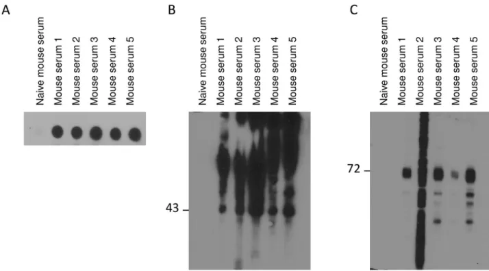
antibodies specific to receptors. Five animals were immunized with the antigen solubilized in 0.1% SDS (mouse 1–5). To select animals optimally responding to the recombinant proteins, reactivity against S1P1was assessed by dot blot and Western blot with sera obtained from the
animals (Fig 2). All sera from immunized mice detected S1P1from stable CHO cell line
express-ing S1P1-GFP by dot blot (Fig 2A). All sera were able to recognize pure S1P1-cmyc fromPichia
yeast (Fig 2B) and four out of five sera detected specifically S1P1-GFP (Fig 2C) by Western blot.
Serum from naïve mouse was unable to detect receptors either by dot or Western blot. Mice 3 and 4 were selected and a fusion was performed between spleen cells and myeloma NS-1 cell line by using standard procedures. From one thousand hybridoma clones screened as above, nine produced S1P1specific antibodies that were further characterized. Here we present results
of one antibody called 2B9. This clone was chosen because it exhibited, by Western blot, the more intense and clean signal among all clone tested on S1P1-GFP membranes. Not all clones
presented the same productivity and 2B9 was one of the best secreting clones.
Specificity of 2B9 was evaluated by Western blot and immunoprecipitation
We first tested the capacity of 2B9 to detect S1P1protein by Western blot. As shown inFig 3A,
2B9 detected S1P1in extracts from CHO cells stably expressing hS1P1-GFP but not from wild
Fig 2. Assessment of mouse sera. A Dot blot of CHO hS1P1-GFP cell membranes (1.6μg of sample was spotted onto the PVDF (polyvinylidene fluoride) membrane and revealed with 1:1,000 v/v diluted sera). B Pure hS1P1-cmyc fromPichia yeast cells (1 μg of sample per lane) was detected by Western blot using sera from five different immunized mice (1:5,000 v/v). C hS1P1 was detected in membrane fractions from CHO expressing hS1P1-GFP cells (20μg of sample per lane) using sera from five different immunized mice (1:5,000 v/v).
type CHO cells that do not express any S1P receptor as formerly described [24]. In HEK cells transiently expressing recombinant human cmyc-hS1P1, cmyc-hS1P2or cmyc-hS1P5, cmyc
antibody detected all receptors whereas 2B9 bound to hS1P1but not hS1P2and hS1P5further
confirming its specificity (Fig 3B). 2B9 also recognized mouse S1P1and human S1P1-GFP
overexpressed in CHO cells (Fig 3C) and was able to immunoprecipitate the recombinant
Fig 3. Detection of receptors in membranes from recombinant CHO and HEK cells by Western blot and in HUVEC and MEF after
immunoprecipitation. A Expression of hS1P1in control CHO WT cells and in CHO cells stably expressing hS1P1-GFP using 2B9 antibodies (1:5,000 w/v dilution). B HEK cells were transfected with vector expressing cmyc-hS1P1, cmyc-hS1P2or cmyc-hS1P5. Expression of the receptors was detected 48 hours later using cmyc (1:1,000 v/v) and 2B9 (1:5,000 w/v) antibodies. C Detection of mouse S1P1in control CHO WT cells, CHO cells stably expressing mS1P1and CHO cells stably expressing hS1P1-GFP using 2B9 antibody (1:1,000 w/v). D Human S1P1was immunoprecipitated from CHO cells stably expressing hS1P1-GFP using 2B9 antibody and detected with rabbit anti-GFP antibodies (1:2,000, v/v). Negative control was realized with no antibodies and positive control with a detergent-solubilized extract. E Endogenous human S1P1was immunoprecipitated from HUVEC cells using 2B9 antibody and detected with rabbit anti-S1P1H60 antibodies (1:1,000, v/v). CHO hS1P1were used as positive control. F Endogenous mS1P1was immunoprecipitated from MEF membrane fraction using 2B9 antibodies and detected with rabbit anti-S1P1H60 antibodies (1:1,000, v/v). 2B9 proteins were used as a control.
human S1P1-GFP as revealed with anti-GFP antibody (Fig 3D). Intensity of Western blot
mouse S1P1detection was low and this is a weakness for direct detection of mS1P1by this
tech-nic. Untagged hS1P1(Fig 3E) stably expressed in CHO cells was also immunoprecipitated by
2B9 and detected with rabbit H60 anti-S1P1antibody, [11,25]. Furthermore, 2B9 precipitated
endogenous human S1P1from HUVECs (Fig 3E) that were previously shown to express S1P1
receptors [9,26]. Lastly, 2B9 was also able to precipitate endogenous mouse S1P1in MEF
while purified antibody protein 2B9 was used as control (Fig 3F).
2B9 detects endogenous S1P
1by immunofluorescence microscopy
We next characterized 2B9 by immunocytochemistry.Fig 4shows that 2B9 recognizes S1P1
-GFP protein since 2B9 colocalized with -GFP signal at the level of the plasma membrane. An IgG2A control antibody did not stain CHO S1P1-GFP cells suggesting the specificity of 2B9. Fig 4. 2B9 specifically recognizes recombinant S1P1. CHO cells stably expressing hS1P1(CHO hS1P1-GFP) or GFP (CHO GFP) and CHO WT cell lines were seeded on coverslips and 24h later were fixed and stained for S1P1(red) with 2B9 or H60 or control IgG2A antibodies (1:100, w/v). Cells were treated with DAPI to visualize nuclei (blue). Colocalization (yellow) is shown between GFP (green) and 2B9 (red). Images are representative of the population examined. Scale bar, 10μm.
Importantly, 2B9 stained plasma membrane similarly to the widely used commercial H60 anti-body [11,25]. Neither 2B9 nor IgG2A stained CHO-GFP cells further confirming that 2B9 interacts with S1P1and the GFP tag did not affect this interaction. Lastly, in agreement with
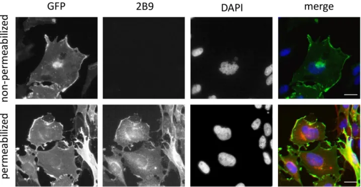
the literature, no staining was observed in WT CHO cells transfected with pcDNA3 plasmid that do not express any S1P receptor [24] (Fig 4). The epitope detected by 2B9 is localized to the intracellular domains of S1P1since strong membrane staining was observed in
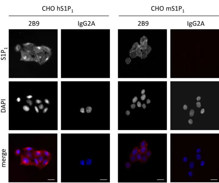
permeabi-lized cells while 2B9 did not stain non-permeabipermeabi-lized cells (Fig 5). We investigated whether 2B9 recognizes untagged human and mouse S1P1. To answer this question, receptors were
sta-bly expressed in CHO cells (CHO hS1P1; CHO mS1P1). 2B9 specifically stained hS1P1at the
level of plasma membrane while no staining was observed with control IgG2A antibodies. Fur-thermore, specific membrane staining was observed in cells expressing mouse S1P1(Fig 6).
These results demonstrate that 2B9 interacts with both human and mouse S1P1receptors. A
major challenge in S1P receptor studies is the detection of endogenous receptors. We have pre-viously shown that S1P1mRNA is present in primary MEF suggesting that protein may be also
expressed [17]. Since 2B9 recognizes mouse S1P1, we asked whether it was able to interact with
the endogenous mS1P1. As shown inFig 7, 2B9 revealed punctate cytoplasmic as well as
mem-brane (arrows and enlarged merged image) S1P1localization while no staining was observed
with the control IgG2A antibody. We next examined whether 2B9 recognized human endoge-nous S1P1. Cytoplasmic as well as plasma membrane S1P1localization was observed with 2B9
in the BT-549 breast cancer cell line (Fig 7). Taken together our results indicate that 2B9 is able to reveal endogenous human and mouse S1P1. To further confirming the specificity of
2B9, we finally tested whether knockdown of the S1P1by specific S1P1small interfering RNA
(si RNA) as previously described [19] leads to loss of S1P1staining. First, CHO S1P1-GFP
cells were transfected with control (scramble, siScr) or si RNAs against S1P1(siS1P1) and Fig 5. 2B9 binds to the intracellular domain of S1P1. CHO S1P1-GFP cells were seeded on coverslips and fixed 24h later. Permeabilized and
non-permeabilized cells were stained for S1P1with 2B9 (red) and for DNA with DAPI (blue). Colocalization (yellow) is shown between GFP (green) and 2B9 (1:100, w/v, red) in permeabilized cells. Non-permeabilized cells are not stained with 2B9 (1:100, w/v, red). Images are representative of the population examined. Scale bar, 10μm.
immunocytochemistry was performed 48 hours later. As shown inFig 8, siS1P1treatment
completely abolished receptor staining as compared to control cells (siScr) demonstrating the efficacy of the S1P1siRNAs. Second, to further validating the specificity of 2B9 we tested
whether S1P1knockdown could trigger loss of endogenous S1P1staining. Membrane S1P1
labeling in MEF was indeed completely abrogated in cells treated with siS1P1as compared to
control cells (Fig 8). These data thus establish that 2B9 specifically recognizes endogenous S1P1.
S1P1receptor undergoes rapid internalization upon agonist stimulation [9,27]. We next
followed endocytosis of endogenous S1P1in HUVEC cells using 2B9. As shown inFig 9,
under serum starvation conditions, 2B9 detected cytoplasmic as well as membrane S1P1in
control cells (Ctrl). White arrows indicate that part of S1P1localized at the level of the plasma
membrane. Treatment with S1P or FTY720-P rapidly induced receptor endocytosis and inter-nalization since S1P1is localized only within the cytoplasm (yellow arrows). Our data confirm Fig 6. 2B9 recognizes human and mouse S1P1. CHO cells stably expressing human (CHO hS1P1) or mouse (CHO mS1P1) S1P1receptors were fixed
and stained with 2B9 (red) or control IgG2A (red) antibodies (1:100, w/v). DNA was stained with DAPI (blue). Images are representative of the population examined. Scale bar, 10μm.
that pharmacological agonists induce receptor internalization and that 2B9 is well suited to study endogenous S1P1endocytosis.
2B9 detects S1P
1in human spinal cord and kidney by
immunohistochemistry
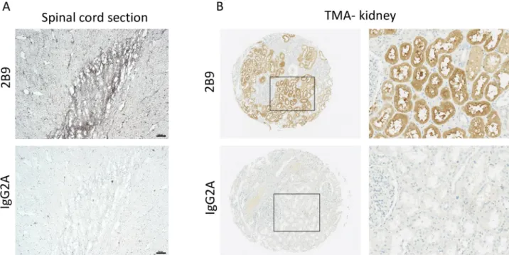
Because expression of S1P1-5is often dysregulated in pathological conditions, detection of
S1P1at tissue level is of crucial importance. Firstly, immunohistochemistry (IHC) on frozen
human spinal cord sections was conducted as the presence of S1P1in human spinal cord
section was previously reported using H60 polyclonal antibodies [28].Fig 10A, revealed the specific 2B9 staining at tissue level as compared to control IgG2A antibodies. Secondly, we showed the specific S1P1staining in the epithelial cells of the collecting duct of human kidney
(Fig 10B) using 2B9 antibodies, as compared to control IgG2A, in line with other studies [25,29].
Discussion
S1P1-5are differentially expressed under various physiological and pathological conditions [30,
31]. Despite the plethora of studies describing the cellular functions of S1P receptors, tools that allow detection, quantification and localization of these receptors are unfortunately scarce. In fact, most of the currently available antibodies directed at GPCRs are not correctly validated
Fig 7. 2B9 recognizes endogenous S1P1. MEF and BT-549 cells were fixed and stained with 2B9 (1:100, w/v, red) or IgG2A (1:100, w/v, red) antibodies
and DAPI (blue). Plasma membrane and cytoplasmic localization is seen with 2B9. Arrows indicate S1P1staining at the level of the plasma membrane. Inserts represent magnified regions. Images are representative of the population examined. Scale bar, 10μm.
neither for specificity nor for the detection of endogenously expressed receptors [16,32,33]. A challenge for GPCR receptor studies and more particularly S1P receptors studies is the specific and accurate detection of endogenous proteins. To this purpose, we developed and character-ized a new monoclonal anti-S1P1antibody, named 2B9. Based on our previous work [14,15,
34], we expressed and purified the human S1P1in thePichia pastoris model, which allows a
large availability of pure proteins. Immunization of mice with a SDS-solubilized S1P1induced
an immune reaction against foreign antigens and nine hybridoma cell lines producing anti-bodies were established. The advantage of the hybridoma model is the unlimited production of the same monoclonal antibody while polyclonal antibody production is animal-dependent. This is the case for the commonly used rabbit anti-S1P1antibody sc-25489 (H60) from Santa
Cruz Biotechnology Company. This antibody was used in a great number of S1P1studies [9,
13,25,26,28,35–42] but is not anymore commercially available. In this work, we show that 2B9 specifically binds to S1P1expressed either as endogenous or recombinant in various cell
lines. 2B9 detected, by Western blot, recombinant mouse and human S1P1receptors expressed
in CHO and HEK cells as well as the endogenous human and mouse receptors present in cell lines of different origin, like primary cells (HUVEC and MEF) and cancer derived cells (BT-549). Variation in the molecular weight of S1P1among cell lines can be explained at least in Fig 8. S1P1knockdown leads to loss of 2B9 staining. CHO cells stably expressing hS1P1-GFP (CHO hS1PR1-GFP) and MEF were transfected with
siRNA against S1P1(si S1P1) or scramble (si Scr). At 48 hours post transfection, CHO cells were fixed and stained for DNA with DAPI (blue). MEFs were fixed and stained for S1P1with 2B9 (1:100, w/v, green) and DNA with DAPI (blue). Images are representative of the population examined. Scale bar, 20μm.
part by posttranslational modification such as glycosylation [43–45]. S1P1exhibit one potential
glycosylation site located at position 30 (asparagine) in the amino-terminus extra-cellular part of the receptor. Asparagine 30 N-glycosylation was formally detected in recombinant HEK cells [44] but not in recombinantPichia pastoris. In general, proteins expressed in P. pastoris
have shorter glycosylation chains than those expressed inSaccharomyces cerevisiae thus
mak-ingP. pastoris a more attractive host for the expression of recombinant proteins.
Some 2B9 binds to the cmyc-hS1P1in HEK cells, but does not interact with cmyc-hS1P2
and cmyc-hS1P5suggesting its specificity. Finally, endogenous receptors in HUVEC and MEF
cells were detected, after immunoprecipitation, as a large protein band, in accordance with
Fig 9. Endogenous S1P1agonist-induced internalization can be followed with 2B9. HUVEC cells were seeded on coverslips and
24h later were serum starved for 2 hours. Cells were then stimulated with either vehicle (Ctrl), S1P (1μM) or FTY720-P (1μM) for 30 min, then fixed, permeabilized and stained with 2B9 (1:100, w/v, green) and DAPI (blue). Under serum starvation conditions (Ctrl) S1P1was localized at the plasma membrane and cytoplasm. In the presence of S1P or FTY720-P, S1P1is localized within cytoplasm. White arrows indicate S1P1staining at the level of plasma membrane in non-treated cells and yellow arrows intracellular staining in S1P and FTY720-P treated cells. Images are representative of the population examined. Scale bar, 10μm.
previous studies [46]. Our results show that 2B9 is suitable for immunoprecipitation. This potential is essential in the context of cell or tissue analyses where variation of very low expres-sion levels correlate with physiopathological conditions. Specific and dynamic subcellular localization of S1P1has been correlated to its cellular functions. S1P1cytoplasmic, membrane,
as well as nuclear localization have been reported in recombinant [47–50] and endogenous cells [38,42,51]. Furthermore, subcellular S1P receptors localization reflects the pharmacolog-ical action of receptor specific agonists and antagonists [27]. Thus developing antibodies appropriate for cell imaging studies contributes to the understanding of S1P receptors traffick-ing at the cellular level. In agreement with previous studies that used the CHO hS1P1-GFP cell
line, we showed that 2B9 detects hS1P1at the plasma membrane [52]. When expressed in
other cell lines [47–50], hS1P1-GFP was also found at the plasma membrane. More
impor-tantly, we showed cytoplasmic and membrane staining of the endogenous human and mouse receptors using 2B9 in various cells (HUVEC, MEF and BT-549 breast cancer cell line). This labeling is correlate well with specific concern since the control IgG2A antibody did not show any staining and knockdown of the S1P1receptor with specific siRNAs [17] led to the loss of
immunofluorescent signal. Taken together, our results demonstrate that 2B9 is suitable for endogenous S1P1immunocytochemistry studies. Immunofluorescence performed on intact
and permeabilized cells demonstrated that 2B9 recognizes an epitope located at the intracellu-lar part of S1P1. Thus, various glycosylation patterns encountered at different cells will have no
impact on the detection of S1P1. S1P receptors are considered as therapeutic targets and more
particularly S1P1in multiple sclerosis. The oral sphingosine-1-phosphate receptor modulator
Fingolimod (FTY720) was described to induce receptor endocytosis and to prevent lympho-cyte egress from lymphoid tissues [53]. For this reason, we investigated whether we can study ligand-induced receptor internalization using 2B9. Indeed, in agreement with the literature
Fig 10. S1P1is expressed in human tissues. S1P1is expressed in human spinal cord. Immunohistochemistry was performed in frozen human spinal cord sections using 2B9 and control IgG2A antibodies as described in methods section. Scale bar, 100μm. A, S1P1is expressed in human kidney. Immunohistochemistry was performed in human TMA kidney samples using 2B9 and control IgG2A antibodies (1:50, w/v) as was previously described [23]. Right panel shows 2mm TMA kidney sections, left panel shows X20 magnification.
[9,11,47,48,50,51,54–56] treatment of cells with S1P or FTY720-P led to the internalization of the plasma membrane S1P1receptor suggesting that 2B9 is suitable for receptor endocytosis
studies upon ligand stimulation.
The detection of the S1P1receptor by immunohistochemistry is important in tissue since
its expression is related to physiopathology [1,10,53,57]. 2B9 antibody detected S1P1in
human adult kidney and spinal cord in agreement with previous studies [25,28,29] suggesting that 2B9 is appropriate for immunohistochemistry studies. The current knowledge of thera-peutic potentials of S1P1in disorders including inflammation, fibrosis, and cancer underlines
the importance of tools that allow the monitoring of deregulated S1P1expression. In
conclu-sion, our work participates to the production and characterization of a new tool for the detec-tion, localization and quantitation of endogenous S1P1receptor expression. We must finally
advise researchers that all results obtained with an antibody on recombinant cells or on tissues or endogenous cells have to be compared with results obtained with a control.
Supporting information
S1 File. Arrive guideline checklist.
(PDF)
Acknowledgments
We thanks Dr Kevin R. Lynch (University of Virginia,School of Medicine, Charlottesville,
VA, USA)for providing S1P1-GFP CHO cells, Dr James R. Van Brocklyn (Department of
Evo-lution, Ecology and Organismal Biology, The Ohio State University, Columbus, OH, USA) for providing cmyc-tagged S1P receptors pcDNA3 plasmids and Dr Raoul Mazar (IPBS-CNRS, Toulouse, France) for providing HUVEC.
Author Contributions
Conceptualization: Franck Talmont.
Formal analysis: Franck Talmont, Anastassia Hatzoglou. Funding acquisition: Franck Talmont.
Investigation: Franck Talmont, Lionel Moule´dous, Marion Baranger, Anne Gomez-Brouchet,
Anastassia Hatzoglou.
Methodology: Franck Talmont.
Project administration: Franck Talmont.
Resources: Franck Talmont, Anne Gomez-Brouchet, Jean-Marie Zajac, Clarence Deffaud,
Olivier Cuvillier.
Supervision: Franck Talmont, Jean-Marie Zajac, Clarence Deffaud, Anastassia Hatzoglou. Validation: Franck Talmont, Anastassia Hatzoglou.
Visualization: Franck Talmont, Anastassia Hatzoglou.
Writing – original draft: Franck Talmont, Anastassia Hatzoglou.
References
1. Blaho VA, Hla T. An update on the biology of sphingosine 1-phosphate receptors. Journal of lipid research. 2014. Epub 2014/01/25.https://doi.org/10.1194/jlr.R046300PMID:24459205.
2. Fukushima N, Ishii I, Contos JJ, Weiner JA, Chun J. Lysophospholipid receptors. Annual review of phar-macology and toxicology. 2001; 41:507–34.https://doi.org/10.1146/annurev.pharmtox.41.1.507PMID: 11264467.
3. Rosen H, Stevens RC, Hanson M, Roberts E, Oldstone MBA. Sphingosine-1-phosphate and its recep-tors: structure, signaling, and influence. Annual review of biochemistry. 2013; 82:637–62.https://doi. org/10.1146/annurev-biochem-062411-130916PMID:23527695.
4. Hla T, Maciag T. An abundant transcript induced in differentiating human endothelial cells encodes a polypeptide with structural similarities to G-protein-coupled receptors. The Journal of biological chemis-try. 1990; 265(16):9308–13. Epub 1990/06/05. PMID:2160972.
5. O’Sullivan C, Dev KK. The structure and function of the S1P1 receptor. Trends Pharmacol Sci. 2013; 34 (7):401–12. Epub 2013/06/15.https://doi.org/10.1016/j.tips.2013.05.002PMID:23763867.
6. Choi JW, Lee CW, Chun J. Biological roles of lysophospholipid receptors revealed by genetic null mice: an update. Biochimica et biophysica acta. 2008; 1781(9):531–9. Epub 2008/04/15.https://doi.org/10. 1016/j.bbalip.2008.03.004PMID:18407842
7. Liu Y, Wada R, Yamashita T, Mi Y, Deng CX, Hobson JP, et al. Edg-1, the G protein-coupled receptor for sphingosine-1-phosphate, is essential for vascular maturation. The Journal of clinical investigation. 2000; 106(8):951–61. Epub 2000/10/18.https://doi.org/10.1172/JCI10905PMID:11032855
8. Allende ML, Dreier JL, Mandala S, Proia RL. Expression of the sphingosine 1-phosphate receptor, S1P1, on T-cells controls thymic emigration. The Journal of biological chemistry. 2004; 279(15):15396– 401. Epub 2004/01/21.https://doi.org/10.1074/jbc.M314291200PMID:14732704.
9. Mullershausen F, Zecri F, Cetin C, Billich A, Guerini D, Seuwen K. Persistent signaling induced by FTY720-phosphate is mediated by internalized S1P1 receptors. Nature chemical biology. 2009; 5 (6):428–34. Epub 2009/05/12.https://doi.org/10.1038/nchembio.173PMID:19430484.
10. Pyne NJ, Tonelli F, Lim KG, Long JS, Edwards J, Pyne S. Sphingosine 1-phosphate signalling in can-cer. Biochem Soc Trans. 2012; 40(1):94–100. Epub 2012/01/21.https://doi.org/10.1042/BST20110602 PMID:22260672.
11. Oo ML, Thangada S, Wu MT, Liu CH, Macdonald TL, Lynch KR, et al. Immunosuppressive and anti-angiogenic sphingosine 1-phosphate receptor-1 agonists induce ubiquitinylation and proteasomal deg-radation of the receptor. The Journal of biological chemistry. 2007; 282(12):9082–9. Epub 2007/01/24. https://doi.org/10.1074/jbc.M610318200PMID:17237497.
12. Garris CS, Wu L, Acharya S, Arac A, Blaho VA, Huang Y, et al. Defective sphingosine 1-phosphate receptor 1 (S1P1) phosphorylation exacerbates TH17-mediated autoimmune neuroinflammation. Nature immunology. 2013; 14(11):1166–72. Epub 2013/10/01.https://doi.org/10.1038/ni.2730PMID: 24076635
13. Silva VR, Micheletti TO, Pimentel GD, Katashima CK, Lenhare L, Morari J, et al. Hypothalamic S1P/ S1PR1 axis controls energy homeostasis. Nature communications. 2014; 5:4859. Epub 2014/09/26. https://doi.org/10.1038/ncomms5859PMID:25255053.
14. Talmont F, Mouledous L, Boue J, Mollereau C, Dietrich G. Denatured G-protein coupled receptors as immunogens to generate highly specific antibodies. PloS one. 2012; 7(9):e46348. Epub 2012/10/03. https://doi.org/10.1371/journal.pone.0046348PMID:23029489
15. Sarramegna V, Muller I, Mousseau G, Froment C, Monsarrat B, Milon A, et al. Solubilization, purifica-tion, and mass spectrometry analysis of the human mu-opioid receptor expressed in Pichia pastoris. Protein Expr Purif. 2005; 43(2):85–93. Epub 2005/08/13.https://doi.org/10.1016/j.pep.2005.05.007 PMID:16095919.
16. Talmont F, Mouledous L. Evaluation of commercial antibodies against human sphingosine-1-phosphate receptor 1. Naunyn-Schmiedeberg’s archives of pharmacology. 2014; 387(5):427–31. Epub 2014/01/ 25.https://doi.org/10.1007/s00210-014-0957-5PMID:24458374.
17. Andrieu G, Ledoux A, Branka S, Bocquet M, Gilhodes J, Walzer T, et al. Sphingosine 1-phosphate sig-naling through its receptor S1P5 promotes chromosome segregation and mitotic progression. Science signaling. 2017; 10(472). Epub 2017/03/30.https://doi.org/10.1126/scisignal.aah4007PMID: 28351953.
18. Gomez D, Guedin A, Mergny JL, Salles B, Riou JF, Teulade-Fichou MP, et al. A G-quadruplex structure within the 5’-UTR of TRF2 mRNA represses translation in human cells. Nucleic acids research. 2010; 38(20):7187–98. Epub 2010/06/24.https://doi.org/10.1093/nar/gkq563PMID:20571083
19. Brizuela L, Martin C, Jeannot P, Ader I, Gstalder C, Andrieu G, et al. Osteoblast-derived sphingosine 1-phosphate to induce proliferation and confer resistance to therapeutics to bone metastasis-derived
prostate cancer cells. Molecular oncology. 2014; 8(7):1181–95. Epub 2014/04/29.https://doi.org/10. 1016/j.molonc.2014.04.001PMID:24768038
20. Kersante F, Mouledous L, Zajac JM, Mollereau C. Modulation by neuropeptide FF of the interaction of mu-opioid (MOP) receptor with G-proteins. Neurochemistry international. 2010; 56(6–7):768–73. Epub 2010/03/10.https://doi.org/10.1016/j.neuint.2010.02.014PMID:20211672.
21. Andrieu G, Quaranta M, Leprince C, Cuvillier O, Hatzoglou A. Gem GTPase acts upstream Gmip/RhoA to regulate cortical actin remodeling and spindle positioning during early mitosis. Carcinogenesis. 2014; 35(11):2503–11. Epub 2014/09/01.https://doi.org/10.1093/carcin/bgu185PMID:25173885.
22. Andrieu G, Quaranta M, Leprince C, Hatzoglou A. The GTPase Gem and its partner Kif9 are required for chromosome alignment, spindle length control, and mitotic progression. FASEB journal: official pub-lication of the Federation of American Societies for Experimental Biology. 2012; 26(12):5025–34. Epub 2012/09/12.https://doi.org/10.1096/fj.12-209460PMID:22964304.
23. Gomez-Brouchet A, Illac C, Gilhodes J, Bouvier C, Aubert S, Guinebretiere JM, et al. CD163-positive tumor-associated macrophages and CD8-positive cytotoxic lymphocytes are powerful diagnostic mark-ers for the therapeutic stratification of osteosarcoma patients: An immunohistochemical analysis of the biopsies fromthe French OS2006 phase 3 trial. Oncoimmunology. 2017; 6(9):e1331193. Epub 2017/09/ 22.https://doi.org/10.1080/2162402X.2017.1331193PMID:28932633
24. Okamoto H, Takuwa N, Gonda K, Okazaki H, Chang K, Yatomi Y, et al. EDG1 is a functional sphingo-sine-1-phosphate receptor that is linked via a Gi/o to multiple signaling pathways, including phospholi-pase C activation, Ca2+ mobilization, Ras-mitogen-activated protein kinase activation, and adenylate cyclase inhibition. The Journal of biological chemistry. 1998; 273(42):27104–10. Epub 1998/10/09. PMID:9765227.
25. Akiyama T, Sadahira Y, Matsubara K, Mori M, Igarashi Y. Immunohistochemical detection of sphingo-sine-1-phosphate receptor 1 in vascular and lymphatic endothelial cells. Journal of molecular histol-ogy. 2008; 39(5):527–33. Epub 2008/09/02.https://doi.org/10.1007/s10735-008-9193-yPMID: 18758970.
26. Gatfield J, Monnier L, Studer R, Bolli MH, Steiner B, Nayler O. Sphingosine-1-phosphate (S1P) displays sustained S1P1 receptor agonism and signaling through S1P lyase-dependent receptor recycling. Cel-lular signalling. 2014; 26(7):1576–88. Epub 2014/04/08.https://doi.org/10.1016/j.cellsig.2014.03.029 PMID:24704119.
27. Liu CH, Thangada S, Lee MJ, Van Brocklyn JR, Spiegel S, Hla T. Ligand-induced trafficking of the sphingosine-1-phosphate receptor EDG-1. Molecular biology of the cell. 1999; 10(4):1179–90. Epub 1999/04/10.https://doi.org/10.1091/mbc.10.4.1179PMID:10198065
28. Nishimura H, Akiyama T, Irei I, Hamazaki S, Sadahira Y. Cellular localization of sphingosine-1-phos-phate receptor 1 expression in the human central nervous system. The journal of histochemistry and cytochemistry: official journal of the Histochemistry Society. 2010; 58(9):847–56. Epub 2010/06/23. https://doi.org/10.1369/jhc.2010.956409PMID:20566754
29. Chae SS, Proia RL, Hla T. Constitutive expression of the S1P1 receptor in adult tissues. Prostaglandins & other lipid mediators. 2004; 73(1–2):141–50. Epub 2004/05/29. PMID:15165038.
30. Mao-Draayer Y, Sarazin J, Fox D, Schiopu E. The sphingosine-1-phosphate receptor: A novel thera-peutic target for multiple sclerosis and other autoimmune diseases. Clin Immunol. 2017; 175:10–5. Epub 2016/11/29.https://doi.org/10.1016/j.clim.2016.11.008PMID:27890706
31. Sukocheva OA. Expansion of Sphingosine Kinase and Sphingosine-1-Phosphate Receptor Function in Normal and Cancer Cells: From Membrane Restructuring to Mediation of Estrogen Signaling and Stem Cell Programming. International journal of molecular sciences. 2018; 19(2). Epub 2018/02/01.https:// doi.org/10.3390/ijms19020420PMID:29385066.
32. Marchalant Y, Brownjohn PW, Bonnet A, Kleffmann T, Ashton JC. Validating Antibodies to the Cannabi-noid CB2 Receptor: Antibody Sensitivity Is Not Evidence of Antibody Specificity. The journal of histo-chemistry and cytohisto-chemistry: official journal of the Histohisto-chemistry Society. 2014; 62(6):395–404. Epub 2014/03/29.https://doi.org/10.1369/0022155414530995PMID:24670796
33. Michel MC, Wieland T, Tsujimoto G. How reliable are G-protein-coupled receptor antibodies? Naunyn-Schmiedeberg’s archives of pharmacology. 2009; 379(4):385–8. https://doi.org/10.1007/s00210-009-0395-yPMID:19172248.
34. Talmont F. Monitoring the human beta1, beta2, beta3 adrenergic receptors expression and purification in Pichia pastoris using the fluorescence properties of the enhanced green fluorescent protein. Biotech-nol Lett. 2009; 31(1):49–55.https://doi.org/10.1007/s10529-008-9840-0PMID:18797996.
35. Akiyama T, Hamazaki S, Monobe Y, Nishimura H, Irei I, Sadahira Y. Sphingosine-1-phosphate receptor 1 is a useful adjunct for distinguishing vascular neoplasms from morphological mimics. Virchows Archiv: an international journal of pathology. 2009; 454(2):217–22. Epub 2008/11/14.https://doi.org/10.1007/ s00428-008-0696-4PMID:19005676.
36. Galvani S, Sanson M, Blaho VA, Swendeman SL, Obinata H, Conger H, et al. HDL-bound sphingosine 1-phosphate acts as a biased agonist for the endothelial cell receptor S1P1 to limit vascular inflamma-tion. Science signaling. 2015; 8(389):ra79. Epub 2015/08/14.https://doi.org/10.1126/scisignal.aaa2581 PMID:26268607
37. Gonzalez-Cabrera PJ, Cahalan SM, Nguyen N, Sarkisyan G, Leaf NB, Cameron MD, et al. S1P(1) receptor modulation with cyclical recovery from lymphopenia ameliorates mouse model of multiple scle-rosis. Molecular pharmacology. 2012; 81(2):166–74. Epub 2011/10/28.https://doi.org/10.1124/mol. 111.076109PMID:22031473
38. Healy LM, Sheridan GK, Pritchard AJ, Rutkowska A, Mullershausen F, Dev KK. Pathway specific modu-lation of S1P1 receptor signalling in rat and human astrocytes. British journal of pharmacology. 2013; 169(5):1114–29. Epub 2013/04/17.https://doi.org/10.1111/bph.12207PMID:23587004
39. Kono M, Tucker AE, Tran J, Bergner JB, Turner EM, Proia RL. Sphingosine-1-phosphate receptor 1 reporter mice reveal receptor activation sites in vivo. The Journal of clinical investigation. 2014; 124 (5):2076–86. Epub 2014/03/29.https://doi.org/10.1172/JCI71194PMID:24667638
40. Lukas S, Patnaude L, Haxhinasto S, Slavin A, Hill-Drzewi M, Horan J, et al. No differences observed among multiple clinical S1P1 receptor agonists (functional antagonists) in S1P1 receptor down-regula-tion and degradadown-regula-tion. Journal of biomolecular screening. 2014; 19(3):407–16. Epub 2013/09/05.https:// doi.org/10.1177/1087057113502234PMID:24003058.
41. Oo ML, Chang SH, Thangada S, Wu MT, Rezaul K, Blaho V, et al. Engagement of S1P(1)-degradative mechanisms leads to vascular leak in mice. The Journal of clinical investigation. 2011; 121(6):2290– 300. Epub 2011/05/11.https://doi.org/10.1172/JCI45403PMID:21555855
42. Wilkerson BA, Grass GD, Wing SB, Argraves WS, Argraves KM. Sphingosine 1-phosphate (S1P) car-rier-dependent regulation of endothelial barrier: high density lipoprotein (HDL)-S1P prolongs endothelial barrier enhancement as compared with albumin-S1P via effects on levels, trafficking, and signaling of S1P1. The Journal of biological chemistry. 2012; 287(53):44645–53. Epub 2012/11/09.https://doi.org/ 10.1074/jbc.M112.423426PMID:23135269
43. Guan Y, Zhu Q, Huang D, Zhao S, Jan Lo L, Peng J. An equation to estimate the difference between theoretically predicted and SDS PAGE-displayed molecular weights for an acidic peptide. Scientific reports. 2015; 5:13370.https://www.nature.com/articles/srep13370#supplementary-information. PMID: 26311515
44. Kohno T, Wada A, Igarashi Y. N-glycans of sphingosine 1-phosphate receptor Edg-1 regulate ligand-induced receptor internalization. FASEB journal: official publication of the Federation of American Soci-eties for Experimental Biology. 2002; 16(9):983–92.https://doi.org/10.1096/fj.01-0809comPMID: 12087059.
45. Unal ES, Zhao R, Qiu A, Goldman ID. N-linked glycosylation and its impact on the electrophoretic mobil-ity and function of the human proton-coupled folate transporter (HsPCFT). Biochimica et biophysica acta. 2008; 1778(6):1407–14. Epub 2008/04/15.https://doi.org/10.1016/j.bbamem.2008.03.009PMID: 18405659
46. Huang YL, Lin HS, Chen SU, Lee H. Tyrosine sulphation of sphingosine 1-phosphate 1 (S1P1) is required for S1P-mediated cell migration in primary cultures of human umbilical vein endothelial cells. J Biochem. 2009; 146(6):815–20. Epub 2009/08/21.https://doi.org/10.1093/jb/mvp131PMID:19692429.
47. Lee MJ, Van Brocklyn JR, Thangada S, Liu CH, Hand AR, Menzeleev R, et al. Sphingosine-1-phos-phate as a ligand for the G protein-coupled receptor EDG-1. Science. 1998; 279(5356):1552–5. Epub 1998/03/21. PMID:9488656.
48. Reeves PM, Kang YL, Kirchhausen T. Endocytosis of Ligand-Activated Sphingosine 1-Phosphate Receptor 1 Mediated by the Clathrin-Pathway. Traffic. 2016; 17(1):40–52. Epub 2015/10/21.https://doi. org/10.1111/tra.12343PMID:26481905
49. Toman RE, Payne SG, Watterson KR, Maceyka M, Lee NH, Milstien S, et al. Differential transactivation of sphingosine-1-phosphate receptors modulates NGF-induced neurite extension. The Journal of cell biology. 2004; 166(3):381–92. Epub 2004/08/04.https://doi.org/10.1083/jcb.200402016PMID: 15289497
50. Watterson KR, Johnston E, Chalmers C, Pronin A, Cook SJ, Benovic JL, et al. Dual regulation of EDG1/ S1P(1) receptor phosphorylation and internalization by protein kinase C and G-protein-coupled receptor kinase 2. The Journal of biological chemistry. 2002; 277(8):5767–77. Epub 2001/12/14.https://doi.org/ 10.1074/jbc.M110647200PMID:11741892.
51. Estrada R, Wang L, Jala VR, Lee JF, Lin CY, Gray RD, et al. Ligand-induced nuclear translocation of S1P(1) receptors mediates Cyr61 and CTGF transcription in endothelial cells. Histochem Cell Biol. 2009; 131(2):239–49. Epub 2008/10/22.https://doi.org/10.1007/s00418-008-0521-9PMID:18936953
52. Schurer SC, Brown SJ, Gonzalez-Cabrera PJ, Schaeffer MT, Chapman J, Jo E, et al. Ligand-binding pocket shape differences between sphingosine 1-phosphate (S1P) receptors S1P1 and S1P3
determine efficiency of chemical probe identification by ultrahigh-throughput screening. ACS chemical biology. 2008; 3(8):486–98. Epub 2008/07/02.https://doi.org/10.1021/cb800051mPMID:18590333
53. Chun J, Hartung HP. Mechanism of action of oral fingolimod (FTY720) in multiple sclerosis. Clinical neuropharmacology. 2010; 33(2):91–101. Epub 2010/01/12.https://doi.org/10.1097/WNF. 0b013e3181cbf825PMID:20061941
54. Jongsma M, Florczyk UM, Hendriks-Balk MC, Michel MC, Peters SL, Alewijnse AE. Validation of a rapid, non-radioactive method to quantify internalisation of G-protein coupled receptors. Naunyn-Schmiedeberg’s archives of pharmacology. 2007; 375(5):329–36. Epub 2007/05/15.https://doi.org/10. 1007/s00210-007-0164-8PMID:17497135
55. LaMontagne K, Littlewood-Evans A, Schnell C, O’Reilly T, Wyder L, Sanchez T, et al. Antagonism of sphingosine-1-phosphate receptors by FTY720 inhibits angiogenesis and tumor vascularization. Can-cer research. 2006; 66(1):221–31. Epub 2006/01/07.https://doi.org/10.1158/0008-5472.CAN-05-2001 PMID:16397235.
56. Valentine WJ, Godwin VI, Osborne DA, Liu J, Fujiwara Y, Van Brocklyn J, et al. FTY720 (Gilenya) phos-phate selectivity of sphingosine 1-phosphos-phate receptor subtype 1 (S1P1) G protein-coupled receptor requires motifs in intracellular loop 1 and transmembrane domain 2. The Journal of biological chemistry. 2011; 286(35):30513–25. Epub 2011/07/02.https://doi.org/10.1074/jbc.M111.263442PMID:21719706
57. Dyckman AJ. Modulators of Sphingosine-1-phosphate Pathway Biology: Recent Advances of Sphingo-sine-1-phosphate Receptor 1 (S1P1) Agonists and Future Perspectives. Journal of medicinal chemis-try. 2017; 60(13):5267–89. Epub 2017/03/16.https://doi.org/10.1021/acs.jmedchem.6b01575PMID: 28291340.