HAL Id: inserm-00177055
https://www.hal.inserm.fr/inserm-00177055
Submitted on 5 Oct 2007HAL is a multi-disciplinary open access archive for the deposit and dissemination of sci-entific research documents, whether they are pub-lished or not. The documents may come from
L’archive ouverte pluridisciplinaire HAL, est destinée au dépôt et à la diffusion de documents scientifiques de niveau recherche, publiés ou non, émanant des établissements d’enseignement et de
France: comparison with HCV monoinfected patients
matched for body mass index and HCV genotype.
Laurent Castéra, Marc-Arthur Loko, Brigitte Le Bail, Patrick Coffie, Victor
de Lédinghen, Pascale Trimoulet, Maria Winnock, François Dabis, Didier
Neau, The Groupe d’Epidemiologie Clinique Du Sida En Acquitaine Gecsa
To cite this version:
Laurent Castéra, Marc-Arthur Loko, Brigitte Le Bail, Patrick Coffie, Victor de Lédinghen, et al.. Hepatic steatosis in HIV-HCV coinfected patients in France: comparison with HCV monoinfected pa-tients matched for body mass index and HCV genotype.. Alimentary Pharmacology and Therapeutics, Wiley, 2007, 26 (11-12), pp.1489-1498. �10.1111/j.1365-2036.2007.03533.x�. �inserm-00177055�
Hepatic steatosis in HIV-HCV coinfected patients in France: comparison with HCV monoinfected patients matched for body mass index and HCV genotype
L. CASTÉRA (1, 2), M. A. LOKO (3), B. LE BAIL (4), P. COFFIE (3), V. DE LEDINGHEN (1), P. TRIMOULET (5), M. WINNOCK (3), F. DABIS (3), D. NEAU (6) & THE GROUPE D’EPIDÉMIOLOGIE CLINIQUE DU SIDA EN AQUITAINE (GECSA)*
(1) Department of Hepatology, Hôpital Haut Lévêque, Centre Hospitalier Universitaire CHU Bordeaux, Pessac, France
(2) Department of Hepatology, Hopital St André, CHU Bordeaux, Bordeaux, France (3) INSERM U593 ISPED, Université Victor Segalen Bordeaux-2, Bordeaux, France (4) Department of Pathology, Hôpital Pellegrin, CHU Bordeaux, Bordeaux, France (5) Department of Virology, Hôpital Pellegrin, CHU Bordeaux, Bordeaux, France
(6) Department of Infectious Diseases, Hôpital Pellegrin, CHU Bordeaux; Bordeaux, France
Corresponding author: Laurent Castera, MD, PhD
Service d’Hépato-Gastroenterologie,
C.H.U. de Bordeaux,
Hôpital Haut Lévêque,
Avenue Magellan, 33604 Pessac, France Tel: 00 33 5 57 65 64 39 Tel: 00 33 5 57 65 64 45 e-mail: laurent.castera@chu-bordeaux.fr +++++++++++++++++++++++++++++++++++++++++++++++++++++++++++++++++++++++++++++++ This article is published in the Alimentary Pharmacology and Therapeutics' OnlineAccepted collection on Blackwell Synergy. The article has been allocated a unique Digital Optical Identifier (DOI), which will remain unchanged
throughout publication.
It must be emphasized that this article has not been edited, and changes are inevitable during this process.
See http://www.blackwell-synergy.com/page/onlineaccepted for details.
Please cite this article as a "Postprint"; doi: 10.1111/j.1365-2036.2007.03533.x +++++++++++++++++++++++++++++++++++++++++++++++++++++++++++++++++++++++++++++++
HAL author manuscript inserm-00177055, version 1
*Composition of the GECSA in charge of the ANRS CO3 Aquitaine Cohort:
Scientific committee: G. Chêne, F. Dabis, M. Dupon, M. Longy-Boursier, P. Morlat, JL. Pellegrin, JM. Ragnaud and R. Salamon Chair.
Methodological coordination: F. Dabis, G. Chêne, R. Thiébaut, C. Lewden and S. Lawson-Ayayi.
Medical coordination: M. Dupon, P. Mercié, JF. Moreau, P. Morlat, JL. Pellegrin, JM. Ragnaud, N. Bernard, D. Lacoste, D. Malvy and D. Neau.
Data Management and Analysis: MJ. Blaizeau, M. Decoin, S. Delveaux, C. Hannapier, S. Labarrère, V. Lavignolle-Aurillac, B. Uwamaliya-Nziyumvira, G. Palmer, D. Touchard, M. Winnock, E. Balestre, A. Alioum, H. Jacqmin-Gadda and R. Thiébaut.
Participating physicians: Bordeaux University Hospital: P. Morlat, N. Bernard, M. Bonarek, F. Bonnet, P. Gellie, D. Lacoste, K. Lacombe; M. Dupon, F. Dauchy, H. Dutronc, S. Lafarie; M. Longy-Boursier, P. Mercié, D. Malvy, T. Pistonne, P. Thibaut; JM. Ragnaud, D. Chambon, C. De La Taille, C. Cazorla, T. Galperine, D. Neau, A. Ochoa; JL.Pellegrin, JF. Viallard, O. Caubet, E. Lazaro C. Nouts; P.Couzigou, L. Castera; H. Fleury, ME. Lafon, B. Masquelier, I. Pellegrin; D. Breilh; JF. Moreau, P. Blanco. Dax Hospital: P. Loste, L. Caunègre. Bayonne Hospital: F. Bonnal, S. Farbos, MC Gemain. Libourne Hospital: J.Ceccaldi, S. Tchamgoué. Mont de Marsan Hospital: S. De Witte.
ABSTRACT
Background: Significance of steatosis in HIV-HCV coinfection remains controversial. Aim: To compare the prevalence and predictors of hepatic steatosis between HIV-HCV and HCV patients matched for steatosis known determinants.
Methods: 564 naive HCV patients undergoing liver biopsy were studied: 137 with HIV-HCV coinfection and 427 with HCV monoinfection, among whom 137 were matched for age, gender, BMI and HCV genotype.
Results: Steatosis of any grade (67.1% vs. 41.6%, p<0.0001), mixed steatosis (55.4% vs. 21.1%, p<0.0001), severe histological activity (A2-A3: 78.1% vs. 55.5%, p<0.0001), and severe fibrosis (F3-F4: 33.1% vs. 15.3%, p<0.0001) were significantly more common in coinfected than in matched monoinfected patients. In multivariate analysis, steatosis was associated only with severe histological activity (odds ratio (OR): 3.1 (95% CI: 1.3-7.1)) in coinfected patients and with elevated BMI (OR; 1.3 (1.1-1.5)), HCV genotype 3 (OR: 5.6 (2.3-13.9)), severe histological activity (OR: 3.1 7.3)) and severe fibrosis (OR: 4.7 (1.3-17.3)) in monoinfected patients.
Conclusions: Steatosis is significantly more common and severe in HIV-HCV coinfected than in HCV monoinfected French patients, even after matching for BMI and HCV genotype. Steatosis is associated only with severe histological activity in coinfected patients and with previously reported factors in monoinfected patients, thus suggesting different underlying mechanisms.
Key words: hepatic steatosis, HIV infection, HCV infection, BMI, HCV genotype
INTRODUCTION
Hepatic steatosis, defined as lipid accumulation in the hepatocyte cytoplasm, is a frequent histological finding in patients with chronic hepatitis C, occurring in more than 50 % of cases [1] and that has been suggested to be associated with fibrosis progression [2-8]. Two distinct forms of hepatocellular steatosis, “metabolic” and “viral”, can be observed in patients with chronic HCV infection [9]. Classical metabolic risk factors for steatosis such as obesity, diabetes type 2, hyperlipidaemia and excessive alcohol intake account for the vast majority of cases of steatosis in patients infected with HCV non 3 genotypes [10-12]. In contrast, in patients infected by HCV genotype 3, steatosis is thought to be induced by the virus itself through a direct cytopathic effect [13].
Liver disease is an important and increasing cause of morbidity and mortality in HIV-HCV coinfected patients [14, 15]. The prevalence and significance of steatosis in HIV-HIV-HCV coinfected patients have been recently investigated, and it has been suggested that it may contribute to the accelerated progression of liver disease observed in HIV-HCV coinfected patients [16-21]. However, despite its potential clinical importance, conflicting results have been published regarding factors associated with steatosis in the context of HIV-HCV coinfection. Some studies [19, 21] suggest that predictors of steatosis in HIV-HCV coinfected patients are the same than those observed in HCV monoinfected patients namely HCV genotype 3 and high body mass index (BMI) whereas other do not [16-18, 20]. Such discordant results may be explained by differences in the clinical features of studied populations, patients’ recruitment biases or ethnic factors. In addition, it must be stressed that two studies [18, 20] only from the US included a control group of HCV monoinfected patients and that patients of this control group were not matched for steatosis known determinants, BMI and HCV genotype.
The aim of this study was to compare the prevalence and predictors of steatosis between HIV-HCV coinfected and HCV monoinfected French patients matched for steatosis known determinants, BMI and HCV genotype as well as age and gender.
PATIENTS AND METHODS Study population
This retrospective study included patients with HIV-HCV coinfection and HCV mono-infection who were referred for liver biopsy prior to HCV antiviral therapy at the University Hospital of Bordeaux between January 1999 and January 2005.
Inclusion criteria were: age over 18, positive serum antibodies to HCV by means of a second- or third-generation HCV enzyme-linked immunosorbent assay (Ortho Diagnostic, Raritan NJ, USA) and detectable serum HCV RNA (AmplicorTM HCV, Roche Molecular Systems, Pleasanton, CA, USA).
Exclusion criteria were: coinfection with hepatitis B, other known causes of liver disease, alcohol intake of more than 50g/day, previous treatment for HCV, decompensated cirrhosis, and liver biopsy specimen <10 mm.
A total of 564 patients met those criteria. They comprised 427 HCV monoinfected patients and 137 HIV-HCV coinfected patients. This latter group is a subgroup of 148 HIV-HCV patients from the Aquitaine Cohort in whom we recently investigated the prevalence and predictors of steatosis [22]. The ANRS CO3 Aquitaine Cohort is a prospective hospital-based cohort of HIV-1-infected patients under routine clinical management which was initiated in 1987 in the Bordeaux University Hospital and four other public hospitals in the Aquitaine region, Southwestern France, by the Groupe d’Epidémiologie Clinique du SIDA en Aquitaine GECSA [23]. All adults who are in- or out- patients of the participating hospital wards with HIV-1 infection, confirmed by Western-Blot testing, and who have given an informed consent are enrolled in the cohort, whatever their clinical stage, gender or HIV transmission category. Also, information from at least one follow-up visit after the baseline assessment, or a known date of death must be available. At each hospital contact, a standardized questionnaire including epidemiological, clinical, biological and therapeutical data is filled in by clinicians
for each patient included in the Cohort and entered into the database. The schedule of follow-up visits is based on clinical practice, and an active search of patients lost to follow-follow-up is performed annually.
Among the 427 HCV mono-infected patients, 137 were matched to the 137 HIV-HCV coinfected patients according to age, gender, BMI and HCV genotype.
Clinical, biological and virological parameters
The following data were recorded for all patients at the time of liver biopsy: age, sex, risk factors for HCV infection, BMI, alaninz amonitransferas (ALT) and HCV genotype (Inno-Lipa II HCV, Innogenetics, Ghent, Belgium). In addition, the following data were specifically recorded for HIV-HCV coinfected patients: route of transmission of HIV infection, antiretroviral therapy, median CD4 lymphocyte count, and plasma HIV RNA load.
Histological studies and steatosis evaluation
Paraffin-embedded biopsy specimens of more than 10 mm were analyzed prospectively during clinical management by an experienced pathologist unaware of clinical and biological data except for the HCV or HIV status. Liver biopsy specimens were stained with hematoxylin-eosin-saffran, or picroSirius red for collagen. Data were collected in a standardized report form developed for hepatitis C before this specific study. In this report, histological activity and fibrosis are scored according to the METAVIR classification [24]. Steatosis is also graded according to METAVIR as previously validated [24]: none; mild: involving 1-10% of hepatocytes; moderate: involving 11-30% of hepatocytes; and severe: >30% of hepatocytes. In addition, the pattern of steatosis was characterized as follows: predominantly macrovesicular; predominantly microvesicular; mixed.
Statistical analysis
For univariate and multivariate statistical analysis, the outcome of interest was the presence of steatosis. Variables considered for analyses of steatosis determining factors were age at liver biopsy, gender, BMI, source of HCV infection, HCV genotype, ALT activity, use of antiretroviral treatment before liver biopsy, histological activity and fibrosis.
Continuous variables were expressed by their mean ± standard deviation (SD) and categorical variables by absolute figures and percentages. Mann-Whitney and Chi-2 tests were used for statistical comparisons of unmatched quantitative and qualitative variables, whereas Wilcoxon test and a conditional logistic regression were used for comparing quantitative and qualitative matched variables, respectively. A p value<0.05 was considered significant.
To assess the independent value of each parameter related to steatosis in univariate analysis with p<0.25, a multivariate analysis was performed by means of a stepwise logistic regression analysis. Statistical analyses were performed using SAS software (SAS institute Inc, Cary, NC, USA, version 9.1).
RESULTS Study population
The baseline characteristics of the 137 HIV-HCV coinfected patients as compared with those of the 427 HCV monoinfected patients are shown in table 1. Among HIV-HCV coinfected patients, the main source of HIV infection was intravenous drug use (68.0%). The median CD4 count was 508 cells/mm3 (78-1644 cells/mm3). A total of 125 (91%) patients were taking antiretroviral therapy: 122 (89%) were taking nucleoside reverse transcriptase inhibitors (NRTI), 55 (40%) protease inhibitors (PI), and 50 (36.5%) non nucleoside reverse transcriptase inhibitors (NNRTI). Plasma HIV RNA was undetectable (<50 copies/ml) in 60 patients (44%).
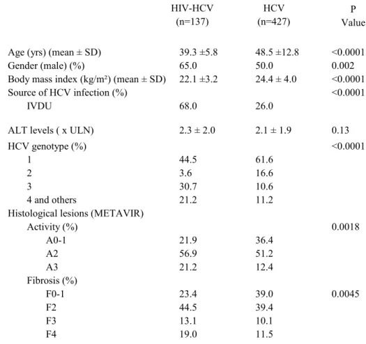
Coinfected patients differed from monoinfected patients in that there were significantly younger, more often male, had lower BMI, were more likely to have a prior history of intravenous drug use and to be HCV genotype 3-infected. They also had higher histological activity grade and more severe fibrosis.
Prevalence, severity and patterns of steatosis in the 137 HIV-HCV coinfected patients and the 427 HCV monoinfected patients
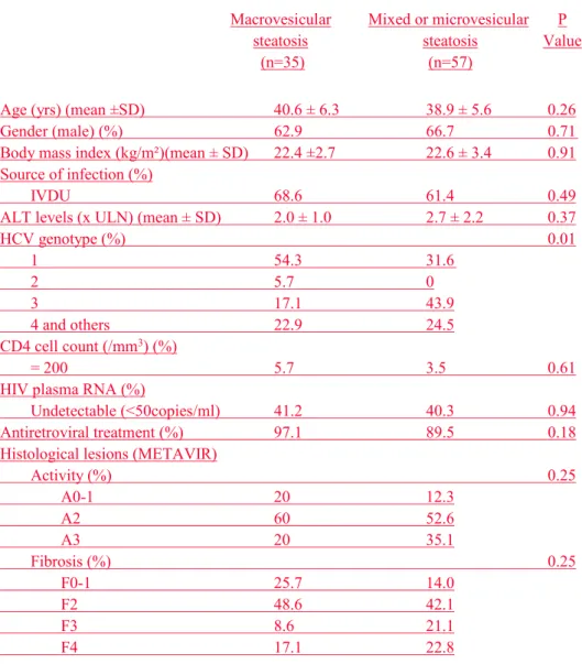
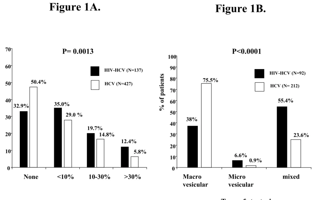
Steatosis of any grade was significantly more common (67.1 % vs. 49.6%, respectively) in coinfected than in monoinfected patients (Figure 1A). Among the 92 HIV-HCV coinfected and the 212 HCV monoinfected patients who had steatosis of any grade on liver biopsy, the pattern of steatosis differed significantly between the two groups (Figure 1B). Although the majority of HCV monoinfected patients had macrovesicular steatosis, coinfected patients were more likely to have a mixed pattern of steatosis (55.4% vs. 23.6%, respectively) or microvesicular steatosis (6.6% vs. 0.9%, respectively). Among the 92 HIV-HCV coinfected patients with steatosis, those with macrovesicular steatosis did not differ from those with
mixed of microvesicular steatosis for most characteristics, except for HCV genotype distribution (genotype 1: 54.3% vs. 31.6 %, respectively; p=0.01) (Table 2).
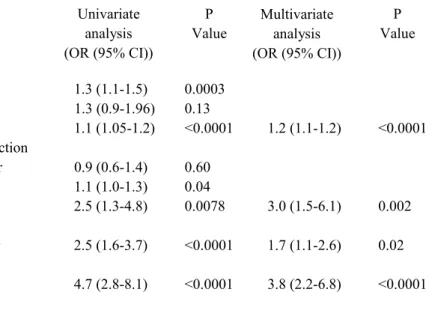
Factors associated with steatosis in the 427 monoinfected patients
In univariate analysis, steatosis was associated with age, BMI, ALT level, HCV genotype 3, severe histological activity and severe fibrosis (Table 3). In multivariate analysis, steatosis was associated with BMI (OR: 1.2 95% CI 1.1-1.2, p<0.0001), HCV genotype 3 (OR: 3.0 1.5-6.1, p=0.002), severe histological activity (A2-A3 OR: 1.7 1.1-2.6, p=0.02) and severe fibrosis (F3-F4 OR: 3.8 2.2-6.8, p< 0.0001) (Table 3).
Prevalence, severity and patterns of steatosis in the 137 HIV-HCV coinfected patients and the 137 matched HCV monoinfected patients
The baseline characteristics according to HIV status of the 137 matched patients are shown in Table 4. Coinfected patients were more likely to have a prior history of injection drug use, higher ALT levels, more severe histological activity and more severe fibrosis.
Steatosis of any grade was significantly more common in coinfected patients than in monoinfected patients (67.1% vs 41.6%, p<0.0001) (Figure 2A). Among the 92 HIV-HCV coinfected and the 57 HCV monoinfected patients who had steatosis of any grade on liver biopsy, the type of steatosis differed significantly between the two groups (Figure 2B). Although the majority of HCV monoinfected patients had macrovesicular steatosis, coinfected patients were more likely to have a mixed pattern of steatosis (55.4% vs. 21.1%, respectively) or microvesicular steatosis (6.6% vs. 1.7%, respectively).
Factors associated with steatosis in the 137 HIV-HCV coinfected patients and the 137 matched HCV monoinfected patients
In univariate analysis, steatosis was associated with severe histological activity A2-3, in coinfected patients whereas it was associated with elevated BMI, HCV genotype 3, severe histological activity (A2-A3) and severe fibrosis (F3-F4) in monoinfected patients (Table 5). In multivariate analysis, steatosis was associated with severe histological activity (A2-3 OR: 3.1 95% CI 1.3-7.1, p=0.008) in coinfected patients whereas it was associated with elevated BMI (OR: 1.3 1.1-1.5; p=0.005), HCV genotype 3 (OR: 5.6 2.3-13.9; p=0.0002), severe histological activity (OR: 3.1 1.3-7.3; p=0.009) and severe fibrosis (OR: 4.7 1.3-17.3; p=0.02) in monoinfected patients (Table 5).
DISCUSSION
The results of the present study, conducted in a large cohort of French patients with chronic hepatitis C, show that, even after adjusting by design for potential confounding factors HCV genotype and BMI, hepatic steatosis is significantly more common and more severe in HIV-HCV coinfected than in HCV monoinfected patients. In multivariate analysis, steatosis was associated only with severe histological activity in coinfected patients whereas it was associated with already reported factors (increased BMI, HCV genotype 3, severe histological activity and severe fibrosis) in monoinfected patients.
Steatosis has been reported in HIV-HCV coinfected patients with a prevalence ranging from 40% to 72% [16-20]. The 67% prevalence in our study is close to those reported in another French study [19] and in two recent American studies [20, 21]. A strength of the present study is that coinfected patients were compared to a control group of HCV monoinfected patients. So far, only two studies used a similar methodology [18, 20]. Both were conducted in American patients but obtained discrepant results: Monto et al. [18] found that steatosis was less common in coinfected patients than in monoinfected patients, 47% vs. 59%, respectively whereas Gaslightwala et al. [20] found that steatosis was more common in coinfected patients than in monoinfected patients (72.1% vs. 52%, respectively). Difference in the studied populations might account for this discrepancy.
It must be stressed that the baseline characteristics age, gender, BMI and proportion of patients infected by HCV genotype 3 of our population of HIV-HCV coinfected patients are similar to those reported in the Ribavic study, a multicenter French trial having included 395 coinfected patients [19]. In addition, the baseline characteristics as well as the prevalence and severity of steatosis in our control group of 427 HCV monoinfected patients are also similar to those previously reported [25, 26]. Finally, the factors independently associated with steatosis in multivariate analysis BMI, HCV genotype 3, severe activity and severe fibrosis
are also consistent with those of previous reports and of a recent meta-analysis in 3068 patients [27], lending further validity to our findings.
Interestingly, when compared with the total group of 427 HCV monoinfected patients, our coinfected patients were younger, more often male, had lower BMI, and were more likely to be infected with HCV genotype 3. Also, steatosis was significantly more common and more severe in coinfected patients, a finding in line with those reported by Gaslightwala et al. [20]. However, one should be cautious when interpreting such findings because the observed difference might be related to differences in the distribution of well known risk factors for steatosis such as HCV genotype 3 infection and increased BMI, between coinfected and monoinfected patients. The only way to avoid such a bias is to match coinfected patients with monoinfected patients according to these two risk factors, which had not been done in any study so far.
An important finding of the present study, is that, even after matching for BMI and HCV genotype, steatosis remained significantly more common and more severe in coinfected patients than in monoinfected patients. Also, coinfected patients were more likely to have a mixed pattern of macrovesicular and microvesicular steatosis than monoinfected patients before and after matching, a finding consistent with those of two recent studies [20, 21]. Although the reason for this finding is unclear, it suggests that different underlying mechanisms may be responsible for steatosis in coinfected patients. Indeed, microvesicular steatosis may be induced by NRTI which inhibit mitochondrial DNA polymerase gamma [28]. Consistent also with the hypothesis of different underlying mechanisms is the fact that factors independently associated with steatosis in multivariate analysis differed between coinfected and monoinfected patients. Indeed, steatosis was associated only with severe histological activity in coinfected patients whereas it was associated with previously reported
risk factors such as BMI, HCV genotype 3, severe activity and fibrosis in monoinfected patients.
In HCV monoinfected patients, two distinct forms of steatosis can be observed: “metabolic” steatosis associated with risk factors such as obesity, diabetes type 2, hyperlipidaemia and excessive alcohol intake and “viral” steatosis in patients infected by HCV genotype 3, through a direct cytopathic effect. The direct responsibility of HCV in the pathogenesis of steatosis has been suggested by several lines of evidence: 1) the independent association with HCV genotype 3 infection in most studies [9, 26, 29]; 2) the correlation between severity of steatosis and HCV genotype 3 replication levels while no such relationship has been found with other HCV genotypes [2, 7, 13, 30, 31]; 3) the disappearance of steatosis in HCV genotype 3 infected patients with sustained viral clearance after antiviral therapy [7, 31-33]; 4) finally, the development of hepatic steatosis in transgenic mouse lines [34] or in cell lines [35] expressing HCV core protein and the inhibition of microsomal triglyceride transfer protein, a key enzyme for hepatic lipoprotein assembly and secretion, in both transgenic mouse [36] and human liver [37].
Interestingly, despite the significant proportion of coinfected patients with HCV genotype 3 infection (30.7%) in our study, which is higher than that reported in studies from the US [16-18, 20] but similar to that reported in a recent French study [19], no correlation was found between genotype 3 and steatosis in coinfected patients. Histological activity was the only factor associated with steatosis. An association between histological activity and steatosis has been reported in HCV monoinfected patients [27, 38] as well as in HIV-HCV coinfected patients [17]. Several mechanisms may account for the relationship between steatosis and histological activity. HCV infection is associated with increased cytokines production enhancing histological inflammation and leading to increased lipid peroxydation [39]. Also, HCV core protein has been shown to induce oxidative stress in vitro and in vivo [40]. In
HCV coinfected patients, histological inflammation may be related not only to HCV infection but also to the use of antiretroviral drugs. Antiretroviral therapy has clearly been implicated in the development of steatosis in patients with HIV [41] and HIV-HCV coinfection [17, 21]. Although no relationship was found between steatosis and antiretroviral treatment in the present study, we cannot rule out the implication of the latter. Indeed, given the fact that nearly all patients were on antiretroviral therapy, we cannot exclude the fact that we did not have enough statistical power to demonstrate a significant relationship between steatosis and antiretroviral treatment. Finally, the retrospective design of our study did not allow to take into consideration the cumulative duration of exposure to antiretroviral drug which may also play an important role.
In conclusion, steatosis is significantly more common and more severe in HIV-HCV coinfected than in HCV monoinfected French patients, even after matching for BMI and HCV genotype. Steatosis is associated only with severe histological activity in coinfected patients and with already reported factors BMI, HCV genotype 3, severe histological activity and fibrosis in monoinfected patients. These results suggest different underlying mechanisms which deserve further investigation.
REFERENCES
1. Scheuer PJ, Ashrafzadeh P, Sherlock S, Brown D and Dusheiko GM. The pathology of hepatitis C. Hepatology 1992;15:567-71
2. Adinolfi LE, Gambardella M, Andreana A, Tripodi MF, Utili R and Ruggiero G. Steatosis accelerates the progression of liver damage of chronic hepatitis C patients and correlates with specific HCV genotype and visceral obesity. Hepatology 2001;33:1358-64.
3. Castera L, Hezode C, Roudot-Thoraval F, et al. Worsening of steatosis is an independent factor of fibrosis progression in untreated patients with chronic hepatitis C and paired liver biopsies. Gut 2003:288-92
4. Fartoux L, Chazouilleres O, Wendum D, Poupon R and Serfaty L. Impact of steatosis on progression of fibrosis in patients with mild hepatitis C. Hepatology 2005;41:82-7
5. Hourigan LF, MacDonald GA, Purdie D, et al. Fibrosis in chronic hepatitis C correlates significantly with body mass index and steatosis. Hepatology 1999;29:1215-9
6. Rubbia-Brandt L, Fabris P, Paganin S, et al. Steatosis affects chronic hepatitis C progression in a genotype-specific way. Gut 2004;53:406-12
7. Patton HM, Patel K, Behling C, et al. The impact of steatosis on disease progression and early and sustained treatment response in chronic hepatitis C patients. J Hepatol 2004;40:484-90
8. Westin J, Nordlinder H, Lagging M, Norkrans G and Wejstal R. Steatosis accelerates fibrosis development over time in hepatitis C virus genotype 3 infected patients. J Hepatol 2002;37:837-42.
9. Castera L, Chouteau P, Hezode C, Zafrani ES, Dhumeaux D and Pawlotsky JM. Hepatitis C virus-induced hepatocellular steatosis. Am J Gastroenterol 2005;100:711-5
10. Camma C, Bruno S, Di Marco V, et al. Insulin resistance is associated with steatosis in nondiabetic patients with genotype 1 chronic hepatitis C. Hepatology 2006;43:64-71
11. Fartoux L, Poujol-Robert A, Guechot J, Wendum D, Poupon R and Serfaty L. Insulin resistance is a cause of steatosis and fibrosis progression in chronic hepatitis C. Gut 2005;54:1003-8
12. Monto A, Alonzo J, Watson JJ, Grunfeld C and Wright TL. Steatosis in chronic hepatitis C: relative contributions of obesity, diabetes mellitus, and alcohol. Hepatology 2002;36:729-36.
13. Rubbia-Brandt L, Quadri R, Abid K, et al. Hepatocyte steatosis is a cytopathic effect of hepatitis C virus genotype 3. J Hepatol 2000;33:106-15
14. Rosenthal E, Poiree M, Pradier C, et al. Mortality due to hepatitis C-related liver disease in HIV-infected patients in France (Mortavic 2001 study). Aids 2003;17:1803-9
15. Salmon-Ceron D, Lewden C, Morlat P, et al. Liver disease as a major cause of death among HIV infected patients: role of hepatitis C and B viruses and alcohol. J Hepatol 2005;42:799-805
16. Marks KM, Petrovic LM, Talal AH, Murray MP, Gulick RM and Glesby MJ. Histological findings and clinical characteristics associated with hepatic steatosis in patients coinfected with HIV and hepatitis C virus. J Infect Dis 2005;192:1943-9
17. Sulkowski MS, Mehta SH, Torbenson M, et al. Hepatic steatosis and antiretroviral drug use among adults coinfected with HIV and hepatitis C virus. Aids 2005;19:585-92
18. Monto A, Dove LM, Bostrom A, Kakar S, Tien PC and Wright TL. Hepatic steatosis in HIV/hepatitis C coinfection: prevalence and significance compared with hepatitis C monoinfection. Hepatology 2005;42:310-6
19. Bani-Sadr F, Carrat F, Bedossa P, et al. Hepatic steatosis in HIV-HCV coinfected patients: analysis of risk factors. Aids 2006;20:525-31
20. Gaslightwala I, Bini EJ. Impact of human immunodeficiency virus infection on the prevalence and severity of steatosis in patients with chronic hepatitis C virus infection. J Hepatol 2006;44:1026-32
21. McGovern BH, Ditelberg JS, Taylor LE, et al. Hepatic steatosis is associated with fibrosis, nucleoside analogue use, and hepatitis C virus genotype 3 infection in HIV-seropositive patients. Clin Infect Dis 2006;43:365-72
22. Neau D, Winnock M, Castéra L, et al. Prevalence and factors associated with hepatic steatosis in patients coinfected with hepatitis C virus and HIV: ANRS CO3 Aquitaine Cohort. J Acquir Immune Defic Syndr 2007:45:168-73
23. Binquet C, Chene G, Jacqmin-Gadda H, et al. Modeling changes in CD4-positive T-lymphocyte counts after the start of highly active antiretroviral therapy and the relation with risk of opportunistic infections: the Aquitaine Cohort, 1996-1997. Am J Epidemiol 2001;153:386-93
24. The French METAVIR Cooperative Study Group. Intraobserver and interobserver variations in liver biopsy interpretation in patients with chronic hepatitis C. Hepatology 1994;20:15-20
25. Castera L. Steatosis, insulin resistance and fibrosis progression in chronic hepatitis C. Minerva Gastroenterol Dietol 2006;52:125-34
26. Asselah T, Rubbia-Brandt L, Marcellin P and Negro F. Steatosis in chronic hepatitis C: why does it really matter? Gut 2006;55:123-30
27. Leandro G, Mangia A, Hui J, et al. Relationship between steatosis, inflammation, and fibrosis in chronic hepatitis C: a meta-analysis of individual patient data. Gastroenterology 2006;130:1636-42
28. Lonergan JT, Behling C, Pfander H, Hassanein TI and Mathews WC. Hyperlactatemia and hepatic abnormalities in 10 human immunodeficiency virus-infected patients receiving nucleoside analogue combination regimens. Clin Infect Dis 2000;31:162-6 29. Clement S, Negro F. Hepatitis C virus: The viral way to fatty liver. J Hepatol
2007;46:985-7
30. Hezode C, Roudot-Thoraval F, Zafrani ES, Dhumeaux D and Pawlotsky JM. Different mechanisms of steatosis in hepatitis C virus genotypes 1 and 3 infections. J Viral Hepat 2004;11:455-8
31. Poynard T, Ratziu V, McHutchison J, et al. Effect of treatment with peginterferon or interferon alfa-2b and ribavirin on steatosis in patients infected with hepatitis C. Hepatology 2003;38:75-85.
32. Castera L, Hezode C, Roudot-Thoraval F, et al. Effect of antiviral treatment on evolution of liver steatosis in patients with chronic hepatitis C: indirect evidence for a role of HCV genotype 3 in steatosis. Gut 2004;53:420-4
33. Kumar D, Farrell GC, Fung C and George J. Hepatitis C virus genotype 3 is cytopathic to hepatocytes: Reversal of hepatic steatosis after sustained therapeutic response. Hepatology 2002;36:1266-72.
34. Moriya K, Yotsuyanagi H, Shintani Y, et al. Hepatitis C virus core protein induces hepatic steatosis in transgenic mice. J Gen Virol 1997;78:1527-31
35. Abid K, Pazienza V, de Gottardi A, et al. An in vitro model of hepatitis C virus genotype 3a-associated triglycerides accumulation. J Hepatol 2005;42:744-51
36. Perlemuter G, Sabile A, Letteron P, et al. Hepatitis C virus core protein inhibits microsomal triglyceride transfer protein activity and very low density lipoprotein secretion: a model of viral-related steatosis. Faseb J 2002;16:185-94.
37. Mirandola S, Realdon S, Iqbal J, et al. Liver microsomal triglyceride transfer protein is involved in hepatitis C liver steatosis. Gastroenterology 2006;130:1661-9
38. Asselah T, Boyer N, Guimont MC, et al. Liver fibrosis is not associated with steatosis but with necroinflammation in French patients with chronic hepatitis C. Gut 2003;52:1638-1643.
39. Neuman MG, Benhamou JP, Malkiewicz IM, et al. Kinetics of serum cytokines reflect changes in the severity of chronic hepatitis C presenting minimal fibrosis. J Viral Hepat 2002;9:134-40
40. Okuda M, Li K, Beard MR, et al. Mitochondrial injury, oxidative stress, and antioxidant gene expression are induced by hepatitis C virus core protein. Gastroenterology 2002;122:366-75.
41. Ristig M, Drechsler H and Powderly WG. Hepatic steatosis and HIV infection. AIDS Patient Care STDS 2005;19:356-65
Figure 1A. Severity of steatosis in the 137 HIV-HCV coinfected and 427 HCV monoinfected patients.
Figure 1B. Type of steatosis in the 92 HIV-HCV coinfected and 212 HCV monoinfected patients who had steatosis of any grade on liver biopsy.
Figure 2A. Severity of steatosis in the 137 HIV-HCV coinfected and 137 matched HCV monoinfected patients.
Figure 2B. Type of steatosis in the 92 HIV-HCV coinfected and 57 matched HCV monoinfected patients who had steatosis of any grade on liver biopsy.
Age (yrs) (mean ± SD) 39.3 ±5.8 48.5 ±12.8 <0.0001
Gender (male) (%) 65.0 50.0 0.002
Body mass index (kg/m²) (mean ± SD) 22.1 ±3.2 24.4 ± 4.0 <0.0001
Source of HCV infection (%) <0.0001
IVDU 68.0 26.0
ALT levels ( x ULN) 2.3 ± 2.0 2.1 ± 1.9 0.13
HCV genotype (%) <0.0001
1 44.5 61.6
2 3.6 16.6
3 30.7 10.6
4 and others 21.2 11.2
Histological lesions (METAVIR)
Activity (%) 0.0018 A0-1 21.9 36.4 A2 56.9 51.2 A3 21.2 12.4 Fibrosis (%) F0-1 23.4 39.0 0.0045 F2 44.5 39.4 F3 13.1 10.1 F4 19.0 11.5
Table 1. Characteristics of the 564 HCV-infected patients at the time of liver biopsy according to HIV status.
HIV-HCV (n=137) HCV (n=427) P Value
IVDU: intravenous drug use; ULN upper limit of normal value SD: standard deviation
Table 2. Characteristics of the 92 HIV-HCV coinfected patients with steatosis of any grade at liver biopsy, according to the type of steatosis.
Macrovesicular steatosis (n=35) Mixed or microvesicular steatosis (n=57) P Value
Age (yrs) (mean ±SD) 40.6 ± 6.3 38.9 ± 5.6 0.26
Gender (male) (%) 62.9 66.7 0.71
Body mass index (kg/m²)(mean ± SD) 22.4 ±2.7 22.6 ± 3.4 0.91
Source of infection (%)
IVDU 68.6 61.4 0.49
ALT levels (x ULN) (mean ± SD) 2.0 ± 1.0 2.7 ± 2.2 0.37
HCV genotype (%) 0.01 1 54.3 31.6 2 5.7 0 3 17.1 43.9 4 and others 22.9 24.5 CD4 cell count (/mm3) (%) = 200 5.7 3.5 0.61
HIV plasma RNA (%)
Undetectable (<50copies/ml) 41.2 40.3 0.94
Antiretroviral treatment (%) 97.1 89.5 0.18
Histological lesions (METAVIR)
Activity (%) 0.25 A0-1 20 12.3 A2 60 52.6 A3 20 35.1 Fibrosis (%) 0.25 F0-1 25.7 14.0 F2 48.6 42.1 F3 8.6 21.1 F4 17.1 22.8
IVDU: intravenous drug use; ULN upper limit of normal value SD: standard deviation
Table 3. Factors associated with steatosis in the 427 HCV monoinfected patients in univariate and multivariate analysis.
Univariate analysis (OR (95% CI)) P Value Age (yrs/10) 1.3 (1.1-1.5) 0.0003 Gender (male) 1.3 (0.9-1.96) 0.13
Body mass index 1.1 (1.05-1.2) <0.0001 1.2 (1.1-1.2) <0.0001 Source of HCV infection IVDU vs. Other 0.9 (0.6-1.4) 0.60 ALT level 1.1 (1.0-1.3) 0.04 HCV genotype 2.5 (1.3-4.8) 0.0078 3.0 (1.5-6.1) 0.002 3 vs. Other Histological activity 2.5 (1.6-3.7) <0.0001 1.7 (1.1-2.6) 0.02 A2-3 vs. A0-1 Fibrosis 4.7 (2.8-8.1) <0.0001 3.8 (2.2-6.8) <0.0001 F3-4 vs. F0-1-2 Multivariate analysis (OR (95% CI)) P Value
IVDU: intravenous drug use OR: odds ratio
CI: confidence interval
Table 4. Characteristics of the 137 matched pairs of HCV-infected patients at the time of liver biopsy according to HIV status.
HIV-HCV (n=137) HCV (n=137) P Value
Age (yrs) (mean ±SD) 39.3 ± 5.8 40.1 ± 6.2 NS
Gender (male) (%) 65.0 65.0 NS
Body mass index (kg/m²) (mean ± SD) 22.1 ±3.2 22.5 ± 2.8 NS Source of infection (%)
IVDU 68.0 51.0 0.005
ALT levels (x ULN)(mean ± SD) 2.3 ± 2.0 1.8 ± 1.5 0.01 HCV genotype (%)
1 44.5 44.5 NS
2 3.6 3.6
3 30.7 30.7
4 and others 21.2 21.2
Histological lesions (METAVIR)
Activity (%) <0.0001 A0-1 21.9 44.5 A2 56.9 48.2 A3 21.2 7.3 Fibrosis (%) <0.0001 F0-1 23.4 48.2 F2 44.5 36.5 F3 13.1 8.0 F4 19.0 7.3
IVDU: intravenous drug use; ULN upper limit of normal value SD: standard deviation
HIV-HCV univariate (OR (95% CI)) 1.2 (0.6-2.3) 1.0 (0.5-2.2) 1.12 (1-1.3) 0.6 (0.3-1.3) 1.2 (0.9-1.5) 1.6 (0.7-3.5) 3.1 (1.3-7.1) 2.0 (0.9-4.7) 0.6 (0.3-1.3) 1.9 (0.6-5.7) 1.3 (0.6-2.8) 1.5 (0.7-3.1) 0.98 (0.2-5.5) P Value 0.55 0.9 0.065 0.18 0.16 0.27 0.008 0.086 0.18 0.23 0.44 0.25 0.98 HCV univariate (OR (95% CI)) 1.7 (0.97-3.0) 1.2 (0.9-4.1) 1.2 (1.1-1.4) 1.6 (0.8-3.2) 1.0 (0.8-1.2) 5.3 (2.4-11.7) 3.8 (1.8-7.9) 8.1 (2.5-25.6) P Value 0.06 0.07 0.0036 0.18 0.94 <0.0001 0.0004 0.0004 Age (yrs/10) Gender (male) Body mass index Source of HCV infection IVDU vs. other ALT level HCV genotype 3 vs. Other Histological activity A2-3 vs. A0-1 Fibrosis F3-4 vs. F0-1-2 Treatment with NNRTI
Yes vs. No Treatment with NRTI
Yes vs. No Treatment with PI Yes vs. No
Plasma HIV RNA (copies/ml) =50 vs. <50 CD4 cell count (/mm3) =200 vs. >200 HIV-HCV multivariate (OR (95% CI)) 3.1 (1.3-7.1) P Value 0.008 HCV multivariate (OR (95% CI)) 1.3 (1.1-1.5) 5.6 (2.3-13.9) 3.1 (1.3-7.3) 4.7 (1.3-17.3) P Value 0.005 0.0002 0.009 0.02 Table 5. Factors associated with steatosis in the 137 HIV-HCV coinfected patients and in the 137 matched HCV monoinfected patients in univariate and multivariate analysis.
IVDU: intravenous drug use; NNRTI; non nucleoside reverse transcriptase inhibitors; NRTI: nucleoside reverse transcriptase inhibitors; PI: protease inhibitors
OR: odds ratio CI: confidence interval
Figure 1A.
P= 0.0013
50.4%
32.9%
35.0%
29.0 %
19.7%
14.8%
12.4%
5.8%
% of
patients
HIV-HCV (N=137) HCV (N=427)0
10
20
30
40
50
60
70
None
<10%
10-30%
>30%
% of
patients
P<0.0001
HIV-HCV (N=92) HCV (N= 212)75.5%
38%
6.6%
0.9%
55.4%
23.6%
0
10
20
30
40
50
60
70
80
90
100
Macro
vesicular
Micro
vesicular
mixed
Figure 1B.
HIV-HCV (N=137) HCV (N=137)