HAL Id: inserm-00800959
https://www.hal.inserm.fr/inserm-00800959
Submitted on 14 Mar 2013
HAL is a multi-disciplinary open access
archive for the deposit and dissemination of
sci-entific research documents, whether they are
pub-lished or not. The documents may come from
teaching and research institutions in France or
abroad, or from public or private research centers.
L’archive ouverte pluridisciplinaire HAL, est
destinée au dépôt et à la diffusion de documents
scientifiques de niveau recherche, publiés ou non,
émanant des établissements d’enseignement et de
recherche français ou étrangers, des laboratoires
publics ou privés.
Neuroprotective and antiepileptogenic effects of
combination of anti-inflammatory drugs in the immature
brain.
Young Se Kwon, Eduardo Pineda, Stéphane Auvin, Don Shin, Andrey
Mazarati, Raman Sankar
To cite this version:
Young Se Kwon, Eduardo Pineda, Stéphane Auvin, Don Shin, Andrey Mazarati, et al..
Neuro-protective and antiepileptogenic effects of combination of anti-inflammatory drugs in the immature
brain.. Journal of Neuroinflammation, BioMed Central, 2013, 10 (1), pp.30.
�10.1186/1742-2094-10-30�. �inserm-00800959�
S H O R T R E P O R T
Open Access
Neuroprotective and antiepileptogenic effects of
combination of anti-inflammatory drugs in the
immature brain
Young Se Kwon
1,2†, Eduardo Pineda
1†, Stéphane Auvin
1,3, Don Shin
1, Andrey Mazarati
1and Raman Sankar
1,4*Abstract
Background: Inflammatory signaling elicited by prolonged seizures can be contributory to neuronal injury as well as adverse plasticity leading to the development of spontaneous recurrent seizures (epilepsy) and associated co-morbidities. In this study, developing rat pups were subjected to lithium-pilocarpine status epilepticus (SE) at 2 and 3 weeks of age to study the effect of anti-inflammatory drugs (AID) on SE-induced hippocampal injury and the development of spontaneous seizures.
Findings: We selected AIDs directed against interleukin-1 receptors (IL-1ra), a cyclooxygenase-2 (COX-2) inhibitor (CAY 10404), and an antagonist of microglia activation of caspase-1 (minocycline). Acute injury after SE was studied in the 2-week-old rats 24 h after SE. Development of recurrent spontaneous seizures was studied in 3-week-old rats subjected to SE 4 months after the initial insult.
None of those AIDs were effective in attenuating CA1 injury in the 2-week-old pups or in limiting the development of spontaneous seizures in 3-week-old pups when administered individually. When empiric binary combinations of these drugs were tried, the combined targeting of IL-1r and COX-2 resulted in attenuation of acute CA1 injury, as determined 24 h after SE, in those animals. The same combination administered for 10 days following SE in 3-week -old rats, reduced the development of spontaneous recurrent seizures and limited the extent of mossy fiber sprouting.
Conclusions: Deployment of an empirically designed ‘drug cocktail’ targeting multiple inflammatory signaling pathways for a limited duration after an initial insult like SE may provide a practical approach to neuroprotection and anti-epileptogenic therapy.
Keywords: Epilepsy, Anti-epileptogenesis, Hippocampus, Status epilepticus, Inflammation, IL-1β, COX-2
Findings
Epilepsy affects approximately 1% of the population. The principal manifestations of the disease (seizures) as well as the associated co-morbidities exert a considerable toll on persons afflicted with this disorder. Despite treatment with anticonvulsant medications aimed at a number of pharmacological targets, approximately one-third of patients remain treatment-resistant [1]. Thus one of the
most important benchmarks for epilepsy research agreed upon has been therapy to prevent the development of epilepsy, or anti-epileptogenesis [2].
At the present time no evidence-based treatment for the prevention of epilepsy and the associated co-morbidities exists. Clinical trials to address the prevention of post-traumatic epilepsy have mainly involved a number of anti-epileptic drugs (AED) and the results have been uniformly disappointing [3]. In the laboratory setting, a number of pharmacological and electrical methods can be employed to produce status epilepticus (SE), which produce hippocampal injury acutely, while spontaneous recurrent seizures (SRS) and neurocognitive and behavioral deficits develop as chronic sequelae. Treatment of experimental animals with AEDs chronically after a bout of SE has
* Correspondence:rsankar@ucla.edu
†
Equal contributors 1
Department of Pediatrics, Division of Neurology, David Geffen School of Medicine at UCLA, 22-474 MDCC in CHS, Los Angeles, CA 90095-1752, USA 4Department of Neurology, David Geffen School of Medicine at UCLA, Los Angeles, CA 90095, USA
Full list of author information is available at the end of the article
© 2013 Kwon et al.; licensee BioMed Central Ltd. This is an Open Access article distributed under the terms of the Creative Commons Attribution License (http://creativecommons.org/licenses/by/2.0), which permits unrestricted use, distribution, and reproduction in any medium, provided the original work is properly cited.
resulted in variable degrees of neuroprotection but has not produced discernible anti-epileptogenic effects [4-7]. A recent review summarizes data suggesting the potential for achieving anti-epileptogenesis by modulating inflam-mation after an initial insult such as SE [8]. A large body of data exists identifying a number of inflammation-associated mechanisms in mediating neuronal injury. We hypothesized that multiple pathways are activated after an insult, and that combination therapy leveraging more than one target may prove more efficacious in achieving neuro-protection and in modifying epileptogenesis.
Chronic post-SE animals with SRS (epileptic animals) demonstrate anatomical and electrophysiological evi-dence of a form of synaptic plasticity known as mossy fiber sprouting [9]. Mossy fibers are axons of the dentate granule cells which make synaptic contacts with the dendrites of the CA3 pyramidal cells and interneurons in the hilus which participate in feedback as well as feed-forward inhibition. In epileptic brains, which dem-onstrate loss of mossy fiber targets, these axons form re-current connections to granule cell dendrites in the inner molecular layer of the dentate gyrus. This form of synaptic plasticity has been demonstrated in experimen-tal models of limbic epilepsy as well as surgically resected hippocampi from humans as a treatment for medication-resistant temporal lobe epilepsy (TLE). The extent of sprouting does not appear to correlate with seizure density [10], while histological data suggest that the robustness of mossy fiber sprouting may reflect the extent of hippocampal injury [11].
Here, we report on the effect of an empirically derived combination therapy directed against inflammatory sig-naling pathways for a limited duration to achieve dis-cernible neuroprotection, decrease in SRS, and mossy fiber sprouting in developing animals. Previous work in our laboratory established that 2-week-old rat pups respond with extensive CA1 injury with minimal accom-panying hilar injury after SE induced by lithium-pilocarpine treatment [12]. Profound hilar injury is encountered in 3-week-old pups after SE and these animals are highly likely to develop SRS and their hippo-campi demonstrate dense mossy fiber sprouting [12,13].
All experiments were performed in accordance with the policies of the National Institutes of Health. In order to study the effect of treatment acute on neuronal injury, we selected 2-week-old (postnatal day 14, P14) Wistar rat pups, which showed highly selective CA1 injury which was enhanced by inflammation induced with lipopolysac-charide (LPS) pretreatment [14]. Treatment with LPS enhanced kindling epileptogenesis at this age and animals followed for 3 months after lithium pilocarpine SE demonstrated more gliosis and a more severe epileptic phenotype [15]. In these experiments, rats were injected with lithium chloride (3 mEq/kg, i.p., given 16 to 18 h
prior to s.c. injection of 60 mg/kg of pilocarpine) as described before [12-15]. In addition, these animals received 50 μg/kg of LPS i.p., immediately followed by i.p. injections of either vehicle (n = 6) or one of the following anti-inflammatory drugs (AID) minocycline (n = 5, 100 mg/kg, Sigma) because of its ability to inhibit microglial activation and attenuation of tumor necrosis factor signaling, as well as inhibiton of expression of caspase-1 and caspase-3, cyclooxygenase 2 inhibitor (COX-2 inhibitor) CAY10404 dissolved in DMSO (1 mg/kg, n = 6 or 10 mg/kg, n = 10) (Cayman Chemical, Ann Arbor, MI, USA), or recombinant interleukin-1 antagonist (rIL-1ra, 100 mg/kg, n = 5) (Amgen Inc., Thousand Oaks, CA, USA). We were the first to describe in detail the age-specific pattern of injury that is selective to the CA1 in P14 pups; the dentate hilus and the area CA3 are spared from SE-induced injury at this very young age [12]. The LPS injection was used to augment the selective CA1 injury in the P14 rat pup [14,15] such that even modest neuroprotection with our AID treatment regimens would be readily discernible. We also hypothesized that more than one inflammatory signaling pathway may participate in mediating acute injury as well as adverse plasticity leading to epileptogenesis. Thus, in a separate set of experiments, these P14 pups were injected with different binary combinations of these AIDs (rIL-1ra + COX-2 hibitor (n = 5); rIL-1ra + minocycline (n = 5); COX-2 in-hibitor + minocycline (n = 5)). Age-matched vehicle controls received DMSO or saline. All animals received diazepam 10 mg/kg i.p. 90 min after pilocarpine injection to improve survival. Histological analysis using hematoxylin and eosin was undertaken as described in our previous papers [12-15].
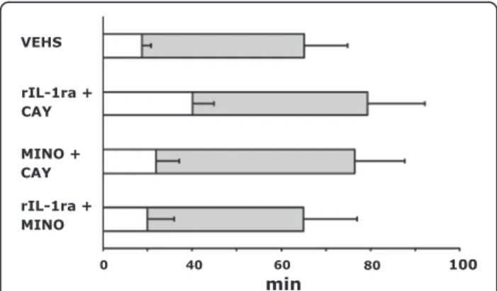
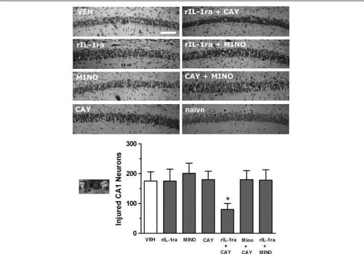
Following systemic administration of pilocarpine, all rats showed typical seizure behaviors progressing to stage 3 or beyond. We found no differences in the latency to seizure onset in the presence of inflammation induced by LPS or when any of the anti-inflammatory drugs was used one at a time (data not shown). When binary combinations were tried, the combination rIL-1ra + COX-2 inhibitor (CAY 10404, 10 mg/kg) resulted in increased latency to seizure onset as compared to those given vehicle alone (40.1 ± 9.70 min vs. 17.5 ± 4.08, P <0.01) (Figure 1). However, the duration of seizure remained similar for all treatments. Figure 2 shows that only the combination of rIL-1ra + COX-2 inhibitor resulted in discernible neuroprotection of CA1 neurons as determined 24 h after SE.
Less than 30% of the rats subjected to SE at P14 de-velop SRS [12], and treatment with LPS increases that fraction to about 50% [15]. Our previous work [12] also showed that by P21, the hippocampal injury produced by lithium-pilocarpine SE extended beyond the CA1 re-gion, involving also the hilar interneurons and CA3 neurons. Because >75% of the 3-week-old animals (P21) Kwon et al. Journal of Neuroinflammation 2013, 10:30 Page 2 of 6 http://www.jneuroinflammation.com/content/10/1/30
subjected to lithium pilocarpine SE developed SRS and dense mossy fiber sprouting in our previous studies [12,13], we deployed rats of this age to evaluate the effi-cacy of the rIL-1ra + COX-2 inhibitor combination in preventing epileptogenesis after lithium-pilocarpine SE. In this set of experiments animals were not primed with LPS because our goal was not to study injury and neuroprotection at 24 h, but to observe the rats in the long term for the development of epilepsy and to evalu-ate mossy fiber sprouting in the chronically epileptic animals. Previous work [12,13] has shown that at least 75% of P21 animals develop epilepsy 3 months or longer after lithium-pilocarpine SE.
Animals were treated with vehicle or one dose of the combination anti-inflammatory therapy immediately prior to the administration of pilocarpine. This sequence of administration was undertaken to ensure that the agents used in intervention are available even as the in-flammatory cascade is being set into motion by the SE, such that a proof of principle as to the validity of the chosen targets can be established. That can set the back-ground to explore in the future the window of opportun-ity for effective intervention after seizures have started. The only combination used was that involving rIL-1ra and the COX-2 inhibitor since none of the other regimens had resulted in discernible neuroprotection in the earlier set of experiments. A separate group of animals continued to receive once daily treatment with the anti-inflammatory cocktail for 10 days following SE. Four months after SE, animals were implanted with epi-dural electrodes as described in our other reports and
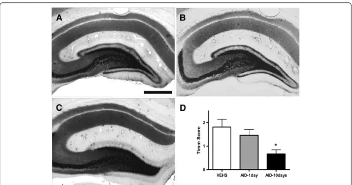
subjected to a continuous EEG and video monitoring for a period of 3 weeks for the purpose of acquisition and analysis of spontaneous recurrent seizures. At the end of monitoring, animals were euthanized and brains were processed for the analysis of mossy fiber sprouting using Timm staining [12,13] and employing a 0–5 scale as pre-viously described [16].
Monitored at 4 months after SE at the age of 3 weeks, a single AID treatment consisting of IL-1ra and CAY did not result in a significant change in the number of rats developing SRS. Spontaneous seizures were observed in six of nine vehicle-treated rats, seven of nine rats with single drug injection, and in six of nine animals with 10-day treatment regimen. However, protracted (10-day) AID cocktail treatment did reduce seizure frequency among those animals with spontaneous seizures (Figure 3). Over 3 weeks of continuous video and EEG monitoring, vehicle-treated rats exhibited minimal-maximal-median seizures of 1-18-3; those which received a single injection: 2-13-3; and animals with 10 once-a-day injections: 2-4-2 (*P <0.05 10 days of drug treatment vs. one AID cocktail treatment or saline treatment).
The analysis of mossy fiber sprouting showed moder-ate synaptic reorganization in vehicle-tremoder-ated rats as well as in animals treated with a single injection of anti-inflammatory drugs (Figure 4, Timm scores, mean scores ± SEM: 1.81 ± 0.32 and 1.46 ± 0.25, respectively). There was a statistically significant decrease in mossy fiber sprouting in the animals treated with anti-inflammatory drugs for 10 days (Timm score 0.67 ± 0.17; P <0.05 vs. one AID cocktail treatment or saline treatment).
The disappointing results with sustained AED treatment of post-SE animals to prevent epileptogenesis have directed research into other molecular targets. Rather than modifying neuronal excitability via influencing ion channel conductance, interest has turned to addressing pathways that may have a greater effect on plasticity resulting from seizures. Of those, the neuroinflammation-related pathways seem to be receiving attention of late [8]. Cyclooxygenase-2 inhibitor celecoxib has been reported to have a beneficial effect on epileptogenesis following lithium pilocarpine SE in both mature [17] and developing [18] rats. However, other investigators found parecoxib, another COX-2 inhibitor, to be neuroprotective but not antiepileptogenic [19] in the pilocarpine model of TLE. However, Holtman et al. [20,21] found that not only was the COX-2 inhibitor SC-58236 ineffective as an anti-epileptogenic agent [20] in a rat model of epilepsy after electrically-induced SE, it actually produced seizure dete-rioration and increased mortality [21]. Number of diffe-rences may account for the discrepancy between the results of Holtman et al. [20,21] and those of ours and other investigators [17-19]. The model of SE employed in the Holtman et al. [20,21] studies involved induction of Figure 1Status epilepticus (SE) after administration of
lipopolysaccharide (LPS) with or without anti-inflammatory drugs. Mean (± SEM) of both time to onset (open bar) and total duration (open + filled bar) after pilocarpine treatment following a prior injection of LPS or LPS + combination of anti-inflammatory drugs. The latency to pilocarpine-induced seizure onset in combination treatment of COX-2 inhibitor plus rIL-1ra with LPS was significantly delayed as compared to those given vehicle (*P <0.01). There were no significant changes in total duration of SE among groups. CAY, COX-2 inhibitor; MINO, minocycline; rIL-1ra = recombinant interleukin-1 receptor antagonist; VEHS, vehicles.
SE electrical stimulation of the hippocampus rather than treatment with lithium-pilocarpine. The SE was much longer in duration (up to 9 h compared to 60 to 90 min in the pilocarpine studies, in which the animals received a di-azepam dose). It is not possible to speculate as to whether there was some unique toxicity to the specific COX-2 in-hibitor used in that study. Consistent with the expected safety and possible benefit of COX-2 inhibition in the treatment of pilocarpine SE, mice with conditional ablation of the COX-2 gene in the forebrain enjoyed diminished mortality and some improvement in memory performance after pilocarpine-induced SE [22].
Blocking the synthesis of interleukin-1β biosynthesis by an interleukin converting enzyme antagonist has shown potential in the model of kindling epileptogenesis [23] as well as epilepsy induced by kainic acid treatment [24]. The availability of the human recombinant interleukin-1
receptor antagonist (rIL-1ra) anakinra prompted us to evaluate its anti-epileptogenic potential, especially since its transport across the blood–brain barrier [25,26] appears to be adequate for modifying IL-1β signaling in the brain. Our experiments showed it to be effective when combined with the COX-2 selective inhibitor, CAY 10404. Despite concerns about the potential for fibroblast growth factor-2 (FGF-2) to increase excitability [27], localized de-livery of FGF-2 and brain-derived neurotrophic factor (BDNF) by employing viral vectors has demonstrated po-tential for anti-epileptogenesis [28,29].
In humans, brain injury from a number of causes such as SE, traumatic brain injury, hypoxic-ischemic encephalop-athy, stroke, and so on, give rise to variable incidences of epilepsy after varying latencies. The classic post-traumatic epilepsy prevention studies [3] subjected patients to prolonged treatment with AEDs with significant toxicities Figure 2Neuronal injury in hippocampal CA1 subfield after co-administration of anti-inflammatory drugs against LPS + pilocarpine-induced SE in 2-week-old rats. Treatment with a single anti-inflammatory compound did not diminish the amount of CA1 hippocampal damage as compared to vehicle (left column). A combination treatment of rIL-1ra + CAY resulted in significant reduction of injured neurons which was not seen with any other combination (right column). Outset shows high magnification of eosinophilic neurons with pyknotic nuclei and irregular cell bodies that were counted as injured CA1 cells in the graph. Bars represent mean ± SEM. Quantification shows significant attenuation of neuronal injury resulted from combined administration of rIL-1ra + CAY administered prior to the onset of status (*P <0.05). CAY, COX-2 inhibitor (10 mg/kg); MINO, minocycline; naïve, age-matched normal rat (no seizures, no treatment); rIL-1ra, recombinant interleukin-1 receptor antagonist; VEH, vehicle. Scale bar is 50 μm.
Kwon et al. Journal of Neuroinflammation 2013, 10:30 Page 4 of 6 http://www.jneuroinflammation.com/content/10/1/30
and side effects, and still failed to prevent the development of epilepsy. Our results highlight that both limited neuroprotection and a modicum of anti-epiletogenic dis-ease modification can be achieved by targeting inflamma-tion. However, the translationally important message is that while evidence exists supporting the role of many indi-vidual inflammatory pathways, only a specific combination therapy provided discernible benefits. Further, the AID cocktail treatment protocol involved a limited duration, unlike the chronic AED regimens that have been tried in humans as well as animal models. The importance of our findings for clinical translation are that our intuition-driven empiric AID cocktail design leverages elucidated mechanisms involving specific pathways while enabling: (1) treatment with drug classes that have already been evaluated for safety and approved for human use; (2) short duration treatment with drug classes that are already in use for chronic conditions; and (3) does not involve intro-duction of viral vectors into the CNS. It remains to be shown if the doses and duration of the regimen can be optimized for greater efficacy, and if such treatment may also modify the evolution of epilepsy-associated co-morbidities that seem to impact on the quality of life of patients even more than seizure frequency.
Figure 3Protracted anti-inflammatory drug treatment reduced the frequency of spontaneous recurrent seizures (SRS) among epileptic animals. Bar graphs represent the average number of seizures observed. Over 3 weeks of continuous video and EEG monitoring, vehicle-treated animals (VEHS) presented an average of 2.9 ± 0.6 seizures per week, while those treated with a single injection of anti-inflammatory drug (AID) did not show significant reduction in seizure frequency and exhibited an average of 2.5 ± 0.4 seizures per week (P >0.5). Animals treated with daily injections of AID for 10 consecutive days showed a marked decrease in SRS frequency (*P <0.05 10 days of drug treatment vs. two other groups, n = 8).
Figure 4Timm staining showing mossy fiber sprouting in supragranular region of the dentate gyrus of saline-treated and AID-treated animals. (A) Vehicle treated. (B) The second group of animals was injected with AID for 1 day. (C) The third group received AID treatment for 10 consecutive days after induction of status epilepticus. Four months after SE, brains were harvested and processed for Timm staining. (D) Protracted AID (10-days) reduced mossy fiber sprouting as observed using Timm staining. Analysis of mossy fiber sprouting showed synaptic reorganization in vehicle-treated rats as well as in animals treated with a single injection of anti-inflammatory drugs (Timm scores 1.81 ± 0.32 and 1.46 ± 0.25, respectively). There was a statistically significant decrease in mossy fiber sprouting in the animals treated with anti-inflammatory drugs for 10 days (Timm score 0.67 ± 0.17); (P <0.05 vs. each of two other groups). Scale bar, 100 um.
Competing interests
None of the authors has any conflicts of interest with any commercial entities.
Authors’ contributions
YSK, EP, SA, and DS performed all the experiments, data analysis, and also collaborated in writing. AM assisted with study design and data analysis. RS participated in the design of the studies, data analysis, and finalization of the manuscript. All authors have read and approved the final version of the manuscript.
Acknowledgements
This study was supported by National Institutes of Health research grants (R01 NS065783 and R21 MH079933, both to A.M.), the Epilepsy Foundation of America (Postdoctoral Research Training Fellowship to EP), Association pour l’Etude des Affections Congenitales (SA), and DAPA Foundation (RS).
Author details
1Department of Pediatrics, Division of Neurology, David Geffen School of Medicine at UCLA, 22-474 MDCC in CHS, Los Angeles, CA 90095-1752, USA. 2Department of Pediatrics, College of Medicine, Inha University, Incheon, Republic of Korea.3Department of Pediatric Neurology, Hôpital Robert Debré, INSERM U676, Paris 75019, France.4Department of Neurology, David Geffen School of Medicine at UCLA, Los Angeles, CA 90095, USA.
Received: 27 September 2012 Accepted: 15 February 2013 Published: 26 February 2013
References
1. Kwan P, Brodie MJ: Early identification of refractory epilepsy. N Engl J Med 2000, 342:314–319.
2. 2007 Epilepsy Research Benchmarks: http://www.ninds.nih.gov/research/ epilepsyweb/2007_benchmarks.htm.
3. Temkin NR: Preventing and treating posttraumatic seizures: the human experience. Epilepsia 2009, 2:10–13.
4. François J, Koning E, Ferrandon A, Nehlig A: The combination of topiramate and diazepam is partially neuroprotective in the
hippocampus but not antiepileptogenic in the lithium-pilocarpine model of temporal lobe epilepsy. Epilepsy Res 2006, 72:147–163.
5. Brandt C, Gastens AM, Sun M, Hausknecht M, Löscher W: Treatment with valproate after status epilepticus: effect on neuronal damage, epileptogenesis, and behavioral alterations in rats. Neuropharmacology 2006, 51:789–804.
6. Brandt C, Glien M, Gastens AM, Fedrowitz M, Bethmann K, Volk HA, Potschka H, Löscher W: Prophylactic treatment with levetiracetam after status epilepticus: lack of effect on epileptogenesis, neuronal damage, and behavioral alterations in rats. Neuropharmacology 2007, 53:207–221. 7. Pitkänen A, Nissinen J, Jolkkonen E, Tuunanen J, Halonen T: Effects of
vigabatrin treatment on status epilepticus-induced neuronal damage and mossy fiber sprouting in the rat hippocampus. Epilepsy Res 1999, 33:67–85.
8. Vezzani A, Friedman A, Dingledine RJ: The role of inflammation in epileptogenesis. Neuropharmacology 2012 [Epub ahead of print, PMID: 22521336].
9. Buckmaster PS: Mossy Fiber Sprouting in the Dentate Gyrus. In Jasper's Basic Mechanisms of the Epilepsies [Internet]. 4th edition. Edited by Noebels JL, Avoli M, Rogawski MA, Olsen RW, Delgado-Escueta AV. Bethesda, MD: National Center for Biotechnology Information (US); 2012.
10. Pitkänen A, Nissinen J, Lukasiuk K, Jutila L, Paljärvi L, Salmenperä T, Karkola K, Vapalahti M, Ylinen A: Association between the density of mossy fiber sprouting and seizure frequency in experimental and human temporal lobe epilepsy. Epilepsia 2000, Suppl 6:S24–29.
11. El Bahh B, Lespinet V, Lurton D, Coussemacq M, Le Gal La Salle G, Rougier A: Correlations between granule cell dispersion, mossy fiber sprouting, and hippocampal cell loss in temporal lobe epilepsy. Epilepsia 1999, 40:1393–1401.
12. Sankar R, Shin DH, Liu H, Mazarati A, Pereira de Vasconcelos A, Wasterlain CG: Patterns of status epilepticus-induced neuronal injury during development and long-term consequences. J Neurosci 1998, 18:8382–8393.
13. Sankar R, Shin D, Mazarati AM, Liu H, Katsumori H, Lezama R, Wasterlain CG: Epileptogenesis after status epilepticus reflects age- and model-dependent plasticity. Ann Neurol 2000, 48:580–589.
14. Auvin S, Shin D, Mazarati A, Nakagawa J, Miyamoto J, Sankar R:
Inflammation exacerbates seizure-induced injury in the immature brain. Epilepsia 2007, Suppl 5:27–34.
15. Auvin S, Mazarati A, Shin D, Sankar R: Inflammation enhances
epileptogenesis in the developing rat brain. Neurobiol Dis 2010, 40:303–310. 16. Cavazos JE, Golarai G, Sutula TP: Mossy fiber synaptic reorganization
induced by kindling: time course of development, progression, and permanence. J Neurosci 1991, 11:2795–2803.
17. Jung KH, Chu K, Lee ST, Kim J, Sinn DI, Kim JM, Park DK, Lee JJ, Kim SU, Kim M, Lee SK, Roh JK: Cyclooxygenase-2 inhibitor, celecoxib, inhibits the altered hippocampal neurogenesis with attenuation of spontaneous recurrent seizures following pilocarpine-induced status epilepticus. Neurobiol Dis 2006, 23:237–246.
18. Zhang HJ, Sun RP, Lei GF, Yang L, Liu CX: Cyclooxygenase-2 inhibitor inhibits hippocampal synaptic reorganization in pilocarpine-induced status epilepticus rats. J Zhejiang Univ Sci B 2008, 9:903–915. 19. Polascheck N, Bankstahl M, Löscher W: The COX-2 inhibitor parecoxib is
neuroprotective but not antiepileptogenic in the pilocarpine model of temporal lobe epilepsy. Exp Neurol 2010, 224:219–233.
20. Holtman L, van Vliet EA, van Schaik R, Queiroz CM, Aronica E, Gorter JA: Effects of SC58236, a selective COX-2 inhibitor, on epileptogenesis and spontaneous seizures in a rat model for temporal lobe epilepsy. Epilepsy Res 2009, 84:56–66.
21. Holtman L, van Vliet EA, Edelbroek PM, Aronica E, Gorter JA: Cox-2 inhibition can lead to adverse effects in a rat model for temporal lobe epilepsy. Epilepsy Res 2010, 91:49–56.
22. Levin JR, Serrano G, Dingledine R: Reduction in delayed mortality and subtle improvement in retrograde memory performance in pilocarpine-treated mice with conditional neuronal deletion of cyclooxygenase-2 gene. Epilepsia 2012, 53:1411–1420.
23. Ravizza T, Noé F, Zardoni D, Vaghi V, Sifringer M, Vezzani A: Interleukin converting enzyme inhibition impairs kindling epileptogenesis in rats by blocking astrocytic IL-1β production. Neurobiol Dis 2008, 31:327–333. 24. Maroso M, Balosso S, Ravizza T, Iori V, Wright CI, French J, Vezzani A:
Interleukin-1β biosynthesis inhibition reduces acute seizures and drug resistant chronic epileptic activity in mice. Neurotherapeutics 2011, 8:304–315. 25. Gutierrez EG, Banks WA, Kastin AJ: Blood-borne interleukin-1 receptor
antagonist crosses the blood–brain barrier. J Neuroimmunol 1994, 55:153–160. 26. Greenhalgh AD, Galea J, Dénes A, Tyrrell PJ, Rothwell NJ: Rapid brain
penetration of interleukin-1 receptor antagonist in rat cerebral ischaemia: pharmacokinetics, distribution, protection. Br J Pharmacol 2010, 160:153–159.
27. Zucchini S, Buzzi A, Barbieri M, Rodi D, Paradiso B, Binaschi A, Coffin JD, Marzola A, Cifelli P, Belluzzi O, Simonato M: FGF-2 overexpression increases excitability and seizure susceptibility but decreases seizure-induced cell loss. J Neurosci 2008, 28:13112–13124.
28. Paradiso B, Marconi P, Zucchini S, Berto E, Binaschi A, Bozac A, Buzzi A, Mazzuferi M, Magri E, Navarro Mora G, Rodi D, Su T, Volpi I, Zanetti L, Marzola A, Manservigi R, Fabene PF, Simonato M: Localized delivery of fibroblast growth factor-2 and brain-derived neurotrophic factor reduces spontaneous seizures in an epilepsy model. Proc Natl Acad Sci U S A 2009, 106:7191–7196.
29. Bovolenta R, Zucchini S, Paradiso B, Rodi D, Merigo F, Navarro Mora G, Osculati F, Berto E, Marconi P, Marzola A, Fabene PF, Simonato M: Hippocampal FGF-2 and BDNF overexpression attenuates
epileptogenesis-associated neuroinflammation and reduces spontaneous recurrent seizures. J Neuroinflammation 2010, 7:81–86.
doi:10.1186/1742-2094-10-30
Cite this article as: Kwon et al.: Neuroprotective and antiepileptogenic effects of combination of anti-inflammatory drugs in the immature brain. Journal of Neuroinflammation 2013 10:30.
Kwon et al. Journal of Neuroinflammation 2013, 10:30 Page 6 of 6 http://www.jneuroinflammation.com/content/10/1/30