HAL Id: hal-02297474
https://hal.sorbonne-universite.fr/hal-02297474
Submitted on 26 Sep 2019HAL is a multi-disciplinary open access archive for the deposit and dissemination of sci-entific research documents, whether they are pub-lished or not. The documents may come from teaching and research institutions in France or abroad, or from public or private research centers.
L’archive ouverte pluridisciplinaire HAL, est destinée au dépôt et à la diffusion de documents scientifiques de niveau recherche, publiés ou non, émanant des établissements d’enseignement et de recherche français ou étrangers, des laboratoires publics ou privés.
Efficacy and tolerability of a custom-made Narval
mandibular repositioning device for the treatment of
obstructive sleep apnea: ORCADES study 2-year
follow-up data
Valérie Attali, Marie-Françoise Vecchierini, Jean-Marc Collet, Marie-Pia
d’Ortho, Frederic Goutorbe, Jean-Baptiste Kerbrat, Damien Leger, Florent
Lavergne, Christelle Monaca, Pierre-Jean Monteyrol, et al.
To cite this version:
Valérie Attali, Marie-Françoise Vecchierini, Jean-Marc Collet, Marie-Pia d’Ortho, Frederic Goutorbe, et al.. Efficacy and tolerability of a custom-made Narval mandibular repositioning device for the treat-ment of obstructive sleep apnea: ORCADES study 2-year follow-up data. Sleep Medicine, Elsevier, 2019, �10.1016/j.sleep.2019.04.021�. �hal-02297474�
Efficacy and tolerability of a custom-made Narval mandibular
repositioning device for the treatment of obstructive sleep apnea:
ORCADES study 2-year follow-up data
Valérie Attali1,2, Marie-Françoise Vecchierini3,4, Jean-Marc Collet5, Marie-Pia d'Ortho,6,7, Frederic Goutorbe 8, Jean-Baptiste Kerbrat5,9, Damien Leger3,4, Florent Lavergne10, Christelle
Monaca11, Pierre-Jean Monteyrol12; Laurent Morin10, Eric Mullens13, Bernard Pigearias14, Francis Martin2, Fabienne Tordjman2, Hauria Khemliche15 , Lionel Lerousseau16, and Jean-Claude Meurice17, on behalf of the ORCADES investigators
1Sorbonne Université, INSERM, UMRS1158 Neurophysiologie Respiratoire Expérimentale et
Clinique, Paris, France; 2AP-HP, Groupe Hospitalier Pitié-Salpêtrière Charles Foix, Service
des Pathologies du Sommeil (Département "R3S"), Paris, France; 3AP-HP, Hôpital Hôtel Dieu, Centre du Sommeil et de la Vigilance, Paris, France; 4Université Paris Descartes, Sorbonne Paris Cité, Paris, France; 5AP-HP, Groupe Hospitalier Pitié-Salpêtrière Charles Foix, Stomatologie et Chirurgie Maxillo-Faciale, Paris, France; 6AP-HP, DHU FIRE, Hôpital Bichat-Claude Bernard, Physiologie et Explorations Fonctionnelles, Paris, France; 7UFR de Médecine, Université Denis Diderot Paris 7, Paris, France; 8Centre Médecine du Sommeil,
Centre Hospitalier de Béziers, Béziers, France; 9Hôpital Charles Nicolle, Stomatologie et Chirurgie Maxillo-Faciale, Rouen, France; 10ResMed Science Center, Saint-Priest cedex, France; 11Hôpital Roger Salengro, Neurophysiologie Clinique, Lille, France; 12Polyclinique du Tondu, Oto-Rhino-Laryngologie, Bordeaux, France; 13Fondation Bon Sauveur,
Laboratoire du Sommeil, Albi, France; 14Laboratoire du Sommeil, Nice, France; 15Groupe
Hospitalier Public Sud de l’Oise, Senlis, France; 16Service de Pneumologie, Centre Hospitalier Antibes, Antibes, France; 17Centre Hospitalier Universitaire, Pneumologie,
Correspondance: Dr Valérie ATTALI, Department of Sleep Medicine ("Service
d’Exploration des Pathologies du Sommeil"), Pitié-Salpêtrière Hospital, 47-83 Bd de
l’Hôpital, 75651 Paris Cedex 13 France. Tel: + 33 1 42167730, E-mail: valerie.attali@aphp.fr
“Take home” message
Mandibular repositioning device therapy was effective in maintaining improvements in sleep
apnoea and quality of life over 2 years of follow-up in patients who refused or were intolerant
of continuous positive airway pressure
Word Count Abstract: 204
Word Count Article: 3266 (excluding references, abstract, figures and tables)
Number of tables: 4
ABSTRACT
Objective/Background: Mandibular repositioning device (MRD) therapy is an alternative to
continuous positive airway pressure (CPAP). The ORCADES study is assessing the long-term
efficacy and tolerability of MRD therapy in OSAS; 2-year follow-up data are presented.
Patients/Methods: OSAS patients who refused or were noncompliant with CPAP were fitted
with a custom-made computer-aided design/computer-aided manufacturing (CAD/CAM) bi-block MRD (ResMed, Narval CC™); mandibular advancement was individually titrated.
Sleep and respiratory parameters were determined at baseline, 3–6 months and 2 years. The
primary endpoint was treatment success (percentage of patients achieving a 50% reduction in the apnoea-hypopnoea index [AHI]).
Results: Of 315 enrolled patients, 237 remained on MRD treatment at 2 years and 197 had
follow-up data. Treatment success rate at 2 years was 67%; AHI <5/h, <10/h and <15/h was
achieved in 30%, 56% and 72% of patients, respectively. On multivariate analysis, 50% decrease in AHI at 3–6 months and absence of nocturia at 3–6 months were significant
predictors of MRD treatment continuation. Adverse events were generally mild and the
majority occurred in the first year of treatment.
Conclusions: Two years’ treatment with an MRD was effective and well tolerated in patients
1. Introduction
Obstructive sleep apnoea syndrome (OSAS) is characterised by recurrent obstructions of
upper airways during sleep, which result in sleep fragmentation and intermittent hypoxia [1].
Moderate to severe OSAS is associated with cardiovascular, metabolic and cognitive
comorbidities, and sleepiness-related accidents [2-4]. Continuous positive airway pressure
(CPAP) remains the gold standard treatment for OSAS [5]. CPAP therapy reduces the
apnoea-hypopnoea index (AHI), improves symptoms and quality of life, reduces the risk of
motor vehicle crashes [6], and potentially reduces cardiovascular events and mortality [7-9].
However, as many as 30–50% of patients prescribed CPAP are non-compliant with therapy
over the long term [10-12].
A mandibular repositioning device (MRD) is recommended as the first alternative to CPAP
[13] in patients requiring treatment for OSAS. The MRD prevents recurrent obstruction of the
upper airways during sleep by maintaining the mandible in a forward position to enlarge [14]
and maintain an open airway [15], and significantly reduces the AHI [16]. Although MRD
therapy is not as effective as CPAP in controlling the occurrence of obstructive events [17],
this is counterbalanced by better adherence to treatment [18]. Therefore, improvement in
symptoms and quality of life after up to 12 months are similar with MRD therapy and CPAP
[19]. However, there is a lack of data on the longer term effects of second-line MRD therapy
in patients with OSAS.
The multicentre, prospective ORCADES study was designed to investigate the long-term
effects of MRD therapy in OSA patients non-compliant with or intolerant of CPAP with
follow-up for 5 years. The first analysis of data after 6 months of follow-up showed a
significant reduction in AHI and symptoms during MRD therapy, which was well tolerated
[16]. Patients were treated either with a custom-made computer-aided design/computer-aided
non-CAD/CAM MRD device (ResMed, Narval™). Early evaluation suggested that the
CAD/CAM device, which allows more accurate adjustment of the vertical opening, was
superior to the non-CAD/CAM MRD [16]. Therefore, this 2-year follow-up of the
ORCADES trial focusses on patients with OSAS treated with the CAD/CAM MRD.
2. Methods
2.1 Study design
ORCADES was a single-arm prospective observational study that was conducted at 28
centres in France (NCT01326143). Full details of the study design have been reported
previously [16]. The study protocol was approved by the relevant ethics committees, and all
procedures were conducted in accordance with the Declaration of Helsinki principles. All
patients received detailed information and gave written informed consent to participate.
2.2 Patients
Adult patients (age ≥18 years) with OSAS (AHI >30/h, or AHI <30/h with excessive daytime
sleepiness and/or an Epworth Sleepiness Scale [ESS] score >10) who refused or were
noncompliant with CPAP (device usage <3 h/night) and had not previously received MRD
treatment were screened by a sleep specialist. Only those without any contraindications to
MRD treatment, as confirmed by a dental sleep specialist, were included. Patient should not
presented dental, periodontal or articular contraindications. A patient completely edentulous
or presenting partial toothless (less than 3 teeth (or implants) by hemi-arch including the
canine at the maxillary level, or presenting less than 4 teeth (or implants) by hemi-arch
Patients included in this analysis were treated with a custom-made CAD/CAM MRD device (ResMed, Narval CC™). The device was fitted by a dental specialist; initial mandibular
advancement that was adjusted over a 15-mm range (the maximal advancement allowed with
MRD depending of the connecting rod size) at subsequent titration visits, to achieve the best
balance between clinical efficacy and tolerability. The first evaluation took place 4–6 months
after treatment initiation [16], then patients were re-evaluated at the 2-year follow-up visit.
MRD replacement during the study was performed based on routine clinical practice.
2.4 Endpoints
The primary endpoint was the treatment success rate, defined as the percentage of patients
achieving a 50% reduction in AHI at the 2-year follow-up visit. Absolute change in AHI from baseline to 2-year follow-up, and from baseline to 3-6 months and 2 years was also
determined. The percentage of patients achieving an AHI below three cut-off values (<5/h,
<10/h and 15/h) was calculated, overall and in patient subgroups based on OSAS severity at
baseline (mild: AHI 5/h to ≤15/h; moderate: AHI 15/h to ≤30/h; severe: AHI >30/h).
Additional nocturnal respiratory endpoints were the oxygen desaturation index (ODI; average number of desaturation episodes per hour, with desaturation defined as a ≥3% decrease in
oxygen saturation [SpO2] from the average value), the lowest SpO2 (nadir SpO2), and total
time with SpO2 <90%. In patients who underwent PSG, total sleep time, sleep latency,
percentage of slow wave and rapid eye movement (REM) sleep, micro-arousal index and
intra-sleep wakefulness were determined. Clinical efficacy, tolerability and device usage were
determined as described below.
Clinical evaluation at the 2-year follow-up included the same endpoints as the 3- to 6-month
follow-up [16]. Briefly, somnolence was evaluated using the ESS, and snoring, nocturia,
libido disorders and nocturnal mouth breathing were self-reported (yes/no). Patients were
asked to rate their sleep quality, state on waking and morning headache on non-graduated 10
cm visual analogue scales (VAS), from "very bad" to "excellent" for sleep quality and state on
waking, and from “absence of pain” to “maximal pain” for morning headache. Quality of life
was evaluated using the Quebec Sleep Questionnaire (QSQ) [20] and a Pichot fatigue scale
questionnaire was administered [21]. Data on MRD-related side effects and their severity
were determined by sleep and dental sleep physicians. Self-reported MRD compliance (hours
per night; nights per week) was assessed.
2.6 Sleep studies
Evaluation of the AHI was based on ventilatory polygraphy (PG) or polysomnography (PSG).
The same test was used in the same patient at baseline, 3–6 months and 2 years. PSG/PG
recordings were manually scored according to the American Academy of Sleep Medicine
(AASM) guidelines [22]. Obstructive apnoea was defined as a ≥10-s cessation of airflow on
the pressure nasal cannula, with or without association with an oro-nasal thermal sensor. Hypopnoea was defined as a ≥50% reduction in airflow, or a <50% airflow reduction on the
nasal pressure cannula accompanied by a ≥3% decrease in arterial oxyhaemoglobin saturation
(SpO2) recorded using finger pulse oximetry or an arousal.
2.7 Statistical analysis
The intention-to-treat (ITT) population for this analysis included all patients using a
changes from baseline to the 2-year follow-up visit were compared using unpaired or paired Student’s t-test or the Wilcoxon–Mann–Whitney nonparametric test depending on normality
of distribution and group comparison. Qualitative changes were described using frequency distribution and compared using Fisher’s exact test or Chi-squared test. Change over time in
AHI, 3% ODI, time with SpO2 <90%, ESS score, symptoms (snoring, nocturia, libido
disorders, nocturnal mouth breathing), QSQ global and sub-scores, and Pichot questionnaire
results was determined using a repeated measures ANOVA; if significant this was followed by a Tuckey’s test to compare visits two by two. Comparisons between patient subgroups
based on baseline OSAS severity, gender and body mass index (BMI) were assessed using the Student’s t-test, ANOVA or Wilcoxon–Mann–Whitney test. Two logistic models were
created and backward stepwise regression analysis was used to determine independent factors
associated with continuation of treatment until the 2-year follow-up in the ITT population
(first model) and achievement of AHI <10/h at year follow-up in patients with available
2-year AHI data (model 2). For both models, variables with a p-value <0.10 in univariate
analysis were entered in the stepwise logistic regressions, and variables with a p-value <0.05
were retained in the final multivariate models. Statistical analyses were performed using SAS
version 9.
3. Results
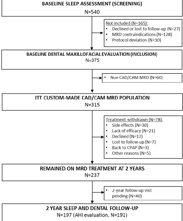
3.1 Population
A total of 540 patients were screened, 165 patients were excluded and 315 were treated with a
CAD/CAM MRD (Figure 1). The majority of patients were male (76%), 20% were obese and
51% had previously been treated with CPAP (Table 1). The number [IQR] of initial MRD
titrations was of 2.0 [1.0, 3.0], and final mandibular advancement was 7.0 [6.0; 8.0] mm. The
with a median follow-up of 24 [25; 28] months; the 2-year follow-up visit was pending for the
remaining 40 patients. Median changes from baseline in weight (0 [–3; 2] kg), BMI (0.28 [–
0.73; 0.99] kg/m2), neck circumference (0 [–1; 1] cm) and waist circumference (0 [–2; 4] cm)
were not statistically significant and only seven patients needed to have their MRD replaced
before the 2-year follow-up visit.
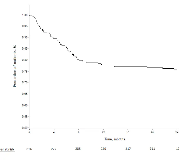
3.2 Withdrawals
A total of 78 patients (25%) were withdrawn before the 2-year follow-up visit, mainly due to
side effects (30 patients), or lack of efficacy (21 patients) (Figure 1). The overall proportion of
withdrawals did not vary by baseline OSAS severity and gender, but the rate of withdrawal
due to adverse events was higher in females than in males (65% vs 32%; p=0.0098).
Withdrawal occurred more frequently in obese versus non-obese patients (40% vs 21%;
p=0.0024) and obese patients were withdrawn more often for lack of efficacy than non-obese
patients (44% vs 19%; p=0.0195). The majority of withdrawals (83%) occurred within the
first 6 months of MRD therapy (Figure 2).
3.3 Sleep study data
AHI data were available for 191 patients (132 underwent PG and 59 had PSG). A 50%
reduction in the AHI was achieved in 67% of participants. The proportion of patients
achieving an AHI of <15/h, <10/h and <5/h was 72%, 56% and 30%, respectively (Figure 3).
After 2 years, the reduction in AHI from baseline was –15 [-23; -7]/h (–64 [–83; –42]%).
AHI, 3% ODI, time with SpO2< 90% and nadir SpO2 values decreased significantly from
baseline to 2-year follow-up (Table 2). In the 59 patients with 2-year PSG data, the change
micro-arousal index was –8 [–13; 1]/h (p=0.0001). No changes in the percentage of slow wave and
REM sleep were observed.
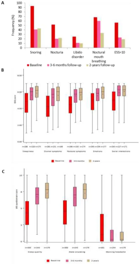
3.4 Symptoms and quality of life
A total of 81% of patients had an ESS score <10 at the 2-year follow-up. The ESS score
decreased from 11 [8; 15] at baseline to 7 [5; 10] at 3–6 months and 7 [4; 9] at 2 years
(p<0.0001); reductions were similar across OSAS severity subgroups. The QSQ global score
increased from 144.0 [111.0; 173.0] at baseline to 180.5 [153.0; 201.0] at 3-6 months and
191.5 [94.0; 205.0] at 2 years (p<0.001), and the Pichot score decreased from 14.0 [7.0; 20.0]
at baseline to 7.0 [3.0; 14.0] at 3–6 months and 6.0 [3.0; 11.0] at 2 years (p<0.001). Changes
over time in symptoms and QSQ subscores are shown in Figure 4.
3.5 Device usage
At the 2-year follow-up, median [IQR] MRD usage was 7 [7; 7] nights/week and 7 [6; 8]
hours/night; 95% of patients used their MRD for 4 hours/night on 4 nights/week, and 85% for 4 hours/night on 7 days/week. Device usage was similar across patient subgroups based on OSA severity, gender or BMI.
3.6 Predictive factors
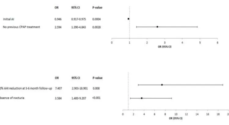
A number of factors were significant predictors of either treatment continuation or AHI <10/h
at 2 years in the univariate analysis (Table 3). Only two variables remained significant
predictors of treatment continuation in the multivariate analysis: a 50% decrease in AHI at 3–
6 months’ follow-up and the absence of nocturia at 3–6 months’ follow-up (Figure 5). There
were also two significant predictors of AHI <10/h at the 2-year follow-up: smaller initial AI
3.7 Tolerability
At least one adverse event was reported by 59% of patients. The most common event was
TMJ disorder (Table 4). Of the 509 adverse events recorded by the 2-year follow-up, 137
(27%) were reported in the first 6 months of therapy and 64 (13%) were reported in the first
year. Only 13% of all events were classified as severe (Table 4). 30 patients withdrawn the
study for side effects before the 2 year evaluation, as follow: dental pain (7 pts), TMJ disorder
(7 pts), gingival pain (5 pts), occlusion change (2 pts), tooth loosening (1 pt), mouth pain (1
pt), discomfort (1 pt), mouth dryness (1 pt), nausea (1 pt), suspected allergy (1 pt) and other
reasons (3 pts).
4. Discussion
Two-year follow-up data from the multicentre, prospective ORCADES study showed that
MRD therapy remained effective and well-tolerated in patients with mild to severe OSAS
who refused, were intolerant of, or non-compliant with CPAP.
Non-compliance with CPAP is an important concern in OSAS management [11]. MRD
therapy is recommended as a potential first-line treatment option for mild to moderate OSA
patients without cardiovascular comorbidities, but guidelines also acknowledge that an MRD
provides a non-surgical second-line treatment option and is better than no treatment for adult
patients intolerant of CPAP or who prefer alternate therapy [23]. Several studies have
investigated the long-term effects of MRD in OSAS, but mainly included only a small number
of mild to moderate OSAS patients [24-29]. Larger comparative [19, 30-32] or
noncomparative [24, 26, 33, 34] trials evaluated an MRD as first-line therapy, but only one
The purpose of the 5-year ORCADES study is to provide long-term evaluation of MRD as
second-line treatment of OSAS in patients with a range of disease severity. The large number
of patients included (n=315) and continuing treatment at 2 years (n=237), the selection of a
homogeneous population intolerant of or refusing CPAP, and the high proportion of
individuals with severe disease are important strengths of this study. In addition, particular
attention was payed to adapting the study design to follow the most recent guidelines on MRD
treatment [23]. Sleep and dental maxillofacial specialists were both involved to ensure
selection of the right patients and to exclude those with contraindications for MRD therapy.
The CAD/CAM MRD was a custom-made titratable device and a titration period was
included to achieve mandibular propulsion that maximised resolution of symptoms,
tolerability and AHI reduction before patients entered the long-term follow-up. In addition,
this 2-year interim analysis provided important information to improve understanding of
therapy withdrawal and adverse event rate evolution over time at a time point in therapy
where patients should, in theory, renew their MRD.
As well as these strengths, the study also has a number of limitations. The most important is
the observational, registry-based design, without random allocation to treatment. However,
patient management in this setting is representative of routine clinical practice and our
findings are similar to those of another observational cohort study [36]. The findings are
therefore likely to have good external validity. Seventy-eight patients withdrew from our
study before the 2-year assessment and 40 were still awaiting assessment. This left a total of
191 patients (60% of the ITT population) who had an AHI evaluation at 2 years, something
that could influence the study results and their interpretation. The reduction in patient
numbers over time highlights the difficulty in maintaining adherence to chronic therapy and
retaining patients in a clinical pathway, even when therapy is reimbursed. Such difficulties
model analysis of treatment continuation, where non-analysed patients were considered as
treatment failures. It is also important to acknowledge that two different types of patients were
enrolled in the study: those intolerant of CPAP therapy and those who refused CPAP. This
could have influenced the study findings, as indicated in the univariate analysis on treatment
success (AHI <10/h). More research is needed to differentiate and identify specific traits of
these two populations. Another study limitation to mention is that two types of sleep test (PG
and PSG) were used in the study to evaluate respiratory events, however each patient was
evaluated with the same sleep test all along the study which limits discrepancy.
In our study, we defined the rate of treatment success as a 50% reduction in AHI because it is
an endpoint that has been widely used in non-CPAP surgical intervention and MRD studies
[31, 38, 39], thus allowing easy comparison of findings, and is suited to evaluating MRD
efficacy related to quality of sleep [39]. The treatment success rate in this analysis (67%),
without any difference between OSAS severity subgroups, is consistent with previous
long-term studies [13]. To improve sensitivity and clinical relevance, we also performed analyses
using three different residual AHI thresholds (<15/h, <10/h and <5/h). Residual AHI <10/h is
commonly related to long-term control of symptoms [40], and was observed in 56% of
patients. In our study, 72% of patients had an AHI <15/h, which has been associated with a
reduction in the risk of new-onset hypertension [40]. The AHI findings were consistent with
the maintenance of good OSAS symptom control, good sleep quality and good quality of life,
including all domains of the QSQ. In the subgroup of patients with severe OSAS at baseline,
the proportion of patients achieving an AHI <10/h and <15/h was 37% and 53%, respectively,
suggesting that long-term MRD treatment is a good alternative to CPAP for some of these
patients. An AHI <5/h is often used to evaluate CPAP efficacy [41], and was achieved by
<5/h cut-off was determined to define OSAS based on historical cohorts [43-45], recent data
suggest that the prevalence of sleep-disordered breathing in the general population based on
an AHI >5/h would be 84% in men and 61% in women [3]. It was suggested that the higher
apparent prevalence of sleep-disordered breathing in recent versus historical studies might be
explained by the increased sensitivity of current recording techniques and scoring criteria. In
addition, revision of the AHI criteria for definition of OSAS may be appropriate based on
recent findings of a lack of association between mild OSAS and cardiac morbidity [2, 3].
In our study, MRD therapy was associated with relatively consistent control of the AHI over
time. However, there was a slight increase in the median AHI at the two-year follow-up in the
absence of weight gain [28] and irrespective of OSAS severity (Figure 3). Only seven patients
had an MRD replacement before the 2-year follow-up, and it is possible that the slight
increase in AHI could simply be due to a worn device even though long-term increases in the
AHI over a median follow-up of 16.6 years have been reported in a small group of patients
using an optimally titrated MRD in the absence of weight change [26]. It is important to note
that increasing age and bite changes over time could influence long-term assessments of MRD
effectiveness [13]. These factors will be taken into account for the 5-year follow-up of the
ORCADES study.
Baseline AI and absence of previous CPAP treatment were independent predictors of a
complete response to MRD treatment (AHI <10/h). Baseline AI and others factors such as
gender or positional OSAS have previously been reported to be related to MRD efficacy [16,
46, 47]. Conversely, the absence of previous CPAP treatment has not previously been
associated with long-term AHI reduction on MRD to the best of our knowledge, although it
was predictive of MRD treatment continuation in one observational study [36]. This suggests
that patients intolerant of CPAP may be at greater risk of not having a long-term response to
use of an MRD could help retain these vulnerable patients in the care network. This was
supported by our observation that some patients accepted a return to CPAP therapy after
MRD treatment cessation. Absence of nocturia and a 50% reduction in AHI at short-term
follow-up were independent predictors of long-term MRD therapy continuation; of these,
relapse of nocturia has previously been associated with MRD treatment cessation [36].
The CAD/CAM MRD used in this study was well tolerated. The majority of adverse events
were of mild intensity, most were observed within the first 6 months of treatment, and the
majority of withdrawals (83%) occurred within 6 months of MRD therapy initiation. Taken
together with the predictors of therapy continuation found in our study, this highlights the
importance of optimal early control of AHI and symptoms and early identification and
management of adverse events for achievement and maintenance of MRD efficacy and
compliance.
This second interim analysis of the 5-year ORCADES study showed that 2 years of MRD
therapy was effective and well-tolerated in patients with mild to severe OSAS who refused or
were intolerant of CPAP. Long-term maintenance of a complete response to MRD therapy
was significantly more likely in patients who refused CPAP compared with those who were
intolerant or noncompliant with CPAP.
Acknowledgements: Editing assistance was provided by Nicola Ryan, independent medical
writer, funded by ResMed.
Study funding and role of the funding source: The ORCADES study was funded by
C.R.O. Clinact (France) mandated by ResMed performed the collection, quality control,
management and analysis of the data. The Executive Steering Committee had full access to all
References
[1] Levy P, Kohler M, McNicholas WT, Barbe F, McEvoy RD, Somers VK, et al. Obstructive
sleep apnoea syndrome. Nat Rev Dis Primers. 2015;1:15015.
[2] Campos-Rodriguez F, Martinez-Garcia MA, de la Cruz-Moron I, Almeida-Gonzalez C,
Catalan-Serra P, Montserrat JM. Cardiovascular mortality in women with obstructive sleep
apnea with or without continuous positive airway pressure treatment: a cohort study. Ann
Intern Med. 2012;156:115-22.
[3] Heinzer R, Vat S, Marques-Vidal P, Marti-Soler H, Andries D, Tobback N, et al.
Prevalence of sleep-disordered breathing in the general population: the HypnoLaus study.
Lancet Respir Med. 2015;3:310-8.
[4] Jordan AS, McSharry DG, Malhotra A. Adult obstructive sleep apnoea. Lancet.
2014;383:736-47.
[5] Barbe F, Duran-Cantolla J, Sanchez-de-la-Torre M, Martinez-Alonso M, Carmona C,
Barcelo A, et al. Effect of continuous positive airway pressure on the incidence of
hypertension and cardiovascular events in nonsleepy patients with obstructive sleep apnea: a
randomized controlled trial. JAMA. 2012;307:2161-8.
[6] Tregear S, Reston J, Schoelles K, Phillips B. Continuous positive airway pressure reduces
risk of motor vehicle crash among drivers with obstructive sleep apnea: systematic review and
meta-analysis. Sleep. 2010;33:1373-80.
[7] Drager LF, McEvoy RD, Barbe F, Lorenzi-Filho G, Redline S. Sleep Apnea and
Cardiovascular Disease: Lessons From Recent Trials and Need for Team Science. Circulation.
2017;136:1840-50.
[8] Sanchez-de-la-Torre M, Campos-Rodriguez F, Barbe F. Obstructive sleep apnoea and
[9] Marin JM, Agusti A, Villar I, Forner M, Nieto D, Carrizo SJ, et al. Association between
treated and untreated obstructive sleep apnea and risk of hypertension. JAMA.
2012;307:2169-76.
[10] Meurice JC, Dore P, Paquereau J, Neau JP, Ingrand P, Chavagnat JJ, et al. Predictive
factors of long-term compliance with nasal continuous positive airway pressure treatment in
sleep apnea syndrome. Chest. 1994;105:429-33.
[11] Weaver TE, Grunstein RR. Adherence to continuous positive airway pressure therapy:
the challenge to effective treatment. Proc Am Thorac Soc. 2008;5:173-8.
[12] Wolkove N, Baltzan M, Kamel H, Dabrusin R, Palayew M. Long-term compliance with
continuous positive airway pressure in patients with obstructive sleep apnea. Can Respir J.
2008;15:365-9.
[13] Marklund M. Update on Oral Appliance Therapy for OSA. Curr Sleep Med Rep.
2017;3:143-51.
[14] Chan AS, Sutherland K, Schwab RJ, Zeng B, Petocz P, Lee RW, et al. The effect of
mandibular advancement on upper airway structure in obstructive sleep apnoea. Thorax.
2010;65:726-32.
[15] Kato J, Isono S, Tanaka A, Watanabe T, Araki D, Tanzawa H, et al. Dose-dependent
effects of mandibular advancement on pharyngeal mechanics and nocturnal oxygenation in
patients with sleep-disordered breathing. Chest. 2000;117:1065-72.
[16] Vecchierini MF, Attali V, Collet JM, d'Ortho MP, El Chater P, Kerbrat JB, et al. A
custom-made mandibular repositioning device for obstructive sleep apnoea-hypopnoea
syndrome: the ORCADES study. Sleep Med. 2016;19:131-40.
[17] Sharples LD, Clutterbuck-James AL, Glover MJ, Bennett MS, Chadwick R, Pittman
devices and continuous positive airway pressure for obstructive sleep apnoea-hypopnoea.
Sleep Med Rev. 2016;27:108-24.
[18] Almeida FR, Henrich N, Marra C, Lynd LD, Lowe AA, Tsuda H, et al. Patient
preferences and experiences of CPAP and oral appliances for the treatment of obstructive
sleep apnea: a qualitative analysis. Sleep Breath. 2013;17:659-66.
[19] Aarab G, Lobbezoo F, Heymans MW, Hamburger HL, Naeije M. Long-term follow-up
of a randomized controlled trial of oral appliance therapy in obstructive sleep apnea.
Respiration. 2011;82:162-8.
[20] Lacasse Y, Bureau MP, Series F. A new standardised and self-administered quality of
life questionnaire specific to obstructive sleep apnoea. Thorax. 2004;59:494-9.
[21] Pichot P, Brun JP. [Brief self-evaluation questionnaire for depressive, asthenic and
anxious dimensions]. Ann Med Psychol (Paris). 1984;142:862-5.
[22] Sleep-related breathing disorders in adults: recommendations for syndrome definition
and measurement techniques in clinical research. The Report of an American Academy of
Sleep Medicine Task Force. Sleep. 1999;22:667-89.
[23] Ramar K, Dort LC, Katz SG, Lettieri CJ, Harrod CG, Thomas SM, et al. Clinical Practice
Guideline for the Treatment of Obstructive Sleep Apnea and Snoring with Oral Appliance
Therapy: An Update for 2015. J Clin Sleep Med. 2015;11:773-827.
[24] Fransson AM, Tegelberg A, Leissner L, Wenneberg B, Isacsson G. Effects of a
mandibular protruding device on the sleep of patients with obstructive sleep apnea and
snoring problems: a 2-year follow-up. Sleep Breath. 2003;7:131-41.
[25] Gong X, Zhang J, Zhao Y, Gao X. Long-term therapeutic efficacy of oral appliances in
treatment of obstructive sleep apnea-hypopnea syndrome. Angle Orthod. 2013;83:653-8.
[27] Rose EC, Barthlen GM, Staats R, Jonas IE. Therapeutic efficacy of an oral appliance in
the treatment of obstructive sleep apnea: a 2-year follow-up. Am J Orthod Dentofacial
Orthop. 2002;121:273-9.
[28] Wiman Eriksson E, Leissner L, Isacsson G, Fransson A. A prospective 10-year follow-up
polygraphic study of patients treated with a mandibular protruding device. Sleep Breath.
2015;19:393-401.
[29] Gauthier L, Laberge L, Beaudry M, Laforte M, Rompre PH, Lavigne GJ. Mandibular
advancement appliances remain effective in lowering respiratory disturbance index for 2.5-4.5
years. Sleep Med. 2011;12:844-9.
[30] Doff MH, Hoekema A, Wijkstra PJ, van der Hoeven JH, Huddleston Slater JJ, de Bont
LG, et al. Oral appliance versus continuous positive airway pressure in obstructive sleep
apnea syndrome: a 2-year follow-up. Sleep. 2013;36:1289-96.
[31] Walker-Engstrom ML, Tegelberg A, Wilhelmsson B, Ringqvist I. 4-year follow-up of
treatment with dental appliance or uvulopalatopharyngoplasty in patients with obstructive
sleep apnea: a randomized study. Chest. 2002;121:739-46.
[32] Ghazal A, Sorichter S, Jonas I, Rose EC. A randomized prospective long-term study of
two oral appliances for sleep apnoea treatment. J Sleep Res. 2009;18:321-8.
[33] Gupta A, Tripathi A, Sharma P. The long-term effects of mandibular advancement splint
on cardiovascular fitness and psychomotor performance in patients with mild to moderate
obstructive sleep apnea: a prospective study. Sleep Breath. 2017;21:781-9.
[34] Marklund M, Sahlin C, Stenlund H, Persson M, Franklin KA. Mandibular advancement
device in patients with obstructive sleep apnea : long-term effects on apnea and sleep. Chest.
[35] Anandam A, Patil M, Akinnusi M, Jaoude P, El-Solh AA. Cardiovascular mortality in
obstructive sleep apnoea treated with continuous positive airway pressure or oral appliance:
an observational study. Respirology. 2013;18:1184-90.
[36] Attali V, Chaumereuil C, Arnulf I, Golmard JL, Tordjman F, Morin L, et al. Predictors of
long-term effectiveness to mandibular repositioning device treatment in obstructive sleep
apnea patients after 1000 days. Sleep Med. 2016;27-28:107-14.
[37] Iglay K, Cartier SE, Rosen VM, Zarotsky V, Rajpathak SN, Radican L, et al.
Meta-analysis of studies examining medication adherence, persistence, and discontinuation of oral
antihyperglycemic agents in type 2 diabetes. Curr Med Res Opin. 2015;31:1283-96.
[38] Bettega G, Pepin JL, Veale D, Deschaux C, Raphael B, Levy P. Obstructive sleep apnea
syndrome. fifty-one consecutive patients treated by maxillofacial surgery. Am J Respir Crit
Care Med. 2000;162:641-9.
[39] Lee WH, Hong SN, Kim HJ, Rhee CS, Lee CH, Yoon IY, et al. A Comparison of
Different Success Definitions in Non-Continuous Positive Airway Pressure Treatment for
Obstructive Sleep Apnea Using Cardiopulmonary Coupling. J Clin Sleep Med.
2016;12:35-41.
[40] Wee JH, Lim JH, Gelera JE, Rhee CS, Kim JW. Comparison of success criteria based on
long-term symptoms and new-onset hypertension in mandibular advancement device
treatment for obstructive sleep apnoea: observational cohort study. BMJ Open.
2018;8:e021644.
[41] Marin JM, Carrizo SJ, Vicente E, Agusti AG. Long-term cardiovascular outcomes in
men with obstructive sleep apnoea-hypopnoea with or without treatment with continuous
positive airway pressure: an observational study. Lancet. 2005;365:1046-53.
[43] Bixler EO, Vgontzas AN, Lin HM, Ten Have T, Rein J, Vela-Bueno A, et al. Prevalence
of sleep-disordered breathing in women: effects of gender. Am J Respir Crit Care Med.
2001;163:608-13.
[44] Duran J, Esnaola S, Rubio R, Iztueta A. Obstructive sleep apnea-hypopnea and related
clinical features in a population-based sample of subjects aged 30 to 70 yr. Am J Respir Crit
Care Med. 2001;163:685-9.
[45] Young T, Palta M, Dempsey J, Skatrud J, Weber S, Badr S. The occurrence of
sleep-disordered breathing among middle-aged adults. N Engl J Med. 1993;328:1230-5.
[46] Marklund M, Stenlund H, Franklin KA. Mandibular advancement devices in 630 men
and women with obstructive sleep apnea and snoring: tolerability and predictors of treatment
success. Chest. 2004;125:1270-8.
[47] Sutherland K, Takaya H, Qian J, Petocz P, Ng AT, Cistulli PA. Oral Appliance
Treatment Response and Polysomnographic Phenotypes of Obstructive Sleep Apnea. J Clin
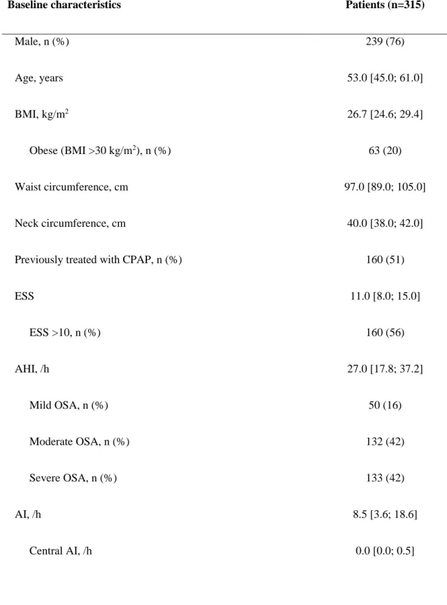
TABLES
TABLE 1 Demographic, respiratory and clinical data at baseline for the intention-to-treat
population
Baseline characteristics Patients (n=315)
Male, n (%) 239 (76) Age, years 53.0 [45.0; 61.0] BMI, kg/m2 26.7 [24.6; 29.4] Obese (BMI >30 kg/m2), n (%) 63 (20) Waist circumference, cm 97.0 [89.0; 105.0] Neck circumference, cm 40.0 [38.0; 42.0]
Previously treated with CPAP, n (%) 160 (51)
ESS 11.0 [8.0; 15.0] ESS >10, n (%) 160 (56) AHI, /h 27.0 [17.8; 37.2] Mild OSA, n (%) 50 (16) Moderate OSA, n (%) 132 (42) Severe OSA, n (%) 133 (42) AI, /h 8.5 [3.6; 18.6] Central AI, /h 0.0 [0.0; 0.5]
Mean SpO2, % 94.0 [93.0; 98.0]
Minimum SpO2, % 84.0 [78.0; 87.0]
Time with SpO2 <90%, min 7.0 [1.0; 22.0]
ODI, /h 17.0 [9.0; 29.0] Dental status, n (%) Good 259 (83) Acceptable 53 (17) Periodontal status, n (%) Good 254 (81) Acceptable 58 (19) Dental mobility, n (%) None 295 (95)
Low and limited 17 (5)
Angle malocclusion, n (%)
Type 1 209 (69)
Type 2 80 (27)
Type 3 13 (4)
Values are median [interquartile range] or number of patients (%)
AHI, apnoea-hypopnoea index; AI, apnoea index; BMI, body mass index; CPAP, continuous positive airway pressure; ESS, Epworth Sleepiness Scale; ODI, oxygen desaturation index; SpO2, oxygen saturation.
TABLE 2 Change in sleep and respiratory parameters over time during MRD therapy.
(n=191) Baseline 3–6 months 2 years
AHI, /h 26 [18; 35] 6 [3; 11]* 8 [4; 16]*
3% ODI, /h 17 [9; 29] 5 [2; 12]* 8 [3; 15]*
Time with SpO2 <90%, % 7 [1; 22] 0 [1; 9]** 0 [1; 9]**
Nadir SpO2, % 84 [87; 94] 87 [90; 95]* 87 [89; 96]*
Values are median [interquartile range].
AHI, apnoea-hypopnoea index; ODI, oxygen desaturation index; SpO2, oxygen saturation. *p<0.0001 vs baseline; **p<0.0004 vs baseline.
TABLE 3: Univariate analysis of predictive factors based on continuation of treatment after two years and achievement of reduction of an apnoea-hypopnoea index AHI of <10/h at the 2-year follow-up
Variable OR (95% CI) p-value
Continuation of treatment after 2 years (ITT CAD/CAM population, n=315)
Neck circumference (cm) 0.924 (0.866-0.986) 0.021
Waist circumference (cm) 0.981 (0.962-1.001) 0.058
Obesity (yes/no) 0.591 (0.338; 1.033) 0.065
ESS score >10 (yes/no) 1.674 (1.048-2.675) 0.031
Episodes of breathing cessation during sleep at inclusion (yes/no) 1.817 (1.066-3.098) 0.028
Compliance to with MRD at the last titration visit (days/week) 1.320 (1.086-1.605) 0.005
Compliance to with MRD at the last titration visit (hours/night) 1.327 (1.108-1.558) 0.002
Pain at the last titration visit (yes/no) 0.419 (0.242-0.727) 0.002
50% reduction in AHI at the 3-6 months’ follow-up (yes/no) 3.542 (1.930-6.499) <0.001
Nocturnal mouth breathing at the 3-6 months’ follow-up (yes/no) 1.859 (1.115-3.101) 0.017
Absence of snoring at the 3-6 months’ follow-up (yes/no) 2.000 (1.089-3.680) 0.026
Absence of nocturia at the 3-6 months’ follow-up (yes/no) 1.930 (1.040-3.578) 0.037
Compliance to with MRD at the 3-6 months’ follow-up (days/week) 1.387 (1.064-1.807) 0.015
Compliance to with MRD at the 3-6 months’ follow-up (hrs/night) 1.270 (1.010-1.514) 0.040
Reduction in AHI to <10/h at the 2-year follow-up (patients with AHI measurement, n=191)
Dental class (class II versus I) 3.542 (1.636-7.668) <0.0001
Dental class (class III versus I) 5.565 (0.630-49.172) <0.0001
Maximal propulsion (mm) 1.117 (0.978-1.276) 0.101
Mandibular propulsion as % of maximal propulsion 0.986 (0.974-0.998) 0.077
Initial AHI by severity group (mild versus severe) 5.552 (2.120-14.538) <0.0001
Initial AHI by severity group (moderate versus severe) 3.574 (1.857-6.878) <0.0001
Initial AI (number/h) 0.941 (0.913-0.970) <0.0001
Initial dorsal AHI (number/h) 0.981 (0.959-1.004) 0.1026
Positional OSA (yes/no) 2.412 (0.941-6.184) 0.063
TABLE 4 Adverse events at the 2-year follow-up visit (n=315) Patients, n (%) All Severe Requiring patient withdrawal TMJ disorder 89 (28.3) 18 (5.7) 7 (2.2)
Gingival pain or gingivitis 61 (19.3) 13 (4.1) 5 (1.6)
Occlusion change 53 (16.8) 1 (0.3) 2 (0.6)
Dental pain 50 (15.9) 6 (1.9) 7 (2.2)
Tooth migration or dental mobility 31 (9.8) 0 (0) 0 (0)
Mouth dryness or hypersalivation 27 (8.6) 0 (0) 1 (0.3)
Mouth pain or irritation 12 (3.8) 2 (0.6) 1 (0.3)
Discomfort 14 (4.4) 1 (0.3) 1 (0.3)
Dental fracture or prothesis loosening 10 (3.2) 7 (2.2) 1 (0.3)
Broken MRD 7 (2.2) 5 (1.6) 4 (1.2)
Nausea or vomiting 4 (1.3) 1 (0.3) 1 (0.3)
Mouth ulcer 4 (1.3) 1 (0.3) 0 (0)
Lack of prothesis retention 4 (1.3) 1 (0.3) 1 (0.3)
Suspected allergy 2 (0.6) 1 (0.3) 2 (0.6)
Other 19 (6.0) 5 (1.6) 3 (0.9)
Figures
FIGURE 1 Study flow chart. CAD/CAM, computer-aided design, computer-aided
manufacturing; CPAP, continuous positive airway pressure; FU, follow-up; ITT,
FIGURE 2 Proportion of patients continuing mandibular repositioning device therapy over
FIGURE 3 Mandibular repositioning device efficacy at 2-year follow-up by obstructive sleep
apnoea syndrome (OSAS) severity (A) (AHI, apnoea–hypopnoea index; Success rate,
percentage of patients with a ≥50% decrease in AHI from baseline to follow-up. Chi-squared
test for AHI achieved; *p<0.001). Change in AHI over time by baseline OSAS severity in
patients remaining in the study at the 2-year follow-up (B) (two-by-two comparisons of AHI (Tuckey’s test) for baseline versus 3-6 months and baseline versus 2 years: p<0.0001 for each
severity subgroup; for 3–6 months versus 2 years: p=0.0082 for mild OSAS, p=0.0001 for
FIGURE 4 Proportion of patients with different symptoms and Epworth Sleepiness Scale
(ESS) score >10 (A), Quebec Sleep Questionnaire (QSQ) scores (B) and visual analogue scale
(VAS) scores (C) at baseline, and after 3–6 months and 2 years of follow-up.
FIGURE 5 Forest plot of multivariate analysis showing predictors of apnoea-hypopnoea
index (AHI) <10/h (A) and continuation of treatment (B) after two years. AI, apnea index; CI,
confidence interval; CPAP, continuous positive airway pressure; OR, odds ratio.
ORCADES Investigators :
Dr Darius ABEDIPOUR Cabinet médical, Lyon. France
Dr Aurélie ALLARD-REDON
Cabinet dentaire, BEHREN LES FORBACH. France Dr Alexandre ARANDA
Clinique de l'Union, Service de Neurologie, Saint Jean. France
Dr Valérie ATTALI
CH de Valence, Service ORL, VALENCE. France
Dr Martine BECU
CHG de Chalons en Champagne, Service de Pneumologie, Chalons en Champagne. France Dr Wally BERUBEN
Cabinet Dentaire, Chalons en champagne. France
Dr Jerome BESSARD
Clinique de l'Union, Service d'Odontologie, Saint Jean. France Dr Isabelle BONAFE
Faculté d'odontologie, Montpellier. France
Dr Mohammed BOUKHANA Centre du sommeil, Metz. France Dr Bruno CHABROL
Cabinet Dentaire, CREIL. France
Dr Gérard CHATTE
Cabinet médical, CALUIRE. France Dr Dominique CHAUVEL-LEBRET
CHU Rennes, Pôle d'Odontologie et Chirurgie Buccale, Rennes. France
Dr Jean-Marc COLLET
Hôpital Pitié Salpêtrière Service de Stomatologie et Chirurgie Maxillo-Faciale, PARIS. France Dr Olivier COSTE
Polyclinique du Tondu, Bordeaux. France
Dr Nathalie DUMONT
Cabinet medical, Marseille. France Dr Sophie DURAND-AMAT
Cabinet médical, Lagny sur Marne. France
Pr Marie-Pia D'ORTHO
Groupe Hospitalier Bichat Service de Physiologie-Explorations fonctionnelles, PARIS. France Dr Jean-Marc ELBAUM
Cabinet Medical, Marseille. France
Clinique Beau Soleil, MONTPELLIER. France
Dr Frédéric GOUTORBES
Centre Hospitalier de Beziers, Service de Pneumologie, BEZIERS. France Dr Thierry GRANDJEAN
Cabinet dentaire, Schœneck. France
Dr Wilma GUYOT
Cabinet Dentaire, VANDOEUVRE LES NANCY. France Dr Doniphan HAMMER
Espace Médical Rabelais, POITIERS. France
Dr Carmen HAVASI
Cabinet Dentaire, Nice. France Dr Pascal HUET
Clinique Bretéché, Nantes. France
Dr Jean-Baptiste KERBRAT
CHRU de ROUEN-Hopital Charles Nicolle, Service de Maxillo-Faciale, Rouen. France Dr Hauria KHEMLICHE
Centre Hospitalier de Senlis Avenue Paul Rougé Unité Sommeil, Senlis. France
Dr Christian KOLTES
Centre du sommeil, Metz. France Pr Damien LEGER
Hôtel Dieu de PARIS Centre de Sommeil, PARIS. France
Dr Laurent LACASSAGNE
Clinique de l'Union, Service de Pneumologie, Saint Jean. France Dr Xavier LAUR
Cabinet Dentaire, CASTRES. France
Dr Lionel LEROUSSEAU
Centre Hospitalier d'Antibes, Service de Pneumologie, Antibes. France Dr Olivier LIARD
Cabinet dentaire Lagny sur Marne. France
Dr Matthieu LONGUET
Centre Hospitalier de Beziers, Service ORL, BEZIERS. France Dr Anne MALLART
Hôpital Roger Salengro Service de Neurologie Clinique, Lille. France
Dr Francis MARTIN
Centre Hospitalier de CompiègneService de Pneumologie, Unité des pathologies du Sommeil, Compiègne. France
Dr Frédéric MERLE-BERAL
Clinique de l'Union, Service d'Odontologie, Saint Jean. France
Pr Jean-Claude MEURICE
CHU de Poitiers, Service de Pneumologie, Poitiers. France Dr Zoubida MOKHTARI
Centre Hospitalier de Senlis Avenue Paul Rougé Unité Sommeil, Senlis. France
Dr Christelle MONACA
Hôpital Roger Salengro Service de Neurologie Clinique, Lille. France Dr Pierre-Jean MONTEYROL
Polyclinique du Tondu, Bordeaux. France
Pr Jean-François MUIR
CHU de ROUEN- Hôpital de Bois Guillaume Service de Pneumologie, ROUEN. France Dr Eric MULLENS
FONDATION BON SAUVEUR, Laboratoire de Sommeil, ALBI. France Dr Dominique MULLER
Cabinet médical, Metz. France Dr Charles PAOLI
CH Montreuil, MONTREUIL. France Dr François-Xavier PETIT
Maison de la Mutualité, Nantes. France Dr Bernard PIGEARIAS
Dr Marc PRADINES
Cabinet Dentaire, TOULOUSE. France Dr Arnauld PRIGENT
Clinique St Laurent, Service de Pneumologie, Rennes. France Dr Gil PUTTERMAN
Hôtel Dieu de PARIS, Service de Stomatologie PARIS. France
Dr Marc REY
CHU Timone, Centre du Sommeil de Neurophysiologie, Marseille. France Dr Mickael SAMAMA
Hôpital Pitié Salpêtrière Service de Stomatologie et Chirurgie Maxillo-Faciale, PARIS. France
Pr Renaud TAMISIER
CHU de Grenoble, Physiologie, sommeil et exercice, Grenoble. France Dr Michel TIBERGE
CHU de RANGUEIL, Service de Neurologie et Explorations Fonctionnelles Neurologiques Toulouse. France
Dr Cyrille TISON
Hôpital Roger Salengro, Service de Stomatologie, Lille. France Dr Fabienne TORDJMAN
Hôpital Pitié Salpêtrière Service du Sommeil, PARIS. France Dr Bernard TRIOLET
Cabinet Dentaire Ribecourt Dreslincourt. France.
Pr Christian VACHER
Hôpital Beaujon Service de Chirurgie Maxillo-Faciale et Stomatologie, Clichy. France Dr Marie-Françoise VECCHIERINI
Hôtel Dieu Centre de Sommeil, PARIS. France
Dr Alain VERAIN