HAL Id: tel-01380068
https://hal.archives-ouvertes.fr/tel-01380068
Submitted on 12 Oct 2016HAL is a multi-disciplinary open access archive for the deposit and dissemination of sci-entific research documents, whether they are pub-lished or not. The documents may come from teaching and research institutions in France or abroad, or from public or private research centers.
L’archive ouverte pluridisciplinaire HAL, est destinée au dépôt et à la diffusion de documents scientifiques de niveau recherche, publiés ou non, émanant des établissements d’enseignement et de recherche français ou étrangers, des laboratoires publics ou privés.
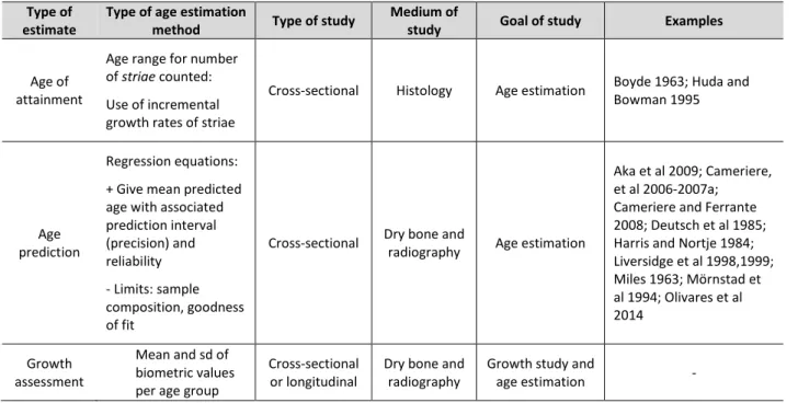
critical review of existing methods and the application
of two standardised methodological approaches
Louise Corron
To cite this version:
Louise Corron. Juvenile age estimation in physical anthropology: A critical review of existing methods and the application of two standardised methodological approaches . Biological anthropology. Aix-Marseille Universite, 2016. English. �tel-01380068�
AIX-MARSEILLE UNIVERSITÉ
FACULTÉ DE MEDECINE DE MARSEILLE
ECOLE DOCTORALE
: Sciences de l’Environnement - ED 251
UMR 7268 Anthropologie bioculturelle, Droit, Ethique et Santé-EFS-CNRS
T H È S E
Présentée et publiquement soutenue devant
LA FACULTÉ DE MEDECINE DE MARSEILLE
Le 17 juin 2016
Par Louise CORRON
Née le 16 juin 1988 à Aubervilliers
JUVENILE AGE ESTIMATION IN PHYSICAL ANTHROPOLOGY:
A CRITICAL REVIEW OF EXISTING METHODS AND THE APPLICATION
OF TWO STANDARDISED METHODOLOGICAL APPROACHES
Pour obtenir le grade de DOCTORAT d’AIX-MARSEILLE UNIVERSITÉ
SPÉCIALITÉ : ANTHROPOLOGIE BIOLOGIQUE
Membres du Jury de la Thèse :
M. Pascal Adalian (Professeur, Aix-Marseille Université, UMR 7268 ADES)
Mme Silvana Condemi (CNRS, UMR 7268 ADES) Directrice
Mme Eugenia Cunha (Professeur, University of Coimbra)
M. François Marchal (CNRS, UMR 7268 ADES) Directeur
M. Hans Christian Petersen (Professeur Associé, University of Southern Denmark) Rapporteur
Acknowledgments
To the members of the jury,
Mr Pascal Adalian (Professor, Aix-Marseille University, UMR 7268 ADES) Mrs Silvana Condemi (CNRS, UMR 7268 ADES),
Mrs Eugenia Cunha (Professor, University of Coimbra), Mr François Marchal (CNRS, UMR 7268 ADES),
Mr Hans Christian Petersen (Associate Professor, IMADA, University of Southern Denmark)
Mr Norbert Telmon (Professor, University Paul Sabatier, Toulouse – Hospital Practitioner, UMR 5288 AMIS)
Thank you for reading and evaluating my (very long) work and for your comments that helped improve this work.
Mr Hans Christian Petersen (Associate Professor, IMADA, University of Southern Denmark) and Mr Norbert Telmon (Professor, University Paul Sabatier, Toulouse – Hospital Practitioner, UMR 5288 AMIS), thank you both for accepting to be the reporters of my work.
To the team of supervisors who directly and consistently contributed to this work, First, thank ou for the effort of reading and correcting this ros eef anuscript.
Mr Pascal Adalian (Professor, Aix-Marseille University, UMR 7268 ADES), your confidence in me, your kindness and admirable clairvoyance have greatly helped me improve during my time in Marseilles and achieve this work. I have learned many things while working with you here. I will try to make the most of what you have taught me.
Mrs Silvana Condemi (CNRS, UMR 7268 ADES), thank you for accepting to supervise this work, for taking the ti e to read and correct ork fro start to finish, e en though the su ject asn t directly in your field of interest. I am grateful for your critical outlook on my work that greatly contributed to improve it and for your helpful advice.
Mr François Marchal (CNRS, UMR 7268 ADES), I have learned a lot from you during my stay in Marseilles. Your efficiency and rigor are impressive and were greatly appreciated during the difficult phase of writing and throughout these past three (and a half) years. I also wish to thank you for helping me perfect (I hope!) the art of tiramisu-making.
To the members and associates of the anthropology teams of the UMR7268 ADES,
Mr Michel Signoli (Director of UMR7268 ADES), I am extremely grateful to you for taking a chance on me and accepting me in the anthropolog fa il of Marseilles four years ago, and for fighting to obtain the funding from the Ministry of Education and Research that allowed me to do my work serenely.
A special thank you to Laetitia Delouis for all your help with administrative paperwork, but especially for your kindness, for helping me staying down-to-earth and for our talks on the stairs in the sun.
I wish to thank Mrs Kathia Chaumoître and Mr Michel Panuel (Hospital Practitioners, APHM, Professors, Aix-Marseille University) for granting me access to the database to collect the samples used in this study.
Thank you to Virginie Astruc, Lydia Carlier, Anne-marie Ferrandes, Tony Garcin, Gisèle Geoffroy, Loïc Lalys, Stéphane Mazières, Cathy Rigeade, Aurore Schmitt, Stefan Tzortzis for their kindness, their help and humour.
Yann Ardagna, thank you for your help, for teaching me the subtleties of the true arseillais , for all the laughs and jokes exchanged during the lunches and numerous goûters we all shared together.
Julien Corny, thank you for your precious outlook, support and for our great philosophical talks. Laurent Puymerail, thank you for your help exploring the virtual world of Avizo®, your good nature, your amazing sense of humour, your kindness and your friendship.
Thank you to all the people who helped me achieve this work,
Mrs Judite Alves and Mrs Maria Susana Garcia, (University of Lisbon), thank you granting me access to the Luis Lopes collection and for your kind welcome in Lisbon.
Mr Hugo Cardoso (Assistant Professor, Simon Fraser University), thank you for your help with obtaining data for the Luis Lopes collection.
Ms Mathilde Daumas, thank you for providing a large part of the sample from Marseilles.
Mr Bruno Foti (PU-PH, Aix-Marseille University), for providing the illustrative orthopantomogram. Mr Pascal Murail, thank you for your support and for vouching for me 4 years ago.
Mr Norbert Telmon (Professor, University Paul Sabatier, Toulouse – Hospital Practitioner, UMR 5288 AMIS), thank you for kindly providing the data for the test sample from Toulouse.
Mr Jean-Luc Voisin, thank you for your advice on the protocol for the study of the clavicles. Mr Tim White (Professor, University of California, Berkeley), thank you for welcoming me to the laboratory, for your advice and for your kindness. Josh Carlson, thank you for your kindness and for being so welcoming during my visits to Berkeley.
Thank you to the army of statisticians who helped me understand the wonderful world of applied statistics:
Mr Stephen Milborrow, I could not have understood or constructed MARS models without your help. Thank you ever so much.
Mr Luis Sagaon-Teyssier and Mr Antoine Vilotitch (Inserm). You helped me a great deal, even though ou didn t ha e to, and for that I a trul grateful. Thank ou for great advice and for helping me achieve what I wanted to do.
Mrs Bérengère Saliba-Serre (UMR 7268 ADES), thank you for your kindness and patience and for pointing me in the right statistical direction.
To my friends,
Adeline, Clémence, Julie, Laetitia and Louise, thank you for pulling me away from work when I needed it and for your support. Thank you to Eric and Antoine for your friendly and useful scientific advice shared around a drink (or more!).
Thank you to my fellow anthropologist friends for the shared laughter. The infernal trio from Bordeaux: Maguelone, Justine and Jeanna (the JL s Gwendal (my favourite Britton), David, Pierre, Sacha.
Alexia, Aurore, Clémence, Emeline S., Emeline V., Florence, Gaëlle, Laetitia, Marie, Marion, Melissa, Sandra, and Sandy. Girls, ou re great. I er happ to kno ou and proud to count ou as future colleagues and as my friends. A lot of the best times I spent these past years in Marseilles were with you. You also helped me cope with the bad ones, so thank you for that.
A special thank you to Emeline V. for being amongst other things my second observer and for providing part of the scans, and to Sandy for her hawk-eye reading of the manuscript and for her ever-wise advice.
To my family,
Thank you all for your love and support through everything, good and bad. Mum, thanks for reading and correcting su stantial prose .
Vincent, thank you for always (and still, it s een a hile!) being there, even virtually, and for teaching me about perspective.
And finally, thank you to Barry, Maurice and Robin; Freddie, Brian, Roger and John for keeping me company while I was writing.
If I have forgotten to explicitly mention anyone, please consider this as a personal, warm and apologetic thank you.
Summary
Introduction ... 9
Juvenile age estimation in physical anthropology: concepts, contexts and methods ... 13
Chapter 1. Age estimation of juvenile individuals in physical anthropology: definitions and contextualisation ______________________________________________________________ 14 1.1. Age differences: biological ages and chronological age ... 14
1.1.1. Chronological age ... 15
1.1.2. Biological age ... 16
1.2. Juvenility: a polyfactorial, contextualised and pluridisciplinary concept ... 18
1.2.1. Bioarchaeology: from bone to past populations, from biological individual to social status ... 18
a. General concepts ... 18
b. The social juvenile: the social expression of biological development ... 19
1.2.2. Forensic anthropology: from bone to individual, from biological individual to legal status ... 21
a. General concepts ... 21
b. Restrictions and regulations concerning the practice of forensic anthropology ... 21
c. The legal juvenile: the minor ... 22
1.2.3. The biological juvenile: an expression of biological development ... 25
1.3. Skeletal development: growth and maturation ... 27
1.3.1. Skeletal development ... 27
1.3.2. Skeletal maturation ... 29
a. Appearance of ossification centres ... 30
b. Morphology and size of ossification centres ... 31
c. Fusion of ossification centres ... 31
1.3.3. Skeletal growth ... 32
a. Growth studies ... 33
b. Skeletal growth phases and patterns ... 36
1.3.4. Dental development: growth and mineralisation ... 38
1.3.5. Variability, secular trends and developmental anomalies ... 40
a. Skeletal growth and maturation ... 40
b. Dental development ... 44
c. Growth and maturation in past populations ... 45
1.4. Juvenile age estimation in physical anthropology: a case in point for the need of methodological standardisation ... 48
1.4.2. Age as an individual identifier of juveniles in forensic anthropology ... 50
1.4.3. The importance of standardised juvenile age estimation protocols and methods ... 52
Chapter 2. A critical review of juvenile age estimation methods ______________________ 56 2.1. Evolution of age estimation concepts and principles: from clinical or empirical studies to statistically robust methods ... 57
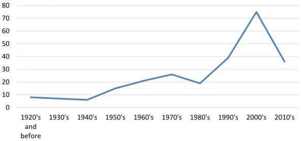
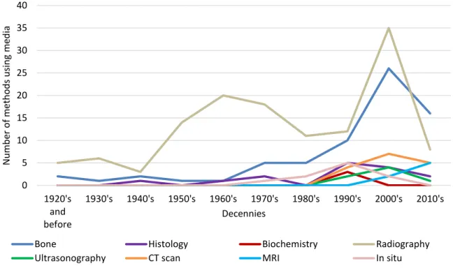
2.1.1. First occurrences of ju enile age esti ation s and efore : understanding iological development ... 57
2.1.2. Ju enile age esti ation s- s : exploring, i pro ing and testing ... 58
2.1.3. Present of ju enile age esti ation s-… : opti ising, har onising and standardising ... 61
2.2. Methodological typology ... 64
2.2.1. Method construction: objectives and exploited data ... 64
a. Types of age estimation ... 65
b. Measurements of ossification centres ... 66
c. Bone maturation: morphology, maturation stages and sequences, fusion states ... 70
d. Dental growth: dental measurements ... 76
e. Dental maturation: mineralisation sequences and tooth eruption ... 77
2.2.2. Method application: methods adapted to the subjects of study ... 81
a. Skeletal age estimation ... 81
b. Dental age estimation ... 83
2.2.3. Limitations of juvenile age estimation ... 84
2.3. Critical review and classifications of juvenile age estimation methods ... 94
2.3.1. Juvenile age estimation: method construction and method selection ... 94
a. Methodological deconstruction to understand method construction ... 94
b. General structure of juvenile age estimation methods ... 101
c. Statistical analysis by Multiple Correspondence Analysis (MCA): from method to bone ... 104
d. Empirical classifications: from bone to method ... 107
i. Approach adopted for constructing the classifications ... 107
ii. Organisation of the empirical classification ... 110
2.3.2. Statistical and empirical classifications: a complementary approach for method evaluation . 113 a. Statistical classification (MCA and clusterisation) ... 113
i. Sampling criteria ... 113
ii. Statistical and methodological criteria ... 123
b. Empirical classifications ... 136
c. Comparison of the results ... 142
2.4. Conclusions on juvenile age estimation ... 144
2.5. Constructing a standardised juvenile age estimation method ... 147
2.5.1. What are the needs in terms of juvenile age estimation from skeletal elements? ... 147
A standardised approach of juvenile age estimation ... 149
Chapter 3. Material _________________________________________________________ 150 3.1. Study sample and variables ... 150
3.1.1. Growth and maturation patterns of the three bones studied ... 151
a. Flat bone: the iliac bone ... 151
b. Short bone: the fifth lumbar vertebra ... 161
c. Long bone: the clavicle ... 165
d. Summary of developmental events of the three bones studied... 170
3.1.2. Study sample from Marseilles ... 170
a. Characteristics of reference samples ... 170
b. Inclusive criteria and sample composition ... 176
3.2. Test samples ... 179
3.2.1. Test sample from Toulouse... 179
3.2.2. Test sample from the Luis Lopes osteological reference collection ... 180
3.3. Taphonomic alterations ... 183 Chapter 4. Methods ________________________________________________________ 185 4.1. Acquired data ... 185 4.1.1. Biometric variables ... 185 a. Iliac variables ... 185 b. Lumbar variables ... 187 c. Clavicular variables ... 189 4.1.2. Non-biometric variables ... 190
4.2. Acquisition of variables on CT scan data ... 193
4.2.1. Segmentation and reconstruction techniques ... 193
4.2.2. Biometric variables: uni- and bi-dimensional data ... 195
a. Iliac variables ... 197
i. Plane definition and landmark positioning ... 197
ii. Variable acquisition protocols ... 198
b. Lumbar variables ... 200
i. Plane definition and landmark positioning ... 200
ii. Variable acquisition protocol ... 201
c. Clavicular variables ... 203
i. Plane definition and landmark positioning ... 203
ii. Variable acquisition protocols ... 206
4.2.3. Non-biometric variables: bone fusion assessment on virtual bones ... 208
4.3. Acquisition of variables on dry bone ... 209
a. Iliac variables ... 210
b. Lumbar variables ... 211
c. Clavicular variables ... 211
4.3.2. Non-biometric variables ... 212
4.3.3. Difficulties in data acquisition on dry bones ... 212
4.4. Validation of data acquisition methods ... 213
4.4.1. Testing scanned bone/dry bone variable consistency ... 213
a. Consistency between variables taken on dry and scanned bone ... 213
b. Equality of geometric and anatomical variables taken on dry and scanned bone ... 213
4.4.2. Repeatability and reproducibility of the variables ... 214
a. Landmark positioning ... 214
b. Biometric variables: measurements ... 215
c. Non-biometric variables: stages and scores... 217
4.5. Descriptive statistics of the samples ... 218
4.5.1. Age and sex ratios of the different samples ... 218
4.5.2. Non-biometric variables: iliac maturation stages ... 218
4.5.3. Biometric variables: iliac, lumbar and clavicular variables ... 218
4.6. Interactions between the variables, age, and biological factors ... 219
4.6.1. Relationship between age and the variables ... 219
4.6.2. Influence of other biological factors: asymmetry, sex, variable collinearity ... 219
a. Bilateral asymmetry ... 219
b. Sexual dimorphism ... 220
4.7. Modelling the relationship of biological variables with age ... 221
4.7.1. Parametric regression models ... 221
a. Univariate linear regression ... 221
b. Polynomial regression ... 221
4.7.2. Selective criteria for parametric regression models ... 222
a. Regression coefficients and tests ... 222
b. Residuals: normality, homoscedasticity and independence ... 222
c. 95% prediction intervals and precision ... 224
4.7.3. Validation of regression models ... 225
a. Cross-validation ... 225
b. Separate independent samples ... 225
4.7.4. Collinearity and Multicollinearity ... 226
4.7.5. Heteroscedasticity ... 227
4.7.6. Multi-variate Adaptive Regression Splines (MARS) models ... 229
a. Principles of MARS models ... 229
4.7.7. Application of predictive models for age estimation ... 231
4.8. Age estimation using Bayesian probabilities on non-biometric ordinal data ... 232
4.8.1. The Bayesian approach ... 232
4.8.2. Choosing the a priori probabilities ... 234
a. Non-uniform priors ... 234
b. Uniform priors ... 235
4.8.3. Choosing the appropriate Bayesian approach for age estimation ... 237
a. Dependent Bayesian method (DBM) ... 239
b. Independent Bayesian method (IBM) ... 240
4.8.4. Evaluation and validation of Bayesian posterior probabilities for age estimation ... 243
Results ... 247
Chapter 5. Results I: Protocol validation and sample descriptions ___________________ 248 5.1. Validation of data acquisition protocols ... 249
5.1.1. Variable consistency on scanned bone surfaces and dry bones ... 249
a. Testing the consistency of variables taken on dry bone and scanned dry bone ... 249
b. Testing the equality of geometric and anatomical variables ... 251
i. Dry clavicles ... 251
ii. Scanned dry bone surfaces ... 252
5.1.2. Repeatability and reproducibility ... 254
a. Landmark positioning ... 254
i. Ilium ... 254
ii. Fifth lumbar vertebra ... 257
iii. Clavicle ... 261
b. Biometric variables ... 263
i. Ilium ... 263
ii. Fifth lumbar vertebra ... 269
iii. Clavicle ... 277
c. Non-biometric variables ... 283
5.2. Descriptive statistics of the sample from Marseilles... 285
5.2.1. Age and sex ratios ... 285
5.2.2. Iliac variables ... 286
a. Biometric variables of the ilium ... 286
b. Maturation stages of the iliac bone ... 288
5.2.3. Lumbar variables ... 289
5.2.4. Clavicular variables ... 291
5.3. Bilateral asymmetry of the variables in the Marseilles sample ... 292
5.3.2. Fifth lumbar vertebra ... 293
5.3.3. Clavicle ... 293
5.3.4. Iliac bone ... 294
5.4. Sexual dimorphism of the variables in the Marseilles sample ... 295
5.4.1. Ilium ... 295
5.4.2. Fifth lumbar vertebra ... 297
5.4.3. Clavicle ... 300
5.4.4. Iliac bone ... 302
5.5. Homogeneity of the variables from the different samples ... 304
Chapter 6. Results II: Age estimation ___________________________________________ 306 6.1. Age estimation using regression models and biometric variables ... 307
6.1.1. Parametric models ... 307
a. Ordinary Least Squares (OLS) regressions ... 310
i. Iliac variables ... 310
ii. Lumbar variables ... 324
iii. Clavicular variables ... 328
iv. Conclusions on OLS models ... 332
b. Heteroscedasticity- and autocorrelation-robust variance-covariance matrices (vcovHAC) - Weighted Least Squares (WLS) regressions ... 335
i. Dealing with heteroscedasticity I: variance-covariance matrices robust to residual heteroscedasticity and autocorrelation ... 335
ii. Dealing with heteroscedasticity II: age prediction using WLS models ... 336
6.1.2. Non-parametric Multi-variate Adaptive Regression Splines (MARS) models ... 342
a. Iliac variables ... 342
b. Lumbar variables ... 350
c. Clavicular variables ... 359
6.1.3. Testing the models ... 366
a. Independent test sample from Toulouse ... 366
b. Independent test sample from Lisbon (Luis Lopes collection) ... 369
i. Iliac variables ... 369
ii. Lumbar variables ... 372
iii. Clavicular variables ... 376
6.2. Age estimation using Independent Bayesian probabilities and maturation stages of the iliac bone . ... 381
6.2.1. Uni-site probabilities ... 381
a. Independent test sample from Marseilles ... 381
b. Independent test sample from Lisbon (Luis Lopes collection) ... 385
a. Independent test sample from Marseilles ... 388
i. Four-digit combination ... 388
ii. Three-digit combinations ... 389
iii. Two-digit combinations ... 392
b. Independent test sample from Lisbon (Luis Lopes collection) ... 396
i. Four-digit combination ... 396
ii. Three-digit combinations ... 397
iii. Two-digit combinations ... 400
6.2.3. Influence of sex on age estimation using posterior probabilities ... 404
a. Independent test sample from Marseilles ... 404
b. Independent test sample from Lisbon (Luis Lopes collection) ... 407
6.3. Comparison with other age estimation methods ... 408
6.3.1. Age esti ation using iliac io etric data: co parison ith Rissech and Malgosa s ethod . 408 a. Independent test sample from Marseilles ... 409
b. Independent test sample from Toulouse ... 409
c. Independent test sample from Lisbon (Luis Lopes collection) ... 410
6.3.2. Age estimation using maximum cla icular length: co parison ith Black and “cheuer s ethod ... 411
6.3.3. Age estimation using iliac maturation stages: comparison ith Co ueugniot and colla orators method ... 412
a. Independent test sample from Marseilles ... 413
b. Independent test sample from Lisbon (Luis Lopes collection) ... 416
Discussion ... 420
Chapter 7. Juvenile age estimation in physical anthropology: standardised methods to express and exploit developmental variability ______________________________________ 421 7.1. Standardised sampling of population variability ... 421
7.1.1. Adopting a standardised and adapted sampling protocol ... 421
7.1.2. Sample characteristics ... 423
7.1.3. Past and present individuals: recalibrating variability ... 424
7.2. Standardised data and data acquisition protocols ... 426
7.2.1. Protocols for data acquisition ... 426
a. Biometric variables ... 426
b. Maturation stages ... 428
7.2.2. Predictor variables ... 429
a. Biometric variables ... 429
b. Maturation of the ischio-pubic ramus and the acetabular region ... 432
7.3.1. Regression models and age estimation: choosing the best approach ... 435
a. A prerequisite: methodological standardisation ... 435
b. MARS models: an approach fitting individual variability ... 436
7.3.2. Age estimation using maturation stages and Bayesian probabilities ... 439
7.4. Juvenile age estimation using two standardised approaches ... 441
7.4.1. MARS models ... 441
a. Biological interpretations of statistical results ... 441
b. Comparison of MARS models with other age estimation methods ... 445
7.4.2. Bayesian posterior probabilities for age estimation: strengths and weaknesses ... 447
7.5. Limitations and perspectives of juvenile age estimation ... 449
7.5.1. Practical use of MARS models and perspectives for improvement ... 449
7.5.2. The influence of context ... 450
a. Age estimation in bioarchaeology and palaeodemography... 451
b. Age estimation in forensic anthropology ... 452
c. Juvenile age estimation: an example of the limitations of biological modelisation?... 454
7.5.3. Age-specific versus non age-specific methods ... 457
7.5.4. Populations of study ... 459
7.5.5. Understanding developmental patterns and variability to improve age estimation ... 461
Conclusion ... 465
List of tables ... 469
List of figures ... 478
French summary ... 487
The study of juvenile individuals in physical anthropology has seen considerable changes since the birth of the discipline at the end of the 19th Century. The numerous contexts of study of juveniles
(medicine, physiology, sociology, psychology, ethics, law) have led to several denominations, each corresponding to subtle variations related to age and/or specific developmental characteristics: sub-adults or non-sub-adults, immatures, juveniles, foetuses, new-borns, infants, children, adolescents, minors, etc. (Scheuer and Black 2000). In physical anthropology, these various terms can relate to a biological and/or social (or biocultural) status that characterises the individuals in question and as such, gives them a biological status and social and/or legal identity, depending on whether anthropologists are working in a bioarchaeological or forensic context, respectively.
Before becoming a particular filed of interest for physical anthropologists, juvenile individuals were almost exclusively the subject of clinical studies alone. These were undertaken to control normal development (growth and maturation) and spot individuals presenting development-related pathologies to treat them. The notion of individual variability already peaks through these early works (Huxley 1924; Pryor 1907). The relationship between age and development then moved from clinical to scientific use, as development was no longer controlled but extensively studied and even predicted or mathematically modelled (Scammon and Calkins 1923). Most of the earliest anthropological studies involving juveniles were purely methodological and quickly made use of the development of medical imaging techniques to refine skeletal and dental analyses (Flecker 1932; Hess et al 1932): bone and dental developmental patterns led to direct application of the findings for juvenile age estimation, both on the living and the deceased. This particular and still very active field of interest, is therefore relatively ancient: the earliest occurrence found for the present study dates from 1894, with the work of L. Wacholz on bone development (Wacholz 1894). However, few studies were dedicated to using these methods for analysing and interpreting the role and status of these individuals in past populations efore the s Johnston, In Brothwell 1968). It is now admitted that juveniles hold a central part in any osteoarchaeological study whenever they are involved. The analysis of juveniles or juvenile remains in a forensic anthropological context has particular implications related to the potential legal status of the individuals, which is determined by the estimated age. Age is therefore a primary source of information for the study of juveniles in physical anthropology, regardless of the context and one of the key elements of an individual s iological profile.
The biological profile of an individual (age, sex, stature, individual characteristics) established from the study of human remains is a key concept in physical anthropology (Cunha et al 2009). In osteoarchaeology or bioarchaeology, the biological profile is constructed for each individual of the study to be analysed along with the other individuals at a global scale to participate in interpretations
of the archaeological, historical, ethnological and/or cultural context in terms of population studies. In a forensic context, the biological component takes on an even bigger role, as the context is set but the interpretation rests almost solely on the information provided by the biological profile. A ju enile s iological profile is quite succinct, because age and stature are the only parameters that can be reliably estimated from the juvenile skeleton (Saunders, In Katzenberg and Saunders, 2008).
Although the goals of juvenile age estimation differ according to context, the methods developed for that purpose follow similar principles. Today, anthropologists can choose from an array of methods to estimate age from juvenile remains (Cunha et al 2009; Wood, In Black and Ferguson 2011) such as dental and skeletal development indicators expressing growth and/or maturation changes that occur between early foetal life and early adulthood. Each growth and maturation event that can be related to age presents intrinsic and extrinsic variability, which needs to be considered to improve age estimation.
Because growth and developmental variability has different levels of expression (individual and population), and is caused by various factors (age, sex, genetic and epigenetic, secular trends, pathologies), it seems difficult to estimate age in juveniles following a standardised methodological approach. Indeed, current methods are often population-, age- or sex- dependent, which limits their practical application and can lead to biased estimates and interpretations. In addition to these methodological biases, the discrepancies observed between estimated (biological) and real (chronological, calendar or legal) age are also the result of the variable correlation between age and the biological indicator used to estimate it.
For several years now, the anthropological community has been advocating method uniformity to respect common statistical and methodological criteria and allow direct comparison between methods and results: reliability, standard error of estimation and accuracy should be known, sampling protocols should be standardised, etc. (Cattaneo 2007; Cunha et al 2009; Dirkmaat et al 2008; Ferembach et al 1979). By respecting these principles, methods could be objectively evaluated, bearing in mind the influence of methodological and population biases for estimating age in both osteoarchaeological and forensic contexts. The principles behind juvenile age estimation methods and the importance of context in both method construction and application are presented in Chapters 1 and 2.
The present study was undertaken to achieve two main goals:
- Present a critical methodological review of a large number of juvenile age estimation methods available to anthropologists today. This will lead to the elaboration of a methodological decisional tool, presented as a set of arborescent decisional trees. It is based
on objective criteria evaluating methodological construction protocols and method application. This practical tool can be used by anthropologists working in a forensic or archaeological context as a guide to the selection of methods according to the data available and highlights methods respecting valid methodological criteria (Chapter 2);
- Elaborate and apply standardised approaches of juvenile age estimation, respecting rigorous methodological protocols and criteria for data acquisition and analysis as well as statistical criteria, taking into account individual variability (Chapters 3 through 6).
The results obtained in answer to these two objectives will be discussed in Chapter 2 and conjointly in Chapter 7.
Juvenile age estimation in physical
anthropology: concepts, contexts and
Chapter 1. Age estimation of juvenile individuals in physical
anthropology: definitions and contextualisation
A sub-adult, non-adult or juvenile sensus largo is an individual who has not reached the biological (physiological, skeletal, dental), psychological, legal, or social status of an adult, i.e. biological, legal, or social maturity. This particular status is defined by the chronological (or calendar) age of an individual, i.e. the time (expressed in years, months, days, etc.) between his/her date of birth (or conception in some cases) and the present date. This simple notion of age as we understand it covers in fact a polyfactorial notion of immaturity that results from biological, cultural, psychological, social (Bogin 1997) and socio-legal parameters.
Age estimation is the principal component of the study of juvenile individuals in physical (or biological) anthropology. Indeed, physical anthropologists rely on indicators of biological immaturity (i.e. growth and development) to estimate chronological age, which is then used to interpret a social or legal status of immaturity. Estimated age is based on skeletal and/or dental remains or elements, so anthropologists work with skeletal and/or dental ages, inferred by maturation indicators or growth parameters (Wood and Cunningham, In Black and Ferguson 2011). Although biological growth and development are both highly correlated with chronological age, they are not perfectly equivalent to it. This correlation presents different levels of variability according to various factors. In addition to this, the polyfactorial definition of immaturity, its context of study, and the information available to anthropologists result in other inconsistencies, which we will present here.
1.1. Age differences: biological ages and chronological age
To be able to compare the different definitions of immaturity, anchor them in a common time frame, and provide clear limitations for grading the different levels of immaturity, a neutral reference unit for numerically grading the level of immaturity and its temporal extent is needed. Developmental growth and maturation phases are strongly correlated with age (Adalian et al 2002; Cardoso 2008b; Fazekas and Kosa 1978; Scheuer and Black 2000; Ubelaker 1987), so age seems to be the best way to grade the appearance, extent and ending of developmental phases. This means that the different levels of biological immaturity are often the reflection of the variations in intensity of developmental activity (growth or maturation accelerations and decelerations, static periods, growth spurts, etc.) and can be anchored in time using age limits. These variations, particularly growth phases, are used to define sub-categories of immaturity. Indeed, two main growth phases are known (Bogin 1999): the prenatal phase (from conception to birth) and the postnatal phase (after birth).
Additional information on maturation and/or other particular growth events can also partake in refining these age categories.
1.1.1. Chronological age
The only neutral unit is the real age, calendar age, or chronological age of the indi idual, as it is not subject to any confusions: chronological age is the mathematical difference between the date of birth and the date of death of the individual or between the date of birth and the date of an event of interest (e.g. the date of a crime) for a living individual. The definition of chronological age is a given and does not present any other possible value if both dates are known and considered accurate. The
notions of i aturit and age are indeed insepara le ut not e ui alent. Along ith context,
they are the core elements of any anthropological study and interpretations of populations involving sub-adults.
Chronological age is usually expressed in years and/or months in official administrative documents, such as hospital or cemetery records and death certificates (Cardoso 2005; Cardoso and Severino 2010; Rissech et al 2013b; Saunders 1992; Scheuer and Black 2000). It can be established in two ways (Insee):
- The age reached during the year of the event, which is equal to the year of the event minus the year of birth. This age is expressed in full years (e.g. 19 years).
- The age in number of past years, which is the mathematical difference between the date of the event and the birth date. This age is more precise, and can be expressed in years, months, and days (e.g. 19 years 5 months 2 days). The exact age of an individual on date d is the difference between that particular date and his/her date of birth
Chronological age can be unknown for individuals of living populations, if birth and death dates are not registered, if the registers have been lost or destroyed or if the individuals refuse or cannot give their chronological age (Rösing et al 2007; Schmeling et al 2006b, 2007). Chronological age is almost never known for individuals of archaeological populations, unless the individuals possess authentic birth certificates, or the dates of birth and death are known. This is the case for the individuals buried in the ce eteries of “t Bride s Church UK , “pitalfields (UK), and Lisbon (Portugal) where birth dates and death dates are registered or engraved on tombstones (Cardoso 2005; Molleson and Cox 1993; Rissech et al 2008, 2013a).
Chronological age is also the information used to determine the legal status of an individual. For juveniles, it is a decisive piece of information as it places an individual below or above a legal threshold. The position of an individual will determine the legal consequences concerning him/her
and the parties involved in the legal case. Chronological age also has social implications for an individual: reaching a particular age has important social and family meaning (Gélis 1986). This relates to the coming-of-age rituals or rites of passage that are still acti el follo ed in so e populations (van Gennep 1909). These rituals define particular statuses that anthropologists aim to interpret by studying the individuals.
1.1.2. Biological age
In physical anthropology, the biological status of an individual is determined by his/her skeletal and/or dental remains, or any other physiological elements in living individuals. Biological parameters are often the only source of information available for determining a juvenile state and esti ate a iological age that is co pared to the real age of the indi idual, hen the latter is available. Biological age is the only information of the biological profile/identity that can be established for juvenile individuals using osteological methods with sufficient reliability (Scheuer and Black 2000). Biological age can refer to several biological/physiological parameters or processes, and takes on as many adjectives, depending on the material used for estimation. This is why, depending on the material used, the terms skeletal age, dental age, physiological age, menstrual age, etc. can be found in literature. The biological status of juvenility is used to interpret a social and/or legal status of juvenility, by comparing the resulting biological age to chronological age groups defining these social or legal statuses (Buchet and Séguy 2008; Garcin 2009).
The concept of biological age was first used in a clinical context to assess the progression of an individual along the continuum of biological development, and check whether or not it was following its normal course (Gertych et al 2007; Lampl and Johnston 1996; Smith, In Kelley and Larsen 1991; Wood and Cunningham, In Black and Ferguson 2011). If development is normal, chronological age and biological age are relatively similar. If development is slow, or on the contrary accelerated, biological age will be respectively lower or higher than chronological age.
The age used by anthropologists is obtained from biological parameters mainly observed on bone and/or dental remains (Scheuer and Black 2000; White and Folkens 2005). Dental age is assessed by the degree of tooth development (dental emergence from the alveolar crypt, radicular closure, degree of enamel mineralisation). Skeletal age is assessed by the timing of appearance and fusion of ossification centres (primary and secondary), and by the changes of the size and shape of the bones with time (Scheuer and Black 2000). Skeletal age expresses the relation between a skeletal developmental state and the time that passed before this state was reached (Garcin 2009; Scheuer and Black 2000). It reflects one of the three phases of skeletal development: time of appearance of the ossification centre; time of appearance of a particular morphology and size of the ossification
centre; time of fusion between two ossification centres of a bone.
Dental or skeletal ages are often the only precise information on age that anthropologists can use to characterise juvenile individuals (Buikstra and Ubelaker 1994; Maples 1989; Scheuer and Black 2000; Quatrehomme 2015; Wood and Cunningham, In Black and Ferguson 2010). Because of the variability inherent to skeletal and dental development, it is important to note that biological age used by anthropologists is an estimated age. It is strongly and highly correlated with chronological age, and is more or less equal to it, depending on the reliability and precision of the estimation and where the individual is located on the normal range of developmental variability (Scheuer and Black 2000). Indeed, if an individual has a relatively slow skeletal developmental rate but is still in the normal range of variability for developmental rates, skeletal age will be lower than chronological age but will not be considered as significantly different. Indeed, his/her skeleton will seem to belong to a slightly or highly younger individual. Therefore, age will be more or less underestimated (Schmeling et al 2003a, 2003b; Schmidt et al 2008). Over- or under-estimation of age is also related to the method used for estimation. The composition of the study sample (age, sex ratio, age ratio, population characteristics) and the resulting method constructed with it can affect the estimated ages of different individuals (Coqueugniot et al 2010; Schillaci et al 2012; White and Folkens 2005).
The central question arising from this chapter is, when comparing chronological and biological ages, where (or rather, when) do we place the limit of normal variability that defines the level of acceptance of biological age as an estimator of chronological age? Anthropologists have provided an answer by proposing prediction intervals of the estimated age (biological age) and giving levels of accuracy for these estimates and intervals. Accuracy indicates the percentage of chronological ages found within the prediction intervals. However, it does not resolve another important question: the social and legal interpretation of biological. If the implications of being a juvenile, both in bioarchaeology and forensic anthropology, are known, related to age, relatively easy to understand and often context-dependent, the assessment of this particular status however, is not.
1.2. Juvenility: a polyfactorial, contextualised and pluridisciplinary
concept
1.2.1. Bioarchaeology: from bone to past populations, from biological
individual to social status
a. General concepts
Biological or physical anthropology is a pluridisciplinary field that aims to study and analyse human diversity and variability in past and present populations, by studying living humans and the remains of Humans and their ancestors (Dutour et al 2005; Johnston 1969; Jurmain et al 2008; Katzenberg and Saunders 2008).
Bioarchaeology focusses on a series of topics related to Human populations such as understanding funerary practices, palaeodemography, population movements and genetics, human activities, diets, and diseases through the study and analysis of human remains and the integration of data found in archaeological or historic contexts (Buikstra, In Buikstra and Beck 2008). This definition of bioarchaeology will be the one considered in our work and it will be used indifferently with osteoarchaeology. Bioarchaeological analyses include age estimation, sex determination, observation of pathologies and other particularities of skeletal and dental elements to characterise individuals of these past populations.
According to R. Hoppa and J. Vaupel (2002), palaeode ograph is the field of in uir that attempts to identify demographic parameters from past populations (usually skeletal samples) derived from archaeological contexts, and then to make interpretations regarding the health and well- eing of those populations . Palaeodemographists use biostatistical approaches to estimate the age structure of past populations and understand their dynamics to construct and interpret demographic models of mortality by combining the reconstruction of the history of past populations from human remains and the information found in contemporary records, if available (Bocquet-Appel and Masset 1982; Lauwers, In Buchet et al 2006). The individuals of a past population are attributed different age groups to construct the demographic profile of the population in question (Bocquet and Masset 1977; Buchet and Séguy 2002, 2008; Milner et al, In Katzenberg and Saunders 2008).
The study of juvenile individuals in archaeological samples, be it for bioarchaeological analysis or palaeodemography, has long suffered from intrinsic, extrinsic and excavation-related sampling biases (Johnston, In Brothwell 1968; Saunders, In Katzenberg and Saunders 2008). However, they have now regained their rightful place as members of past populations in the same way as their adult peers (Garcin 2009) and they are often the main subject of anthropological studies.
b. The social juvenile: the social expression of biological development
The social status of children (the generic social term used for juveniles) relies on both biological and cultural parameters (Hanawalt 2002). The place, image and role of the child are different according to populations, cultures, and historical periods (Halcrow and Tayles 2008). Considering the social place of juveniles brings additional information to the biological characteristics of juvenility and allows a global analysis of a population depending on its temporal and spatial contexts (Garcin 2009). In bioarchaeology, there are two complementary definitions for children: the biological juvenile, whose immature state is assessed by the skeletal remains of the individual, and the cultural child, represented by the artefacts associated with the grave or particular burial practices (Sofaer-Derevenski 2000). These definitions hide in fact several other concepts that participate in the definition of juveniles.
In Western societies, se eral social phases can e identified for juveniles:
- Infanc : infants or a ies are ju eniles for ho eaning is not co plete. Weaning age is greatly socio-economically dependent, as it is directly linked to the type of food available and its relative abundance. Mean age of complete weaning is around 36 months (Dettwyler 1995).
- Childhood: B. Bogin (1997) gives a definition of childhood based on both biological and social factors: […] childhood is the period follo ing infanc , hen the oungster is eaned fro nursing but still depends on older people for sur i al, feeding and protection. […] . This phase lasts fro to 7 years of age. The socio-cultural definition of childhood also englobes the biological juvenile stage (Pereira and Altmann, In Watts 1985), although B. Bogin differentiates them biologically. Juvenility lasts from 3 to 10- ears Bogin . Generall , ju eniles are sociall considered as older children.
- Adolescence: adolescence is a relatively recent social concept. It designates a transitional period between childhood and adulthood when important developmental and psychological changes occur, before biological and social maturity is reached. It is marked by events of physical development leading to, and in some societies marking the entrance in, adulthood (puberty, pubertal growth spurt, development of secondary sexual characteristics). It is the period when sexual, social, economic and political behaviors start approaching those of adult individuals (Bogin 1997) and lead the way to accessing the rights and obligations related to adulthood (marriage, political and legal rights…). The age limits of this category depend on socio-cultural context.
For most periods in the past, the position of children in the family was at best equivocal. There was no concept of childhood; as soon as it was independent of the mother, the child was treated as a little adult (Molleson, In Buchet 1997). As seen above, the social progression of the juvenile
approximately corresponds to developmental phases, but not all of them were used to mark this progression in past societies. In Roman and Medieval Western Europe, only three age groups defined juveniles: infantia (0-7 years), puertitia (7-14 years) and adolescentia (14-21 years) (Buchet and Séguy 2008). In medieval and modern societies, the status of newborns was linked to biological fragility and social exclusion (Séguy, In Buchet 1997) because infant and child mortality were a given until the beginning of the 19th Century in all pre-industrialised populations (Alduc-Le Bagousse, In
Buchet et al 2006; Molleson, In Buchet 1997; Morel 2004; Schurr 1998). These individuals had no social status.
Indeed, until the late Middle-Ages, childhood had neither value nor specific place in society (Molleson, In Buchet 1997). Children belonged to the domestic sphere rather than the public sphere (Séguy, In Buchet 1997). During late medieval times, the growing influence of Christianity, mainly through baptism, greatly changed the social recognition of infants and children to finally abolish their social marginalisation and install a durable more positive attitude towards children (Treffort, In Buchet 1997). These changes can be seen in the appearance and progressive homogenisation of funerary treatments of new-borns throughout that period (Alduc-Le Bagousse, In Buchet et al 2006; Gourdon et al 2009; Séguy, In Buchet 1997; Treffort, In Buchet 1997). This positive attitude continued throughout the centuries, although the Church lost more and more of its influence after the Modern Period (18th-19th Centuries) as the social role of children became more and more characterised by its
parent s and family circle affect.
In spite of this, child labour remained very common until the 20th Century. It began as soon as the
individual was physically able to learn a trade to provide for the family (from 7-8 years onwards) (Orme 2003). Child labour has been regulated since the 19th Century, and progressively diminished
with the instalment of free and mandatory education until the age of 16. These regulations are not always respected, particularly in developing countries where child labour is a real social problem (International Labour Organization). These cases can be the subject of legal proceedings requiring juvenile age estimation, to assess whether or not the child is old enough to be working.
The social child as we know it today results from gradual but major societal changes that have occurred since the 19th Century. These changes concern education, medical care, economics, culture,
psychology, etc. that provoked the transition of the child as a collective body to a private, individual body (Gélis 1986). Based on the previous observations, it seems that all juveniles are defined as such by their biological characteristics and by the treatment and consideration they receive from their peers in the society to which they belong (Bogin 1997). As all these aspects are intrinsically variable, it is no surprise that the resulting definition of juveniles sensus largo would be too.
1.2.2. Forensic anthropology: from bone to individual, from biological
individual to legal status
a. General concepts
Forensic Anthropology (FA) is the application and/or development of physical anthropology methods in a forensic context on a single individual. Its objective is twofold (Quatrehomme 2015). First, the identification of a partially or completely skeletonised individual from his/her skeletal and/or dental remains. This is done by constructing a biological profile that includes age, sex, stature, geographic origin (with some reservations, depending on authors), and other particularities (pathologies, asymmetries, other remarkable features) (Dirkmaat 2012; Lewis and Rutty 2003; Scheuer 2002; White and Folkens 2005). The second objective consists in finding the cause and circumstances of death or of the committed crime through trauma or lesion analysis, and assessing whether or not a second party could be involved. This aspect mainly implies analysing the context in which the remains or evidence were found. The most important element of the FA report is the construction of the biological profile of the individual from his/her skeletal remains, as it is the primary base of information used to guide the investigators in their search of a missing person, and ultimately formally identify the individual in question (Black and Ferguson 2011; Cunha et al 2009; Dirkmaat 2012; Franklin 2010; Quatrehomme 2015; Scheuer 2002; White and Folkens 2005).
The temporal frame of FA is defined by the statute of limitations from Penal law as inferior or equal to 10 years after the death of the concerned individual (article 213-5 of the French Code of Criminal Procedure (CCP), with a few exceptions (articles 7 and 8 of the CCP). For a minor, the number of years until majority is added to the time of the statute of limitation. In some cases, the legal time frame is overlooked and ethical principles take over to determine the forensic nature of the case (when dealing with identification of missing persons, or disaster victim identification for example). In practice, a forensic context rarely concerns periods of more than several decades after the date of death (Quatrehomme 2015).
b. Restrictions and regulations concerning the practice of forensic anthropology
The practice, methods and results of a FA investigation and their interpretation have to rely on solid scientific arguments to be valid in front of a Court. In the United States, several institutions were created for such purposes: the American Academy of Forensic Sciences (AAFS) created a Physical Anthropology Section in 1972; the American Board of Forensic Anthropology (ABFA), founded in 1977, delivers certifications to FA experts.
In Northern America, the methods and protocols used by forensic experts must respect the Daubert criteria. This standard is used by a trial judge to make a preliminary assessment of whether an expert s scientific testi on is ased on a reasoning or ethodolog that is scientificall alid and can properl e applied to the case Rule : Testi on Expert itnesses , Dau ert . Merrell Dow Pharmaceuticals (92-102), 509 U.S. 579 (1993)).
Even if the FA community of Europe empirically follows the same precepts as the northern American regulations, no official legal restrictions and regulations exist for FA practice at a European level (Cattaneo 2007; Kranioti and Paine 2011). FA practitioners are mainly submitted to international and European recommendations for autopsy practice (e.g. recommendation n° R (99)-3 for harmonised practices, Rougé et al 2001), and regulations of the International Penal Court (IPC), installed in 1998 by the UN, but no regulations are explicitly dedicated to FA practice. However, the active role played by forensic anthropologists in the series of environmental or human international mass disaster events that occurred during the second half of the 20th Century (WWII, genocides, the
Spanish and Balkan wars, etc.) and these past few years (the Tsunami in East Asia of 2002, plane crashes in Europe) has led to the official creation and optimisation of international cooperation in case of mass disasters (e.g. Belgium and UK Disaster Victim Identification (DVI) teams.
c. The legal juvenile: the minor
If the ter ju enile is the iological adjecti e for su -adults, inor is its legal counterpart. Minority is defined vis-à-vis a specific threshold of chronological age: a minor is an individual younger than that threshold, and an individual older than the threshold has reached legal majority. This upper age limit determines the civil and criminal status of the individual following the corresponding articles in the Civil and Penal Codes. It refers to the notion of individual and penal responsibility in case of a crime.
The age of penal majority, i.e. the age from which an individual falls under common penal law is established at 18 in almost all European countries, except Denmark (15 years), Germany and Portugal (21 years). In the United States, it is either established at 18 or 21, depending on the state in question (International Juvenile Justice Observatory / IJJO). These differences can be problematic, especially in a world where international immigration (legal and illegal) is exploding (Focardi et al 2014). Indeed, the creation of the European Union and the free circulation of its inhabitants have blurred political and economic boundaries. More specifically where the European Union is concerned, based on the differentiating aspects and the common points that converge on the map of all European juvenile justice systems, the objective of the European Juvenile Justice Observatory (EJJO), founded on July 13th 2008 by the IJJO, is to create a European space for reflecting on and
developing initiatives, establishing codes and standards for good practices in serving the education and integration of minors in conflict with the law. This work in progress shows that the question of knowing and verifying legal minority is a national and international legal, social and ethical issue.
Another legal issue concerns minor individuals exclusively. Indeed, the civil and penal status of a minor change according to his/her age. Legislators adapt the legal consequences to the minority status of the concerned individual, be she/he a victim or a suspected culprit (Service of European Affairs 1999; Service of Legal Affairs 2007). The age of penal or criminal responsibility, i.e. the age after which minors are considered sufficiently old to be submitted to penal law, varies once again greatly between European countries and within the United States. It is an absolute notion in certain countries, where the minor who has not reached the age of penal responsibility cannot be held legally responsible for his/her actions. Age of penal responsibility ranges from a minimum of 10 years (Switzerland, England and Wales) to a maximum of 16 years (Portugal). In the United States, criminal responsibility ranges from 7 years (North Carolina) to 10 years (Wisconsin) (IJJO).
When a minor is sentenced, priority is given to educational measures. However, because individual responsibility is greatly variable from 0 to 18 years, several sub-age thresholds exist to distinguish specific levels of criminal or civil responsibility or legal representation of minors according to chronological age. Some exceptions to this principle can be found. For example, in Germany, an individual aged between 18 and 21 will be judged as minor or adult, depending on his/her mental and moral development state (Schmeling et al 2003a, 2007).
These sub-thresholds also deter ine the underage status of an indi idual. In France, minors are not judged by ordinary penal jurisdictions (criminal court, crown court), but they can be judged by a juvenile court judge, in a juvenile court for criminal cases, surveillance and education. If the minor indi idual is the icti of a cri e, the culprit s sentence ill ar according to the position of the individual in regard to these thresholds. If there is a grave suspicion that a crime was committed by a minor, he/she can be taken into custody pending trial (Order n°45-174 of February 2nd, 1945,
modified by law n°2011-392 of April 14th 2011).
In France, jurisprudence considers that children as young as 8 or 10 have developed enough discernment to be legally responsible for their actions. Minors younger than 10 years can receive an educational sanction if deemed necessary by the legal authorities. Individuals younger than 13 can onl e the o ject of protection, assistance, and can in no a e taken into custod . Article 122-8 of the French Penal Code states that minors capable of discernment are considered legally responsible, but only minors aged between 13 and 18 can be concerned by criminal penalties, if the circumstances and personality apply. The French Civil Code adds the 16-year threshold as the maximum age for a minor to be legally represented by a parent or tutor (Article 17-3 of the French
Civil Code). Minors aged between 13 and 16 years can be taken into custody for a maximum duration of 6 months, and custody can last up to one year for individuals aged between 16 and 18 years. However, the duration of the final sentence cannot be more than half the duration of the same sentence applied to an adult individual. Legal age thresholds for minors are only several years apart, which means that age needs to be precisely and reliably known, or precisely and reliably estimated by experts, in order to carry out a just and adapted sentence.
A special legal case concerns the status of children whose death occurs before the acquisition of a civil birth certificate. With the Circular of March 3rd 1993, a "still birth" certificate is drawn up only if it has not been established that the child was born alive and viable. Three situations are concerned by this circular:
- If the child was born alive, but was not viable, and died before the acquisition of a birth certificate. The civil status officer draws up the certificate upon production of a medical certificate regardless of the gestation period;
- If the child was born alive and viable, but died before the acquisition of a birth certificate, without a medical certificate stating it was born alive and viable;
- If the child was still-born. Since the 30th November 2001 circular, a still-life bulletin can be
established if the gestation period was at least 22 gestational weeks (GW) or if the child had reached a weight of 500 grams (foetal viability criterion of the World Health Organization, WHO 2006b).
If these conditions are met, the child can be legally registered on a civil status certificate. This threshold applies for the acquisition of a birth certificate, a death certificate if the child was born alive but died within three days after birth, or for establishing a certificate of still-born birth (Circular n°2001/576 of November 30th 2001 and decree of July 19th 2002). A still-born child of less than 22 GW
and eighing less than gra s is legall considered as an anato ical speci en and cannot be
civically registered.
Almost all other countries use criteria of weight and gestation duration to establish foetal viability. However, the definition of a lifeless child varies between countries and does not always originate from a legal institution (for Germany, Switzerland, and Belgium). Since Decree n°2008-800 of August 20th, 2008 redefined the notion of juvenile deaths, the death certificate of a child has been established on the basis of a medical certificate obtained at childbirth. Gestation duration (22 gestational weeks), or a weight of 500 grams are no longer taken into account as viability criteria.
This overview of the notion of legal minority indicates how the legal and social status of juvenile individuals is partly defined (and almost entirely defined for foetuses and new-borns) by biological parameters, and mainly indicators of biological development that are accessible to anthropologists.
1.2.3. The biological juvenile: an expression of biological development
Development is the result of a series of mechanisms occurring in a living organism, from the time of fecundation to the attainment of its definitive morphology (Eveleth and Tanner 1990). According to S. Gould (1977), studying biological development aims to comprehend the evolution of human ontogeny. Development is the progressive change in the size and conformation of an element with time. Biological immaturity is therefore a state of ongoing development, with changes influenced by internal and external factors, but which, in the end, always result in the individual attaining complete maturity (Scheuer and Black 2000). Ontogenic changes can be fully perceived by studying the evolution of the form of an immature organism until it reaches its mature form. Form is the result of the combination between two components: size and shape. Shape is the result of geometric characteristics of an object, independently of its size, position and rotation (Kendall 1977). Size is a more variable concept, as its evaluation is always relative to a reference unit or object. Changes in the size and shape of an element are the result of two biological dynamic processes: growth and maturation respectively (Figure 1.1).Figure 1.1 Parameters, processes and their characterisations involved in the biological development of a juvenile or immature form to attain an adult or mature form
B. Bogin (1997) suggested characterising human biological development using five stages or phases that precede the final stage of adulthood or biological maturity. Each phase is characterised by a particular growth pattern or trajectory, or by specific developmental events. Human development can therefore be separated into six biological phases that correspond to variably precise age ranges:
mineralisation occurs, and the structure of the skeleton is formed (Boller 1964; Jeffery and Spoor 2004; Jit 1957; Morimoto et al 2008; Noback 1943; Ogata and Uhthoff 1990; Scheuer and Black 2000, 2004). It is characterised by a very high growth rate (Bareggi et al 1996; Bertino et al 1996; Deter and Harrist 1992; Fazekas and Kosa 1978; Ford 1956; Guihard-Costa 1991, 1993; Lampl and Jeanty 2003). It ranges from the 7th month in utero (age limit of foetal viability) to the 28th day after birth.
- Infancy: infancy is the phase when the deciduous dentition emerges and progressively appears in the buccal cavity, when the individual progressively masters voluntary movements and locomotion, implying significant changes in that regard in the pelvic girdle and the lower limb bones (Reynolds 1945). It is characterised by rapid growth in height (by extension of post-cranial bones, and long bones in particular) until the age of one, followed by progressive deceleration (Johnston 1962, 1996; Maresh and Deming 1939; Reynolds 1945). It ranges from the 2nd postnatal month to the 3rd year.
- Childhood: this period is characterised by moderate growth (Johnston 1962, 1996; Reynolds 1947; WHO multicentre Growth Reference Study Group 2006b), but a high need of energetic food: indeed, childhood is the phase when the brain develops the most and the most rapidly, and demands a high caloric intake to do so. Of course, the bony structures of the skull follow the growth in size and the development of the brain (Madeline and Elster 1995a). A small increase in general growth velocity called the mid-growth spurt (a sudden and rapid increase in growth rate, Bogin 1997) appears at the end (Tanner 1962). This mid-growth spurt marks the transition from childhood to the juvenile stage. It starts around three years and ends with the emergence of the first permanent molars and the upper permanent incisors (around 7 years, Bogin 1999). Children present characteristically mixed dentitions: both deciduous and permanent teeth are present in the maxillary and mandibular crypts and/or have erupted (Clements et al 1953; Hurme 1948, 1949; Nanda and Chawla 1966; Nolla 1960). Ossification centres continue to mature (Elgenmark 1946; Tupman 1962).
- Juvenile stage: the main biological events of this phase are the emergence of the first permanent molars and the end of brain growth. Growth continuously decelerates to reach its lowest rate around 10-12 years, when the next phase starts.
- Adolescence: this phase is characterised by an important growth spurt in stature, and by extension, of skeletal elements (Grave and Brown 1976; Smith and Buschang 2005; Tanner 1962, 1981), followed by the progressive cessation of growth. Once growth is over, an active phase of bone maturation takes over and results in the fusion of the majority of secondary ossification centres to form mature bone elements (Baughan et al 1980; Moss and Noback 1958; Tanner 1981).
As seen previously (see section 1.2.1.), childhood and adolescence are defined not only by biological events and developmental milestones, but also by several social parameters. In that way, these two