HAL Id: tel-03191618
https://tel.archives-ouvertes.fr/tel-03191618
Submitted on 7 Apr 2021HAL is a multi-disciplinary open access archive for the deposit and dissemination of sci-entific research documents, whether they are pub-lished or not. The documents may come from teaching and research institutions in France or abroad, or from public or private research centers.
L’archive ouverte pluridisciplinaire HAL, est destinée au dépôt et à la diffusion de documents scientifiques de niveau recherche, publiés ou non, émanant des établissements d’enseignement et de recherche français ou étrangers, des laboratoires publics ou privés.
Stimulation cérébrale multi-sites : modèles dynamiques
et applications aux crises d’épilepsie
Marouan Arrais
To cite this version:
Marouan Arrais. Stimulation cérébrale multi-sites : modèles dynamiques et applications aux crises d’épilepsie. Traitement du signal et de l’image [eess.SP]. Université Rennes 1, 2020. Français. �NNT : 2020REN1S055�. �tel-03191618�
T
HESE DE DOCTORAT DE
L'UNI
VERSITE DE RENNES 1
ECOLE DOCTORALE N°601
Mathématiques et Sciences et Technologies de l'Information et de la Communication Spécialité : AST – Signal, image et vision Par
Marouan ARRAIS
Stimulation cérébrale multi-sites :
Modèles dynamiques et applications aux crises d’épilepsie.
Thèse présentée et soutenue à Rennes, le 15 Décembre 2020 Unité de recherche : LTSI – INSERM UMR_S 1099
Université de Rennes 1 – Laboratoire Traitement du Signal et de l’image
Rapporteurs avant soutenance :
Jonathan TOUBOUL Professeur Associé, Brandeis University, USA
Frédéric ALEXANDRE Directeur de Recherche, INRIA, Université de Bordeaux
Composition du Jury :
Président : Lotfi SENHADJI Professeur – Université Rennes 1
Rapporteur : Jonathan TOUBOUL Professeur Associé, Brandeis University, USA
Rapporteur : Frédéric ALEXANDRE Directeur de Recherche, INRIA, Université de Bordeaux Examinateur : Laure BUHRY Enseignant-Chercheur, Université de Lorraine
Dir. de thèse : Fabrice WENDLING Directeur de Recherche, INSERM, Université Rennes 1 Co-dir. de thèse : Julien MODOLO Chargé de Recherche, INSERM, Université Rennes 1
2
Résumé en français
L'épilepsie est l'un des troubles neurologiques les plus répandus, qui touche plus de soixante-dix millions de personnes dans le monde (environ 1 % de la population mondiale) (Katchanov and Birbeck, 2012; Ngugi et al., 2011). Elle se caractérise par des crises récurrentes (Kwan and Brodie, 2000) qui nuisent considérablement à la qualité de vie des patients. Les crises sont principalement liées à des décharges neurales excessives et synchronisées dans une ou plusieurs structures du cerveau (Badawy et al., 2012). La thérapie la plus immédiate pour traiter et contrôler l'épilepsie est l'utilisation de médicaments, ou éventuellement une combinaison de médicaments (Sankaraneni and Lachhwani, 2015). Cependant, un tiers des patients épileptiques ne répond pas à la thérapie médicamenteuse (Kobau et al., 2008). La chirurgie peut représenter une option pour ces patients, cependant, une grande partie des patients (60-70%) n'est pas éligible en raison d'un rapport bénéfice/risque défavorable. Par conséquent, il existe un besoin urgent de thérapies alternatives qui pourraient réduire de manière significative la fréquence des crises. Parmi les approches candidates, la stimulation cérébrale fait l'objet d'une attention croissante de la part de la communauté scientifique.
En effet, il a été démontré il y a plusieurs décennies (Upton and Cooper, 1976) que la stimulation électrique du cerveau peut impacter l'activité épileptiforme, ce qui a motivé les efforts de recherche visant à identifier ses mécanismes d'action et à optimiser ses effets thérapeutiques. De plus, des études expérimentales ont identifié un lien entre l'amélioration de l'efficacité de la stimulation cérébrale pour mettre fin aux crises d'épilepsie, et le choix des sites de stimulation quand une stimulation multi-site est appliquée (Sobayo and Mogul, 2016). Cependant, l'utilisation clinique de la stimulation cérébrale dans le contexte de l'épilepsie est encore limitée et largement basée sur une approche par essai-erreur (Hoang et al., 2017). En outre, les études axées sur les paramètres (intensité, fréquence, forme d'onde, ...) sont encore peu nombreuses, essentiellement empiriques et basées sur des évaluations qualitatives. Ce problème est dû au fait que la caractérisation précise de la réponse à la stimulation au chevet du patient ne peut pas être réalisée, car cela nécessiterait des séances
3 prolongées qui ne sont pas compatibles avec le temps et la tolérance limités des patients. Une autre approche consiste à réaliser des expériences in vivo et in vitro sur des animaux pour identifier les paramètres les plus efficaces, ce qui n'est pas une approche optimale car l'espace total des paramètres de stimulation est trop grand pour être pratique à explorer.
Dans ce contexte, une possibilité d'identifier les paramètres de stimulation optimaux, tout en évitant ces longues séances de test irréalistes des paramètres de stimulation, consiste à utiliser des modèles bio-informatiques neuro-inspirés. Au cours des dernières décennies, de tels modèles ont été développés pour simuler l'activité cérébrale à différentes échelles spatiotemporelles. Les modèles microscopiques (Hodgkin and Huxley, 1952) décrivent l’activité d'un seul neurone, tandis que les modèles mésoscopiques, tels que les modèles de masse neurale (NMM) (Jansen and Rit, 1995; Wendling et al., 2002a) ou les modèles dits de champs neuraux (Spiegler and Jirsa, 2013) décrivent l'activité moyenne de populations de neurones. Dans le domaine de l'épilepsie, les modèles bio-informatiques sont de mieux en mieux acceptés et sont maintenant reconnus comme une approche efficace pour obtenir des informations sur les mécanismes physiopathologiques qui sous-tendent l'activité épileptiforme (F. Wendling et al., 2016). Parmi ces modèles, le choix des NMM pour modéliser l'activité épileptiforme est motivé par leur facilité d'utilisation (petit nombre de paramètres par rapport aux modèles microscopiques), tout en conservant les principales propriétés neuro-anatomiques et neurophysiologiques. En outre, les NMM permettent de simuler des signaux à la même échelle que les signaux électrophysiologiques enregistrés en clinique, à partir de signaux de scalp (EEG) ou intracérébraux (EEG de profondeur, SEEG ou ECoG). L'un des modèles pionniers des NMM est le modèle de Jansen et Rit (Jansen and Rit, 1995), initialement développé pour étudier les potentiels évoqués visuels, puis adapté pour générer des activités épileptiformes avec des valeurs appropriées de paramètres liés à l'excitation et à l'inhibition (Wendling et al., 2000).
En utilisant les NMM et des techniques mathématiques dérivées de la théorie des systèmes dynamiques, nous avons tenté identifier les mécanismes conduisant à l'initiation, la propagation et la terminaison des crises. La stimulation électrique cérébrale a été incluse dans les NMM sur la base de connaissances physiologiques et
4 biophysiques. Par conséquent, la réponse en fréquence d'une population de neurones épileptiques à une perturbation électrique externe a été étudiée, et une conception plus rationnelle des protocoles de stimulation cérébrale a été suggérée. En plus d'étudier les effets de la stimulation d'une seule région, nous avons abordé la question de la stimulation multi-sites, qui est une technique émergente consistant à stimuler plusieurs sites cérébraux simultanément pour obtenir un effet neuromodulateur. Nous avons étudié l'efficacité de la stimulation multi-sites par rapport à la stimulation d'un seul site, ainsi que le choix des cibles et du moment de la stimulation. Cette recherche nous a conduit à proposer une méthode pour concevoir des méthodes optimales de stimulation multi-site visant à faire avorter l'activité épileptiforme.
Dynamique des populations neuronales locales dans des conditions spontanées (sans stimulation)
La dynamique des masses neurales est étroitement liée aux gains synaptiques. Chaque état d'excitabilité (résultant de la combinaison des gains synaptiques) d'une sous-population neuronale a été associé à un schéma d'activité neuronale. Un tel mode de représentation a permis d'identifier les sous-populations neuronales impliquées dans la génération de types d'activité spécifiques. Les résultats établissent un lien entre l'apparition d'oscillations de haute fréquence et de faible amplitude (dite fast onset activity), qui caractérisent l'apparition rapide de l'épilepsie, et une activité accrue d'interneurones projetant vers le soma (interneurones inhibiteurs rapides). Cette activité accrue est représentée par une augmentation du gain synaptique correspondant. De plus, l'activité de fond est liée à des niveaux élevés d'activité d'interneurones lents projetant vers les dendrites. Cette sous-population neuronale inhibe à la fois les interneurones inhibiteurs projetant vers le soma et les cellules pyramidales. Par conséquent, elle entraîne la suppression des oscillations épileptiques et diminue les oscillations de grande amplitude générées par les neurones excitateurs en influençant les cellules pyramidales.
5
Dynamique de populations neuronales locales dans des conditions de stimulation
Comprendre comment la variation des paramètres de stimulation affecte la dynamique d’une région neuronale est essentiel pour la conception d'un protocole de stimulation rationnel, et fournir de tels diagrammes de bifurcation pour un modèle réaliste de l'activité neuronale sous stimulation électrique est un pas dans cette direction. Ces diagrammes ont fourni des résultats cohérents avec les travaux précédents relatifs à l'identification de paramètres de stimulation efficaces capables d'interrompre les crises d'épilepsie (Beurrier et al., 2001; Filali et al., 2004; Shen et al., 2003). Les résultats ont également confirmé que l'utilisation de hautes fréquences (plus de 90 Hz) associées à une amplitude spécifique (2 mV dans le modèle) a le potentiel de supprimer l'activité épileptiforme à basse fréquence. Des travaux futurs permettront de valider cette prédiction in vivo.
Conception d'une stimulation multi-sites capable d’arrêter les crises d'épilepsie au niveau réseau
Les recherches menées au niveau d'une seule population neuronale ont été étendues pour optimiser la neurostimulation multi-sites et générer des hypothèses expérimentalement vérifiables. La perspective d'effectuer une stimulation multi-sites dans l'épilepsie est en partie motivée par les effets rapportés de la stimulation multi-sites chez l'homme, par exemple dans l'amélioration de la mémoire de travail (Alagapan et al., 2019). Dans notre étude, nous avons développé un modèle décrivant un réseau neuronal épileptogène et étudié l'impact de la stimulation multi-sites sur les régions neuronales connectées générant des décharges interictales; une activité reconnue comme un marqueur électrophysiologique des systèmes neuronaux épileptogènes (Wendling et al., 2002). Nos résultats ont confirmé l'efficacité de la stimulation multi-sites pour réduire la fréquence des décharges épileptiques, et ont montré qu'il est possible d'orienter le choix des cibles de stimulation en se basant sur une métrique de la théorie des graphes (à savoir la centralité de vecteur propre). Nous montrons ainsi que l'efficacité de la stimulation multi-sites est directement liée à la structure du circuit et à la connectivité. Ainsi, nous avons présenté une méthode de sélection et de limitation du nombre de régions cibles basée sur les potentiels de champs locaux (LFP) "enregistrés", qui devrait être réalisable expérimentalement sur
6 la base des LFP enregistrés. Ces « hubs » de connectivité choisis sont caractérisés par une connectivité élevée, et leur stimulation a un impact important sur la dynamique du réseau, contrairement à la stimulation d'autres nœuds moins centraux. En outre, il convient de mentionner que la stimulation multi-sites de quelques régions a été identifiée comme optimale, et a surpassé la stimulation du réseau entier ou d'une seule région du réseau.
7
Acknowledgments
I was fortunate to have two thesis supervisors who were remarkable not only for their scientific but also for their human qualities. It is therefore with immense gratitude that I would love to thank Messrs. Fabrice Wendling and Julien Modolo, to who I am heavily indebted and who have expertly guided me during these last three years. Thank you for your trust, your support, your encouragement and your help that have been invaluable. I would also like to thank Professor Lotfi Senhadji for welcoming me in his research laboratory.
Of course, I would love to thank all the jury members for accepting to participate to my thesis examination. I would also like to thank Maxime Yochum and Elif Koksal Ersoz, with who I enjoyed discussing, for their helpful advice and support whenever needed. My appreciation also extends to my colleagues and laboratory staff for their support and company over the past few years.
Undoubtedly, I am indebted to my family, whose value to me only grows with age. Thank you for being always by my side.
8
Table of contents
Introduction ... 10
Chapter 1: Literature review and problem statement ... 14
1.1 Electrophysiological activity ... 14
1.2 Epileptic syndromes ... 19
1.3 Neurostimulation ... 22
1.4 Gap of Knowledge ... 26
1.5 Challenges ... 28
1.6 Computational models of brain activity ... 30
1.7 Mathematical methods for analysis of brain models ... 36
Problem statement ... 41
Chapter 2: Neural mass models of epilepsy ... 44
2.1 Jansen and Rit neural mass model ... 44
2.2 Wendling’s neural mass model ... 50
2.3 Coupling of models ... 57
2.4 How we added the perturbation ... 62
Chapter 3: Dynamics of local neuronal populations under spontaneous and stimulated conditions ... 65
3.1 Spontaneous condition: Investigation of models parameters ... 65
3.2 Stimulated condition: Investigation of stimulation parameters ... 76
3.3 Particular case: weak perturbations ... 83
3.4 Perturbed condition: dynamics of a local neuronal region receiving afferents from another neuronal region ... 100
Chapter 4: Optimization of multi-site stimulation ... 106
4.1 Optimal stimulation strategy: Single- vs multi-site stimulation and identification of stimulation targets ... 113
4.2 Optimal stimulation strategy: Open- vs closed-loop and impact of stimulation timing with respect to epileptic discharge onset ... 118
General discussion and Future perspectives ... 122
List of Figures ... 128
List of Publications ... 132
10
Introduction
Epilepsy is one of the most prevalent neurological disorders, affecting more than seventy million people worldwide (approx. 1% of the world population) (Katchanov and Birbeck, 2012; Ngugi et al., 2011). It is characterized by recurrent seizures (Kwan and Brodie, 2000) that dramatically impair patient’s quality of life. Seizures are primarily related to excessive and synchronized neural discharges in one or several brain structures (Badawy et al., 2012). The most immediate therapy for treating and controlling epilepsy is the use of antiepileptic drugs (AED), or possibly a combination of drugs (Sankaraneni and Lachhwani, 2015). However, one third of epileptic patients do not respond to drug therapy and are classified as patients with drug-resistant epilepsy (DRE) (Kobau et al., 2008). DRE is defined by the International League Against Epilepsy as the failure of adequate trials of 2 tolerated, appropriately chosen, and used AED schedules, whether as monotherapies or in combination to achieve sustained seizure freedom for 12 months, or 3 times the inter-seizure interval before the treatment started (Kwan et al., 2010). Surgery can be an option for those drug-refractory patients, however, a large fraction of patients (60-70%) is not eligible due to several factors, including the location of the epileptogenic zone. Regions involved in key functions cannot be resected since their removal may result in a highly unfavorable benefit/risk ratio. Hence, there is a pressing need for alternative therapies that could decrease seizure frequency, or even preventing them completely. Among the therapies that could represent an alternative to drugs for those patients, brain stimulation has
11 been receiving increasing attention by the biomedical engineering and clinical communities.
Brain stimulation has been proved as an effective method to modulate neural activity (Davis et al., 1982; Upton and Cooper, 1976; Wright and Weller, 1983). Consequently, studies have been conducted to identify its mechanisms of action and optimize its therapeutic effects, which are still not fully understood today. Stimulation parameters such as amplitude, frequency, waveform, timing and anatomical target play a critical role in neural tissue response and potential seizure abortion. Depending on the combination of those parameters, the net effect has been reported to be either null, able to abort seizures, or even induce seizures. Therefore, a major clinical challenge is identifying the optimal set of parameters among all the possibilities that would result in seizure abortion. An additional challenge is the considerable variation, between patients, of the underlying etiology, location and extent of epilepsy.
Due to this difficulty to rationally provide efficient sets of parameters for epilepsy (i.e., with therapeutic effects), the clinical use of brain stimulation in the field of DRE has remained limited. Accurate characterization of the stimulation response at the bedside cannot be achieved, since this would require extensive testing sessions which are not compatible with patients’ and clinicians’ limited time and tolerance. An alternative approach is to perform animal in vivo and in vitro experiments to identify the most effective parameters, however exploring the entire stimulation parameter space is still a major roadblock. In this context, one possibility to identify optimal stimulation parameters, while avoiding such unrealistic extended testing sessions of stimulation parameters, consists in using computational models taking into account the physiological characteristics of brain tissue.
Over the past decades, different types of models, either neuro-inspired of purely mathematical, have been developed to simulate brain activity at different spatiotemporal scales. Microscopic models (Hodgkin and Huxley, 1952) describe single neuron dynamics; while mesoscopic models, such as neural mass models (NMMs) (Jansen and Rit, 1995; Wendling et al., 2002b) or neural field models (Spiegler and Jirsa, 2013) describe the averaged activity of neuronal assemblies. In the field of epilepsy, computational models have gained acceptance and are now recognized as an efficient approach to get insights into the pathophysiological mechanisms
12 underlying epileptiform activity (Fabrice Wendling et al., 2016). In the present thesis, the choice of NMMs to model epileptiform activity has been motivated by their ease of use (small number of parameters as compared to microscopic models), while retaining key neuroanatomical and neurophysiological properties. Furthermore, NMMs simulate signals at the same spatial scale than the electrophysiological signals typically recorded in clinics, from the scalp level using electroencephalography (EEG), or intracerebral using stereo-electroencephalography (SEEG).
Using NMMs and mathematical techniques derived from dynamical systems theory, we have attempted to elucidate the mechanisms underlying the mechanisms leading to seizure initiation, propagation and termination. Brain electrical stimulation was included in NMMs based on physiological and biophysical knowledge. Therefore, the frequency response of an epileptic neuronal population to an external electrical perturbation was studied, and a more rational design for brain stimulation protocols was determined. In addition to investigating the stimulation effects of a single region, we tackled the issue of multi-site stimulation, which is an emerging technique consisting in stimulating several brain sites simultaneously to achieve a neuromodulatory effect. We investigated the effectiveness of multi-site stimulation as compared to single-site stimulation, and the choice of stimulation targets and timing. This investigation led us to propose a method to design optimal multi-site stimulation methods aiming at aborting epileptiform activity.
This thesis is organized as follows:
- Chapter 1: We provide a state-of-the-art regarding electrophysiological activity, the experimental techniques used for measuring neuronal activity, epileptic syndromes and the classification of epilepsy seizures. Then, we present an overview of neurostimulation, along with the computational models and mathematical techniques used to study the brain activity. We also introduce the main research question tackled in this thesis, and present the gap of knowledge limiting the therapeutic effects of brain stimulation. An outline of the proposed approach is provided.
13 - Chapter 2: We introduce the mathematical models used for simulating the electrical activity of a single neuronal population in the absence or presence of electrical stimulation. The means of coupling neuronal populations and constructing a large neuronal network and including stimulation within the model are presented.
- Chapter 3: First, we present the dynamics of a single neuronal population under spontaneous and stimulation conditions. Neuronal dynamics are linked to the activity and role of specific neuronal types. Furthermore, we present qualitative changes in dynamics while applying electrical stimulation, and identify optimal settings to replace pathological activity with a more physiological activity. Second, we attempt at deriving a transfer function for a non-linear system, with the objective to provide a fast and accurate identification of candidate stimulation frequencies that could effectively abort the generation of epileptiform activity.
- Chapter 4: In this chapter, we focus on networks of coupled neuronal populations. The dynamics of these networks are studied to determine a rational design for efficient (i.e., able to abort epileptiform activity) stimulation protocols. The previous investigation (Chapter 3) is then extended to optimize multi-site neurostimulation by taking into account additional factors such as network effects. Experimentally testable hypotheses are also proposed.
Finally, we conclude this thesis by discussing our results in the context of the existing literature, and provide future perspectives and challenges along our lines of research.
14
Chapter 1
Literature review and
problem statement
1.1 Electrophysiological activity
The brain is the organ located in the cranium enabling perception, thoughts, emotions, consciousness, and sustaining key vital functions. The brain is composed of two main types of cells: neurons (involved in information processing, propagation and storage) and glial cells (mainly involved in regulating metabolic processes and synapses). Neurons communicate by sending and receiving electrical and chemical signals, and are composed of four main parts: dendrites, cell body (soma), axon and synapses, as shown in Figure 1.1. Dendrites are appendages responsible for the reception of information from other cells, and are organized as a tree-like structure that receives inputs from other neurons. The soma supports and maintains the functioning of the neuron, since it produces necessary proteins to the function of dendrites and axons (Marieb and Hoehn, 2010). When the sum of inputs exceeds a threshold within a limited temporal window, the neuron will trigger an action potential that will be conducted through the axon until reaching the neuron ending and releasing at the synaptic level neurotransmitters (chemical messengers) that will bind on post-synaptic receptors (Lovinger, 2008).
15 Figure 1.1. Main components of a neuron (adapted from Santiago Ramón y Cajal).
Action potentials play a fundamental role in neuronal communication. This transient phenomenon is characterized by a drastic and short (millisecond scale) depolarization of the membrane. Once the total incoming postsynaptic potentials exceeds the so-called firing threshold, typically around -55 mV, an action potential is triggered. The generation of action potentials is possible thanks to ionic currents that have different kinetics. The ionic channels present on the dendrites’ membrane regulate extra/intracellular concentrations of specific ions such as sodium "#$,
potassium %$ and chloride &'(. The opening of channels induces positive ions flow
into the cell, particularly the entering of sodium, which depolarizes the membrane. Once the firing threshold is exceeded, a rapid depolarization occurs driven by the rapid opening of voltage-gated "#$ channels, leading further influx of sodium ions within the
cell, contributing to increase the depolarization. This reversal causes the membrane potential to approach the "#$ equilibrium potential at approximately +50 mV. This peak
is followed by a slower repolarization, caused by a current of %$ ions from the
intra-cellular space to the extra-intra-cellular space. The conductance of %$ ionic channels
evolves more slowly than sodium channels, and remains high after that the membrane potential has returned to its resting state (typically -55 mV), resulting in a membrane potential that is transiently more negative than the resting potential, which is called hyperpolarization. It is worth mentioning that intra-cellular and extra-cellular ions concentrations do not change significantly during an action potential: only a small
16 fraction of ions move across neuron membrane (Chrysafides et al., 2020; Lodish et al., 2000). The action potential then propagates through the axon until reaching the synapse, where neurotransmitters are released and bind on the post-synaptic receptors of afferent neurons, potentially inducing action potentials in those target neurons. Figure 1.2 presents the time course of an action potential along with the aforementioned terminology regarding variations of the membrane potential.
Figure 1.2. Action potential time course (adapted from Wikipedia/Action_potential). Once the firing rate threshold is exceeded, following a rapid summation of inputs, a rapid depolarization occurs, followed by repolarization and hyperpolarization phase of the membrane potential.
The electrical activity of an assembly of neurons, and the summation of their post-synaptic potentials, originate the brain rhythms that can be experimentally
17 recorded in brain tissue. The different electrophysiological patterns generated by neuronal activity can be recorded using several techniques that have various degrees of invasiveness. For non-invasive recordings, the most common modality is electroencephalography (EEG), which consists in measuring neuronal activity from cortical sources using scalp electrodes, as presented in Figure 1.3. Although it offers a poor spatial resolution (on the order of the square centimeter), it has an excellent temporal resolution and no serious safety restrictions; as opposed to invasive modalities such as stereo-electroencephalography (SEEG, involving recordings through intracranial electrodes). Given the distance between cortical regions and scalp electrodes, EEG measures the synchronized oscillations of a neuronal population, instead of individual action potentials (Nunez and Srinivasan, 2006). These rhythms are classically classified according to their dominant frequency (Buzsáki and Silva, 2012; Nunez and Cutillo, 1995).
Figure 1.3 Example of a typical recording session of EEG signals (here, a high-resolution EEG cap with 256 electrodes).
Brain rhythms refer to the oscillatory activity patterns that can be recorded for example with EEG or SEEG. Five main oscillatory patterns can be associated with specific behaviors, excitability levels and consciousness states (Mackay, 1997). 1)
18 Delta corresponds to oscillations of frequency lower than 4 Hz with a high amplitude, are typically generated by thalamo-cortical circuits (Dossi et al., 1992), and are involved in sleep states (Steriade et al., 1993). 2) Theta corresponds to oscillation of a frequency between 4 and 7 Hz, and can be observed in the hippocampus and frontal cortex (Buzsáki, 2002; Cavanagh and Frank, 2014). Moreover, theta activity participates in the process of short-term storage of information also known as working memory (Lega et al., 2012), and in the regulation of emotions (Ertl et al., 2013). 3) Alpha oscillations are between 8 and 12 Hz, and are the first rhythm that has been observed by Hans Berger in 1929, due to its significant amplitude. Alpha oscillations are typically observed in posterior regions. Those oscillations have been related, among others, to awareness and visual attention (Niedermeyer’s Electroencephalography, 2005; “The Brain’s Alpha Rhythms and the Mind - 1st Edition,” 2003). 4) Beta oscillations cover the 13-30 Hz frequency band, and are present during normal, waking consciousness for example. 5) Gamma oscillations (>30 Hz) are associated with cognitive processes such as learning and perception (Kucewicz et al., 2014). Those main rhythms are represented below in Figure 1.4.
19
1.2 Epileptic syndromes
According to the International League Against Epilepsy (ILAE), a seizure can be defined as a “transient occurrence of signs and/or symptoms due to abnormal excessive or synchronous neural activity in the brain” (Fisher et al., 2005). The occurrence of at least two unprovoked or reflex seizures within more than 24 hours leads to the diagnosis of the individual as suffering from epilepsy (Fisher et al., 2014). This chronic neurological disorder can affect people of all ages, and can result in social, behavioral, health and economic consequences to patients and their families.
The symptoms associated with epileptic seizures differ depending on the affected region(s). Common seizure symptoms include temporary confusion, loss of consciousness or awareness, jerking movements and muscle contractions (Pack, 2019). Epileptogenic neuronal regions responsible for this paroxysmal alteration of neurologic function can be observed and detected using several techniques. Computed tomography (CT) and magnetic resonance imaging (MRI) enables detecting epileptic foci caused by morphological abnormalities. In the absence of morphological changes, functional and electrophysiological data are required. Magneto-encephalography (MEG) enables the identification epileptic foci with a good spatial resolution by detecting the (extremely small) magnetic fields generated by neuronal activity, however this neuroimaging modality is sensitive to radial sources and is limited by its high cost and low penetration (Ebersole and Ebersole, 2010). Electroencephalography (EEG) is a commonly used neuroimaging modality proving a good temporal resolution but a relatively poor spatial resolution, unlike intracranial EEG (sEEG) which is an invasive method that has a much improved spatial resolution since electrode contacts are directly at the contact of brain tissue. A combination of EEG and MRI may assist neurologists further for the diagnosis of specific epilepsy syndromes (Pohlmann-Eden and Newton, 2008).
Although the predictive value of interictal spikes has been debated, these brief paroxysmal discharges that occur between seizures have been considered as a biomarker of epileptogenicity (Roehri et al., 2018). Another marker of epileptogenicity is high-frequency oscillations (HFOs) (Jacobs et al., 2009, 2008), for which several studies have related better postsurgical outcomes to the resection of neuronal areas
20 with higher rates of HFOs (Fedele et al., 2016; Höller et al., 2015; Jacobs et al., 2010, 2008). However, the difficulty to differentiate between physiological and pathological HFOs limits their clinical use as a unique biomarker of epileptogenic tissue (Jacobs et al., 2012; Jefferys et al., 2012; Roehri et al., 2018).
EEG/SEEG signals exhibit typical patterns during seizures, such as spike-waves events, fast onset activity and after seizure slow spike-waves (Worrell et al., 2008). In Figure 1.5, SEEG signals recorded during epileptic seizures in human and mice are presented.
a)
21 Figure 1.5 a) SEEG signals recorded in human hippocampus at the onset of a temporal lobe seizure. During interval a-1, no epileptiform activity is present, while in a-2 spikes are detected followed by fast onset activity in a-3, and finally slow waves during a-4. b) SEEG signals recorded from different mice brain regions at the beginning of a seizure (left subiculum (LSub), left hippocampus (LHip), left thalamus (LTha), left enthorinal cortex (LEC), right enthorinal cortex (REC), right subiculum (RSub)).
Epileptic seizures are described as either partial or generalized (Pack, 2019). Partial seizures involve one of multiple brain foci belonging to the same hemisphere, and fall into two categories; with and without loss of consciousness. Conversely, generalized seizures involve the entire brain, they can be classified into 6 types; 1) absence seizures causing a brief loss of awareness, 2) tonic seizures causing a stiffening of the muscles, 3) atonic seizures causing a loss of muscle control, 4) clonic seizures associated to repeated or rhythmic, jerking muscle movements, 5) myoclonic seizures, and 6) tonic-clonic seizures that are the most dramatic and can cause an abrupt loss of consciousness and loss of bladder control.
Epileptic syndromes are defined as the group of clinical characteristics that consistently occur together (Scheffer et al., 2017). This cluster of features incorporates seizures types, age at which seizures begin, EEG characteristics, imaging findings, triggering factors, and response to anti-epileptic drugs (AEDs) among others. Those features provide information regarding etiology and the type of drug that could efficiently reduce seizures. Syndromes can vary greatly: some are called ‘benign’, as reference to seizure- and drug-free after a certain age; while other syndromes are severe and difficult to control. The most common syndromes, based on their frequency, are benign rolandic epilepsy, childhood absence epilepsy and temporal lobe epilepsy (Boyer, 2016; Cendes, 2004; Guerrini and Pellacani, 2012). Benign rolandic epilepsy (BRE) is one of the most common types of epilepsy, accounting more than one-third of all epilepsy cases. Symptoms appear during childhood between the ages of 3 and 10, and cause partial seizures during sleep, accompanied of tingling feeling in the mouth and an inability to speak. Anti-epileptic drugs are recommended as a treatment for patients suffering from BRE, but are not necessary. Most children suffering from this syndrome become seizure-free by the age of 16. Childhood absence epilepsy (CAE) is characterized by a brief loss of consciousness and affects children between
22 3 and 6 years. Seizures are often controlled and the patients can regain their normal life with anti-epileptic drugs. Up to 90% of children with CAE become seizure-free by the age of 12.
One of the most classic syndromes that is of interest in the context of this thesis is temporal lobe epilepsy (TLE), which is common in adults (Tatum, 2012). In TLE, the epileptogenic zone is found in temporal structures such as hippocampus or amygdala. Seizures begin in late childhood and adolescence and are mostly focal, although some TLE patients have generalized seizures. One feature of this type of epilepsy is their resistance to antiepileptic drugs, which might require resective surgery (Bernhardt et al., 2013; Thom et al., 2010) if the candidate satisfies certain factors. However, a large fraction of TLE patients (60-70%) is not eligible to resective surgery due to 1) multiple seizure foci involving one or both cerebral hemispheres, and/or 2) unfavorable benefit/risk ratio. Thus, there is a pressing need to develop and adopt alternative therapies that could significantly relieve seizures. Among the candidate approaches, brain stimulation has been receiving increasing attention in the last decade, which is reviewed in the next section.
1.3 Neurostimulation
Neurostimulation refers to the modulation of neuronal activity through electric, magnetic, or pharmacologic means. In this thesis, we focused on the use of electric stimulation in particular, which consists in delivering an electric current and associated electric field in brain tissue to induce a modulation of neuronal activity. This therapeutic technique has provided promising results in drug-refractory patients suffering from epilepsy in whom surgery would have an unfavorable benefit/risk ratio, and also other neurological diseases such as Parkinson’s disease (Deuschl et al., 2006; Weaver et al., 2009), and chronic pain (Kumar et al., 2008, 2005) for example. Moreover, it has been used even for memory enhancement, relieve depression and eating disorders (Akhtar et al., 2016; Dalton et al., 2018; Meisenhelter and Jobst, 2018).
The ability of electrical stimulation to alter neuronal activity has been documented since the mid-20th century (Bailey and Bremer, 1938). Since then, significant research efforts have been conducted to determine its mechanisms, effects,
23 potential applications and optimal parameters. The effects of an induced electric field are categorized as 1) sub-threshold changes in ongoing neural activity (Bikson et al., 2004); delivering of a weak electric current assuring and preserving the main properties of neuronal function (Lundstrom et al., 2017), or 2) supra-threshold stimulation that directly triggers action potentials (Lopez et al., 1991) by depolarizing the neuron membrane (Ranck, 1975; Rattay, 1989), and thus activating voltage sensitive ion channels responsible for action potential generation (Hodgkin and Huxley, 1952).
Neurostimulation can be delivered through a variety of modalities differing by their invasiveness, targeted neuronal structures and adaptability of its parameters, as reviewed below.
Invasive versus non-invasive
Neurostimulation can either be invasive (targeting deep structures), or non-invasive (targeting the cortex, i.e. brain surface). Invasive neurostimulation modalities require surgical interventions and include deep brain stimulation (DBS), responsive neurostimulation (RNS), vagal nerve stimulation and chronic sub-threshold cortical stimulation. Non-invasive approaches include transcranial magnetic or electrical stimulation. By applying these techniques, the induced currents spread through a large portion of neuronal tissue (Miranda et al., 2006), as compared to the focal stimulation reached using invasive methods as DBS (Butson et al., 2006). However, non-invasive methods are safer, since they do not require a surgical procedure that can induce a number of complications such as hemorrhages (Zewdie et al., 2020).
Closed-loop versus Open-loop paradigms
Most neurostimulation devices are open-loop, i.e. stimulation is delivered according to a predefined pattern regardless of underlying electrophysiological activity. Stimulation parameters are then fixed based on patients’ response and seizure frequency. In contrast, other devices use are based on a closed-loop approach, such as the RNS® device (Neuropace, USA) that analyzes in real–time electrophysiological
24 patterns and delivers stimulation only when epileptiform patterns are detected (Heck et al., 2014; Jobst et al., 2017; Skarpaas et al., 2019).
Transcranial magnetic stimulation (TMS)
TMS refers to the application of a magnetic field which, in turn, causes electric currents to flow in brain tissue according to Maxwell-Faraday’s law. In order to induce the magnetic field, high-level pulses of electric current are delivered to a coil placed on the patient’s head. TMS is a form of supra-threshold stimulation, since neurons are forced to trigger action potentials. TMS is used to treat drug-refractory depression (Reddy and Vijay, 2017; Rizvi and Khan, 2019) and chronic pain (Hamid et al., 2019), and is explored for a wide range of potential applications.
Transcranial electrical stimulation (TES)
TES is a non-invasive neurostimulation technique, and depicts a direct application of electric current via electrodes placed on the scalp, in correspondence of a specific cortical region. TES covers different techniques, which include transcranial direct current stimulation (tDCS), transcranial alternative current stimulation (tACS) and random noise stimulation (tRNS). As opposed to TMS, TES techniques uses a low-intensity current unable to elicit an action potential, thereby only affecting cortical excitability (Radman et al., 2009).
Vagal nerve stimulation (VNS)
Unlike TMS and TES, VNS is an invasive neurostimulation consisting in the implantation of a pulse generator called stimulator in the thoracic chest that supplies electrodes threaded around the vague nerve. Used in humans for the first time in 1990 (Penry and Dean, 1990), VNS is recognized as an effective neurostimulation techniques for treating epilepsy (Ben- Menachem et al., 1994; Handforth et al., 1998).
Deep brain stimulation (DBS)
DBS involves the insertion of intracranial electrodes to stimulate deep, specific brain structures. The implantation of intracranial electrodes to record electrical brain activity and stimulation was first introduced by Delgado (Delgado et al., 1952). Since then, clinical studies were conducted to explore neurostimulation targets to provide relief and alter pathological activity. Although that the mechanisms of DBS are not well
25 understood, it is proven as an effective therapeutic neurostimulation technique to treat several neurological disorders, notably Parkinson’s disease (Benabid et al., 1987; Cooper et al., 1980). DBS is also being actively explored as a possible therapy for epilepsy (Laxpati et al., 2014; Zangiabadi et al., 2019).
Responsive neurostimulation (RNS)
The RNS system is an invasive, closed-loop neuromodulation approach involving implantation of the stimulator in the skull connected to leads placed in up to two seizure onset areas. Brain activity is monitored and stimulation is delivered as response to the detection of precursor epileptic biomarkers (Morrell and Halpern, 2016; Sun et al., 2008). Unlike open-loop approaches which do not include a feedback loop, this is an adaptive approach takes into account changes and fluctuations in ongoing electrophysiological activity by employing recording sensors.
Chronic sub-threshold cortical stimulation (CSCS)
CSCS represents another type of invasive stimulation that is still under investigation and not widely available. It is an experimental form of stimulation that delivers continuous, low-level stimulation at the level of seizure onset zones (Lundstrom et al., 2016).
Figure 1.6. Neurostimulation techniques for the treatment of neurological disorders. Illustrations present examples of stimulation targets and devices used. (Figure adapted from (Edwards et al., 2017)).
26
1.4 Gap of Knowledge
Significant advances related to recording techniques, molecular and computational investigations have enriched our understanding about brain functioning in physiological and pathological states. However, several questions remain unanswered. Knowledge about the mechanisms of action at the level of neural networks, the bridging between neural network and a single neuron, and the factors impacting therapeutic effects of a neurostimulation is still limited. In this section, we expose the present gaps of knowledge regarding electrical brain stimulation in epilepsy.
Causes of epilepsy
Clinical and molecular biochemistry, electrophysiological and neuroimaging exploration techniques such as EEG, computed tomography, and (functional) magnetic resonance imaging have contributed in identifying abnormal tissue (Shorvon, 1994) and mechanisms of many inherited and acquired disorders. Below, we present several possible epilepsy etiologies.
- Structural etiology: this refers to abnormalities visible on structural neuroimaging. Neuroimaging techniques as CT and MRI enable the detection of structural abnormalities such as malformations, stroke and trauma. For instance, temporal lobe epilepsy is associated to hippocampal sclerosis, which is a severe cell loss that can be detected using MRI. The association of epileptiform activity with a structural etiology can inform potential surgical interventions.
- Genetic etiology: this refers to genetic mutation involved in the development of epilepsy. Molecular genetics can identify these pathogenic mutation, nevertheless, in the most cases the underlying genes are unknown. This etiology can be suggested based on family history and patients’ age, and is more detected in neonatal, childhood and juvenile epilepsies.
27 - Infectious etiology: this is the most common cause of epilepsy (Vezzani et al., 2016). Infections such as cerebral malaria, viral encephalitis, tuberculosis and cerebral toxoplasmosis can lead to triggering epileptic seizures.
- Metabolic etiology: here, epilepsy results from biochemical changes occurring throughout the body such as porphyria, uremia and aminoacidopathies. The identification of specific metabolic causes allows the suggestion of efficient therapies, note that antiepileptic drugs are ineffective against metabolic epilepsy.
- Immune etiology: the increasing interest to antibody tests has led to the recognition of several immune epilepsies both in adults and children (McKnight et al., 2005; Peltola et al., 2000). This etiology is associated with central nervous system inflammation that increases the risk of causing epilepsy.
Let us mention that the unicity of etiology is not guaranteed, and that the identified etiology can be caused by another etiology. For instance, metabolic disorders or structural abnormalities can be caused by a genetic defect. Moreover, one third of adult epilepsy patients are of unknown etiology (Ramanathan et al., 2014). This lack of knowledge impairs treatment efficacy since patients’ condition remain refractory to conventional medications. Recent studies assume that up to 20% of these epilepsies could be explained by autoimmune encephalitis (Dubey et al., 2017).
Electrical brain stimulation
Despite the widespread use of electrical brain stimulation, either invasive or non-invasive, a knowledge gap persists regarding its mechanisms of action, potential targets and stimulation parameters, which we briefly review below.
Mechanisms: Electrical brain stimulation induces neurochemical and neurophysiological changes at the membrane and synaptic levels, thereby affecting activity changes at the network level via the structural connectome. The mechanisms underlying these alterations are still poorly understood. However, the therapeutic
28 effects of brain stimulation are generally linked to either a release of inhibitory transmitters (Dostrovsky and Lozano, 2002), or an inactivation of neurons through depolarization blockade (Pollo and Villemure, 2007).
Stimulation targets: Among the factors influencing stimulation efficacy, stimulation targets and parameters are crucial. Stimulation can be either applied directly to the identified seizure onset zone (SOZ), or indirectly by stimulating a neuronal structure indirectly connected to the so-called epileptogenic network. Direct stimulation of the SOZ alters tissue excitability and neuronal synchronization, and can exert inhibitory effects (Jobst et al., 2010). Conversely, indirect stimulation aims to suppress neuronal structures that favor seizure emergence by perturbing neuronal networks. Moreover, electrical stimulation can be single-site, i.e. focusing on one brain region; or multi-site, i.e. targeting multiple neuronal regions. In the case of seizures involving multiple seizure onset zones, multi-site stimulation could be more adequate and efficient than single-site stimulation. Multiple targets have been evaluated regarding DBS, including the anterior nucleus of the thalamus (ANT), which is one of the most targeted structures for the treatment of drug refractory epilepsy (Fisher et al., 2010), but also hippocampus, subthalamic nucleus (STN) and centromedian thalamic nucleus (CMTN) (Rahman et al., 2010). Typical stimulation settings are: frequency ≥ 100 Hz and voltage between 1 and 10 V for ANT stimulation; frequency ≥ 130 Hz and voltage between 1 and 5 V for hippocampal and STN stimulation; high-frequency stimulation (~100 - 250 Hz) at voltage between 1 and 10 V for CMTN (Li and Cook, 2018). Nevertheless, the chosen stimulation parameters are empirically fixed during post-operative sessions while optimal values remain unknown. Furthermore, there is still a lack of clinical randomized control trials comparing these different paradigms.
1.5 Challenges
Electrical brain stimulation is recognized as a promising therapeutic technique to relieve patients suffering from neurological diseases such as epilepsy. The increasing attention paid to this neuromodulation strategy has led to the development of various stimulation approaches. As aforementioned, the main factors differentiating these protocols are the degree of invasiveness, number and type of stimulation targets
29 and stimulation properties including intensity, frequency and waveforms. In addition, stimulation can be either single-site or multi-site, invasive targeting deep neuronal structures, or non-invasive applied on the scalp and targeting cortical regions. Due to this significant diversity in stimulation protocols (Li and Cook, 2018), their comparison is especially challenging.
Only the delivered electrical field can be directly comparable between those techniques, but the impact on neuronal tissue and thus the responses to this external perturbation are varied and unpredictable. For example, while applying a tDCS, although the delivered electrical currents are identical, variables such as the neuronal networks involved and the orientation of neurons with respect to the injected electric field influence the effects of stimulation and challenge the comparison between clinical trials. Various factors, including biological variation, measurement reproducibility and the ongoing activity of the stimulated neuronal tissue, which can be affected by factors such as past and present neurological activity, influence the response to a specific electrical brain stimulation protocol. Those factors limit the identification of optimal strategies tailored for specific diseases and patients.
During the last decades, several computational models have been developed and gained acceptance thanks to their ability to 1) simulate brain activity at different scales, and 2) reproduce a large range of neuronal rhythms and patterns. Neuro-inspired models provide access to variables that are difficult or impossible to record clinically such as firing rates (FRs) or post-synaptic potentials (PSPs). The monitoring of these quantities enables testing hypotheses about the underlying mechanisms, and elucidate the general understanding about the function of those neural circuits and their modulation by electrical stimulation. In addition, it becomes possible to perform extensive exploration of the stimulation parameters space in a relatively short time. In the following, the main modeling approaches to simulate brain electrical activity in both physiological and pathological states are presented.
30
1.6 Computational models of brain activity
Computational models have been developed with the objective to understand, simulate and predict brain activity at various spatial scales, from the change of ions’ gradient along the membrane to electrical oscillations observed on scalp recordings. These mathematical formulations can integrate detailed knowledge coming from neurobiological research to explain experimental findings, to generate experimentally testable hypotheses about possible interaction mechanisms, and analyze overall dynamics such as the stability with respect to oscillations / perturbations in the case of epileptic systems.
Microscopic approach
Over the past decades, several models have been constructed at the individual neuronal level, and involve a large number of individual neurons interacting through synaptic projections. The dynamics of each neuron can be described by models with biological relevance such as Hodgkin-Huxley or Morris-Lecar models, or more phenomenological representations such as FitzHugh-Nagumo and Hindmarsh-Rose models. In the field of epilepsy, these models have advanced our understanding of how hyperexcitability develops, how hypersynchronization leads to paroxysmal activity and how seizure-like events emerges (van Drongelen et al., 2007).
Hodgkin-Huxley model
The Hodgkin-Huxley model is a conductance-based model that reproduces accurately the generation of action potentials (Hodgkin and Huxley, 1952). This biophysical model describes the evolution of the different ionic channels underlying the generation and propagation of action potentials in the axon using a set of differential equations describing membrane potential dynamics. The relatively slow dynamics across the neuron’s membrane is assumed to be explicitly dependent from sodium "#$ and potassium %$ channels that govern the flow of those ions through the cell
membrane, and from the leakage current, primarily related to chloride &'(. The
31 &) = + − -./01ℎ ) − 3./ − -456 ) − 34 − -78 ) − 378 9:0 = 0; ) − 0 9<5 = 5; ) − 5 9=ℎ = (ℎ; ) − ℎ) (1.1)
The constants 3@, -@, B ∈ {"#, %, &E} represent the reversal potentials and conductance, respectively. The variables 0, 5 and ℎ represent the probability that a channel is activated, while the constants 9@, B ∈ {0, 5, ℎ} correspond to time constants. 0;, 5;, ℎ; represent values at equilibrium. & denotes the membrane’s capacity, and +
is the externally applied current.
FitzHugh-Nagumo model
As opposed to the Hodgkin-Huxley model, the FitzHugh-Nagumo model (FitzHugh, 1961) is a phenomenological model. It is written under an analytical form allowing the simulation of the neurons membrane potential without distinguishing the contributions of different ion channels. It is represented by the following equations:
) = 9 + + ) −)1
3 + I I = −1
9() − # + KI)
(1.2)
where ) represents the neurons’ membrane potential, I is a recovery variable and + is the external input denoting the magnitude of the stimulus current that may come from other neurons. # and K are constant parameters usually fixed at 0.7 and 0.8. In the case of # = K = 0, the FitzHugh-Nagumo model becomes the Van der Pol oscillator (B Van der Pol, 1927).
Hindmarsh-Rose model
The Hindmarsh model extends the FitzHugh-Nagumo model by adding a third dimension. The system representing this model is given by:
32 M = N − #M1+ KMO − P + +
N = Q − RMO− N
P = S[U M − MV − P]
(1.3)
where M, N, P denote the membrane potential, the fast and slow ion channels dynamics, respectively. + represents the external input, similarly to the FitzHugh-Nagumo model. The variation of model parameters leads to a large variety of dynamics (Barrio and Shilnikov, 2011; Lainscsek et al., 2013). This model is known for its chaotic nature, since it is able to generate different spiking patterns while starting from the same initial conditions and having the same afferences (Freeman, 2000). Moreover, it enables the simulation of neuronal bursting.
Morris-Lecar model
The Morris-Lecar model (Morris and Lecar, 1981) is a spiking model compromising between the biophysically detailed Hodgkin-Huxley and phenomenological FitzHugh-Nagumo or Hindmarsh-Rose models. The difference of voltage ) depends on the conductance of ionic channels and reversal potential of different ions; however, the probabilities of activation or deactivation of ions channels are replaced by hyperbolic approximations. The model is given by:
&) = + − -78 ) − )78 − -7/0; ) − )7/ − -45 ) − )4 5 = (5;− 5)/9< (1.4) Where 0; =1 2 1 + tanh ) − )^ )O 5; =1 2 1 + tanh ) − )1 )6 9< = 1 _ cosh ) − )1 2)6
33 ) represents the membrane potential, and 5 recovery variable, i.e. the probability that potassium channel is open. + is the external current, and & depicts the membrane capacitance. )@, -@, B ∈ {&E, &#, %} are conductance and equilibrium potential of chloride, calcium and potassium. Other parameters are constants.
Macroscopic approach
Although neuronal network models constructed at the level of a single neuron offer the advantage of investigating ongoing mechanisms both at the cellular and network levels, they include a large amount of variables and are computationally expensive and time consuming. To overcome this issue, mesoscopic models have been developed, representing neurons in terms of populations without describing explicitly cellular-level mechanisms and considering instead averaged, mean field dynamics. Electrophysiological signals that can be experimentally recorded, such as the EEG, can be generated and result from interactions between interconnected neuronal subpopulations. Neural field models and neural mass models constitute two classes of mesoscopic models describing how a quantity characterizing neural activity evolves in a spatiotemporal space and only temporal space, respectively.
Epileptor
Epileptor is a phenomenological model developed to reproduce brain electrical activity recorded at seizure onset and offset, such as the abrupt transition to fast spiking, and pre-ictal spikes before seizure (Jirsa et al., 2014). It comprises one subsystem responsible for generating fast discharges and another responsible for generating sharp-wave events. Its mathematical formulation is given by:
c^ = d^− e^ c^, cO − f + +ghij d^ = Q^− R^c^O− d ^ cO = −dO+ cO− cO1+ + ghik+ 0.002- − 0.3 f − 3.5 dO = −dO+ eO cO SO f = S U c^− cn − f − 0.1fo Be f < 0 S U c^− cn − f Be f ≥ 0 (1.5)
34 Where e^ c^, cO = #c^1− Kc^O Be c^ < 0 − 0 − cO+ 0.6 f − 4 O c ^ Be c^ ≥ 0 eO cO = 0 Be c# O < −0.25 O cO+ 0.25 Be cO ≥ −0.25 - c^ = t(u i(v c ^ 9 R9 i iw
The variables c^ and d^ represent the subsystem responsible for the generation of fast discharges, while cO and dO represent the system generating spike-wave events. The variable f represents a slow adaptation variable that drive the system to and out of a seizure. All other parameters are constants.
xy model
The Pz model is an analytical model that does not represent directly a
physiological reality, however, its parameters can be interpreted as realistic variables such as overall excitability of the neuronal population and the balance between excitation and inhibition. It is described by a second-order nonlinear ordinary differential equation: RP R{ = − |} P, P |P + ~ { } P, P = −1 3# P z− 1 2K P 6− Q P O− B P O (1.6)
where a, b and c are real parameters and represent the refractory and shunting properties regulating the occurrence of activation rates, the overall excitability of the neuronal population and the control parameter, respectively. The variable ~({) represents an additive noise following a normal distribution of mean of 1 and standard deviation of 0.2. This model features a bistability, i.e. it can change state from the initial
35 steady state to the limit cycle state describing a seizure-like activity following a perturbation.
This model has been used to investigate the impact of an external random noise and study the transition between steady-state and limit-cycle state (Koppert et al., 2016). This model includes only a few parameters, and its dynamics is similar to physiological models. However, this model does not allow distinguishing between contributions of specific physiological properties. The results show the possibility to abort ongoing seizures using random noise.
Wilson-Cowan model
Wilson and Cowan (Wilson and Cowan, 1973) have considered the excitatory and inhibitory properties of a neural mass as two distinct neuronal populations that were connected to each other. These excitatory and inhibitory populations are represented by two differential equations comprising nonlinear function to couple them and mimic an average synaptic effect. This model is written under the following form:
RÄÅ R{ = − ÄÅ 9Å + &ÅÅÇÅÄÅ + &ÉÅÇÉÄÉ RÄÉ R{ = − ÄÉ 9É + &ÉÉÇÉÄÉ+ &ÅÉÇÅÄÅ (1.7)
where 9 is the population dynamics time constant, Ä is the population activity, &/Ñ is the coupling strength from population # to K, and Ç is a sigmoid function representing the population average rate
Ç Ä = 1
1 + exp −à Ä − â (1.8)
with â and à representing the threshold and slope, respectively.
This model had then been extended and used to investigate the mechanisms that influence the success of single-pulse stimulation in noise-induced spike-wave
36 (SW) seizures characterizing generalized absence seizures. The model represents the thalamo-cortical neural population, and was fixed on the bi-stable region in the parameter space. Results predicted that SW can be aborted through the application of single-pulse stimulation. Moreover, this study pointed at the influence of the direction of the stimulus in state space, in addition to the amplitude and phase of (SW), on the success of response to stimuli (Taylor et al., 2014).
In this thesis, our main focus is temporal lobe epilepsy, which is different from general absence epilepsy studied by Taylor BN and colleagues, where thalamo-cortical loops play the major role. We will use bio-inspired neural mass models, which include knowledge from neuroanatomy and neurophysiology. These models have the capacity to produce EEG-like signals, while most variable represent a precise physiological quantity. Our aim is to advance our understanding about the mechanisms leading to the generation, propagation and abortion of epileptogenic activity in brain tissue. Mathematical techniques will be used to relate seizure dynamics to the activity of specific neuronal types and properties. Moreover, stimulation protocols beyond from single-pulse stimulation will be explored, and the variables influencing the efficiency of external perturbation to decrease / abort epileptogenic activity will be discussed and studied. Those models are presented in details in the next Chapter (Neural mass models of epilepsy: extension to stimulation-like perturbations).
1.7 Mathematical methods for analysis of brain models
The variation of model parameters can lead to sudden changes in neuronal dynamics. To describe these qualitative changes in system dynamics in response to quantitative changes of model parameters, a mathematical method derived from the theory of dynamical systems, and known as bifurcation analysis, is used. This technique enables a complete visualization of the dynamical repertoire under the variation of a parameter, and highlights bifurcation points. Therefore, such bifurcation diagrams provide insights regarding physiological factors responsible for these alterations, and formulating hypotheses regarding the mechanisms underlying neuronal dynamics.
37 The dynamics of neuronal populations can be described by non-linear systems of equations. Since these systems are challenging to solve analytically, they are often approximated by linear systems near equilibrium points. For instance, a non-linear dynamical system, M = e(M), with M({) ∈ ℝ< and e: ℝ< → ℝ< is a non-linear function,
at an equilibrium point ç (ensuring e(ç) = 0), can be approximated and studied through the approximated linear system at this point, written as M = éM. The state portrait and stability are then investigated based on the eigenvalues and eigenvectors of the Jacobian matrix é.
A bifurcation is defined as the point where the system undergoes a transition from one dynamic mode into another non-topologically equivalent mode under the change of one or more parameters. This is closely related to the equilibria of the dynamical system and the qualitative change of its eigenvalues at those equilibria. In contrast, stability is related to the signs of the real part of the eigenvalues of the linearized system. An equilibrium is stable if all eigenvalues have a negative real part (Kuznetsov, 1998), otherwise the equilibrium is unstable. This notion of stability includes other closed trajectories, known as limit cycles, which appear when the imaginary part of eigenvalues is nonzero. These limit cycles denote oscillations in brain models. Furthermore, the eigenvalues position in the complex plane classify the equilibrium point. Equilibrium can be:
- Stable nodes attract neighboring points. They occur when all eigenvalues are negative and real.
- Unstable nodes repel neighboring points. They occur when all eigenvalues are positive and real.
- Stable focuses attract neighboring points by spiraling inward. They occur when eigenvalues are complex conjugate with negative real parts.
- Unstable focuses attract neighboring points by spiraling outward. They occur when eigenvalues are complex conjugate with positive real parts.
38 - Saddles attract along two directions and repel along two directions. They occur
when eigenvalues are real with an opposite sign.
A bifurcation occurs when the stability or number of equilibrium points changes. Below, we present the most important types of bifurcations on which we will focus in this manuscript.
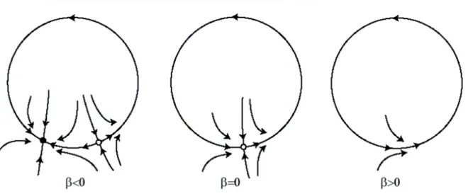
- Saddle-node bifurcation: depicts the collision and disappearance or sudden creation of two equilibria (one stable and one unstable). It is also known as “fold” or “limit point bifurcation”.
Figure 1.7. Saddle node bifurcation. While variable è is increasing, one stable equilibrium points denoted by black dot approaches an unstable equilibrium point denoted by a white dot. For è = 0, the two equilibrium point collide and then disappear. - Hopf bifurcation: This is an important point when analyzing the dynamics of a neuronal model, since it marks the transition from rest to periodic states; appearance or disappearance of oscillations. A Hopf bifurcation occurs when an equilibrium point changes stability via a pair of imaginary eigenvalues. Furthermore, it can be either supercritical (a stable equilibrium becomes unstable and a stable limit cycle appears), or subcritical (resulting in an unstable limit cycle).
39 Figure 1.8. a) Super- and b) sub-critical Hopf bifurcation. Regarding the supercritical Hopf bifurcation, a stable equilibrium point becomes unstable while a stable limit cycle emerges. Conversely, during subcritical Hopf bifurcation, an unstable limit cycle emerges or disappears, depending on the variation of the control variable represented in the figure by è. (Figure adapted from Kuznetsov YA)
- Period doubling: denotes a point involving the appearance or disappearance of a periodic oscillation with twice the period of the original period. A new limit cycle emerges from an existing one, while the period is doubled.
- Torus or Neimark-sacker bifurcation: This bifurcation denotes the appearance of an invariant closed curve from an equilibrium point. It can be detected near limit-Hopf bifurcation.
Other bifurcations can be detected, such as saddle-node on a limit cycle, or more generally saddle-homoclinic bifurcation. These bifurcations represent another type